The document is an extensive overview of the book 'Hemodynamic Monitoring', edited by Michael R. Pinsky, Jean-Louis Teboul, and Jean-Louis Vincent, aimed at enhancing understanding and management of cardiovascular insufficiency in critically ill patients. It discusses the importance of personalized approaches to hemodynamic monitoring, with chapters covering physiology, clinical assessments, monitoring techniques, and decision-making in critical care. The work is intended for a range of healthcare professionals in intensive care, providing evidence-based insights to improve patient outcomes.


![4
1
The primary goal of intensive care medicine is the prevention, reduction, and removal of
temporary risk of death in acutely ill patients, including patients exposed to risk of death
due to surgery and other therapeutic interventions. Cardiovascular organ dysfunction or
failure is, after respiratory failure, the most common organ function problem in intensive
care unit (ICU) patients [1]. The central role of hemodynamic monitoring in the ICU
armamentarium is therefore self-evident. In this context monitoring implies observing
continuously or continually changes in physiologic variables over time to reveal changes
in organ function, to prompt therapeutic interventions, and to evaluate response to thera-
peutic interventions. Monitoring per se cannot be expected to improve patient outcomes –
only timely applied right interventions can do so [2].
Hemodynamic monitoring and diagnostics are different entities, sharing common fea-
tures and overlapping, if diagnostics are frequently repeated. Monitoring tools, such as
cardiac output monitors or pulmonary artery catheter, may help to establish diagnosis,
and diagnostic tools, such as echocardiography, can be used repeatedly to monitor cardio-
vascular function and response to treatment at least over short periods of time.
Measurements and diagnostic evaluations that were intermittently done in the past (e.g.,
cardiac output, venous oximetry) can now be performed continually or continuously.
Echocardiography, traditionally a diagnostic tool, has an established role in perioperative
monitoring of cardiac surgery patients. Barriers for its use for monitoring ICU patients are
disappearing with increased availability of equipment and trained operators, although
operator dependence and the need for frequent repetitions remain limitations. The intro-
duction of miniature transesophageal echocardiography probes is likely to facilitate
echocardiography-
based continual monitoring also in the ICU [3].
The use of dynamic assessment of circulation is a fundamental component of hemody-
namic monitoring. The principle of observing the physiology, inducing a perturbation,
and observing the response was emphasized by Max Harry Weil in 1965, when he
described the use of fluid challenge in shock: “The effect of fluid replacement on the clini-
cal status of the patient in shock is gauged by objective changes in circulation, such as
blood pressure, mental alertness, urine flow, peripheral venous filling, and appearance and
texture of the skin” [4]. In this elegant paper, the today well-known limitations of static
values of hemodynamic variables are discussed with great insight. In the last decades, the
physiology underlying dynamic hemodynamic assessments and their limitations in mon-
itoring the circulation have been established. Instead of using the fluid challenge to per-
turb the circulation, many of the current approaches try to predict the response to a fluid
challenge in order to avoid unnecessary fluid loading. All these dynamic approaches are
based on the principle of assessing “preload dependence.” This can be done by observing
respiratory cycle-dependent variations in intravascular pressures, vascular diameters, and
stroke volume or its surrogates or by directly observing the effect of a volume shift induced
by passive leg raising on these variables. The practical aspects of these of methods as well
as their limitations are discussed elsewhere in this book. Two major issues deserve to be
mentioned already here: first, to be preload or volume responsive is normal and does not
indicate the need for volume; second, hypovolemia and right heart failure may both man-
ifest as left heart preload dependence.
The quest for less invasive hemodynamic monitoring has been driven by the goal to
reduce the risks of invasive techniques, to reduce the need of special skills and resources,
and to make hemodynamic monitoring more widely available. This has been facilitated by
major evolution in signal processing, transducer and imaging technology, and in under-
standing physiology. Wireless transducers and biosensors, and body area networks make
J. Takala](https://image.slidesharecdn.com/monitoreohemodinamico-240424015329-4e755034/75/Monitoreo-Hemodinamico-de-Pinsky-EN-LA-UCI-23-2048.jpg)
![5 1
remote monitoring technically possible, although their routine clinical application is still
confronted with technical and logistic problems [5].
Another trend in hemodynamic monitoring has been the focus on microcirculation.
Research tools used for studying pathophysiology of microcirculation and peripheral tis-
sue perfusion have so far failed to break through into clinical monitoring. In order to
monitor peripheral tissue perfusion in the clinical setting, traditional clinical variables to
monitor circulation have had a renaissance. These include skin temperature, central to
peripheral skin temperature difference, capillary refill time, and evaluation of skin mot-
tling [6]. These simple measurements can be used for monitoring hemodynamics without
any special equipment, and at same time, they are amenable for new senor technologies.
Integration of hemodynamic monitoring data to provide relevant information for
therapeutic decisions becomes a major challenge, when the amount of available data
increases. At the moment, such integration can be achieved using clinical information
systems to display pathophysiologically relevant combinations of data. The development
of intelligent alarms is the next step and can help to apply hemodynamic monitoring out-
side the ICU [7].
Despite all the exciting new developments in technology, the variety of available monitor-
ing devices, and the improved understanding of pathophysiology, the most important chal-
lenge remains: What should be the hemodynamic targets? Hemodynamic monitoring can
only reveal changes in cardiovascular function, and the interpretation of such changes may
prompt therapeutic interventions. What are the right interventions and what should be their
targets remain disappointedly unclear. The application of fixed hemodynamic targets in large-
scale randomized controlled trials has given little if any definitive answers [8]. The risks of
overzealous hemodynamic support with fluids and vasoactive drugs have also been demon-
strated. Given the complexity of hemodynamic pathophysiology, it is very unlikely that any
fixed numeric targets for all patients are appropriate. Rather, assessing response to treatment
should consider changes in the individual patient’s clinical status and signs of tissue perfusion,
such as mental alertness, skin temperature and capillary refill, and urine flow, and objective
changes in hemodynamic variables provided by hemodynamic monitoring and imaging.
References
1. Moreno R, Vincent JL, Matos R, Mendonça A, Cantraine F, Thijs L, et al. The use of maximum SOFA score
to quantify organ dysfunction/failure in intensive care. Results of a prospective, multicentre study.
Intensive Care Med. 1999;25:686–96.
2. Takala J. The pulmonary artery catheter: the tool versus treatments based on the tool. Crit Care.
2006;10:162. https://doi.org/10.1186/cc5021.
3. Vignon P, Merz TM, Vieillard-Baron A. Ten reasons for performing hemodynamic monitoring using
transesophageal echocardiography. Intensive Care Med. 2017;43:1048–51. https://doi.org/10.1007/
s00134-017-4716-1.
4. Weil MH, Shubin H, Rosoff L. Fluid repletion in circulatory shock: central venous pressure and other
practical guides. JAMA. 1965;192:668–74.
5. Rathore MM, Ahmad A, Paul A, Wan J, Zhang D. Real-time medical emergency response system:
exploiting IoT and big data for public health. J Med Syst. 2016;40:283. https://doi.org/10.1007/s10916-
016-0647-6.
6. Lima A, Bakker J. Clinical assessment of peripheral circulation. Curr Opin Crit Care. 2015;21(3):226–31.
7. Kang MA, Churpek MM, Zadravecz FJ, Adhikari R, Twu NM, Edelson DP. Real-time risk prediction on the
wards: a feasibility study. Crit Care Med. 2016;44:1468–73.
8. The PRISM Investigators. Early, goal-directed therapy for septic shock — a patient-level meta-analysis.
N Engl J Med. 2017;376:2223–34. https://doi.org/10.1056/NEJMoa1701380.
Introduction to “Hemodynamic Monitoring”](https://image.slidesharecdn.com/monitoreohemodinamico-240424015329-4e755034/75/Monitoreo-Hemodinamico-de-Pinsky-EN-LA-UCI-24-2048.jpg)

![8
2
Learning Ojectives
In this chapter, we will discuss the definition of shock both from the physiological and clini-
cal point of view. We will categorize shock states according to patient cardiac output. Then
we will analyze the available tools to diagnose shock and to start appropriate therapies.
2.1 Introduction
Circulatory Shock is one of the most common cause of admission to the ICU with a preva-
lence of 30% for patient already in the ICU [1].
Patients are defined in shock when tissue oxygen demand is not coupled with oxygen
supply [2]. From a clinical point of view, shock is often associated with low blood pres-
sure. Hypotension is one of the most common clinical presentations of the shock states,
even though its presence does not always represent a “conditio sine qua non.” Markers
of peripheral hypoperfusion [3] or other signs such as tachycardia not related to pain or
anxiety or fever may be other alert signs to identify patients in shock [2].
The main feature of shock condition is the decrease in oxygen utilization at cellu-
lar level with impaired cellular metabolism and consequent derangements from normal
physiology. If this situation is not promptly corrected, it leads to cellular “energetic failure”
[4] which implies arrest of all metabolic functions and multiple organ failure.
In this chapter we will discuss the shock definitions, referring both to the classical defi-
nition and exploring cellular and metabolic alterations that characterize the shock states.
The different types of shock will be analyzed, trying to highlight their main features and
recognition criteria reported into the most recent guidelines.
2.2 Definition
Many diseases may ultimately lead to a condition of shock, with impairment of the organ
perfusion and onset of multiple organ failure (MOF). From a pathophysiological point
of view, shock is classically defined as a condition in which oxygen supply is inadequate
to peripheral oxygen demand [3]. However, shock may be also defined as a condition in
which hypotension is associated with a variable degree of organ derangement (i.e., oligu-
ria, mottled skin, confusion, dyspnea, etc.). Regardless of the definition, the relationship
between oxygen delivery (DO2) and oxygen consumption (VO2) remains essential [5, 6].
DO2 is the amount of oxygen delivered by the heart to the cells:
DO CO CaO
2 2
= ´ (2.1)
where CO is the cardiac output and CaO2 is the arterial oxygen content, calculated as
shown in the following Eq. (2.2):
CaO Hb SaO PaO
2 2 2
1 34 0 003
= ´ ´
( )+ ´
( )
. . (2.2)
where Hb is the hemoglobin concentration, SaO2 the arterial O2 saturation, and PaO2
arterial partial pressure of oxygen. These equations underline how, besides CO, Hb and
SaO2 play the major role in determining DO2, more than PaO2, which contributes to a
minor extent. Similarly, VO2 is calculated as follows:
A. M. Dell’Anna et al.](https://image.slidesharecdn.com/monitoreohemodinamico-240424015329-4e755034/75/Monitoreo-Hemodinamico-de-Pinsky-EN-LA-UCI-26-2048.jpg)

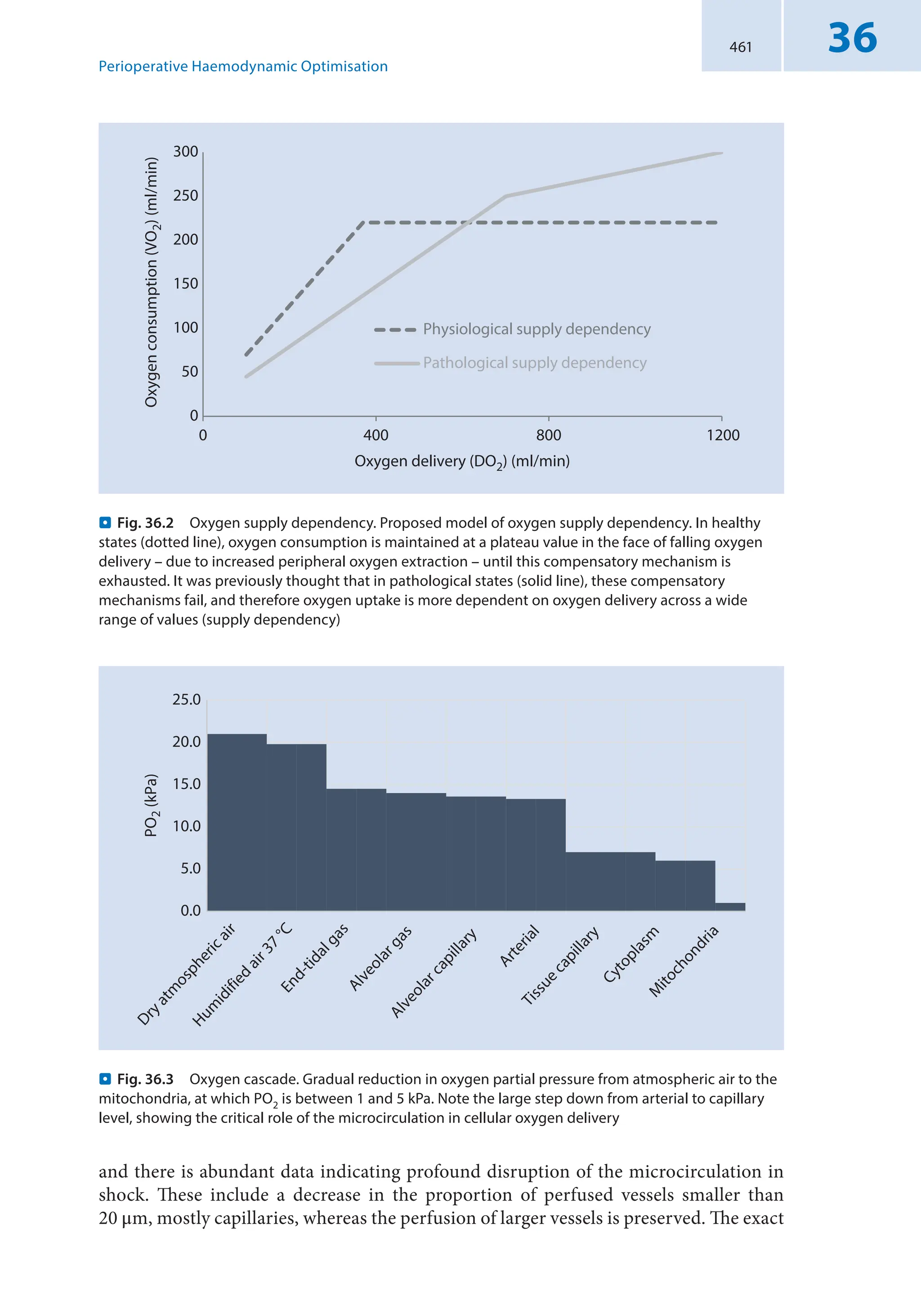
![10
2
increasing O2 extraction. After critical O2ER has been reached, aerobic metabolism begins
to be impaired, and a shift toward anaerobic metabolism occurs with increased lactic acid
production (. Fig. 2.1a). This kind of behavior of DO2/VO2 is preserved in many shock
conditions except for septic shock which is the most common distributive shock (see sub-
sequent paragraph – . Fig. 2.1b). Indeed, in that case, critical O2ER is shifted higher and
rightward, while the slope of the curves is much more pronounced (. Fig. 2.1b).
However, the earliest phases of the shock states may not imply an alteration of global
VO2. Peripheral organs react very differently to reduced blood flow, because of their dif-
ferent physiology. The heart, for instance, has a very high O2ER so that oxygen consump-
tion is essentially dependent upon coronary blood flow, whose decline will generate a
decrease of the aerobic metabolism, with impaired contractility [7]. On the contrary, the
kidneys that receive a very high amount of blood flow (approximately 25% of CO) extract
a very low amount of oxygen, being able to tolerate a longer period of cold ischemia [8].
Finally, during septic shock, especially after the initial phases, very low O2ER along with
high DO2 may occur, and this condition has been associated with poor survival [9, 10].
Considered all the limits of the classical definitions, it may be useful reformulating the
shock definition moving from general hemodynamics to the cellular level. Regardless the
cause and the feature of each type of shock (that we will discuss in the next paragraph),
shock states may be all defined as conditions implying an altered oxygen utilization at
cellular level. Focusing the attention on the last part of the oxygen distribution chain, we
can understand that two main mechanisms are responsible for shock establishment: the
reduction of the oxygen amount available to the cells and the inability to use the oxygen
delivered by the capillary blood flow.
The reduction of the oxygen amount given to the cells may be primarily due to a
decrease of DO2 consequent to a low CO or Hb decrease. In septic shock, despite normal
or high DO2, the capillary blood flow alterations determine very low cellular oxygen con-
centrations. This is in large part due to the increased distance between perfused vessels
and cells with peripheral shunt and reduced cellular O2 availability.
Some experimental and clinical models have shown that even in condition of nor-
mal capillary blood flow, mitochondrial activity may be consistently reduced, due to an
increased production of inflammatory cytokines, eminently common in septic shock [4].
Whatever the cause, the poor oxygen utilization, if not promptly treated, rapidly trans-
lates in an energetic problem that impairs cellular metabolism. In this context, the deli-
cate balance between ATP supply and demand may be considered as the central part of
a complex equilibrium [11]. While ATP supply is essentially related to O2 and substrate
(glucose, lipid, protein) availability, ATP demand is attributable to many cellular functions
such as DNA and RNA synthesis, protein production, and transmembrane pump activity
(particularly Na/K ATPase). When an altered oxygen utilization occurs, ATP concentra-
tion decreases, and energetic cellular activities are somehow hierarchically “hibernated,”
to allow cellular survival. ATP demand due to transmembrane pump function is in general
preserved until the last phases of shock with the aim of keeping constant the transmem-
brane physiological electrical gradient. If the oxygen utilization impairment is not rapidly
resolved, a progressive decrease in cytoplasmic ATP concentration occurs with increased
lactic acid production. The loss of functionality of the transmembrane pumps induces
cellular alterations, leading to organ dysfunction with the classical clinical signs of shock.
However, during the early phases of shock, the signs of hemodynamic impairment such
as hypotension and tachycardia may not always be so evident. Therefore, oxygen deficit and
the increased anaerobic metabolism with a certain degree of organ dysfunction may occur
A. M. Dell’Anna et al.](https://image.slidesharecdn.com/monitoreohemodinamico-240424015329-4e755034/75/Monitoreo-Hemodinamico-de-Pinsky-EN-LA-UCI-28-2048.jpg)
![11 2
before the classical hemodynamic signs. This has been highlighted in the latest guidelines
regarding septic shock definitions, where the recognition of altered mentation, tachypnea,
and systolic blood pressure below 100 mmHg (the so-called quick SOFA, qSOFA) [12] may
anticipate the occurrence of the classical septic shock symptoms. In other cases, hemody-
namic impairment is the cause of cellular oxygen deficit, and hemodynamic alterations
slightly precede or go along with signs of anaerobic metabolism and organ failure.
In all cases, early treatment of the cause of shock and prompt correction of its hemo-
dynamic derangements may stop the progression toward MOF and death.
2.3 Shock Classification
Shock states have always been classified according to patient’s CO. The rationale behind
this approach is that CO is representative of O2 delivery in the condition of stable hemo-
globin and arterial saturation.
SvO2 is another parameter which may be used to assess the existence of an imbalance
between oxygen demand and supply, as well as the adequacy of CO. Normal value is about
65–70% [13]. In low-CO shock, SvO2 values are typically decreased, while in distributive
shock, they are increased.
According to the underlying cause, we can identify four principal types of shock, each one
characterized by difference in hemodynamic parameters such as CO, SvO2, central venous
pressure(CVP),systemicvascularresistance(SVR),andechocardiographicsigns(. Table2.1):
2.3.1 Low-CO States
In low-CO shock the common problem is the inadequacy of oxygen transport.
2.3.1.1 Hypovolemic Shock
It occurs in about 16% of ICU patients. It is attributable to internal or external fluid loss,
and it is the most common cause of shock in trauma patients.
In hypovolemic shock, CO is usually low, because of the decreased preload; SvO2
is low because O2 extraction increases in response to the decrease in DO2, as shown in
. Fig. 2.1a; CVP is also low; and SVR are high in the attempt to keep mean arterial pres-
sure (MAP) at normal or quasi-normal value.
Echocardiography signs are the small volumes of the cardiac chambers and normal or
high contractility.
2.3.1.2 Cardiogenic Shock
It regards about 16% of ICU patients. It derives from ventricular failure caused by differ-
ent pathological conditions (i.e., acute myocardial infarction, end-stage cardiomyopathy,
arrhythmias, valvular heart disease, myocarditis).
In this kind of shock, CO is low, because contractility is impaired; SvO2 is low, because
O2ER is increased, like in hypovolemic shock; CVP is high because of the increased end-
diastolic volume caused by both the inability of the failing heart to empty cardiac cham-
bers at the end of systole and to a certain degree diastolic impairment related to the loss
of ATP production, causing an increase in end-diastolic pressure. SVR are usually high in
order to keep MAP at normal values.
Shock: Definition and Recognition](https://image.slidesharecdn.com/monitoreohemodinamico-240424015329-4e755034/75/Monitoreo-Hemodinamico-de-Pinsky-EN-LA-UCI-29-2048.jpg)

![13 2
Echocardiographic picture of tamponade is characterized by pericardial effusion,
small right and left ventricles, and dilated inferior vena cava; in pulmonary embolism,
dilated right ventricle and small left ventricle are present; in tension pneumothorax, the
hallmark is the compression of the right and left ventricles with small cardiac chambers.
2.4 High-CO States
In high-CO shock, the main problem is in the periphery, as DO2 is generally preserved
but O2ER is impaired.
2.4.1 Distributive Shock
It represents the most common type of shock in ICU patients, accounting for 64% of
admission for shock (62% septic and 2% non-septic). It is characterized by a systemic
vasodilation due to either a release of inflammatory factors during sepsis or anaphylaxis
or a decrease in sympathetic tone in neurogenic shock.
Clinical signs and parameters are the opposite from other types of shock. CO is typi-
cally high, because of the hyperdynamic state caused by the decrease in SVR. SvO2 is high
because of the decrease in O2ER in the periphery and to the increase in DO2 related to the
high CO. CVP can be low or normal. MAP is typically low, at least in the latest phases.
Echocardiography in general shows normal cardiac chambers and preserved or increased
contractility (unless septic cardiomyopathy occurs).
Septic shock is the most common shock an intensivist has to deal with. Unfortunately,
in some cases the correction of hemodynamic derangements may not be effective.
Underlying mechanisms of septic shock are complex and not entirely clear. The altera-
tion of peripheral O2 metabolism can be either caused by a primary pathological mito-
chondrial and cellular dysfunction or indirectly caused by microvascular alterations with
consequent hypoxic cellular damage.
2.5 Shock Recognition
Early recognition of patient in shock is of a paramount importance in order to reduce
morbidity and mortality [12]. Prompt interventions aiming to restore normal hemody-
namics and correcting the cause of shock may effectively change the clinical course of the
disease [14] (. Table 2.2).
The clinical evaluation and physical exam are the first steps to individuate patients at
risk or patients already in shock.
Medical history can often suggest the underlying cause, for example, a history of coro-
nary artery disease may suggest a cardiogenic shock, while elevated body temperature
and dyspnea may indicate a septic shock. Similarly, after a trauma a patient is likely to
suffer from hypovolemic shock because of blood loss or obstructive shock due to tension
pneumothorax. Various types of shock can occur in combination, like the distributive
(neurogenic) shock after a traumatic spinal injury.
Some alert signs may be useful clinical tools for an early identification of shock.
Hypotension is in most of cases the sign that draws the attention of the clinician and is
Shock: Definition and Recognition](https://image.slidesharecdn.com/monitoreohemodinamico-240424015329-4e755034/75/Monitoreo-Hemodinamico-de-Pinsky-EN-LA-UCI-31-2048.jpg)
![14
2
often considered one of the principal manifestations of shock. However, it can be a rela-
tively late sign in some circumstances and the degree of hypotension does not necessarily
correlate with the degree of shock if it is not accompanied by other markers of hypoxia.
Clinical examination should be complete and accurate when shock is suspected. Many
clinical alterations are common in shock states, and some of them reflect organ dysfunc-
tion due to tissue hypoperfusion, while others are related to whole body response [2].
Some of the typical signs are:
5
5 Mottled and clammy skin (especially in low-CO states)
5
5 Altered mental status (confusion, disorientation, epilepsy, coma)
5
5 Oliguria (urine output 0.5 ml per kilogram of body weight per hour).
Further signs like tachycardia, dyspnea, increased respiratory rate, jugular venous
distention, and peripheral edema are often present but are related more to the response of
the body to the ongoing shock conditions more than to the shock per se.
The importance of a thorough clinical examination has been recently highlighted in the
last published guidelines for sepsis and septic shock management, where the new “quick
SOFA” (qSOFA) score has been introduced to individuate those patients who are likely to be
septic [12] and at risk of shock mainly outside the ICU. As mentioned, this score includes
.
. Table 2.2 Diagnostic tools available to diagnose shock state
Diagnostic
tool
Advantages Limits
Clinical signs Available bedside
Easy to detect
Low specificity
Lactate Good marker of tissue hypoperfusion
Available with ABG point of care
Reliable prognostic value
Trend over time has a prognostic value
Possibility of false positive
Relatively slow normalization (hours)
ScvO2-SvO2 Available with ABG point of care
Good marker of O2 debt in conditions
of low DO2
Normal values do not guarantee
adequate perfusion
CO2 gap Available with ABG point of care
Correlates CO to metabolism
pH and temperature derangements
may alter its interpretationABGs must
be drawn exactly at the same time
Respiratory
quotient
Reliable marker of anaerobic CO2
production
May predict response in terms of O2
consumption
CO2 content is complex to calculate
bedside
Echocar-
diography
Available bedside
Useful to identify different types of
shock
It requires a skilled operator
It does not give any functional
information
ABG arterial blood gas analysis, CO cardiac output, ScvO2 central venous oxygen saturation, SvO2
mixed venous oxygen saturation, CO2 gap difference in central venous-to-arterial carbon dioxide
tension, respiratory quotient ratio between venous and arterial carbon dioxide content and
difference in arterial-to-venous oxygen content [R = (CvCO2 − CaCO2)/(CaO2 − CvO2)]
A. M. Dell’Anna et al.](https://image.slidesharecdn.com/monitoreohemodinamico-240424015329-4e755034/75/Monitoreo-Hemodinamico-de-Pinsky-EN-LA-UCI-32-2048.jpg)
![15 2
systolic arterial pressure below 100 mmHg, respiratory rate above 22/min, and altered men-
tal status. If two out of three of such signs are present, sepsis should be suspected.
Arterial hypotension, though very common in shock, deserves a brief comment,
because its role in diagnosis and as a target of therapy has been extensively discussed over
the last years.
The first question to be addressed is whether all the patients in shock are actually hypo-
tensive. Looking at the most recent guidelines on septic and cardiogenic shock, a positive
answer should be given. Indeed, septic shock may be defined as a septic condition in which
hypotension persists despite adequate volume resuscitation, with lactate 2 mmol/L [12].
Cardiogenic shock is defined as a state of ineffective cardiac output caused by a primary
cardiac disorder with both clinical and biochemical manifestations of inadequate tissue
perfusion [15]. In recent trials, pragmatic definition always included a systolic arterial
pressure below 90 mmHg. Considering shock with low CO, hypotension may be mostly
evident only during the most severe phases, as the homeostatic mechanisms will try to
keep mean arterial pressure at a normal level by increasing SVR. In this situation, the
increase in SVR is useful to maintain a minimal level of cardiac and cerebral perfusion
but may be detrimental for other organ perfusion, decreasing blood flow and increasing
oxygen debt [16]. Many patients with acute cardiac failure may develop signs of peripheral
anaerobic metabolism without hypotension [17]. On the contrary, elevated MAP may be
detrimental because it will impair systolic ejection of the failing heart, worsening oxygen
delivery. Along the same way, patients with acute hemorrhage will try to keep MAP within
the normal value by increasing adrenergic tone which will recruit unstressed volume from
venous reservoir and increase arterial tone. However, if the hemorrhage is not promptly
controlled, this compensatory mechanism becomes deleterious, and it will lead to mul-
tiple organ failure (MOF) and death. Hypotension can be evident either only in the last
phase or if blood loss is more than a half of the circulating blood volume [18].
In conclusion we may say that hypotension is frequently associated with shock condi-
tions, but in many cases, shock may initiate without low blood pressure, and it may be a
very late sign of shock.
Another point is if all the hypotensive states may be considered as shock states. On
this purpose, though most of acute hypotensions are attributable to shock, not necessar-
ily low blood pressure is related to the impaired organ perfusion. For example, in case
of deep sedation, whole body metabolic functions are put at rest and oxygen demand
decreases significantly. Hence lower blood pressure might be tolerated without alterations
of organ perfusion. However, if very low MAP (i.e., 45–50 mmHg) can be safely accepted
for patients without clear signs of organ hypoperfusion is still on debate [19, 20], so it is
reasonable to check the causes of low blood pressure for their prompt correction.
The last, and maybe more important, aspect is the clear definition of hypotension. In
most guidelines and trials [12, 15, 21], hypotension is defined as systolic blood pressure
below 90 mmHg or MAP below 65 mmHg. Such definition comes from the assumption
that below a certain MAP, organ blood flow becomes dependent upon pressure, while
above that point the blood flow can be considered constant [22]. The thresholds represent
a mean among thousands of values and cannot be considered adequate for all patients.
While patients with normal or constitutional low blood pressure may easily tolerate MAP
below 65 mmHg, those who are hypertensive may not be able to adapt their blood flow
regulation even when the MAP is above 65 mmHg [23]. In addition, one threshold can be
adequate for some organs, but is not necessarily considered appropriate for the whole body.
Rules allowing autoregulation for each organ are different [22], and the most important
Shock: Definition and Recognition](https://image.slidesharecdn.com/monitoreohemodinamico-240424015329-4e755034/75/Monitoreo-Hemodinamico-de-Pinsky-EN-LA-UCI-33-2048.jpg)
![16
2
regulator of organ blood flow – i.e., metabolic control – can vary widely in shock condi-
tion, changing completely normal physiology. Even if a general threshold of 65 mmHg
may be considered theoretically adequate, only a thorough examination of the patient that
includes the evaluation of peripheral hypoperfusion can really define the lower acceptable
value of MAP in particular cases. This is even more important considering that the need
of a specific blood pressure essentially derives from the need of our cardiovascular system
to preferentially direct the blood flow toward the organs, according to their metabolic
needs [24]. Accordingly, during sport, a decrease of SVR to increase the peripheral blood
flow in certain organs is coupled with a CO increase, to keep MAP stable and guarantee a
good organ perfusion. On the contrary, when a decrease of SVR is not due to an increase
in metabolic demand but to a dysregulated response (i.e., like in septic shock), the CO
increase is not coupled anymore to the real metabolic demands of our peripheral tissue.
In summary, hypotension is the most common alert sign during shock; however it can be
evident sometimes only in the latest phases of the disease. Similarly, a general threshold of
MAP to define shock has been adopted, but it can vary widely from patient to another, and
only other signs can tell the clinician if that blood pressure needs to be increased or not.
Other data, coming from blood sample analysis, may lead toward the diagnosis of shock
even in the absence of other clear clinical signs or can help to confirm the clinical suspicion.
Among them, arterial lactate level is the most commonly used in clinical practice.
Lactic acid is produced also in physiological conditions, but its production is increased
in condition of anaerobic metabolism and/or when its hepatic elimination is impaired
[25, 26]. Absolute blood lactate values have been associated with a worse outcome in all
kinds of shock, and their persistence over time despite treatments has been associated to
higher mortality rate [27–29]. Values above 2 mmol/L are considered alert signs associ-
ated with increased mortality up to 40%, especially if associated with hypotension [12].
Values above 4 mmol/L have been associated with higher mortality rate in septic shock
regardless of other clinical conditions [30].
If the patient has a central venous line or a pulmonary artery catheter, central or mixed
venous SO2 may be measured (ScvO2 and SvO2, respectively). In clinical practice, mea-
surement of ScVO2 is used as a surrogate of SvO2. ScVO2 reflects the oxygen extraction
only from the upper part of the body (since it is measured in the superior vena cava).
Even though their values are not completely interchangeable [31], both represent periph-
eral O2ER of the body and may guide therapies. If they are very low (70% and 65%,
respectively), they may indicate that an increase in DO2 is mandatory [32]. In 2001 a
pivotal paper showed that patients with septic shock and very low ScvO2 had lower mor-
tality if a protocol aiming to keep SvO2 above 70% was applied in the first 6 h after emer-
gency admission [33]. Unfortunately, three more recent trials failed to replicate the same
results [21, 34–36] so that ScvO2 optimization has been waived from the last guidelines.
However, in such trials, baseline ScvO2 was already 70%, making useless a protocol aim-
ing at ScvO2 optimization [37]. In many shock conditions, particularly in septic shock,
ScvO2 appears normal or even high even if an oxygen debt is developing, as shown by an
increase in other markers of organ perfusion (i.e., lactate). Some papers have even shown
that higher ScvO2 in septic patients are related to higher mortality [10], maybe because
it mirrors an impairment of peripheral oxygen extraction [38]. In conclusion, we should
keep in mind that low ScvO2 values need to be corrected, while normal or high values do
not necessarily correlate a good organ perfusion in shock condition.
The difference between mixed venous and arterial pCO2 (CO2 gap) and CO is inversely
correlated [39], so that an increase in CO2 gap is a marker of inadequate CO. More recently
A. M. Dell’Anna et al.](https://image.slidesharecdn.com/monitoreohemodinamico-240424015329-4e755034/75/Monitoreo-Hemodinamico-de-Pinsky-EN-LA-UCI-34-2048.jpg)
![17 2
some authors have shown that central venous CO2 may be a valid surrogate of mixed
venous CO2 for CO2 gap calculation [40]. A value of CO2 gap 6 mmHg is an alert sign
indicating a low cardiac output with peripheral CO2 stagnation [41]. CO2 gap varies more
rapidly than lactate and ScvO2 could be used as an early marker of peripheral oxygen sup-
ply impairment. It has been proposed as a tool for diagnosis and a goal for therapy [42].
The ratio between CO2 gap and arterial-venous difference in oxygen content (C(a-v)O2)
can be considered as a surrogate of the respiratory quotient (R), where the numerator is
the difference in CO2 content instead in CO2 partial pressure. Many authors [43, 44] have
shown that a ratio 1.7 can effectively individuate patients who are developing an oxy-
gen debt and require CO increase. Recent papers [45] have also shown that patients with
higher ratio and higher lactate are more likely to die in course of septic shock.
Echocardiography is a very useful diagnostic tool in patients in shock. It can be per-
formed bedside and allows to confirm diagnosis of shock (i.e., myocardial infarction or
pericardial tamponade) and to define the type of shock, as discussed previously. It can also
show left and right ventricular function, stroke volume, and possible valvular diseases.
Most severe shocks require advanced hemodynamic monitoring. Data coming from
such tools may help to classify the shock and adopt the most appropriate therapies. In
particular, CO may differentiate between low and high flow shock states, while cardiac
filling pressure or intrathoracic volumes may indicate if the cause of low CO is primarily
cardiac or hypovolemic.
2.6 Self-Evaluation Questions
?
? 1. What is the definition of shock?
?
? 2. How many types of shock exist and what are the differences among them?
?
? 3. How does hypotension correlate to shock states?
?
? 4. What are the main clinical signs of shock?
?
? 5. How many diagnostic tools do we have and how can we use them?
?
? 6. What is the role of lactate in shock diagnosis and treatment?
Take-Home Messages
5
5 Shock is a particular condition in which a problem in oxygen utilization at cellular
level induces a variable degree of organ dysfunction.
5
5 Shock states are usually classified according to CO and etiology of the low CO. Par-
ticularly three shocks with low CO (hypovolemic, cardiogenic, and obstructive) and
one with high CO (distributive) are classically defined.
5
5 Some clinical signs evaluable at the bedside may help clinician to define shock
conditions.
5
5 Hypotension is the most common sign of shock but is not always the first sign.
5
5 The lactate increase and the CO2 gap are important markers of shock states and may
guide therapy and reassessment along with echocardiography.
Shock: Definition and Recognition](https://image.slidesharecdn.com/monitoreohemodinamico-240424015329-4e755034/75/Monitoreo-Hemodinamico-de-Pinsky-EN-LA-UCI-35-2048.jpg)


![23 3
Cardiac output is an adaptive value: it can and must adjust constantly according to the
oxygen requirements of the body. Physiologically, it changes continuously in every indi-
vidual: it is lowest in the middle of the night when asleep and highest during strenuous
exercise. If a critically ill patient is sedated and anesthetized, cardiac output may therefore
be relatively low but still perfectly adequate. An apparently “low” cardiac output therefore
does not necessarily mean that treatment is needed to increase it—it may be sufficient for
that patient under those conditions.
Cardiac output will increase in response to anxiety and exercise, as oxygen require-
ments increase. It must also increase to compensate for a reduction in arterial oxygen
content. For example, when climbing a high mountain, the hypoxemia that occurs as a
result of the lower atmospheric oxygen will be compensated for by an increase in cardiac
output in a healthy, fit individual, so that there is virtually no tissue hypoxia, even at the
top of Everest [1]. Likewise, normovolemic anemia can be very well tolerated by compen-
satory increases in cardiac output, with tissue hypoxia sufficient to cause hyperlactatemia
only occurring when hemoglobin levels decrease as low as 4 g/dL [2].
3.3
Essential Questions When Interpreting
a Cardiac Output Value
When assessing adequacy of cardiac output, it is not sufficient to look just at the cardiac
output value—other factors need to be taken into account, notably related to the presence/
absence of adequate tissue blood flow and to the degree of compensation already present.
We will consider these two aspects separately, although clearly they may overlap.
3.3.1
Is There a Critical Reduction in Tissue Perfusion?
Many individuals walk around with a low cardiac output. They may not walk fast, but
they can complete all their usual activities at their own pace. The real problem arises when
cardiac output is reduced to such a degree that there is a critical reduction in oxygen
availability to the cells. Clinically this often arises when there is hypotension, but clinical
signs also include the effects of altered tissue perfusion as shown through the three clinical
“windows” of perfusion [3]: altered cutaneous perfusion, altered mentation with disori-
entation/confusion, and decreased renal perfusion with resultant oliguria. Biochemically,
the key indication that this critical level has been reached is the presence of increased
blood lactate levels. The usual cutoff value for abnormal blood lactate is set at 2 mEq/L [3],
but any value greater than the normal range, i.e., greater than 1.3 to 1.5 mEq/L, is already
associated with increased mortality rates [4].
A critical reduction in cardiac output can be due to only one of three mechanisms:
5
5 Severe hypovolemia, for example, in hemorrhage or severe dehydration
5
5 Altered cardiac pump function, i.e., critical reduction in myocardial contractility
(e.g., massive myocardial infarction, major arrhythmia [rapid supraventricular
tachyarrhythmia or ventricular tachycardia]), or severe valvular disease
5
5 Major obstruction, for example, cardiac tamponade, massive pulmonary embolism,
or tension pneumothorax
Assessing the Adequacy of Cardiac Output](https://image.slidesharecdn.com/monitoreohemodinamico-240424015329-4e755034/75/Monitoreo-Hemodinamico-de-Pinsky-EN-LA-UCI-41-2048.jpg)
![24
3
3.3.2
Have Compensatory Mechanisms Already Been Activated?
The activation of mechanisms to compensate for poor tissue oxygenation can indicate
whether or not cardiac output is adequate. In the outpatient context, for example, accord-
ing to the New York Heart classification, the severity of heart failure is assessed by the
capacity of exercise of the patient. The less able a patient is to compensate for increased
exercise, the greater the degree of heart failure. Similarly in critically ill patients, if com-
pensatory mechanisms have already been activated to a maximum, any worsening of the
condition will result in tissue hypoxia.
3.3.2.1 Cardiac-Level Compensation
A first, quite non-specific mechanism to compensate for reduced tissue perfusion is an
increase in heart rate. As cardiac output is the product of heart rate and stroke volume, an
increased heart rate can compensate for a decrease in stroke volume. The tissues are still
perfused but at the price of an increased adrenergic response.
Another compensatory mechanism at the cardiac level is an increase in cardiac size.
This is an important compensatory mechanism in heart failure enabling stroke volume to
be maintained somewhat by an increased ventricular preload, through the Frank-Starling
mechanism. These patients have enlarged ventricles on echocardiographic examination
and elevated cardiac filling pressures, which can result in lung and systemic edema.
3.3.2.2 Peripheral-Level Compensation
If DO2 is reduced, tissues can still maintain oxygen consumption by increasing oxygen
extraction, with a reduction in the venous saturation of the hemoglobin. The SvO2, reflect-
ing the oxygen saturation in the mixed venous blood in the pulmonary artery, can be
easily assessed using a pulmonary artery catheter, but these are less widely used than pre-
viously. A central venous catheter provides access to blood in the superior vena cava and
offers a surrogate for ScvO2. Although ScvO2 is a relatively gross estimate of SvO2 [5], an
inadequate cardiac output is typically associated with an ScvO2 well below 70%.
Therapeutic Implications
Cardiac output is determined by four factors, and treatment of inadequate cardiac
output therefore involves optimizing these four aspects: heart rate, preload, afterload,
and myocardial contractility. The analogy has been made with the effort needed to
increase speed when riding a bicycle [6] (. Fig. 3.2): pushing harder on the pedals,
cycling with the wind from behind, cycling on the road with least resistance, and
changing gears.
Heart rate: An increase in heart rate is not very effective unless there is profound
bradycardia [7]; otherwise the increase in heart rate is compensated for by a decrease
in stroke volume. Moreover, in a patient with inadequate cardiac output, the heart
rate may already be increased as a physiological response, so increasing it further is
unlikely to bring additional benefit.
Preload: Increasing preload, i.e., the end-diastolic ventricular volume, can increase
myocardial contractility by increasing myocardial fiber stretch; this in turn increases
cardiac output. Fluid administration should be attempted in all patients with a critical
J.-L. Vincent](https://image.slidesharecdn.com/monitoreohemodinamico-240424015329-4e755034/75/Monitoreo-Hemodinamico-de-Pinsky-EN-LA-UCI-42-2048.jpg)
![25 3
alteration in tissue perfusion (shock) in order to increase preload, even though clinical
benefit is unlikely in cases of obstructive shock. Even in cardiogenic shock with pulmo-
nary edema, the acute nature of the lung edema is associated with a relative decrease
in blood volume secondary to the extravasation of water into the extracellular fluid.
To optimize fluid administration without causing fluid overload and its associated
harmful effects, efforts should be taken to determine the patient’s likely response to
fluid administration. The most effective means of achieving this is by repeated fluid
challenges, in which the clinical response is evaluated together with a cardiac filling
pressure as a safety measure during the rapid administration of a limited amount of
fluid [8]. A passive leg raising test, effectively an“internal”fluid challenge, represents
an alternative method of evaluating response, although the procedure is not as simple
as it may first seem [9].
Afterload: Afterload represents the forces working to prevent ventricular emptying,
effectively represented by systemic vascular resistance. Afterload can be decreased
using vasodilators, but this is possible only when the arterial pressure is adequate.
Myocardial contractility: A direct increase in myocardial contractility can be
achieved with inotropes. Dobutamine is the first choice for this purpose, and
administration of just a few mcg/kg/min can sometimes have dramatic effects, so
that infusions should be started at low doses. This is particularly the case when
vascular tone is not very high (e.g., in sepsis). Nevertheless there are always risks
associated with increased adrenergic stimulation. Importantly, some interventions
can act on more than one determinant. For example, phosphodiesterase inhibi-
tors (milrinone, enoximone) or levosimendan can increase myocardial contractility
(through an inotropic effect) and reduce ventricular afterload (through a vasodilat-
ing effect).
Importantly, the other determinants of DO2 must not be neglected. Hypoxemia
should always be corrected, as it is always associated with a hyperadrenergic response,
which adds strain to the heart and increases catabolism. If there is associated anemia,
blood transfusion can be considered even when the hemoglobin level is between
7 and 9 g/dL.
.
. Fig. 3.2 The four determi-
nants of cardiac output, using an
analogy to the speed of a bicycle.
(Reproduced from [6])
Assessing the Adequacy of Cardiac Output](https://image.slidesharecdn.com/monitoreohemodinamico-240424015329-4e755034/75/Monitoreo-Hemodinamico-de-Pinsky-EN-LA-UCI-43-2048.jpg)


![28
4
Learning Objectives
After reading this chapter, the reader will learn:
1. Which are the main determinants of venous return and cardiac output?
2. What is the mean systemic filling pressure, what is its importance and what is the
gradient of venous return?
3. What is the meaning of central venous pressure in the context of venous return?
4. What is an effective fluid challenge, using Pmsf?
4.1 Introduction
Cardiovascular failure is one of the most common reasons for admission to intensive care
units. The main function of the cardiovascular system is the transport of oxygen (O2) to
the tissues. If the concentration of haemoglobin is stable, the main determinant of O2
transport is the cardiac output (CO).
CO is total volume of blood mobilised by the heart per unit of time, and it is measured
in units of flow (L/min). In stable conditions, the cardiovascular system is a close loop
system, and the heart can only eject the amount of blood that it receives. Thus, the total
volume ejected over a period of time would equal the total volume of blood returning
from the venous system. Therefore, venous return equals cardiac output.
In this chapter we will analyse the determinants of venous return, their impact on the
cardiovascular physiology and the practical implications that those factors may play a role
in the treatment of critically ill patients. The main factors that determine the venous return
to the heart from the systemic circulation are:
1. The degree of filling of the circulation
2. The ability of the heart to maintain a low right atrial pressure
3. The resistance to blood flow between the peripheral vessels and the right atrium
4. The resistance to blood flow between the heart and the capillaries
4.2
Blood Volume and Mean Systemic Filling Pressure
The venous system contains about 70% of the total blood volume, whereas the arterial
system contains only 13–18% and the capillaries 7% [1, 2]. The venous system is a blood
reservoir, able to adjust his capacity according to the haemodynamic conditions. The
venous wall is much thinner than the arterial wall, as blood is circulating at low pressure,
but it still contains smooth muscle fibres, able to contract and expand according to the
haemodynamic situation. During hypovolaemia, sympathetic nervous reflexes cause
venoconstriction, sending blood back to the central circulation, increasing preload and
increasing cardiac output. Actually, even after 20% of the total blood volume has been lost,
the circulatory system functions almost normally because of this variable reservoir func-
tion of veins [1].
The heart pumps blood continuously into the aorta keeping the mean arterial pressure
high, averaging 80–100 mmHg. This pressure decreases progressively as the blood flows
into the systemic circulation, as low as the level of the right atrial pressure (Pra). This fall
in pressure is mainly caused by the increasing total cross-sectional area in each level of the
vascular tree (. Fig. 4.1). When the heart stops, the arterial pressure decreases, and the
RAP progressively increases. At certain point, blood will not be flowing, and if the arteri-
H. D. Aya and M. Cecconi](https://image.slidesharecdn.com/monitoreohemodinamico-240424015329-4e755034/75/Monitoreo-Hemodinamico-de-Pinsky-EN-LA-UCI-46-2048.jpg)
![29 4
oles do not collapse trapping blood in the arterial space, the pressure will be the same in
all territories of the circulatory system. This pressure is the mean systemic filling pressure
(Pmsf). This pressure was described by Bayliss and Starling [3], and they figured that
somewhere in the circulation, there must be a point where the pressure is not changing
when the heart stops. Actually, during a cardiac arrest, the pressure in the small veins
(1 mm) and venules do not change substantially; they are the “pivoting point” of the
system (. Fig. 4.1) [4]. This pressure is less than the capillary pressure, close to the portal
venous pressure and greater than the RAP. Its anatomic location is not necessarily at the
same venous branching level in every organ. The importance of this pressure, rather than
its anatomical location, is that it provides a quantitative measurement of the intravascular
filling status independent from cardiac function: its value is equal to the Pmsf.
Let us imagine the “blood reservoir” as a distensible compartment. The volume
required to fill a distensible tube, such as a tyre or a blood vessel, with no pressure rise is
called the “unstressed” volume (Vo). Further volume expansion will imply necessarily a
pressure rise and an elastic distension of the wall of the tube, which depends on the com-
pliance (C) of the wall. This volume is the “stressed” volume (Vs) and is related to the
pressure in the next equation:
Pmsf s
= V C
/
250
2500
Pivot point
Cross
sectional
area
(cm
2
)
Pmsf
Pressure
40
Small arteries Arterioles Capillaries Venules Small veins
80
20
RAP
.
. Fig. 4.1 Pressures and cross-sectional area across cardiovascular system. Pmsf mean systemic filling
pressure. This is the pressure at all points in the cardiovascular system when the heart stops. During
normal circulation, there is a point (pivot point) where the pressure equalises the Pmsf. At that point, the
pressure is independent of flow and theoretically localises at the venule territory. The drop in pressure is
mainly related to the increase in the total cross-sectional area and the compliance of the vascular wall
Determinants of Venous Return](https://image.slidesharecdn.com/monitoreohemodinamico-240424015329-4e755034/75/Monitoreo-Hemodinamico-de-Pinsky-EN-LA-UCI-47-2048.jpg)
![30
4
4.3
The Right Atrial Pressure
In order to move a fluid across a tube or tubular system, the variables described in
Poiseuille’s Law apply. Therefore, it is the gradient of pressure between two points in the
system, not any single pressure at any particular point, which determines the rate of flow
[5, 6]. Given that most of the blood is in the venous reservoir, the pressure at this point is
particularly interesting. Following Poiseuille’s Law equation, Guyton pointed out that
venous return could be defined by three parameters: the mean systemic filling pressure
(Pmsf), Pra and the resistance to venous return (RVR). This can be also mathematically
represented as follows:
VR Pmsf Pra RVR
= -
( )/
Guyton [7] drew venous return curves in recently dead dogs. The heart was replaced with
a pump, and the Pra was controlled by increasing or decreasing the minute capacity of the
pump. Increasing or decreasing the total quantity of blood controlled the mean circula-
tory filling pressure. From these curves (. Fig. 4.2), one can see that for a given Pra, the
greater the Pmsf, the greater the venous return is. Importantly, under isovolumetric con-
ditions, the greater is the Pra, the lower is the venous return. Thus, given this linear rela-
tionship, if venous return and Pra can be measured and changed without changes in the
volume status, the slope of the line could be calculated (the RVR), and the Pmsf could be
estimated. Under isovolaemic conditions, Pra depends mainly on the right ventricular
function and its ability to accommodate and pump blood to the pulmonary circulation.
Any decrease in the heart function will increase Pra and therefore create congestion in the
venous circulation.
A second factor that affects the Pra is the intrathoracic and the pericardial pres-
sure. Under normal circumstances, intrathoracic pressure is negative or equal to the
0
Venous
return
(L/min)
a b c Pra(mmHg)
Pmsf a
Pmsf b
Pmsf c
.
. Fig. 4.2 Venous return and Frank-Starling curves. Pmsf mean systemic filling pressure, Pra right atrial
pressure. Each venous return curve represents different levels of volume status. To move across a venous
return curve, changes in cardiac function are required, without changes in the volume status. The black
line represents the Frank-Starling curve for a particular level of cardiac function. In order to move from
the blue to the red curve, Pmsf must change from a to c without changes in resistance
H. D. Aya and M. Cecconi](https://image.slidesharecdn.com/monitoreohemodinamico-240424015329-4e755034/75/Monitoreo-Hemodinamico-de-Pinsky-EN-LA-UCI-48-2048.jpg)
![31 4
atmospheric pressure. Any increase in this pressure that translates in an increase of
Pra will result in a decrease of venous return. Similar phenomenon is observed if
there is an increase in pericardial pressure, which will increase Pra, and will result in
a decrease of venous return.
4.4
Arteriolar Resistance and Metabolic Demand
As the venous system can only transport the amount of volume that arrives from the
capillary system, the regulation of the capillary circulation will also determine the venous
return. Each tissue in the body has the ability to control its own local blood flow in pro-
portion to its metabolic needs. Rapid changes in vasoconstriction or vasodilation of arte-
rioles, meta-arterioles and precapillary sphincters may happen within seconds to provide
adequate local tissue blood flow. The main factors involved in the acute control of local
blood flow are:
1. Tissue metabolism: this is by far the most powerful factor. The higher is the local
oxygen demand, the higher is the blood flow.
2. Availability of oxygen: whenever there is a deficit of oxygen (hypoxia), there is an
increase in local blood flow. Oxygen deficit can occur in several ways, such as (1)
failure in oxygenation of blood (ARDS, pneumonia, etc.), (2) failure in transport of
oxygen by haemoglobin (CO poisoning) or (3) failure in the ability of tissue to use
oxygen (septic shock, cyanide poisoning).
3. Deficit of other nutrients: under special conditions, the lack of glucose, thiamine,
niacin and riboflavin can cause vasodilation.
4.5
Control of Venous Tone: Resistance to Venous Return
Certain parts of the venous system are particularly compliant: these include the spleen,
the liver, the large abdominal veins and the venous plexus beneath the skin. Splanchnic
and cutaneous veins have a high population of α1- and α2-adrenergic receptors, so they
are very sensitive to adrenergic stimulation, contrary to skeletal and muscle veins [8]. The
control of the venous system has been extensively studied in animal models. There are
nerve terminations in the proximity of many small vein smooth muscles [9] but not in the
veins of the skeletal muscle [10]. However, circulating catecholamines can induce contrac-
tion of venules and veins of the skeletal muscle and mesentery [9, 10]. Thus, probably
catecholamines released from the sympathetic nerve termination of the arterial side may
pass through the capillary bed and affect the venous system.
Smooth muscles of the veins and arteries do not respond necessarily in the same way to
chemical signals. Dihydroergotamine can activate the veins but not the arteries [11]. The
venoussystemprimarilyhasα-adrenergicreceptors[12–15].Stimulationoftheβ-adrenergic
receptors of arterioles cause vasodilation but has little effect on the veins [16, 17].
Angiotensin can increase Pmsf [16, 18]. Isoproterenol, a β-adrenergic agonist, causes a
decrease in Pmsf when veins are constricted with angiotensin. On the other hand, vasopres-
sin has very little effect on Pmsf [19] or on vascular capacity once reflex blockade [20] and
similar results were reported regarding natriuretic peptides [21].
Nitroglycerin and nitroprusside decrease Pmsf and increase unstressed blood volume
but do not change vascular compliance in ganglion-blockade dogs [22]. Verapamil and
Determinants of Venous Return](https://image.slidesharecdn.com/monitoreohemodinamico-240424015329-4e755034/75/Monitoreo-Hemodinamico-de-Pinsky-EN-LA-UCI-49-2048.jpg)
![32
4
nifedipine increase venous return by reducing the resistance to venous return without
changing the Pmsf, whereas nitroglycerin in small doses can reduce Pmsf without changes
in resistance to venous return [23]. Diltiazem reduces both resistance and Pmsf increasing
CO [23].
Moderate hypercapnia and hypoxia have little direct non-reflex effect on CO and Pmsf
[24]. Severe hypercapnia (PaCO2 to 114 mmHg (15.2 KPa)) caused an increase in Pmsf by
5.5 mmHg, whereas a PaO2 of 34 mmHg (4.5 KPa) caused an increase in Pmsf by
2.5 mmHg [25].
4.6
Measurement of Pmsf in Humans with Intact Circulation
The Pmsf is not easy to measure in patients with an intact circulation. Schipke et al. [26]
performed a fibrillation-defibrillation sequence in 82 patients during cardioverter/defi-
brillator implantation to measure the Pmsf over 13 s. A true equilibrium pressure was not
achieved, and the arterial-central venous pressure difference was 13.2 ± 6.2 mm Hg, and
differences still persisted in sequences of 20 s.
Pinsky [27] proposed a model in animals with an intact circulation to construct
venous return curves observing the relationship between isovolumetric changes in CO
and Pra during intermittent positive pressure recruitment manoeuvres. Pmsf was esti-
mated by calculation of the slope and extrapolation of the Pra value to zero CO. Pmsf
calculated was found similar to Pmsf measured during circulatory arrest. Other studies
[28–30] have confirmed this linear relationship between VR and Pra and derived Pmsf
from the regression equation in animal models with intact circulation. Maas and col-
leagues [31] applied the same rationale to study the effect of a 12-second inspiratory hold
manoeuvre to three different steady-state levels on central venous pressure (CVP), as an
estimate of Pra, and blood flow (CO) measured via the pulse contour method during the
last 3 s in mechanically ventilated postoperative cardiac patients. This study showed
again a linear relationship between changes in CVP and CO, and importantly, Pmsf
could be estimated at bedside in intensive care patients with an intact circulation.
Obviously this technique is only feasible in fully sedated patients under mechanical ven-
tilation. Keller and colleagues [32] used this method to assess the changes on venous
return with passive leg raising (PLR) manoeuvre: they observed nine postoperative car-
diac patients at baseline, during PLR and after volume expansion (500 ml of hydroxy-
ethyl starch). They reported a Pmsf at baseline of 19.7 mmHg. This increased to 22 mmHg
after PLR and to 26.9 mmHg after volume expansion (VE). Although CO increased after
PLR and VE, the gradient of pressure of venous return (difference between Pmsf and
CVP) increased by 2 mmHg after PLR and by 5.8 mmHg after VE. This could explain
why a PLR test does not consistently increase CO in fluid-responsive patients [33], or
even for a fluid challenge, the increase in Pmsf is an essential condition to effectively test
the cardiac response.
Parkin and Wright [34] proposed a method for estimating a mean systemic filling
pressure analogue (Pmsa) using the mean arterial pressure (MAP), Pra, CO and anthro-
pometric data. Pmsa algorithm is fully described in other publications [35]. In essence,
they build a mathematical model that uses the patient’s data as predictors of Pmsa. The
clinical validity of this approach was tested in ten patients in acute renal failure receiving
continuous vein-venous haemofiltration [36]. Fluid replacement therapy was electrome-
chanically controlled to a target value of Pmsa. This method was also used to analyse
H. D. Aya and M. Cecconi](https://image.slidesharecdn.com/monitoreohemodinamico-240424015329-4e755034/75/Monitoreo-Hemodinamico-de-Pinsky-EN-LA-UCI-50-2048.jpg)
![33 4
haemodynamic changes after a fluid challenge (250 ml of colloids or crystalloids in 5 min)
in patients admitted to intensive care [37]: Pmsa increased similarly in responders and
nonresponders, as expected, but interestingly Pra increased more in nonresponders, neu-
tralising the changes in the gradient of pressure of venous return as described by Guyton.
Recently, Gupta et al. [38] used Pmsa to investigate the performance of cardiac power
(defined as the product of arterial pressure and cardiac output) relative to Pmsa (CPvol).
CPvol represents a measurement of cardiac performance adjusted to the vascular tone.
According to the authors, values below 0.047 of CPvol have a high sensitivity (97%) and not
so high specificity (57.5%) to predict fluid responsiveness.
Anderson [39] proposed a non-invasive technique to measure Pmsf by a rapid occlu-
sion of the circulation in the arm (Pmsf-arm). Once the arterial (Pa) and venous pressures
(Pv) in the arm equilibrate, the pressure measured would be Pmsf (. Fig. 4.3). Maas et al.
[40] compared these three methods in 11 postoperative cardiac surgery patients. Bland-
Altman analysis for the difference between Pmsf-arm and Pmsf showed a bias of −1.0
(±3.1) mmHg (p = 0.06) and a coefficient of variation (CV) of 15%. Although there was a
statistically non-significant bias, one may think that this is actually quite significant con-
sidering the small sample size of this study. Regarding the difference between Pmsf and
Pmsa, there was a bias of −6.0 (±3.1) mmHg (p 0.001) and a CV of 17%. The three
methods were useful to track changes after volume expansion.
The precision of the Pmsf-arm technique has been recently studied [41]. Four repeated
measurements were performed in 20 patients after cardiac surgery. Pa and Pv equalised
after 60 s of cuff inflation. For a single measurement, the coefficient error (CE) was 5%
(±2%), and the least significant change (LSC) was 14% (±5%). Averaging two measure-
ments, the CE improves to 4% (±1%), and the LSC was reduced to 10% (±4%).
Cuff inflation
Arterial pressure
Peripheral venous pressure
Pmsf-arm
Time
60 sec
0
Pressure
.
. Fig. 4.3 Arterial-venous equilibrium method for measuring Pmsf-arm at bedside with a pneumatic
tourniquet
Determinants of Venous Return](https://image.slidesharecdn.com/monitoreohemodinamico-240424015329-4e755034/75/Monitoreo-Hemodinamico-de-Pinsky-EN-LA-UCI-51-2048.jpg)
![34
4
Practical Implications
Although the measurement of the vascular tone in the venous side of the circulation
may have a lot of potential applications, there is still very little evidence about the
clinical impact of this information on the management of critically ill patients.
1. Understanding venous return improves management of haemodynamically unstable
patients. Rangappa et al. [42] investigated the potential of a computerised decision
support system (Navigator™, Applied Physiology, Sidney, Australia) to improve the
consistency of haemodynamic evaluation and treatment decisions by intensive care
unit clinical staff with different levels of expertise and experience in 20 patients
admitted after elective cardiac surgery. The authors concluded that this system
improves consistency in decision-making. Sondergaard et al. [43] carried out a small
pilot clinical trial in 27 postoperative patients requiring goal-directed therapy to
evaluate the efficiency of the Navigator™ system in achieving haemodynamic targets
(measuring the percentage time in target zone and the average standardised
distance (ASD) from the centre of the target and time to achieve targets) and the
level of concordance between the therapy suggested by the system and an expert
clinician. The mean percentage time in the target zone was 36.7% for control and
36.5% for intervention, and the ASD was 1.5 in control and 1.6 in intervention (no p
value was reported). There was a high level of concordance between decision
support recommendation and anaesthetist action (84.3%). The authors concluded
that the treatment recommended by the Navigator™ system mirrored that of a senior
anaesthetist in the achievement of therapeutic goals. Unfortunately, this study is
probably underpowered to show differences in the efficiency measurements, fluid
balance or vasoactive medications.
2. The changes in Pmsf can be used to assess systemic compliance and guide the
choice between fluids or vasopressors. The current consensus on circulatory shock
and haemodynamic monitoring recommends that even in the context of fluid-
responsive patients, fluid management should be carefully titrated, especially in
the presence of elevated intravascular filling pressures [44]. The similar principle
applies to the Pmsf. A fluid challenge can be used to assess fluid responsiveness
and also, as spotted by Maas and colleagues [45], to assess systemic compliance. In
this study, systemic compliance is reported from 15 postoperative cardiac surgery
patients around 64 mL/mmHg. Systemic venous compliance could be very useful
information to prioritise treatment: a high compliance after a fluid challenge may
indicate the early use of vasoconstrictors instead of infusion of a large amount of
fluids. Another study [46] showed that administration of noradrenaline (an
α1-adrenergic agonist) increased CO in preload-
responsive patients. Noradrenaline
increased Pmsf either by reducing venous compliance or by venoconstriction
(reduction of venous capacity and shifting unstressed volume to stressed compart-
ment; see . Fig. 4.2). Unfortunately, the authors did not assess the effect of
noradrenaline on venous compliance. In the rest of the patients, noradrenaline had
predominantly an arterial vasoconstrictive effect, increasing cardiac afterload. This
study stressed the importance of monitoring venous tone and CO when using
vasopressors.
3. Pmsf can also be used to assess the effect of IV fluid in the circulation, regardless the
cardiac response and the efficacy of a fluid challenge. In a recent study about fluid
challenges [47], the observation of the Pmsa along with other haemodynamic
H. D. Aya and M. Cecconi](https://image.slidesharecdn.com/monitoreohemodinamico-240424015329-4e755034/75/Monitoreo-Hemodinamico-de-Pinsky-EN-LA-UCI-52-2048.jpg)
![35 4
variables described the short living effect of this technique and pointed out that the
maximal change in CO is about 1 min after the end of IV fluid infusion. Pmsf-arm has
been also used to evaluate the minimal volume required to challenge the cardiovas-
cular system. This is a quasi-randomised clinical trial with 80 patients who received
between 1 and 4 ml/kg of IV fluids in 5 min. Pmsf-arm only increased significantly in
the group of 4 ml/Kg, and the proportion of fluid responders increased significantly
from 20% in the group of 1 mL/Kg to 65% in the group of 4 mL/Kg.
4. Since venous return equals CO, in practice CO and CVP changes can provide most
of the information about the Guytonian view of the circulation. However, without
the understanding how the venous tone works, the values of CVP can be misun-
derstood. Proof of this is the number of studies that looked at the CVP as a fluid
responsiveness predictor [48]. CVP preforms as the meeting point between venous
return and the cardiac function: a high CVP can be related to a high Pmsf or a low
cardiac function or both. Thus, knowing Pmsf would help clinicians to better
understand the haemodynamic status of critically ill patients at bedside.
5. Any cardiovascular intervention in critically ill patients should take into account
that the main regulation of cardiac output occurs in the peripheral tissues.
Therefore, therapy should also be guided by tissue perfusion signs, and not only by
haemodynamic measurements.
Conclusion
The venous system plays an important role in the haemodynamic stability. Most of blood
volume is stored and regulated in the venous territory. The mean systemic filling pressure
can be now measured, and it is the pressure of the pivot point of the circulation, where the
pressure is independent of blood flow. This pressure is the driving pressure of the circula-
tion and affects, along with the cardiac function, venous return. Three methods have been
described to measure Pmsf at bedside, in patients with intact circulation. This variable can
be now integrated as another piece of information that helps to understand patient’s condi-
tions and to guide haemodynamic therapy in accordance to patient’s physiology.
Take-Home Message
5
5 Venous return, which is equivalent to cardiac output, is finely controlled at the
microcirculatory level in the peripheral tissues. Always remember to put haemody-
namics in that context.
5
5 The two main factors that influence the blood flow in peripheral tissues are the
metabolic level and the availability of oxygen. These two are also determinants of
venous return.
5
5 α1-Adrenergic agonist causes venoconstriction, increasing the availability of blood
volume from the venous reservoir to increase cardiac output and blood pressure. This
is very useful in the anaesthetic induction of unstable patients, as most anaesthetic
drugs may cause profound vasodilation and a severe decrease in venous return.
5
5 In order to do an effective fluid challenge, it is necessary to infuse enough volume to
increase Pmsf; otherwise, there is a possibility of a false-negative response. 4 mL/Kg is
an adequate dose in most postoperative patients.
Determinants of Venous Return](https://image.slidesharecdn.com/monitoreohemodinamico-240424015329-4e755034/75/Monitoreo-Hemodinamico-de-Pinsky-EN-LA-UCI-53-2048.jpg)
![36
4
Conflict of Interest Hollmann D. Aya received financial support for educational programmes
and for attending symposia from LiDCO. Maurizio Cecconi has received honoraria for speak-
ing at symposia, financial support for educational programmes and honoraria for advisory
board from Edwards Lifesciences, LiDCO, Deltex, Applied Physiology, Massimo, Bmeye,
Cheetah, and
Imacor.
References
1. Guyton AC. Textbook of medical physiology. 12th ed. Philadelphia: Elsevier Saunders; 2011.
2. Rothe CF. Reflex control of veins and vascular capacitance. Physiol Rev. 1983;63(4):1281–342.
3. Bayliss WM, Starling EH. Observations on venous pressures and their relationship to capillary pres-
sures. J Physiol. 1894;16(3–4):159–318 7.
4. Rothe CF. Mean circulatory filling pressure: its meaning and measurement. J Appl Physiol (1985).
1993;74(2):499–509.
5. Guyton AC, Lindsey AW, Kaufmann BN, Abernathy JB. Effect of blood transfusion and hemorrhage on
cardiac output and on the venous return curve. Am J Phys. 1958;194(2):263–7.
6. Guyton AC, Lindsey AW, Kaufmann BN. Effect of mean circulatory filling pressure and other peripheral
circulatory factors on cardiac output. Am J Phys. 1955;180(3):463–8.
7. Guyton AC. Determination of cardiac output by equating venous return curves with cardiac response
curves. Physiol Rev. 1955;35(1):123–9.
8. Rowell LB. Human cardiovascular control. New York: Oxford University Press; 1993.
9. Furness JB, Marshall JM. Correlation of the directly observed responses of mesenteric vessles of the
rat to nerve stimulation and noradrenaline with the distribution of adrenergic nerves. J Physiol.
1974;239(1):75–88.
10. Marshall JM. The influence of the sympathetic nervous system on individual vessels of the microcircu-
lation of skeletal muscle of the rat. J Physiol. 1982;332:169–86.
11. Mellander S, Nordenfelt I. Comparative effects of dihydroergotamine and noradrenaline on resis-
tance, exchange and capacitance functions in the peripheral circulation. Clin Sci. 1970;39(2):183–201.
12. Appleton CP, Lee RW, Martin GV, Olajos M, Goldman S. Alpha 1- and alpha 2-adrenoceptor stimula-
tion: changes in venous capacitance in intact dogs. Am J Phys. 1986;250(6 Pt 2):H1071–8.
13. Patel P, Bose D, Greenway C. Effects of prazosin and phenoxybenzamine on alpha- and beta-receptor-
mediated responses in intestinal resistance and capacitance vessels. J Cardiovasc Pharmacol.
1981;3(5):1050–9.
14. Ruffolo RR Jr. Distribution and function of peripheral alpha-adrenoceptors in the cardiovascular sys-
tem. Pharmacol Biochem Behav. 1985;22(5):827–33.
15. Shi AG, Ahmad S, Kwan CY, Daniel EE. Characterization of alpha-adrenoceptor subtypes by [3H]prazo-
sin and [3H]rauwolscine binding to canine venous smooth muscle membranes. Can J Physiol Pharma-
col. 1989;67(9):1067–73.
16. Hirakawa S, Itoh H, KotooY, Abe C, Endo T, Takada N, et al. The role of alpha and beta adrenergic recep-
tors in constriction and dilation of the systemic capacitance vessels: a study with measurements of
the mean circulatory pressure in dogs. Jpn Circ J. 1984;48(7):620–32.
17. Rothe CF, Flanagan AD, Maass-Moreno R. Role of beta-adrenergic agonists in the control of vascular
capacitance. Can J Physiol Pharmacol. 1990;68(5):575–85.
18. Lee RW, Lancaster LD, Buckley D, Goldman S. Peripheral circulatory control of preload-afterload mis-
match with angiotensin in dogs. Am J Phys. 1987;253(1 Pt 2):H126–32.
19. Pang CC, Tabrizchi R. The effects of noradrenaline, B-HT 920, methoxamine, angiotensin II and vaso-
pressin on mean circulatory filling pressure in conscious rats. Br J Pharmacol. 1986;89(2):389–94.
20. Martin DS, McNeill JR. Whole body vascular capacitance response to vasopressin is mediated by auto-
nomic function. Am J Phys. 1991;261(2 Pt 2):H493–9.
21. Chien Y, Pegram BL, Kardon MB, Frohlich ED. ANF does not increase total body venous compliance in
conscious rats with myocardial infarction. Am J Phys. 1992;262(2 Pt 2):H432–6.
22. Ogilvie RI, Zborowska-Sluis D. Effects of nitroglycerin and nitroprusside on vascular capacitance of
anesthetized ganglion-blocked dogs. J Cardiovasc Pharmacol. 1991;18(4):574–80.
23. Ito H, Hirakawa S. Effects of vasodilators on the systemic capacitance vessels, a study with the mea-
surement of the mean circulatory pressure in dogs. Jpn Circ J. 1984;48(4):388–404.
H. D. Aya and M. Cecconi](https://image.slidesharecdn.com/monitoreohemodinamico-240424015329-4e755034/75/Monitoreo-Hemodinamico-de-Pinsky-EN-LA-UCI-54-2048.jpg)


![40
5
Learning Objectives
5
5 Mean arterial pressure (MAP) is usually maintained constant as cardiac output varies
along a physiologic range of flows by the process of autoregulation.
5
5 Central autoregulatory control reflects stretch receptors in the aortic wall and carotid
body called baroreceptors that directly link to brain stem sympathetic and parasympa-
thetic nuclei altering their output.
5
5 Local metabolic demands define local blood flow over the autoregulatory MAP range;
thus both hypotension and hypertension represent a failure of normal autoregulation.
5
5 Hypertension to levels above the autoregulatory range causes excess end-organ
vascular pressurization leading to end-organ injury from both vascular strain and
edema (e.g., type II non-ST elevation myocardial infarction, cerebral edema, acute
kidney injury).
5
5 Hypotension represents a failure of normal host adaptive mechanisms and must be
associated with end-organ hypoperfusion of some organ tissues relative to their
metabolic needs.
5
5 Normal autoregulatory range is shifted rightward and upward with pressure on the
x-axis in patients with chronic arterial hypertension; thus otherwise normal-but-lower-
than-usual blood pressure may cause end-organ hypoperfusion and injury.
5
5 The only way to know that a given MAP is adequate for organ perfusion is to assess
organ function. This may be difficult, so surrogates of perfusion like assessment of
lactate kinetics and central venous-to-arterial oxygen and carbon dioxide gradients
can be used to assess adequacy of blood flow.
5.1 Introduction
The systemic vascular system is highly heterogeneous. Central arterial pressure is kept
high owing to both a low central arterial compliance and high outflow resistance in the
peripheral arterioles. The primary role of the left ventricle is to eject its stroke volume
against these high pressures to keep this central arterial capacitor pressurized. However,
most of the blood volume resides in the post-capillary venous circulation, while the
majority of the vessel cross-sectional area is dominated by the capillary system [1], as this
is where diffusion of gases and exchange of metabolic substrates occur. Tone of the arterial
(precapillary) side of the circulation determines relative organ blood flow and perfusion.
If downstream blood flow is inadequate to meet the tissue’s metabolic demands, local
arteriolar and precapillary arteriolar tone decreases so that local blood flow can increase.
It is downstream organ oxygenation that determines this tone by direct retrograde vascu-
lar endothelial cell signaling via nitric oxide and other endothelium-derived vasodilating
factors. Similarly, independent of local metabolic needs, stress and disease states (e.g.,
adrenal insufficiency, intracranial hypertension) can cause general arterial tone to vary
independent of local metabolic demands, resulting in a generalized impairment of the
distribution of blood flow within and among organs. Autoregulation can be blunted in
sepsis and can be hyperactive in essential hypertension, even if blood pressure is effec-
tively controlled with antihypertensive medication. Importantly, not only is arterial pres-
sure controlled as much as possible, organ blood flow is also controlled even if arterial
pressure varies, with the primary goal of sustaining organ blood flow at required levels to
sustain their metabolic activity. Thus, arterial pressure autoregulation reflects one of the
A. Kobzik and M. R. Pinsky](https://image.slidesharecdn.com/monitoreohemodinamico-240424015329-4e755034/75/Monitoreo-Hemodinamico-de-Pinsky-EN-LA-UCI-57-2048.jpg)
![41 5
controlling input processes to sustain blood flow regulation, and the lack thereof is par-
ticularly of interest to the intensivist. This chapter will dwell on this important topic from
that perspective.
5.2
Blood Pressure Autoregulation and Its Relation
to Organ Blood Flow
In normal states, including both at rest and increased metabolic demand (e.g., agitation,
hyperpnea, digestion), the human body controls central blood pressure allowing organ-
specific regional blood flow autoregulation through multiple mechanisms that tend to
match local blood flow to the functional needs of the specific tissue. These central blood
pressure-controlling processes can be divided into short-term, intermediate-term, and
long-term mechanisms. Though all three are important, the most pertinent for the inten-
sivist is the short-term, rapidly acting mechanism that involves three pathways for control:
localischemiaresponse,baroreceptorfeedback,andchemoreceptorfeedback.Importantly,
changes in arterial tone alone are only part of this complex response. When metabolic
need increases, an associated increased global sympathetic tone causes blood flow to be
diverted away from less active tissues, and venomotor tone increases to decrease vascular
unstressed volume increasing mean systemic pressure and the upstream pressure for
venous return and thus increasing cardiac output. This results in an increase in venous
return and cardiac output to sustain the increased local blood flow demands without an
undue need for increased arterial tone or changes in blood pressure.
Independent of central arterial pressure, local ischemia decreases local arteriolar tone
in that ischemic tissue. Two theories prevail as to the mechanism causing this local arterial
vasodilation: a vasodilator theory and a lack-of-oxygen theory. The former postulates that
a decrease in oxygen availability causes tissue ischemia that increases local adenosine and
lactic acid levels. Local release of adenosine and lactic acid causes immediate vasodilation.
The other theory is that local tissue ischemia results in arteriolar vasodilation via retro-
grade endothelial signaling to those feeding arterioles from the ischemic capillaries. Other
nutrients implicated in this mechanism include thiamine (e.g., beriberi disease), ribofla-
vin, fatty acids, and glucose [1]. Beyond these two mechanisms, nitric oxide, formally
known as endothelium-derived relaxing factor [2], contributes to upstream vasodilation
once local flow is increased. Increased flow causes shear stress on the local endothelium,
causing direct release of nitric oxide to give distal arterioles appropriately increased capac-
itance [3]. Other plasma substrates involved in local control of vascular tone are electro-
lytes (e.g., calcium and magnesium), hydrogen ions, carbon dioxide, and other anions
such as acetate.
There are exceptions to this local regulation in specific tissues and organs that use
blood flow for other purposes. Specifically, renal blood flow is much higher than the met-
abolic demands of the kidney because the role of the kidneys is to filter solute and intra-
vascular volume control. Thus, renal vein oxygen saturation is usually very high (close to
90%). If an otherwise healthy subject is given a large intravascular fluid bolus, although
cardiac output may transiently go up during the active infusion time, blood pressure will
remain constant, and renal blood flow and urine output will both go up proportionally.
Similarly, the skin increases and decreases its blood flow to sustain a constant internal core
temperature independent of dermal metabolic demand. Both skin and renal blood flow
Arterial Blood Pressure Regulation](https://image.slidesharecdn.com/monitoreohemodinamico-240424015329-4e755034/75/Monitoreo-Hemodinamico-de-Pinsky-EN-LA-UCI-58-2048.jpg)
![42
5
above their metabolic need level are highly dependent of arterial input pressure, and sys-
temic hypotension causes marked decreases in skin and renal blood flow.
Hence, these local metabolic demands altering local blood flow occur within the con-
text of systemic blood pressure autoregulation. As long as systemic blood pressure is above
some minimal level, local blood flow autoregulation allows tissues to maintain adequate
flow to meet their metabolic demand. However, if arterial pressure falls below a level that
would allow a totally vasodilated periphery to sustain its blood flow at levels needed to
sustain its metabolic needs, then tissue hypoperfusion occurs with its associated ischemic-
induced loss of organ-specific function. If hypotension is sustained, organ injury and
death ensue. Thus, prevention of systemic hypotension, even for short intervals, is a car-
diovascular regulatory priority.
5.3 Systemic Vasomotor Control
As a whole, the nervous system contains many different excitatory and inhibitory signal-
ing systems to maintain a regulated central blood pressure and reginal blood flow. Under
normal conditions, the net effect of these signals is one of the partial vasoconstrictions
[1]. The central blood pressure control is primarily driven by two fast-acting mecha-
nisms: over seconds by baroreceptors in the aortic arch and carotid sinus and over min-
utes to hours by left and right atrial stretch receptors and cerebral baroreceptors. When
activated, the atrial and cerebral receptors stimulate the release of atrial and brain natri-
uretic peptides (ANP and BNP, respectively) that not only change renal tubular resorp-
tion rate but also arterial tone. Finally, the aldosterone-angiotensin system fine-tunes
arterial pressure and effective circulating blood volume on a more chronic (hours to
days) time line.
Globally, the rapid response blood pressure control is mediated through the auto-
nomic nervous system, primarily through the sympathetic (and to a lesser extent, para-
sympathetic) chains that exit the thoracolumbar spinal column. Hypotension stimulates
the baroreceptor stretch receptors to increase their output to the brain stem to increase
sympathetic output. Stimulation of the sympathetic nerve fibers will lead to arterial vaso-
constriction via release of norepinephrine. This sympathetic network affects the arterial
system up to, but not including, the precapillary sphincter and capillaries [1]. Moment-to-
moment activation of this systemic sympathetic response is primarily driven by the baro-
receptor response [4, 5]. These receptors are located in the aortic arch and carotid sinus;
when these areas are stretched, afferent inhibitory signals are sent to the sympathetic path-
way leading to a decrease in systemic vascular resistance [6]. Conversely, an increase in
parasympathetic signaling is noted, commonly demonstrated as bradycardia during
carotid massage.
These baroreceptor responses are not generalizable to all disease states. With extrinsic
carotid massage, hypotension has been observed without evidence of venodilation.
However, models of hemorrhage and negative intravascular pressure (the opposite physi-
ologic stimulus to the carotid sinus) have repeatedly demonstrated venoconstriction, but
only in the splanchnic circulation and not the periphery [5].
Other cardiopulmonary pressure-sensitive receptors lie in the atria, the left ventricle,
and the lung parenchyma. Animal studies have demonstrated that lack of atrial stretch
will lead to arterial vasoconstriction [7], and to a similar effect but through an opposite
A. Kobzik and M. R. Pinsky](https://image.slidesharecdn.com/monitoreohemodinamico-240424015329-4e755034/75/Monitoreo-Hemodinamico-de-Pinsky-EN-LA-UCI-59-2048.jpg)
![43 5
mechanism, activation of ventricular stretch receptors will lead to vasodilation and bra-
dycardia [8], pulmonary congestion activates juxtapulmonary capillary receptors to
induce tachycardia, and inflation of the lungs leads to vasodilation [9]. In summary,
experimental models have demonstrated multiple, fast-acting, pressure-related mecha-
nisms of vasomotor control consistent with protective mechanisms to sustain a constant
perfusion pressure.
Chemoreceptors located in the aortic and carotid bodies can also modulate sympa-
thetic tone. These chemoreceptor organs are sensitive to lack of arterial oxygen, excess
arterial carbon dioxide, and excess arterial hydrogen ion concentration. Detection of any
of these three conditions will yield an increase in vasomotor tone. Interestingly, these bod-
ies also respond to decreases in arterial pressure, but they are less sensitive than the aortic
arch and carotid sinus.
The central nervous system (CNS) also plays a destabilizing role in arterial pressure
regulation when the brain experiences isolated hypoperfusion due to increased intracra-
nial pressure as part of the Cushing reflex [10]. A lack of blood flow to cerebral tissue
significant enough to cause ischemia increases local concentration of carbon dioxide,
stimulating the medullary vasomotor center, ultimately sending a powerful positive stim-
ulus to the peripheral sympathetic nervous system [1]. This causes systemic hypertension
in order to sustain central blood flow in the setting of increased intracranial pressure and
reflex bradycardia because the sympathetic output carotid and aortic body baroreceptors
are inhibited, whereas parasympathetic output is increased. Furthermore, remainder of
the vascular beds is exposed to a very high arterial pressure which often exceeds the auto-
regulatory range arterial pressure control, causing pressure-dependent hyperperfusion of
that tissue-inducing end-organ edema and injury.
Finally, central arterial pressure is not an organ perfusion pressure. Organ perfusion
pressure is defined as the input pressure minus the outflow pressure. Arterial input pres-
sure can be lower than mean arterial pressure if arterial resistance beyond the central
arterial capacitor is high. For example, hepatic arterial pressure is about 20% lower than
mean arterial pressure owing to the high hepatic arterial resistance. Furthermore, cardiac
blood flow cannot occur into a contracting heart muscle. Thus, coronary arterial input
pressure is closer to diastolic arterial pressure than to mean arterial pressure. In most non-
active tissues, the increased resting tone in the arterioles and precapillary sphincters
defines a critical closing pressure greater than venous pressure which is the back pressure
to organ flow. Importantly, this critical closing pressure is not constant, varies across and
among capillary beds and organs, and changes with changes in metabolic activity. Under
normal conditions, this allows local blood flow to be tightly controlled so as to maximize
flow efficiency. However, as hypotension develops or metabolic activity of the tissue
increases, the arteriolar and precapillary tone diminishes such that once fully dilated tis-
sue or outflow venous pressure becomes the back pressure to organ blood flow. For the
brain, the outflow pressure is either intracranial pressure or venous pressure in the head,
whichever is higher. For the left ventricle, it is left ventricular diastolic pressure or right
atrial pressure, whichever is highest. For the intra-abdominal organs, it is either intra-
abdominal pressure or central venous pressure, whichever is higher. For the kidneys, it is
intracapsular pressure, intra-abdominal pressure, or central venous pressure, whichever is
higher. Since all these back pressures can vary independent of each other in disease states,
targeting a minimal mean arterial pressure along may not insure adequate organ perfu-
sion pressure among and within organs.
Arterial Blood Pressure Regulation](https://image.slidesharecdn.com/monitoreohemodinamico-240424015329-4e755034/75/Monitoreo-Hemodinamico-de-Pinsky-EN-LA-UCI-60-2048.jpg)
![44
5
5.4
Disturbance of Normal Blood Pressure Autoregulation
In normal physiologic states, even though the above mechanisms aim at keeping blood
pressure relatively constant, organ flow is also maintained over a wide range of mean arte-
rial pressures, through the similar feedback mechanisms described above. In extremes of
flow regulatory blood pressure, control mechanisms are overwhelmed. This explains the
end-organ injury seen with acute, accelerated hypertension (e.g., posterior reversible
encephalopathy) and the scattered tissue ischemia seen in states of excess vasodilator-
induced hypotension. In the case of significant hypotension, local vasodilation can only
provide increased flow with adequate inflow pressures. Thus, when the baroreceptor gen-
eralized vasoconstrictor response can no longer augment systemic vascular tone enough
to sustain arterial pressure due to inadequate cardiac output, local tissue demands invari-
ably cannot be met by their selective vasodilation; the supply of oxygenated blood is effec-
tively defined by the organ perfusion pressure, and regional tissue vasodilation will either
be ineffective or divert flow from another vascular bed in need of flow. These conditions
are collectively called circulatory shock and underscore the concept that systemic hypo-
tension is always pathological because it reflects a failure of normal adaptive mechanisms
to sustain organ perfusion pressure.
Circulatory shock can be caused by conditions that limit cardiac output due to inade-
quate blood volume (e.g., hemorrhage), vascular obstruction (e.g., pulmonary emboli), or
impaired ventricular pump function (e.g., myocardial infarction). Similarly, circulatory
shock can be caused by vasoplegia (e.g., sepsis) even if blood flow is high. But in all these
conditions, if hypotension develops, control of local blood flow distribution is impaired
and end-organ dysfunction also develops. Circulatory shock, or the syndrome of inade-
quate organ perfusion, is a state when baroreceptors are maximally stimulated by hypo-
tension. The resultant sympathetic discharge (primarily norepinephrine release) leads to
alpha-adrenergic vasoconstriction. Relative flow is then primarily a result of alpha-
receptor concentration in the tissue-specific vascular region. The skin and skeletal muscle
have the most alpha-receptors, while the coronary arteries have very few and the brain
none [11]. Clinically this can be appreciated by skin mottling in states of shock without
pathologic vasodilation (e.g., cardiogenic shock), or skin necrosis after infusion of alpha-
adrenergic agents, such as norepinephrine [12]. This mechanism of alpha-receptor-
mediated preferential vasoconstriction not only allows for preferential blood flow to
critical organs but is an essential mechanism of efficient oxygen delivery (DO2). When
DO2 drops below a critical threshold (DO2c), local tissue DO2 becomes pressure-depen-
dent and tissue ischemia usually develops [13].
Sepsis, a form of vasodilatory shock, induces a vasoplegic state wherein despite
increased circulating levels of catecholamines (e.g., norepinephrine and epinephrine),
arterial vasomotor tone is decreased. Presumably, either alpha-adrenergic receptors on the
vasculature becomes unresponsive and require higher levels of catecholamines to increase
tone, or local mediators overwhelm the vasoconstrictive capacity of endogenous catechol-
amines, or both. The exact mechanisms by which this vasoplegia occurs in sepsis are not
completely understood but probably include (1) hyperpolarization of the vascular smooth
muscle cells rendering them less responsive to changes in transmembrane potential
limiting intrinsic contraction; (2) pharmacologic vasodilation by the local presence of
excess nitric oxide produced by sepsis-induced activation of inducible nitric oxide synthe-
tase and the local release of other vasoactive mediators related by local inflammatory
responses, like bradykinin, prostaglandin, and leukotriene species [12]; and (3)
A. Kobzik and M. R. Pinsky](https://image.slidesharecdn.com/monitoreohemodinamico-240424015329-4e755034/75/Monitoreo-Hemodinamico-de-Pinsky-EN-LA-UCI-61-2048.jpg)
![45 5
alpha-adrenergic receptor downregulation either due to autodigestion or simple internal-
ization from the associated dysfunctional membrane glycocalyx [11].
On the other end of the spectrum, chronic essential hypertension presents a different
autoregulatory challenge. Essential hypertension resets the normal autoregulatory pres-
sure and flow relations upward and to the right with pressure on the x-axis (. Fig. 5.1).
Thus, pressure-dependent low flow below the autoregulatory range will occur in patients
with essential hypertension at mean arterial pressures much above the minimal threshold
values of otherwise healthy individuals.
Several studies have documented the “J-shaped” curve of cardiovascular and cerebral
risk in blood pressure control in patients with essential hypertension [14]. Cerebral blood
flow regulation has been demonstrated as having new set points in chronic arterial hyper-
tension [15]. If systemic blood pressure is brought too low by antihypertensive therapy,
even though the actual pressures would be in the otherwise normal range (e.g.,
120/80 mmHg), there is an increased risk of stroke, renal injury, and myocardial ischemia
[16]. Since essential hypertension is one of the most common chronic diseases seen in
patients over 40 years of age, understanding this interaction between autoregulation
changes and blood flow is important.
A recent clinical trial by Asfar et al. [13] in the control of arterial pressure in septic
shock patients using norepinephrine illustrates interaction between the two states of
altered or impaired autoregulation, sepsis and essential hypertension. In this study patients
with septic shock were treated with mean arterial pressure targets of either 65–75 or
85–95 mmHg, along with usual care. They found that in otherwise normal patients with-
out prior histories of essential hypertension, there was no evidence that targeting a mean
arterial pressure of 85–95 mmHg was better at restoring organ blood flow or minimizing
risk of death as compared to the lower mean arterial pressure range of 65–75 mmHg.
However, patients treated at the high mean arterial pressure range had more arrhythmias,
presumably because of the high levels of vasoactive drug infusion they required to sustain
Mean arterial pressure (mmHg)
Global
blood
flow
0 50 100 150 200
Hypertensive
auto-regulatory zone
Normal
auto-regulatory zone
Tissue
hypoperfusion
Tissue
hyperperfusion
.
. Fig. 5.1 Effect of varying Mean Arterial Pressure on Global Blood Flow in normal subjects and those
with essential hypertension
Arterial Blood Pressure Regulation](https://image.slidesharecdn.com/monitoreohemodinamico-240424015329-4e755034/75/Monitoreo-Hemodinamico-de-Pinsky-EN-LA-UCI-62-2048.jpg)
![46
5
these higher mean arterial pressure levels. Importantly, in those patients with both septic
shock and a history of essential hypertension, targeting the lower mean arterial pressure
range was associated with more evidence of renal hypoperfusion (increase in serum cre-
atinine) and injury (need for dialysis).
5.5
Clinical Detection of Inadequate Autoregulation
The findings of the study by Asfar et al. mentioned above cannot yet be generalized to all
patients in practice, but they are consistent with our understanding of lower limits of
autoregulation and thresholds for organ injury. The exact range of arterial blood pressure
autoregulation for any given patient cannot be determined exactly at bedside. Measuring
the perfusion of specific tissue beds is cumbersome if not impossible for patients in the
ICU. Thus the only way to know if a given mean arterial pressure is adequate for tissue is
to measure organ function. Even this can be difficult, so surrogates of organ perfusion can
be helpful in determining if a measured mean arterial pressure is adequate for the patient
at bedside.
Venous oxygen saturation, either a true mixed venous sample from a pulmonary artery
catheter (SvO2) or its surrogate from a central venous catheter in the superior vena cava
(ScvO2), can be used as a surrogate marker of organ perfusion. These values represent the
amount of oxygen bound to hemoglobin returning to pulmonary circulation after extrac-
tion from local tissues. Values below the normal values (70% and 75% for SvO2 and ScvO2,
respectively) are indicative of one of three possible states: arterial hypoxemia, anemia, or
decreased blood flow. In the last state, tissues are in contact with hemoglobin molecules
longer, allowing for more oxygen extraction. Since venous oxygen saturation has many
determinants, a low SvO2 value by itself does not equate to inadequate autoregulation. In
fact, SvO2 may be high in states of high-output shock associated with tissue hypoperfu-
sion, due to maldistribution of blood flow as commonly seen in sepsis [17].
Concerns regarding high-output hypoperfusion have led to an interest in the measure
of arteriovenous gradients of oxygen and carbon dioxide. The mixed venous-to-arterial
carbon dioxide tension difference (Pv-aCO2) has been suggested as a cardiac output-
independent marker of tissue bed perfusion. In states of decreased perfusion, anaerobic
production of CO2 increases [18]. An increasing gradient of CO2 concentration between
the venous and arterial circulation is suggestive of tissue ischemia, despite a potentially
normal cardiac output [19, 20].
Serum lactate levels can also be used as a surrogate of inadequate organ perfusion.
Under normal conditions, lactate is generated by the redox conversion of pyruvate, which
itself is generated by anaerobic metabolism. Blood lactate levels have been recommended
as a marker of adequate organ resuscitation [21]. However, in disease states, multiple other
mechanisms, most prominently beta-agonist stimulation, contribute to rises in lactate
production [22]; the utility of following lactate as a marker of perfusion has been some-
times questioned [23, 24]. Nevertheless, monitoring blood lactate levels over time has
been recommended to assess the adequacy of resuscitation [25].
Organ-specific markers of injury are generally nonspecific to the cause, and hypoperfu-
sion is only one possibility. Nonetheless, historically markers of renal function have been
used to guide perfusion-oriented resuscitation. In states of systemic hypotension and resul-
tant decreased renal blood flow, multiple mechanisms lead to a decrease in glomerular fil-
tration rate and rise in serum creatinine with a decrease in urine output [26]. Unfortunately,
A. Kobzik and M. R. Pinsky](https://image.slidesharecdn.com/monitoreohemodinamico-240424015329-4e755034/75/Monitoreo-Hemodinamico-de-Pinsky-EN-LA-UCI-63-2048.jpg)
![47 5
a rise in serum creatinine concentration suggestive of injury can be delayed by up to 48 h
after injury [27]. Similarly, urine output measurements have been found to be nonspecific
[28]. Multiple other markers of early renal injury have been investigated, the most promis-
ing of which is the product of urinary concentrations of tissue inhibitor of metalloprotein-
ases (TIMP-2) and insulin-like growth factor-binding protein-7 (IGFBP-7) [29]. It remains
to be seen, however, how utilization of this test translates to clinical practice.
Take-Home Messages
Arterial blood pressure is maintained through multiple systemic and local mechanisms.
Mean arterial pressure is maintained systemically by multiple rapid-acting mechanisms.
Along a wide range of systemic pressures, local vascular beds are able to regulate local
perfusion based on metabolic demands. However, disease states associated with
marked physiologic stress, with or without a chronic shift of the normal autoregulatory
range, can lead to local tissue and systemic organ dysfunction. It is of paramount
importance that measuring systemic blood pressure alone does not represent failure or
success of autoregulation. Organ hypoperfusion can be appreciated clinically, albeit
imperfectly, with the clinical exam, serum biomarkers, blood gas measurements and
gradients, and blood lactate levels.
References
1. Guyton AC, Hall JE. Textbook of medical physiology. Philadelphia: W. B. Saunders; 2005.
2. Loscalzo J. The identification of nitric oxide as endothelium-derived relaxing factor. Circ Res.
2013;113:100–3.
3. Tibballs. The role of nitric oxide (formerly endothelium-derived relaxing factor-EDRF) in vasodilata-
tion and vasodilator therapy. Anaesth Intensive Care. 1993;21:759–73.
4. Thrasher T. Baroreceptors and the long-term control of blood pressure. Exp Physiol. 2004;89:331–5.
5. Abboud F, Heistad D, Mark A, Schmid P. Reflex control of the peripheral circulation. Prog Cardiovasc
Dis. 1976;18:371–403.
6. Bronk S. Afferent impulses in the carotid sinus nerve I. the relation of the discharge from single end
organs to arterial blood pressure. J Cell Compar Physl. 1932;1:113–30.
7. Johnson JA, Moore WW, Segar WE. Small changes in left atrial pressure and plasma antidiuretic hor-
mone titers in dogs. Am J Physiol. 1969;217:210–4.
8. Salisbury P, Cross C, Rieben A. Reflex effects of left ventricular distention. Circ Res. 1960;8:530–4.
9. Ott S. Vasodepressor reflex from lung inflation in the rabbit. Am J Physiol. 1971;221:889–95.
10. Fodstad H, Kelly P, Buchfelder M. History of the cushing reflex. Eur Man Med. 2006;59:1132–7.
11. Bucher M, Kees F, Taeger K, Kurtz A. Cytokines down-regulate α1-adrenergic receptor expression dur-
ing endotoxemia. Crit Care Med. 2003;31:566.
12. Mentzelopoulos S, Gkizioti S. Vasogenic shock physiology. Open Access Emerg Med. 2011;3:1–6.
13. Asfar P, Meziani F, Hamel J-F, et al. High versus low blood-pressure target in patients with septic shock.
N Engl J Med. 2014;370:1583–93.
14. Verdecchia P, Angeli F, Mazzotta G, Garofoli M, Reboldi G. Aggressive blood pressure lowering is dan-
gerous: the J-curve response to aggressive blood pressure lowering is dangerous: the J-curve: con
side of the argument. Hypertension. 2014;63:37–40.
15. Strandgaard S, Olesen J, Skinhoj E, Lassen NA. Autoregulation of brain circulation in severe arterial
hypertension. Br Med J. 1973;1:507–10.
16. Gifford R. Management of Hypertensive Crises. JAMA. 1991;266:829–35.
17. Robin E, Costecalde M, Lebuffe G, Vallet B. Clinical relevance of data from the pulmonary artery cath-
eter. Crit Care. 2006;10:S3.
18. Mallat J, Lemyze M, Tronchon L, Vallet B, Thevenin D. Use of venous-to-arterial carbon dioxide tension
difference to guide resuscitation therapy in septic shock. World J Crit Care Med. 2016;5:47–56.
Arterial Blood Pressure Regulation](https://image.slidesharecdn.com/monitoreohemodinamico-240424015329-4e755034/75/Monitoreo-Hemodinamico-de-Pinsky-EN-LA-UCI-64-2048.jpg)


![50
6
Learning Objectives
The pulmonary circulation is a low-pressure high-flow circuit hidden behind the thorax and
therefore difficult to assess. The characteristics of the pulmonary circulation favours gas
exchange which is crucial for preserving cell metabolism and hence organ function. In this
chapter you will learn about structural and functional characteristics of the pulmonary cir-
culation and its major physiologic differences with the systemic circulation. In addition we
will review its haemodynamic properties that are of importance when you assess the inter-
action between mechanical ventilation and pulmonary circulation or when you treat heart
and lung diseases at the bedside. Pulmonary vascular motor tone is crucial to avoid
ventilation-
perfusion mismatch. On the other hand, elevated pulmonary vascular motor
tone may lead to pulmonary hypertension, hence jeopardising right ventricular function.
You will learn about the key factors that influence pulmonary vascular motor tone and if
elevated over time cause pulmonary vascular remodelling and pulmonary hypertension. At
the end we will review the spectrum of pulmonary hypertension in the setting of the inten-
sive care unit.
6.1 Introduction
The discovery of the pulmonary circulation began with the rejection of Galen’s and Ibn
Sina’s (Avicenna’s) doctrine, which stated that the liver produces blood that is enriched
with air from the lungs and heat from the heart being evaporated or consumed by the
other organs of the body. It was the seminal work of Ibn Al-Nafis (1212–1288) in the
Arab world and Michael Servetus (1511–1553) and Renaldus Columbus (1516–1559) in
the European world that led to the discovery of the pulmonary circulation, as we under-
stand it today. Based on this work, William Harvey (1578–1657) later discovered the
“motion of the heart” and the circulation of blood and Marcello Malpighi (1628–1694)
the pulmonary capillaries [1–3]. However it took another 300 years until the German
physician Julius Kolb (1865) made the first anatomic description of pulmonary vascular
disease. He reported on a high-grade pulmonary artery sclerosis with consequent hyper-
trophy of the right side of the heart found during the autopsy of a 24-year-old male [4].
Nevertheless it took another 80 years to understand the relationship between cardiac
and pulmonary physiology. André F. Cournand and Dickinson W. Richards (1945) were
the first to report results of a right heart and pulmonary artery catheterisation via a
cubital vein [5]. Together with Werner Forssmann, who introduced a urinary catheter
into his right heart in 1929 [6], they were jointly awarded with the Nobel Prize in
Physiology of Medicine in 1956. The bedside investigation of the pulmonary circulation
became only a routine after the cardiologists Harold J. Swan and William Ganz (1970)
designed a balloon-tipped pulmonary artery catheter that could be introduced in the
pulmonary circulation without fluoroscopy [7]. The use of the pulmonary catheter in the
intensive care unit is important for the understanding of the physiology and the patho-
physiology of the pulmonary circulation but requires extensive teaching of intensivists.
This became evident after the publication of the first reports upon intensivists poor
knowledge of the technology [8, 9]. In the present chapter, we will provide the basic
knowledge on the physiology and pathophysiology of the pulmonary circulation from
the perspective of the clinician taking care of his patient at the bedside in the intensive
care unit.
M. Maggiorini](https://image.slidesharecdn.com/monitoreohemodinamico-240424015329-4e755034/75/Monitoreo-Hemodinamico-de-Pinsky-EN-LA-UCI-67-2048.jpg)
![51 6
6.2
Structural Characteristics of Pulmonary Vessels
Compared to other organs, the pulmonary circulation is unique due to its flexible struc-
ture that is able to meet the needs for gas exchange under different environmental condi-
tions both at rest and during exercise. In the air-filled lung, the configuration of alveoli
and capillaries is able to change rapidly in response to alterations of transmural pressure
(pressures in the air, arteries and veins). Moreover the pulmonary circulation is able to
cope with positive inspiratory pressures (within a certain limit of inspiratory pressure and
tidal volume) as they are applied during lung-protective mechanical ventilation.
The pulmonary circulation differs in its shape, structure and compliance from the sys-
temic circulation [10]. The vessels of the systemic circulation can be classified in large
conducting arteries and veins that differ from each other in diameter, wall structure and
wall thickness. These large vessels connect consecutively to small arteries and veins (with
minimal media layer) and capillaries. The muscle layer of the media of the pulmonary
arteries and veins is much thinner compared to vessels of the same size of the systemic
circulation. This difference reflects the lower pressure in the pulmonary circulation. The
difference in blood pressure between the two systems is related to the existence of high-
resistance arterioles of the systemic circulation, which are missing in the pulmonary cir-
culation [10]. In their absence much of the pressure in the pulmonary artery is transmitted
in a highly pulsatile flow to the permeable precapillary arteries and the alveolar capillaries
(. Fig. 6.1). Does the hydrostatic pressure transmitted from the pulmonary artery to the
precapillary arteries and capillaries exceed their permeability threshold, then pulmonary
oedema is formed [11]. This is the mechanism of high-pressure and high-flow pulmonary
oedema as it is found at high altitude or consequently to the resection of more than two-
thirds of the lung [12].
pressure
mmHg
Veins
Arteries
Aorta Vena Cava
Large
Arteries
Large
Veins
Peripheral resistance/cm
Systemic circulation Pulmonary circulation
120
100
80
60
40
20
80
60
40
20
pulsatile
flow
non-pulsatile
flow
.
. Fig. 6.1 Functional differences between the systemic and the pulmonary circulation. The pulmonary
circulation differs in its shape, structure and compliance from the systemic circulation. The difference in
blood pressure between the two is related to the high-resistance arterioles of the systemic circulation that
are missing in the pulmonary circulation. In their absence, much of the pressure in the pulmonary artery is
transmitted in a highly pulsatile flow to the permeable precapillary arteries, the alveolar capillaries and the
pulmonary veins
Pulmonary Circulation](https://image.slidesharecdn.com/monitoreohemodinamico-240424015329-4e755034/75/Monitoreo-Hemodinamico-de-Pinsky-EN-LA-UCI-68-2048.jpg)
![52
6
Furthermore and unique for the lung is the presence of extra-alveolar vessels, both
arteries and veins, which are surrounded by a sheath of loose connective tissue containing
the origin of the pulmonary lymphatics (pulmonary lymph vessels are crucial for the
drainage of the excess interstitial fluids within the lung) [13]. Alveolar septa insert radial
to the sheath of loose connective tissue surrounding extra-alveolar vessels [14]. Conse-
quently the radial pool exercised by the alveolar wall during inspiration results in the
creation of a perivascular subatmospheric pressure leading to an increase in extra-alveolar
vessel diameter (. Fig. 6.2). The second unique element found in the pulmonary circula-
tion are corner vessels that are located inside the alveolar parenchyma. Due to their loca-
tion, they are not surrounded by a connective tissue sheath of their one [15, 16].
Functionally they differ from the other alveolar wall capillaries in that they are protected
against high alveolar air pressures. Corner vessels are characteristically found in West
zones 1 and 2 but not in zone 3 of the lung where all alveolar capillaries are open and have
a rounded outline (. Fig. 6.3). Under zone 2 conditions, corner vessels may act as short-
cuts between precapillary arteries and veins increasing the pulmonary dead space (venti-
lated but not perfused alveoli) and right-to-left shunt [17, 18]. Importantly, most of the
patients undergoing positive pressure ventilation are in zone 2 conditions. In zone 2 the
applied PEEP opens collapsed alveoli but may also over-distend opened alveoli at the
same time.
Deflation Inflation
A
V
Alveolar septal
capillaries
Alveolus
Extra-alveolar
vessel
Lymph vessel
Air
Corner vessel
V A
.
. Fig. 6.2 Effect of inflation on alveolar and extra-alveolar vessels. Extra-alveolar vessels, both arteries
(A) and veins (V), are surrounded by a sheath of loose connective tissue containing the origin of the
pulmonary lymphatics. Alveolar septa insert radial to the sheath of loose connective tissue surrounding
extra-alveolar vessels. Consequently the radial pool exercised by the alveolar wall during inspiration
results in the creation of a perivascular subatmospheric pressure leading to an increase in extra-alveolar
vessel diameter. Corner vessels are located inside the alveolar parenchyma and are therefore not
surrounded by a connective tissue sheath. They are protected against high alveolar air pressures and
therefore stay open in deflation and inflation. Corner vessels are characteristically found in West zones
1 and 2 and not in zone 3 of the lung
M. Maggiorini](https://image.slidesharecdn.com/monitoreohemodinamico-240424015329-4e755034/75/Monitoreo-Hemodinamico-de-Pinsky-EN-LA-UCI-69-2048.jpg)
![53 6
6.3 Pulmonary Vascular Function
Compared to the systemic circulation, the pulmonary circulation is a high-flow low-
pressure circuit of increased frailty allowing blood gas exchange to occur [12, 19, 20]
(. Table 6.1). Consequently, the pulmonary circulation is very sensitive to mechanical
influences that may cause capillary walls to leak and under extreme conditions to rupture
with consecutive leak of plasma, proteins and red blood cells into the interstitial space.
The real filtration pressure in the lung is not the pulmonary artery occlusion pressure
(Ppao) but the pulmonary capillary pressure (Pc). Studies using the double-occlusion
techniques in animals [21, 22] and the single-occlusion technique in men [12] identified
the Pc being approximately 1–4 mmHg above the Pla (left atrial pressure). In the ICU set-
ting, the Pc can be estimated by the analysis of the pulmonary artery curve after arterial
occlusion by the balloon at the tip of the pulmonary artery catheter as shown in . Fig. 6.4.
In healthy individuals, Pc is around 10 mmHg, and the threshold for a hydrostatic leakage
in the absence of inflammation is around 18 mmHg. Systemic and local inflammation
decreases the hydrostatic threshold for the leak of fluid into the pulmonary interstitium.
Plasmatic oncotic pressure and pulmonary lymphatics play a crucial role clearing fluid out
of the lung. Based on the measured distribution of resistances in perfused normal lungs,
with 60% arterial resistance and 40% capillary-venous resistance, Pc can be estimated
from the simple equation of Gaar et al. [23], Pc = Pla – 0.4 × (mPpa – Pla), where mPpa is
mean pulmonary artery pressure. An increase of the capillary-venous resistance has been
Lung height (cm)
0
5
10
15
20
25
30
Blood flow
Zone 1: Palv Part
Zone 2: Part Palv
Zone 3: Part Pven Palv
Zone 4: Pinter Part
Collapse
Waterfall
Distension
Interstitial pressure
.
. Fig. 6.3 Zonal distribution of perfusion in an upright lung as determined by the interrelationship
between arterial, alveolar, venous and interstitial pressure. Gravity-dependent lung perfusion progressively
increases from zones 1 and 2 to 3. In zone 1 the flow is limited to the alveolar corner vessels, whereas most
alveolar wall capillaries are closed. Zone 2 is similar to zone 1 except that alveolar wall perfusion is
increased by patches of open capillaries. In zone 3 alveolar capillaries are all open because venous pressure
exceeds alveolar air pressure. In zone 4, the most dependent part of the lung, interstitial pressure exceeds
venous pressure, which causes alveolar wall capillaries to close hence perfusion to decrease
Pulmonary Circulation](https://image.slidesharecdn.com/monitoreohemodinamico-240424015329-4e755034/75/Monitoreo-Hemodinamico-de-Pinsky-EN-LA-UCI-70-2048.jpg)
![54
6
described, i.e. in high-altitude pulmonary oedema (hypoxic venous constriction) [12] and
in the late phase of ARDS [24], leading to high capillary filtration pressures.
The pulmonary blood flow generated by the thin-walled right ventricle is driven by the
pressure difference between the pulmonary artery (inflow pressure) and the pressure in
the left atrium (outflow pressure). Assuming that the flow through the pulmonary circula-
tion behaves as a laminar fluid within rigid straight and cylindrical capillary tubes of
Newtonian fluids (Hagen-Poiseuille law), the functional state of the pulmonary circula-
tion can be approximated by a single number, the pulmonary vascular resistance (PVR):
PVR mPpa Pla cardiac output
= ( )
- /
The difference (mPpa – Pla) describes the driving pressure of the pulmonary circulation.
The PVR is a good indicator of the state of constriction or dilatation of the pulmonary
resistive vessels and is useful for detecting changes in the arteriolar vessel calibre due to
changes in tone and/or structure. PVR increases with age. This is due to a slight increase
in mPpa and more importantly a decrease in cardiac output leading to doubling of the
PVR over five decades [25]. Body position affects PVR due to changes in venous return
and in the forces exercised over the lung tissue by gravity that result in a de-recruitment of
pulmonary vessels (. Fig. 6.3). Accordingly haemodynamic measurements of the pulmo-
nary circulation in the critically ill patient should always be performed in a supine posi-
tion at rest, where a large proportion of the lung is within zone 3 conditions.
The limitation of a single PVR determination is its non-reliability for the evaluation of
the functional state of the pulmonary circulation at variable flow rates. The PVR calcula-
tion assumes that the relationship between pulmonary artery pressure and cardiac output
(Ppa/CO) is linear and crosses the pressure axis at the value of Pla allowing the PVR to be
constant and independent of the absolute level of pressure or flow. However, in a number
.
. Table 6.1 Limits of normal of pulmonary blood flow and pressure
Variables Mean Limits of normal
Q L/min 6.4 4.4–8.4
Ppa systolic, mmHg 19 13–26
Ppa diastolic, mmHg 10 6–16
Ppa mean, mmHg 13 7–19
Pcap, mmHg 10 8–12
Ppao, mmHg 9 5–13
Pra, mmHg 5 1–9
PVR, dyn.s.cm−5 50 11–99
From Refs. [12, 19, 20]
These results were obtained in 55 healthy resting volunteers. Q flow, Ppa pulmonary artery
pressure, Pcap pulmonary capillary pressure (estimated by arterial occlusion pressure method),
Ppao pulmonary artery occlusion pressure (wedge pressure), Pra right atrial pressure, PVR
pulmonary vascular resistance
M. Maggiorini](https://image.slidesharecdn.com/monitoreohemodinamico-240424015329-4e755034/75/Monitoreo-Hemodinamico-de-Pinsky-EN-LA-UCI-71-2048.jpg)
![55 6
of conditions such as hypoxia, ARDS and other pulmonary as well as cardiac diseases, it
has been shown that both the slope and the towards zero-flow extrapolated y-axis inter-
cept of the multipoint Ppa/CO plot do increase [26–28] (. Fig. 6.5). While the increase in
slope of the Ppa/CO relationship can be explained by a decrease of the cumulated surface
section area of the pulmonary resistive vessels, the cause for the increase of the extrapo-
lated intercept of the Ppa/CO plot is less clear. As a possible explanation, Permutt et al.
proposed a model made of parallel collapsible vessels with various closing pressures which
are progressively recruited while cardiac output increases (the waterfall model) [29].
Accordingly the extrapolated pressure intercept is likely to represent the weighed mean of
closing pressures, hence under normal conditions Pla. However, in some particular cir-
cumstances frequently found in the intensive care settings, the mean closing pressure
exceeds left atrial pressure, i.e. the de-recruited upper lung zone 1 [30], pulmonary hyper-
tension in ARDS [28] and left heart failure [27]. This is a tentative explanation for the
0
10
20
30
40
50
60
70
80
0 1000 2000 3000 4000 5000 6000 7000 8000
pressure
(mmHg)
time (msec)
T0, occlusion
Ppao
Ppa
T0 + 150 msec, Pc
Arterial portion Capillary-venous
portion
Ppa
R1
Pc' Ppao
C
R2
Visual inflection point
.
. Fig. 6.4 Pulmonary artery pressure (Ppa) trace obtained in a person with high-altitude pulmonary
oedema at 4559 m showing a capillary pressure (Pc) of 22 mmHg. The Pc is estimated either by visual
inspection of the pressure trace after occlusion of the pulmonary artery with the balloon of the
Swan-Ganz catheter (visual inflection point of the curve) or extrapolation of the exponential fitting of
the slow component of the pressure decay curve to the moment of occlusion (T0) plus 150 ms. The
inflection point of the pressure decay curve is the intersection point between the rapid and slow part
of the curve trace. The first fast component of the curve corresponds to the stop of flow through the
resistance of the arteries (R1), whereas the second part of the pressure decay curve corresponds to
the emptying of the compliant capillaries (C) through the resistance of the veins (R2). The pressure at
the end of the pressure decay curve is the pulmonary artery occlusion pressure (Ppao)
Pulmonary Circulation](https://image.slidesharecdn.com/monitoreohemodinamico-240424015329-4e755034/75/Monitoreo-Hemodinamico-de-Pinsky-EN-LA-UCI-72-2048.jpg)
![56
6
increase in the towards zero-flow extrapolated y-axis intercept of the multipoint Ppa/CO
plot in these and other similar conditions.
In the pulmonary circulation, an increase in Pla is transmitted to the Ppa. The PVR
equation assumes that this is a 1:1 ratio at any level of cardiac output. However, the PVR
equation does not take into account the distensibility of the pulmonary resistive vessels,
which cause under steady flow conditions an increase in Pla to be transmitted upstream in
a slightly less than 1:1 ratio [31, 32]. In pulsatile flow conditions of the pulmonary circula-
tion, an increase in Pla causes pulmonary perfusion pressure (the difference between sys-
tolic pulmonary artery pressure (sPpa) and dPpa) to increase. Accordingly mPpa increases
more than Pla causing the transpulmonary pressure gradient (TPG), which is the numer-
ator of the PVR equation (mPpa – Pla), to rise with both an increase in stroke volume and
Ppa-Ppao,
mmHg
Pulmonary blood flow, l/min
1 2 3 4 5
0
10
20
30
C
B
A
PVR
.
. Fig. 6.5 Multipoint transpulmonary artery pressure (Ppa – Ppao)/flow (Q) relationship at two levels
of pulmonary vascular resistance (PVR). At the flow rates observed in the clinical setting (2 l/min), the
Ppa/Q relationship is almost linear to the crossing point of the y-axis representing the outflow pressure
of the pulmonary circulation (left atrial pressure). Pulmonary hypertension increases the slope and the
outflow pressure (y-axis intercept) of the pulmonary circulation. The increase in slope is explained by the
increase in vascular resistance. The increase in y-axis intercept illustrates the emergence of a pulmonary
closing pressure which is higher than left atrial pressure (i.e. de-recruited upper lung zone 1 or increased
pulmonary venous resistance). The dotted lines represent the curvilinear fitting of the multipoint
relationship towards zero flow, which takes into account the natural distensibility of the pulmonary
vessels. The thin lines starting from the zero-
intercept connecting to B and A and C illustrate the
limitation of PVR calculation. From A to B, PVR does not change, while from A to C, PVR decreases in the
presence of aggravated pulmonary hypertension, as assessed by higher pressure at a given flow
M. Maggiorini](https://image.slidesharecdn.com/monitoreohemodinamico-240424015329-4e755034/75/Monitoreo-Hemodinamico-de-Pinsky-EN-LA-UCI-73-2048.jpg)
![57 6
Pla (. Fig. 6.6) [31, 32]. In chronic left heart failure, the TPG is traditionally used to assess
the resistive properties of the pulmonary circulation, the upper limit of normal being
12 mmHg. Above this threshold, an “out-of-proportion” increase in mPpa is diagnosed.
However, it has been recently reported that the TPG is often higher than 12 mmHg even
in patients with purely passive transmission of mPpa [34]. To overcome this problem, it
has been proposed to use the diastolic pulmonary artery pressure (dPpa)–Pla gradient
(DPG). In fact, in chronic left heart failure with purely passive transmission of the Pla to
Ppa, the DPG does not increase, whereas in patients with a remodelling of the pulmonary
resistive vessels, DPG increases, because dPpa increases more than Pla (. Fig. 6.6). A DPG
cut off value of 7 mmHg is proposed for the differential diagnosis between poorly passive
and out-of-proportion pulmonary hypertension in left heart disease [33, 35].
6.4 Hypoxic Pulmonary Vasoconstriction
The ability of the pulmonary circulation to react to hypoxia by constriction of the pul-
monary arteries and divert blood flow away from poorly oxygenated areas of the lung
has been first described by von Euler and Liljestrand [36]. Hypoxic pulmonary vasocon-
striction involves mainly resistive arterioles of 1000 mμ in diameter or less but also non-
muscular arterioles and pulmonary veins [37–39]. Globally, hypoxic pulmonary
vasoconstriction is uneven leading to inhomogeneous distribution of blood flow [40].
Chronic global hypoxic pulmonary vasoconstriction causes remodelling of pulmonary
vessels that involves all layers of the vessel wall, including fibroblasts [41]. Structural
changes of the vascular wall lead to sustained elevation of Ppa [12, 42]. To resolve nor-
moxia, it takes weeks or months [43, 44]. Hypoxic pulmonary vasoconstriction is
enhanced by acidosis, cyclooxygenase inhibition and certain drugs such as the periph-
eral chemoreceptor stimulator almitrine or low-dose serotonin. On the other hand,
hypoxic pulmonary vasoconstriction is inhibited by inhaled nitric oxide. Alkalosis,
hypercapnia without associated acidosis, inflammation, endotoxin and intravenous
vasodilatorssuchasprostaglandinsandnitratesandoralvasodilatorssuchasCa-channels
blockers or phosphodiesterase 5 inhibitors abrogate hypoxic pulmonary vasoconstric-
tion and cause pulmonary shunt to increase [44].
Hypoxic pulmonary vasoconstriction is quasi immediate and evolves in two phases, an
initial constrictor response that starts within minutes and a second sustained phase that
reaches its plateau within approximately 120 min [45, 46]. Alveolar capillaries have been
previously proposed as oxygen sensing with propagation of the hypoxic signal by endo-
thelial membrane depolarisation to upstream arterioles. However, current evidence links
preferably oxygen sensing to both pulmonary artery smooth muscle cell mitochondria
and nicotinamide adenosine dinucleotide (phosphate) oxidases. A change in reactive oxy-
gen species seems to be important, but there is disagreement whether the signal is an
increase or decrease in reactive oxygen species [45, 46]. The second phase of the hypoxic
pulmonary vasoconstriction is linked to the endothelium that releases different vasome-
diators including endothelin-1, prostaglandins and nitric oxide [46]. In hypoxia endothe-
lin-1 synthesis is increased and nitric oxide availability is decreased. Finally, an increase of
intracellular Ca++ levels further contributes to sustained hypoxic pulmonary vasocon-
striction [46].
In the setting of the critically ill patient with acute lung injury, hypoxic pulmonary
vasoconstriction is essential to prevent the distribution of blood flow to the nondependent
Pulmonary Circulation](https://image.slidesharecdn.com/monitoreohemodinamico-240424015329-4e755034/75/Monitoreo-Hemodinamico-de-Pinsky-EN-LA-UCI-74-2048.jpg)

![59 6
non-ventilated lung regions [47, 48]. Interventions that have been shown to reinforce the
effect of hypoxic pulmonary vasoconstriction are prone positioning leading the redistri-
bution of pulmonary blood flow to the nondependent parts of the lung, the inhalation of
nitric oxide and the almitrine administration. The simultaneous use of all three measures
has a synergistic effect [49].
6.5
Pulmonary Hypertension in the ICU Setting
In healthy persons resting pulmonary artery pressure is on average 14 mmHg, the upper
limit being 20 mmHg [25]. However, pulmonary hypertension has been historically
defined as a mean mPpa equal or above 25 mmHg at rest as assessed by right heart cath-
eterisation. The clinical significance of mPpa values between 21 and 24 mmHg remains
unclear, because these persons were always excluded from clinical trial. However, it may
represent a subgroup of persons prone to develop pulmonary hypertension, particularly
during exercise and in hypoxia [25, 32]. Pulmonary hypertension is haemodynamically
classified in precapillary (Ppao ≤ 15 mmHg) and postcapillary (Ppao 15 mmHg) pulmo-
nary hypertension, the latter being further divided in two subgroups, one with an isolated
postcapillary hypertension and the other with a combined pre- and postcapillary pulmo-
nary hypertension as shown in . Table 6.2 [33–36]. Based on pathophysiological, clinical
and therapeutical considerations, the population with pulmonary hypertension is divided
into five major classes: pulmonary arterial hypertension (class 1) which includes patients
with veno-occlusive disease and/or pulmonary capillary haemangiomatosis and new-
borns with persistent pulmonary hypertension; pulmonary hypertension due to left heart
disease (class 2); pulmonary hypertension due to lung disease and/or hypoxia (class 3);
chronic thromboembolic pulmonary hypertension and other pulmonary obstructions, i.e.
malignant cells (class 4); and pulmonary hypertension with unclear and/or multifactorial
mechanisms (class 5) [50, 51]. In the intensive care settings of Europe, North America,
Australia and New Zealand, the most frequent condition associated with pulmonary
hypertension is left heart disease particularly those patients with preserved left ventricular
ejection fraction and aortic stenosis. This group is followed by pulmonary hypertension
due to lung disease or hypoxia. In developing countries, where worldwide approximately
80% of the affected population lives, pulmonary hypertension is mainly associated with
.
. Fig. 6.6 Effects of pulmonary artery occlusion pressure (Ppao) and stroke volume (SV) on systolic (s),
mean (m) and diastolic (d) pulmonary artery pressure (Ppa) in two left heart failure patients, one with
passive (continuous line) and the other with“out-of-proportion”(reactive) (dashed line) pulmonary
hypertension (PH). If Ppao is directly transmitted to dPpa, there is a disproportional increase in sPpa and
mPpa depending on SV, whereas if the dPpa increases more than Ppao, there is an out-of-proportion
increase in mPpa and sPpa that is a function of SV (A). In the passive patient, the transpulmonary
pressure gradient (TPG) increases depending on SV, but the diastolic pressure gradient (DPG) is
independent of both Ppao and SV (continuous line in B). In the reactive left heart failure patient, TPG
increases out of proportion, whereas DPG only slightly increases and is independent of SV (dashed line
in B). (Adapted from Ref. [33])
Pulmonary Circulation](https://image.slidesharecdn.com/monitoreohemodinamico-240424015329-4e755034/75/Monitoreo-Hemodinamico-de-Pinsky-EN-LA-UCI-76-2048.jpg)
![60
6
congenital heart disease, rheumatic heart disease and various infection diseases including
schistosomiasis and HIV [52]. Pulmonary arterial hypertension and chronic thromboem-
bolic pulmonary hypertension are both rare conditions. Disorders within the pulmonary
arterial hypertension group share a similar pulmonary angioproliferative vasculopathy
that predominantly affects the precapillary arterioles. A few patients with chronic throm-
boembolic pulmonary hypertension may present with the involvement of the precapillary
arterioles as in class 1; this precludes a surgical treatment of these patients [53].
Treatment options are limited for patients with pulmonary hypertension that arrive at
the intensive care unit haemodynamically unstable due to an acutely decompensated cor
pulmonale. These patients are at high risk of death [54].
Practical Implications
To understand the physiology and the pathophysiology of the pulmonary circulation, a
low-pressure high-pulsatile flow system in close relationship with the alveolar space
and the right and the left side of the heart, the clinician at the bedside needs a good
and profound understanding of pulmonary right heart catheterisation. Echocardiogra-
phy adds important information but is not a technology fit for continuous monitoring
in the intensive care unit and does not allow the estimation of pulmonary capillary
pressure, hence the partitioning between arterial and venous components of pulmo-
nary vascular resistance. Some practical implications for frequent clinical conditions
around the assessment of pulmonary haemodynamics in the intensive care settings
will be presented.
.
. Table 6.2 Haemodynamic definitions of pulmonary hypertension at rest
Definition Characteristics Clinical groups
Pulmonary hypertension mPpa ≥25 mmHg All
Precapillary PH mPpa ≥25 mmHg
Ppao ≤15 mmHg
WHO class 1: pulmonary arterial
hypertension
WHO class 3: PH due to lung diseases
WHO class 4: Chronic thromboembolic PH
WHO class 5: PH with unclear and/or
multifactorial mechanisms
Postcapillary PH mPpa ≥25 mmHg
Ppao 15 mmHg
WHO class 2: PH due to left heart disease
WHO class 5: PH with unclear and/or
multifactorial mechanisms
Isolated postcapillary PH DPG 7 mmHg
PVR ≤ 3 Wood units
Combined post- and
precapillary PH
DPG ≥ 7 mmHg
PVR 3 Wood unitsa
PH pulmonary hypertension, mPpa mean pulmonary artery pressure, Ppao pulmonary artery
occlusion pressure, DPG diastolic pressure gradient (diastolic Ppa – Ppao), PVR pulmonary
vascular resistance
aUnits for PVR are Wood units (mmHg * min)/L or dyn.s.cm−5 (80 * (mmHg * min)/L). Limits of
normal: Wood units (0.25–1.9), dyn.s.cm−5 (20–150)
M. Maggiorini](https://image.slidesharecdn.com/monitoreohemodinamico-240424015329-4e755034/75/Monitoreo-Hemodinamico-de-Pinsky-EN-LA-UCI-77-2048.jpg)
![61 6
1. The cardiac output within the pulmonary circulation is generated by the right heart
and equals the cardiac output (CO) of the systemic circulation. Changes in venous
return and changes in right ventricular afterload (increase in pulmonary vascular
resistance) both affect the CO of the pulmonary and the systemic circulation
equally according to the formula CO = pressure * resistance. Accordingly, in the
pulmonary circulation, an increased PVR, i.e. following hypoxia or pulmonary
embolism, increases mPpa as long as CO is maintained by the right ventricle. When
the right ventricle fails, the increase in mPpa may be less pronounced, or mPpa is
even unchanged due to the decreasing CO. The opposite may be observed during
the inhalation of nitric oxide. Under these circumstances, mPpa may not decrease
because CO is increasing due to the decrease in right ventricular afterload (PVR).
Thus changes in mPpa should always be set in relation with the change in CO
(. Fig. 6.5).
2. Chronic left heart failure is the most frequent cause of pulmonary hypertension in
the ICU setting. In this condition remodelling of pulmonary resistive vessels may
occur leading to an out-of-proportion increase in mPpa. The use of the pulmonary
artery catheter is indispensable to differentiate a purely passive from a reactive
(out-of-
proportion) increase in mPpa. Since the TPG (mPpa – Ppao) is affected by
changes in CO and left atrial pressure, the use of the DPG (dPpa – Ppao) is sug-
gested by the guidelines [51]. In passive transmission of an elevated left atrial
pressure, Ppao equals dPpa irrespective of the actual value of Ppao. Hence dPpa
can be used as a surrogate marker of Ppao (. Fig. 6.6). An increased resistance
across the resistive arterioles of the pulmonary circulation increases dPpa, but not
Ppao, hence the DPG (limit of normal 7 mmHg). Under these circumstances, dPpa
cannot be used a surrogate marker of Ppao.
3. Pulmonary hypertension of any class leads to right ventricular failure at the final
stage. Dysfunction of the right ventricle causes CO to decrease and right ventricu-
lar end-
diastolic pressure (RVEDP) to increase, hence right atrial pressure (Pra) to
increase. Consequently, in decompensated cor pulmonale, mPpa may decrease
due to the CO that decreases while Pra increases. In the decompensated pulmo-
nary hypertension patients of the WHO classes 1, 3 and 4, urgent therapeutic
intervention is needed when Pra exceeds Ppao. Monitoring the relationship
between Pra and Ppao during the treatment of a decompensated cor pulmonale is
crucial for monitoring treatment response. In the healthy person, Pra is 4–5 mmHg
lower than Ppao.
4. In the ARDS patient, high levels of PEEP are used to recruit the dependent part of
the lungs, hence improving oxygenation. In the dependent part of the lung, the
increase in PEEP permutes regions of zone 4 conditions to zone 3 conditions,
whereas in the independent part of the lung, regions in zone 3 change into
zones 2 and 1 conditions. Consequently, in the dependent part of the lung,
pulmonary shunt is decreased, but in the nondependent parts, lung perfusion is
increased. After recruitment with PEEP, if the tip of the pulmonary artery catheter
is facing merely lung regions in zones 1 and 2, your Ppao will be determined by
alveolar pressure (outflow pressure) and not by left atrial pressure [55]. You can
solve this issue by decreasing PEEP and turning your patient from supine to
prone.
Pulmonary Circulation](https://image.slidesharecdn.com/monitoreohemodinamico-240424015329-4e755034/75/Monitoreo-Hemodinamico-de-Pinsky-EN-LA-UCI-78-2048.jpg)


![66
7
Leaning Objectives
The objectives of this chapter are to review the pathophysiology, causes, and approach to
the treatment of both sinus tachycardia and sinus bradycardia in ICU patients.
7.1 Introduction
It is by no accident that the four vital signs, namely, blood pressure, heart rate, respiratory
rate, and temperature, are called vital signs. Yet, many clinicians do not appreciate the
importance of these vital signs nor how to interpret them. The blood pressure (mean arte-
rial pressure—MAP) and heart rate are the most important of the vital signs, while the
temperature is the “least vital” of all the vital signs. Any patient with an abnormal vital sign
is at an increased risk of death. The risk of death is compounded by derangements of
multiple vital signs [1]. In addition, the trends in the vital signs are vitally important in
tracking a patient’s progress.
7.2
Physiology of Heart Rate
All cardiac myocytes in the embryonic heart have pacemaker properties. Some myocytes
synthesize large amounts of contractile proteins to become “working” myocardium.
Others retain pacemaking ability and generate impulses spontaneously; the mammalian
heart region that ordinarily generates impulses at the greatest frequency is the sinoatrial
(SA) node; it is the natural pacemaker of the heart. The SA node is the phylogenetic rem-
nant of the sinus venosus of lower vertebrate hearts. In humans it is about 8 mm long and
2 mm thick. It lies in the groove where the superior vena cava joins the right atrium.
The autonomic nervous system controls various aspects of cardiac function, including
the frequency at which the heart beats. However, cardiac function does not require intact
nervous pathways as a completely denervated heart (a cardiac transplant recipient) can
adapt well to stressful situations. Ordinarily, the frequency of pacemaker firing is con-
trolled by the activity of both divisions of the autonomic nervous system. Increased sym-
pathetic nervous activity, through the release of norepinephrine, raises the heart rate
principally by increasing the slope of the pacemaker potential. This mechanism of increas-
ing heart rate operates during physical exertion, anxiety, and certain illnesses, such as
febrile infectious diseases. Increased vagal activity, through the release of acetylcholine,
diminishes the heart rate by hyperpolarizing the pacemaker cell membrane and by reduc-
ing the slope of the pacemaker potential.
7.3 Tachycardia
Cardiac output (CO) is a function of heart rate (HR) and stroke volume (SV); Cardiac
output = heart rate × stroke volume, with an increase in heart rate being the most impor-
tant mechanism of increasing cardiac output. Tachycardia, defined as a heart rate 100/
min, therefore occurs in situations of increased oxygen demand with the need for an
increased cardiac output or in conditions associated with decreased SV. Tachycardia also
occurs in situations of increased sympathetic tone, i.e., anxiety and fight-flight response.
Stroke volume may be reduced due to decreased preload (volume depletion) or impaired
P. E. Marik](https://image.slidesharecdn.com/monitoreohemodinamico-240424015329-4e755034/75/Monitoreo-Hemodinamico-de-Pinsky-EN-LA-UCI-83-2048.jpg)
![67 7
systolic heart function. Since tachycardia reduces diastolic time during which ventricular
filling occurs, stroke volume may decrease at high heart rates; this however only becomes
clinically significant in patients with diastolic dysfunction who have impaired diastolic
filling. This implies that unless the patient has predominant diastolic dysfunction, slowing
the heart rate (e.g., with a beta-blocker) will reduce cardiac output and oxygen delivery.
There is decreased responsiveness to beta-adrenergic receptor stimulation and
decreased reactivity to baroreceptors and chemoreceptors with aging. Fibrosis and calcifi-
cation of the fibrous skeleton of the heart, composed of the annular rings and fibrous
trigones, together with calcification of the bases of the aortic cusps develop. These changes
contribute to the high incidence of sick sinus syndrome, atrial arrhythmias and bundle
branch blocks. In younger persons, cardiac output is increased predominantly by increas-
ing heart rate in response to beta-adrenergic stimulation. With aging there is a relative
“hyposympathetic state” in which the heart becomes less responsive to sympathetic stimu-
lation, possible secondary to declining receptor function. The aging heart, therefore,
increases cardiac output predominantly by increasing ventricular filling (preload) and
stroke volume rather by an increase in heart rate.
Sinus tachycardia is always an ominous sign, and its cause must always be determined.
A presenting heart rate 105/min and a sustained heart rate 90/min in patients with
hemodynamic compromise are associated with an increased risk of death [2, 3]. The
higher the heart rate, the more life-threatening the situation, and a tachycardia 110/min
in an elderly patient is a very worrying sign. Tachycardia is most commonly due to a low
stroke volume and/or a hypermetabolic state with an increased oxygen demand and in
most instances represents an appropriate compensatory response. However, the clinical
context and derangements of the other vital signs are important in assessing the implica-
tions of a tachycardia. Tachycardia in combination with hypotension (SBP 110 or MAP
75 mmHg) and a high respiratory rate (20/min) is a deadly trio [1]. A tachycardia in
the setting of left- (systolic heart failure) or right-sided heart failure (e.g., pulmonary
embolism) [4] is a particularly foreboding sign being indicative of severely diminished
stroke volume. It is important to emphasize that in almost all circumstance, one should
treat the underlying cause (if possible) of the tachycardia and not the tachycardia itself.
Always determine the cause of a sinus tachycardia (echocardiogram, stroke volume deter-
mination, etc.) and never treat an unexplained sinus tachycardia with a beta-blocker. The
higher the heart rate, the more life-threatening the situation, and a tachycardia 110/min
in an elderly patient is a very ominous sign. ICU patients with cardiac risk factors and a
persistent tachycardia (HR 95/min) are at an increased risk of having an acute cardiac
event [5].
The role of a short-acting cardioselective beta-blocker (esmolol) in resuscitated septic
shock patients who remain tachycardiac is controversial. While Morelli et al. demon-
strated a benefit from this approach [6], this study has a number of limitations, including
the fact that all patients required high inotropic support with levosimendan and that the
overall mortality was very high, which may have concealed a potential detrimental impact
of beta-blockade. This approach may be of benefit in patients with demonstrated diastolic
dysfunction; however, this therapy is best attempted under continuous cardiac output
monitoring.
Inappropriate sinus tachycardia is defined as a sinus heart rate 100 bpm at rest (with
a mean 24-h heart rate 90/min not due to primary causes) [7, 8]. Patients are primarily
young women, and clinical symptoms range from intermittent palpitations to general
multisystem complaints.
The Pulse: An Essential Vital Sign](https://image.slidesharecdn.com/monitoreohemodinamico-240424015329-4e755034/75/Monitoreo-Hemodinamico-de-Pinsky-EN-LA-UCI-84-2048.jpg)
![68
7
The more common causes of sinus tachycardia in the ICU setting include:
5
5 Hypovolemia
5
5 Blood loss (hemorrhagic shock)
5
5 Myocardial dysfunction
5
5 Sepsis
5
5 Fever
5
5 Hypoxemia
5
5 Anxiety/delirium/agitation
5
5 Substance withdrawal; alcohol, opiates, etc.
5
5 Alcohol intoxication
5
5 Thyrotoxicosis
5
5 Pulmonary embolism
5
5 Severe anemia
5
5 Drug induced; dopamine, epinephrine, etc.
5
5 Drug toxicity with sympathomimetic agents (cocaine, amphetamines), synthetic
cannabinoids, etc.
7.4 Bradycardia
Sinus bradycardia is defined as a heart rate less than 60 beats/min. Patients with a sinus
bradycardia usually have a rate between 45 and 59 beats/min, but on rare occasion it may
be as slow as 35 beats/min. Sinus bradycardia is often benign and does not necessarily
indicate sinus node dysfunction. In the ICU, sinus bradycardia is most commonly due to
a drug reaction, but it may occur in patients with intrinsic disease of the conducting tis-
sues of the heart. Bradycardia may also occur with hypothermia, hypothyroidism, and
raised intracranial pressure. The most commonly implicated drugs include beta-blockers,
calcium channel blockers, dexmedetomidine, propofol, clonidine, and digoxin.
Dexmedetomidine, an alpha-2 receptor agonist, decreases the production and response to
catecholamines and leads to bradycardia from these sympatholytic effects. Propofol
induces bradycardia by blocking calcium and potassium channels in cardiac cells. While
propofol and dexmedetomidine alone have a relatively low incidence of bradycardia when
combined with other AV nodal blocking medications, the risk of bradycardia increases
substantially [9].
7.4.1 Treatment
Asymptomatic bradyarrhythmias do not carry a poor prognosis, and in general no ther-
apy is indicated. Recommended initial therapy for bradycardia inducing end organ perfu-
sion problems is atropine. Atropine is an anticholinergic medication with parasympatholytic
properties leading to enhanced SA node automaticity and AV node conduction. The ini-
tial intravenous dose of atropine is 0.5–1.0 mg, which can be repeated every 5 min to a
total dose of 0.04 mg/kg (3 mg for the average adult). Dopamine and isoproterenol are
alternative agents in patients who have responded poorly to atropine. Dopamine is the
preferred catecholamine for symptomatic bradycardia refractory to atropine. Glucagon
P. E. Marik](https://image.slidesharecdn.com/monitoreohemodinamico-240424015329-4e755034/75/Monitoreo-Hemodinamico-de-Pinsky-EN-LA-UCI-85-2048.jpg)
![69 7
may be beneficial in the treatment of bradycardia associated with β-blocker or calcium
channel blocker toxicity. An initial intravenous dose of 0.05–0.15 mg/kg is recommended.
Emergency cardiac pacing is indicated for patients with hemodynamically unstable bra-
dycardia, especially for patients who have failed medical therapy. The presence of syncope,
heart failure, or other symptoms accompanying bradycardias is an indication for pace-
maker implantation.
Conflicts of Interest None of the authors have any real or potential conflicts of interest with
regards to this manuscript.
References
1. Bleyer AJ, Vidya S, Russell GB, et al. Longitudinal analysis of one million vital signs in patients in an
academic medical center. Resuscitation. 2011;82:1387–92.
2. Parker MM, Shelhamer JH, Natanson C, et al. Serial cardiovascular variables in survivors and nonsurvi-
vors of septic shock: heart rate as an early predictor of prognosis. Crit Care Med. 1987;15:923–9.
3. Vellinga NA, Boerma C, Koopmans M, et al. International study on microcirculatory shock occurrence
in acutely ill patients. Crit Care Med. 2015;43:48–56.
4. Qaddoura A, Digby G, Kabali C, et al. The value of electrocardiography for prognostication of acute
pulmonary embolism: a systematic review and meta-analysis [abstract]. J Am Coll Cardiol. 2016;67:830.
5. Sander O, Welters ID, Foex P, et al. Impact of prolonged elevated heart rate on incidence of major
cardiac events in critically ill patients with a high risk of cardiac complications. Crit Care Med.
2005;33:81–8.
6. Morelli A, Ertmer C, Westphal M, et al. Effect of heat rate control with esmolol on hemodynamic and
clinical outcomes in patients with septic shock. A randomized clinical trial JAMA. 2013;310:1683–91.
7. Sheldon RS, Grubb BP, Olshansky B, et al. 2015 Heart Rhythm Society expert consensus statement on
the diagnosis and treatment of postural tachycardia syndrome, inappropriate sinus tachycardia, and
vasovagal syncope. Heart Rhythm. 2015;12:e41–63.
8. ShenWK. How to manage patients with inappropriate sinus tachycardia. Heart Rhythm. 2005;2:1015–9.
9. Handler J. Adverse effects using combined rate-slowing antihypertensive agents. J Clin Hyperten.
2011;13:529–32.
Take-Home Messages
5
5 Sinus tachycardia, defined as a heart rate 100/min is an ominous prognostic
sign in the critically ill and injured patient.
5
5 The underlying cause of the tachycardia must be determined in all cases, with
treatment directed at the underlying cause. Treatment with a beta-blocker may
be a hazardous complication.
5
5 Sinus bradycardia, defined as a heart rate 60/min, is usually a benign rhythm
that occurs most commonly due to an adverse drug reaction.
The Pulse: An Essential Vital Sign](https://image.slidesharecdn.com/monitoreohemodinamico-240424015329-4e755034/75/Monitoreo-Hemodinamico-de-Pinsky-EN-LA-UCI-86-2048.jpg)

![72
8
Learning Objectives
Circulatory shock is accompanied, and likely to be preceded, by profound alterations in
autonomic function. In stable conditions, sympathetic and parasympathetic limbs of the
autonomic nervous system work in a highly coordinated manner, by virtue of physical and
biochemical afferent signals being transduced into coordinated neural activity to maintain
homeostasis in multiple organs innervated by specialized autonomic nerves. In this chap-
ter, we assess how normal autonomic activity is regulated and the practical implications of
how perturbation of the autonomic nervous system fuels further detrimental changes in
shock. We will also highlight how autonomic regulation of cardiovascular and extra-
cardiovascular physiology contributes to circulatory shock and define autonomic dysfunc-
tion practically in a clinical context.
8.1 Introduction
The autonomic system, comprising sympathetic and parasympathetic limbs, plays a cru-
cial role in the homeostatic control of the cardiovascular system. Key neurotransmitters
and receptors mediating each limb are summarized in . Fig. 8.1. In healthy subjects,
physiological variability in heart rate and blood pressure is controlled by the interplay
between the two limbs of autonomic system: parasympathetic and sympathetic system.
Contrary to established physiological teaching, advances in autonomic experimental tech-
niques have shed new light on the conventional model of opposing autonomic limbs [1].
Sympathetic Parasympathetic
Acetylcholine
A
c
e
t
y
l
c
h
o
l
i
n
e
Preganglionic
neuron
Postganglionic
neuron
Adrenal
medulla
Acetylcholine
N
o
r
e
p
i
n
e
p
h
r
i
n
e
E
p
i
n
e
p
h
r
i
n
e
A
c
e
t
y
l
c
h
o
l
i
n
e
(
s
w
e
a
t
g
l
a
n
d
s
)
End-organ
.
. Fig. 8.1 Basic anatomic organization of neurotransmitters of the autonomic nervous system
G. L. Ackland](https://image.slidesharecdn.com/monitoreohemodinamico-240424015329-4e755034/75/Monitoreo-Hemodinamico-de-Pinsky-EN-LA-UCI-88-2048.jpg)
![73 8
Optimal cardiac output requires the simultaneous co-activation of both autonomic limbs,
which permits both a longer time for ventricular filling and more efficient contraction of
the myocardium [2, 3]. The maintenance of cardiovascular variability in response to rou-
tine physiological perturbation is mediated by two key physiological reflexes: the arterial
baroreflex and peripheral chemoreflex.
8.2 Arterial Baroreflex
The arterial baroreflex mechanism buffers acute fluctuations in blood pressure [4]. The
afferent limb of the baroreflex transduces changes in arterial pressure within the aortic
arch and carotid sinus into electrical signals, relayed to the brainstem via aortic and glos-
sopharyngeal nerves. These electrical signals are integrated by neurons within the brain-
stem, principally the nucleus tractus solitarius. Even small increases in blood pressure
result in increased parasympathetic activity which, via the vagus nerve, slows the heart.
Conversely, falls in arterial blood pressure lead to central neural activation of the sympa-
thetic nervous system and consequent catecholamine release (epinephrine and norepi-
nephrine). Reduced baroreceptor sensitivity is associated with excess morbidity [5] and
increased mortality in critical illness [6].
8.3 Chemoreflex
Respiratory autonomic control is regulated by central and peripheral chemoreceptors,
the latter of which are located in the carotid and aortic bodies. Changes in partial pres-
sure of oxygen and carbon dioxide are transduced into neural signals [7]. Through cen-
tral integration of this information initially within the nucleus tractus solitarii in the
brainstem and other higher respiratory centres, changes in ventilation alter efferent
autonomic neural signals acting on a diverse array of target organs. Peripheral chemo-
receptors influence cardiovascular regulation directly, through hypoxia causing
increased heart rate and sympathetic vasoconstrictor nerve activity within skeletal
muscle vascular beds, which contributes to the progression of chronic cardiac failure
[8]. Acute hypoxia results in resetting of the arterial baroreflex to higher pressures and
higher levels of heart rate and muscle sympathetic nerve activity [9]. These effects occur
without altering arterial baroreflex sensitivity and are also independent of breathing
rate and tidal volume. Thus, autonomic regulation of cardiovascular and respiratory
function is very likely to interact during acute shock states. Indeed, peripheral chemo-
receptors play a role beyond that of detecting changes in ventilation, since they also
sense a broad range of metabolic and inflammatory molecules [10]. Inflammatory
mediators robustly increase peripheral chemoreceptor discharge, which is likely to an
important reason why respiratory rate is such a strongly predictive clinical parameter
for detecting sepsis [11]. Moreover, feedback from the lungs, through hyperventilation
driven by hypoxia, acidosis and/or inflammation, also impacts on efferent autonomic
activity. Loss of chemoreflex [12] and baroreceptor sensitivity [6] is associated with
increased mortality in critical illness [13].
Autonomic Dysfunction in Shock](https://image.slidesharecdn.com/monitoreohemodinamico-240424015329-4e755034/75/Monitoreo-Hemodinamico-de-Pinsky-EN-LA-UCI-89-2048.jpg)
![74
8
8.4
Autonomic Variability: An Intrinsic Feature of Health
The successful maintenance of homeostasis, from cells through to the whole organism,
requires dynamic interaction between multiple control systems leading to highly complex,
variable patterns that are not reflected by static clinical measures such as heart rate and
arterial pressure [14]. The maintenance of autonomic variability is a crucial element con-
tributing to this dynamic control of homeostasis, reflecting the ability of afferent, central
and efferent autonomic components to detect, and act upon, subtle physiological altera-
tions [15]. While often related to cardiovascular homeostasis, a wealth of recent data
shows that the autonomic nervous system also regulates the biological activity of other cell
types, including immune cells [16, 17]. Of direct relevance to shock states, both limbs of
the autonomic nervous system alter release of inflammatory mediators through the
immunomodulatory actions of epinephrine (sympathetic), acetylcholine and vasoactive
intestinal peptide (parasympathetic). Although beyond the scope of this chapter, the
extra-cardiovascular regulation of inflammation may very well contribute to the magni-
tude, persistence and/or reversibility of shock states (. Fig. 8.2).
T1
T2
T3
T4
T5
T6
T7
T8
T9
T10
T11
Skin
T12
L1
L2
L3
L4
L5
S1
S2
S3
S4
S5
T1
T2
T3
T4
T5
T6
T7
T8
T9
T10
T11
T12
L1
L2
L3
L4
L5
S1
S2
S3
S4
S5
External genitalia
Gonads
Bladder
Kidney
Rectum
Large
intestine
Small intestine
Adrenal gland
Pancreas
Liver and
bile duct
Abdominal
blood vessels
Stomach
Esophagus
Bronchi
Larynx
Heart
Parotid gland
Mucous membrane-
mouth
Mucous membrane-
nose and palate
Lacrinal gland
Eye
Sympathetic Parasympathetic
Sublingual gland
Submaxillary gland
Trachea
.
. Fig. 8.2 Physiologic interaction at the organ level between sympathetic and parasympathetic
components of the autonomic nervous system.
G. L. Ackland](https://image.slidesharecdn.com/monitoreohemodinamico-240424015329-4e755034/75/Monitoreo-Hemodinamico-de-Pinsky-EN-LA-UCI-90-2048.jpg)
![75 8
8.5
Key Features of Autonomic Dysfunction in Shock
(Circulatory Failure)
Profound changes in autonomic function accompany shock, although the type of shock
(cardiogenic, haemorrhagic, septic) may impact upon the precise autonomic phenotype.
The autonomic profile of redistributive shock, such as that occurs under general anaes-
thesia, is further complicated by the peripheral and central actions of pharmacological
agents [18]. Human (clinical) studies in humans at the onset of septic or haemorrhagic
shock are understandably rare and challenging, so the confounding influence of therapy/
sedation needs to be taken into account when understanding the autonomic changes that
accompany shock. This means that much of our human physiological understanding of
autonomic changes in “pure” shock states is derived from sophisticated physiological
experiments where lower body negative pressure is used to produce controlled hypoten-
sive shock [19]. However, it is worth bearing in mind that in patients most susceptible to
acquiring infections (e.g. cardiac failure), pre-existing autonomic dysfunction is com-
mon and likely to exacerbate the early features of the shock state through cardiovascular
and non-cardiovascular mechanisms [20]. Regardless of the model or clinical type of
shock, the most ubiquitous autonomic feature of shock is the dramatically heightened
activation of the sympathetic nervous system, leading to increased release of catechol-
amines which spill over into the circulation [21, 22]. Central neural processing of affer-
ent signals further coordinates the emergent neuroendocrine release of vasopressin and
angiotensin, amongst other neurohormones, to counteract relative hypovolaemia.
Experimental data demonstrate, through direct measurement of neural activity, that
sympathetic increases in renal, hepatic, adrenal, splenic and cardiac vascular beds occur
during the early phases of shock [23]. Even at very modest levels of hypovolaemia (abso-
lute or redistributive) when heart rate and arterial blood pressure remain unchanged, a
baroreflex-mediated increase in muscle sympathetic nerve activity serves to compensate
for acute hypovolaemic changes. Non-hypotensive hypovolaemia reduces the diameter
of both major arteries containing the stretch-sensitive aortic and carotid arterial barore-
ceptors, the deactivation of which drives heightened sympathetic drive and parasympa-
thetic withdrawal [24].
8.6
Autonomic Dysfunction and Cardiovascular Collapse
After the onset of shock, in the absence or presence of clinical intervention, circulatory
collapse may develop. This most likely occurs as a result of the acute impairment of arte-
rial baroreflex control, rather than loss of sympathetic vasomotor activity. Once coher-
ence between arterial blood pressure and sympathetic nerve activity is lost, profound
vasodilation and decreased systemic vascular resistance occur [24]. Other physiological
stressors that are common features of acute critical illness, including pain, anxiety and
sympatholytic anaesthetic/analgesic agents disrupt autonomic coherence in clinically
unpredictable ways. A similar phenomenon is attributable to circulatory collapse follow-
ing pathological cardiovagal reflex activity [25], where the sudden attenuation of barore-
flex function occurs before haemodynamic decompensation. The often unpredictable,
sudden circulatory collapse that ensues once shock is established may be underpinned by
individual differences in autonomic function. Multiple experimental studies demonstrate
highly variable individual differences in the ability of healthy humans to tolerate central
Autonomic Dysfunction in Shock](https://image.slidesharecdn.com/monitoreohemodinamico-240424015329-4e755034/75/Monitoreo-Hemodinamico-de-Pinsky-EN-LA-UCI-91-2048.jpg)
![76
8
hypovolemia. This observation is likely to be even more pertinent in patients at risk of
shock who frequently have pre-existing comorbidity associated with autonomic impair-
ment [5, 26]. Genetic variability has been linked to toleration of shock [27], as well as
differences in the release of vasoactive hormones [28], baroreflex gain of sympathetic
nerve activation [29] and speed of onset of increased sympathetic neural activity [30].
Myocardial cell injury is readily induced by excessive sympathetic activity during shock
[31], and thus persistent sympathetic activation may limit cardiac output through this
secondary mode of insult.
8.7
Autonomic Dysfunction as a Feature of Persistent Shock
The traditional notion of autonomic impairment is exemplified by abnormal responses
to the Valsalva manoeuvre and neurological syndromes characterized by paradoxical
hypotension or hypertension. Contrary to these clinically defined phenotypes, a far more
sophisticated, biologically relevant model has developed with the understanding of how
adrenoreceptor expression is regulated. Signalling mechanisms following G-protein-
coupled receptor activation (e.g. beta-adrenoreceptor) require dynamic regulatory
mechanisms that enable rapid adaptation to meet cellular demands. Within minutes of
agonist activation, the process of desensitization of GPCRs begins. In the heart, catechol-
amines bind to β1 and β2- GPCRs leading to conformational changes in GPCR structure
that trigger the dissociation of heterotrimeric G-proteins into α and βγ subunits. In turn,
this activates signalling via various downstream proteins. Deactivation of GPCR-elicited
signalling is essential for efficient receptor-mediated signalling, requiring GRKs to
desensitize the receptor to agonist stimulation. GRKs (chiefly GRK2 AND 5 in the heart)
firstly phosphorylate the active receptor to enable binding of β-arrestin, which subse-
quently may lead to clathrin-induced endocytosis, reactivation or degradation of the
receptor. GTPases promote G-protein trimer reformation, which reprimes the GPCR in
readiness for further agonist stimulation. When the balance between activation and
deactivation is disrupted, cardiac dysfunction develops. In clinical practice, this phe-
nomenon is manifest by acutely raised sympathetic drive (as seen at the onset of septic
shock [32]) leading to “cardiac uncoupling” where there is a functional disconnection
between sympathetic autonomic function and the physiological response of the cardiac
myocyte [33]. Moreover, experimental models, and translational studies, demonstrate
that baroreflex dysfunction is associated with reduced cardiac contractility. Reduced
baroreceptor sensitivity leads to unrestrained release of (higher) angiotensin. High levels
of plasma angiotensin generate injurious release of reactive oxygen species through acti-
vation of nicotinamide adenine dinucleotide phosphate oxidase subunit 2 leading to
reduced cardiac contractility [26]. This cardiac dysfunction is associated with the upreg-
ulation of G-protein-coupled receptor kinase expression in cardiomyocytes [26].
Persistent exposure to elevated sympathetic activity, and hence endogenous catechol-
amine release, is a core feature of critical illness following shock even after apparently
successful resuscitation. For example, prolonged bed rest is likely to contribute to persis-
tent autonomic baroreflex dysfunction [34].
G. L. Ackland](https://image.slidesharecdn.com/monitoreohemodinamico-240424015329-4e755034/75/Monitoreo-Hemodinamico-de-Pinsky-EN-LA-UCI-92-2048.jpg)
![77 8
Practical Implications
Although the measurement of autonomic function may have a lot of potential
applications, there are several inherent challenges in capturing processing and
interpretation these data. Furthermore, pre-existing autonomic impairment is
common in surgical patients with a similar clinical profile as those who develop
critical illness for different reasons. This suggests that a more detailed understanding
of these patients’ autonomic physiology may help us understand rational, targeted
treatments either to prevent, or reverse, shock. However, there is still very little
evidence about the clinical impact of this information on the management of critically
ill patients. For example, heart rate variability has been explored in the critical care
setting (including in the early stages of septic shock and traumatic hypovolaemia),
but has not been widely adopted because of technical limitations and lack of
outcome data. Dynamic tests of beta-adrenoreceptor responsivity appear to hold
most promise [35–38], where the cardiometabolic response to catecholamine infusion
appears to identify a relationship between beta-adrenoreceptor signalling/physiology
and outcome. Across several studies, the failure of patients to respond to β-1 adreno-
receptor agonists is strongly predictive of outcome. Even where many patients
required treatment with vasopressors for persistent circulatory shock, graded
dobutamine challenge revealed that preserved cardiac responsivity to dobutamine
stimulation was more frequently present in survivors. Early studies, using an intrave-
nous infusion of dobutamine at 10 microg/kg/min for 1 h after resuscitation, demon-
strated that survivors were far more likely to increase oxygen consumption by more
than 15%. In a study where the majority of patients continued to require pressor
support, dobutamine not only increased oxygen delivery and consumption in
responders but also exerted a significant metabolic effect as reflected by a greater
temperature increase. Thus, an intact cardiometabolic response following a dobuta-
mine “stress test” is consistent with the idea that disruption of beta-adrenoreceptor
physiology is pivotal in determining outcome from shock. Further evidence is
provided by experimental models of baroreflex dysfunction and clinical measurement
of spontaneous baroreflex sensitivity, which further support the hypothesis that
disruption of beta-adrenoreceptor recycling underlies the development, and persis-
tence, of circulatory shock.
8.8 Clinical Interventions
The developing interest in critical care of controlling heart rate with beta-blockers
[39], novel sedative agents [40] and early mobilization [41] is likely to exert profound
effects on autonomic control. For the reasons outlined previously, the benefits of
many of these apparently unconnected interventions may centre on reversing the
detrimental effects of prolonged sympathetic activation on receptor recycling
mechanisms.
Autonomic Dysfunction in Shock](https://image.slidesharecdn.com/monitoreohemodinamico-240424015329-4e755034/75/Monitoreo-Hemodinamico-de-Pinsky-EN-LA-UCI-93-2048.jpg)


![83 9
9.2.1
Oxygen Bound to Hemoglobin
Before analyzing oxygen dissociation curve, it is necessary to know which is the maxi-
mum capability of hemoglobin to carry oxygen. At a hypothetical hemoglobin saturation
of 100%, each mole of hemoglobin is able to carry four moles of oxygen. Given a molecu-
lar weight of hemoglobin of 64,500 Da, corresponding to a molar weight of 64,500 g, the
total oxygen bound at 0 °C should be 22.4 L of oxygen for each hemoglobin mole equal to
89.6 oxygen liters in total [1]. In the Eq. 9.1, k represents the volume of oxygen that is
transported by a single gram of completely saturated hemoglobin and is referred as hemo-
globin carrying capacity.
k = =
89 600
64 500
1 389
,
,
. /
L
g
mL g (9.1)
The hemoglobin carrying capacity is 1.39 mL/g; this means that each gram of completely
saturated hemoglobin is able to bind 1.39 mL of oxygen. Surprisingly, however, the litera-
ture provides different coefficients, ranging from 1.32 to 1.39, which is due to the different
molecular weights that are attributed to the hemoglobin molecule as a result of the exis-
tence of several hemoglobin subtypes with their own molecular weights. The amount of
oxygen carried by hemoglobin is easily computed using Eq. 9.2.
cHbO mL dL Hb SO
2 2
1 39
/ .
( ) = ´ ´ (9.2)
The hemoglobin oxygen binding capacity depends on the possible structural changes that
the hemoglobin molecules undergo in different conditions. This hemoglobin behavior is
graphically explained by hemoglobin dissociation curve (. Fig. 9.1). The sigmoid shape of
the curve comes from changes in protein structure caused by progressive oxygen binding
as the more oxygen is bound by the hemoglobin, the more hemoglobin affinity for oxygen
increases [2]. This is true until hemoglobin has reached its maximum binding capacity
that corresponds to the plateau part of the curve. At this point even big changes in blood
PO2 will produce only a small difference in oxygen saturation. There are many other fac-
tors that affect hemoglobin affinity for oxygen determining a shift of the curve toward the
right or the left (i.e., decreasing or increasing its oxygen affinity). These factors include pH,
temperature, carbon dioxide (CO2) tension, and the concentration of 2,3-diphosphoglyc-
erate. In the presence of low concentration of 2,3-DPG, lower CO2 tension, high pH levels,
and low temperature, the hemoglobin curve shift toward the left. This phenomenon is
translated in a reduction in hemoglobin affinity for oxygen and an increased oxygen dis-
sociation. In the opposite conditions (high 2,3-DPG, higher CO2, low pH, and high tem-
perature), the curve will shift to the right. The value that better describes this behavior is
the P50 which represents the partial oxygen pressure at which the hemoglobin saturation
is 50%. For example, if P50 increases from its normal value (26 mmHg in standard condi-
tion, i.e., pH 7,4, PaCO2 40 mmHg, 37 °C), the Hb dissociation curve shifts to the left,
meaning that in order to obtain the same oxygen saturation of 50%, oxygen partial pres-
sure must increase. These changes in hemoglobin oxygen affinity produce relevant conse-
quences both on the arterial side, where oxygen is loaded and CO2 is released, and on the
capillary side, where oxygen is delivered and CO2 is charged.
Oxygen Delivery](https://image.slidesharecdn.com/monitoreohemodinamico-240424015329-4e755034/75/Monitoreo-Hemodinamico-de-Pinsky-EN-LA-UCI-99-2048.jpg)
![84
9
The sigmoid shape of the curve and its affinity has been extensively studied in the 1970s,
and several equations are available in order to compute hemoglobin oxygen saturation start-
ing from oxygen partial pressure and correcting for pH, base excess, temperature, and PCO2
[3]. The best known of these equations is the one developed by Kelman in 1966 [4]: this
equation uses seven different coefficients in order to generate a curve. Most of these models,
however, have intrinsic biases coming from the fact that they have been built not consider-
ing the possible presence of “abnormal” types of hemoglobin, such as methemoglobin, sulf-
hemoglobin, and fetal hemoglobin. Furthermore, their reliability decreases at low PO2 levels
and with most of the conditions that may cause a shift in the curve position. All these sce-
narios are quite common in clinical practice while referring to critical care patients and even
more common while analyzing different kind of blood such as the arterial and the venous
one. This means that results provided by different authors may be very similar when regard-
ing to arterial blood, but great discrepancy may be observed when these equations are used
to compute venous saturation. This problem comes from the fact that every single body
district has his own hemoglobin dissociation curve: in other words, each given PO2 corre-
sponds to a hemoglobin saturation in each blood compartment. Therefore, when there is the
need to compute other variables (i.e., oxygen content) starting from saturation, it is far bet-
ter to rely on saturation measurements instead of computed ones [3].
0
10
20
30
40
50
60
70
80
90
100
0 20 40 60 80 100
SatO
2
(%)
PO2 (mmHg)
P50
Left shift
¯ PCO2
pH
¯ 2,3 DPG
¯ Temp
Right shift
PCO2
¯ pH
2,3 DPG
Temp
.
. Fig. 9.1 Hemoglobin dissociation curve. The table displays an arterial hemoglobin dissociation curve
at PCO2 40 mmHg, pH 7.4, normal values of 2,3-DPG (5 mmol/L circa), and blood temperature 37 °C
(black line). An increase in PCO2, 2,3-DPG, or temperature or a decrease in pH produces a shift of the
curve to the right (blue line). A decrease in PCO2, 2,3-DPG, or temperature or an increase in pH produces
a shift of the curve to the left (blue line)
E. Duscio et al.](https://image.slidesharecdn.com/monitoreohemodinamico-240424015329-4e755034/75/Monitoreo-Hemodinamico-de-Pinsky-EN-LA-UCI-100-2048.jpg)

![86
9
9.3.1
Oxygen Blood Partial Pressure (Polarography)
This technique was firstly described by Clark in 1956 [5]. Clark’s sensor, using amperom-
etry principles, is able to quantify oxygen concentration in biological fluids. The system
can be simplified as composed by an anode, a cathode, and an oxygen-permeable mem-
brane between the sample and the electrolyte solution. Thanks to the oxygen-permeable
membrane, the oxygen concentration quickly equilibrates between electrolyte solution
and the sample, and when a potential difference is applied to the conductor, the electricity
current that passes through the system will be directly proportional to the oxygen concen-
tration of the system. Many factors regarding the collection and conservation of blood
samples could affect the accuracy of the measurement such as temperature, analysis delay,
and even syringe type. Therefore, attention to the pre-analytic phase is needed in order to
maintain this measurement accuracy.
9.3.2
Oxygen Blood Saturation (SatO2)
Modern blood gas analyzer measures Hb oxygen saturation using spectral analysis of the
hemoglobin released from a sample of hemolyzed arterial blood. Indeed, using dedicated
wavelengths for different hemoglobin species (oxyhemoglobin, HbO2; reduced hemoglo-
.
. Table 9.1 Blood gas analyzer parameters DO2 related
Parameter Method
PO2 (mmHg) Measured (polarography)
HHb (g/dL) Measured (spectral analysis)
O2Hb (g/dL) Measured (spectral analysis)
COHb (g/dL) Measured (spectral analysis)
MetHb (g/dL) Measured (spectral analysis)
tHb (g/dL) Computed from the above values
HHb (%) HHb/tHb*100
COHb (%) COHb/tHb*100
MetHb (%) MetHb/tHb*100
SO2 (%) O2Hb/(O2Hb + HHb)
(Eq. 9.7)
O2Hb (%) O2Hb/tHb*100
cSO2 (%) Computed from PO2 and corrected for PCO2, BE, and pH (see text for details)
In this table we briefly summarize blood gas analysis parameters related to DO2 and how they are
obtained. Note that there are three ways to compute cHbO2: 1,39*Hb*SO2 (1); 1,39*Hb*O2Hb%
(2); 1,39*Hb*cSO2 (3). The reliability of the values obtained varies depending on the clinical
condition, but we recommend not to use the third one (see text for details)
PO2 oxygen partial pressure, HHb deoxyhemoglobin, O2Hb oxyhemoglobin, COHb carboxyhe-
moglobin, MetHb methemoglobin, tHb total hemoglobin, SO2 hemoglobin oxygen saturation,
cSO2 computed hemoglobin oxygen saturation, BE base excess
E. Duscio et al.](https://image.slidesharecdn.com/monitoreohemodinamico-240424015329-4e755034/75/Monitoreo-Hemodinamico-de-Pinsky-EN-LA-UCI-102-2048.jpg)
![87 9
bin, HHb; carboxyhemoglobin, COHb; and methemoglobin, MetHb), modern blood gas
analyzer is able to measure total hemoglobin (tHb), (Eq. 9.7).
tHb HHb O Hb COHb MetHb
= + + +
2 (9.7)
After the machine has measured the concentrations of hemoglobin subtypes, it gives three
values related to hemoglobin oxygen saturation (HbO2, SO2, and cSO2) using the follow-
ing equations (. Table 9.1).
HbO
cHbO
HHb O Hb COHb MetHb
2
2
2
%
( ) =
+ + +
(9.8)
SO
cHbO
cHbO cHHb
2
2
2
%
( ) =
+
(9.9)
Or:
SO
cHbO
tHb COHb MetHb
2
2
%
( ) =
- +
( )
(9.10)
The last value is computed oxygen saturation (cSO2). This value comes (as previously
discussed) from one of the available equations that compute hemoglobin oxygen satura-
tion as a function of PO2, pH, base excess, temperature, and PCO2 [3].
It must be noted that among all the detected hemoglobin species, COHb and MetHb
are not involved in oxygen transport as they are not able to bind oxygen. In normal condi-
tions, considering a negligible amount of COHb and MetHb, HbO2 and SO2 values should
overlap. However, in some pathological conditions (e.g., carbon oxide intoxication), HbO2
and SO2 will be significantly different. In this particular condition, a patient may have a
normal SO2 (or a SpO2) and arterial oxygen partial pressure (PaO2), but the effective oxy-
gen saturation may be extremely low. This condition is brought by a very low value of
HbO2 and high levels of COHb.
9.3.3
Pulse Oximetry (SpO2)
Pulse oximetry is a noninvasive and simple technique that allows, using spectral analysis,
continuous measurement of hemoglobin oxygen saturation at the bedside. The two wave-
lengths commonly used are at 660 nm and at 940 nm. These two different wavelengths are
able to distinguish between reduced hemoglobin and oxyhemoglobin, as the first one
adsorbs the first wavelength (660 nm) ten times more than O2Hb, while the opposite hap-
pens with the 940 nm wavelength. The pulse oximetry probe emits these two different
wavelengths through the cutaneous vascular bed, and the system analyzes the pulsatile
characteristics of arterial blood flow neglecting all the background stationary signals com-
ing from tissues, venous blood, and the non-pulsatile arterial blood. A limitation of the
traditional two wavelengths pulse oximetry is the capability of measuring only O2Hb and
HHb, assuming a blood concentration of COHb and MetHb of zero. In the presence of
these two altered hemoglobin forms, SpO2 becomes less reliable as COHb causes a falsely
high level of SpO2, and MetHb in significant concentrations forces SpO2 result toward
85% regardless of real hemoglobin saturation [6]. Some modern pulse oximeters, using a
more wave lengths, are able to measure both COHb and MetHb [7].
Oxygen Delivery](https://image.slidesharecdn.com/monitoreohemodinamico-240424015329-4e755034/75/Monitoreo-Hemodinamico-de-Pinsky-EN-LA-UCI-103-2048.jpg)
![88
9
9.4 Cardiac Output
Once oxygen has been transferred to the blood, bound or unbound to hemoglobin, the
cardiocirculatory function is of crucial importance to the delivery of oxygen to the periph-
eral tissues. Cardiac output (CO) represents the volume of blood that is ejected by the
heart each minute and is generally measured as the product between heart rate (HR) and
stroke volume (SV):
CO L HR SV
/ min
( ) = ´ (9.11)
Cardiac output is the most important determinant of oxygen delivery, and its modulation
represents the best compensatory mechanism in bioenergetic crises. Cardiac output is
modulated by the autonomic nervous system and by many chemical and mechanical
stimuli. Among chemical factors, PO2, PCO2, and pH are key factors in the hemodynamic
response to tissue hypoperfusion and hypoxia [8]. Indeed, a decrease in oxygen arterial
content is promptly compensated by an increase in cardiac function. The opposite usually
doesn’t happen, as normal arterial saturation (very near to 100%) lies on the plateau por-
tion of hemoglobin dissociation curve and hemoglobin concentration cannot change in
acute if sudden impairment of hemodynamic function happens.
Cardiac output can be measured using several methods. The more common are briefly
summarized in . Table 9.2 and recently reviewed by Laher [9].
9.5
Oxygen Delivery (DO2) and Extraction
Oxygen delivery is the oxygen volume transferred from lungs to the tissues in 1 min time.
Therefore, it may be represented as
DO mL caO CO
2 2
/ min
( ) = ´ (9.12)
In normal conditions at rest (CO equal to 5 L/min, SatO2 100%, and PaO2 100 mmHg),
therefore, the volume of oxygen transferred from lungs to peripheral tissues is about
1000 mL/min. If tissue oxygen consumption is, at rest, about 250 mL/min, the oxygen
amount that returns to the lung will be 750 mL/min. This means that only 25% of oxygen
is extracted by the tissues. Oxygen extraction ratio (O2ER) can be expressed as
O ER
VO
DO
2
2
2
100
%
( ) = ´ (9.13)
Therefore, oxygen that remains in mixed venous blood corresponds to 75% of total oxygen
that has been delivered. This percentage corresponds approximately to normal mixed
venous blood hemoglobin saturation (SvO2). SvO2 can be precisely computed as an oxy-
gen fraction function, using the equation below:
SvO SaO
VO
DO
2 2
2
2
1
%
( ) = ´ -
æ
è
ç
ö
ø
÷ (9.14)
E. Duscio et al.](https://image.slidesharecdn.com/monitoreohemodinamico-240424015329-4e755034/75/Monitoreo-Hemodinamico-de-Pinsky-EN-LA-UCI-104-2048.jpg)

![90
9
threatening. If mixed venous blood sampling is not possible, central venous blood satura-
tion (ScvO2) is an acceptable surrogate even if, depending on the clinical condition, it may
be higher or lower than SvO2 [10].
9.6
Step Toward Energetic Crisis
In patients suffering from conditions that may affect oxygen transport, appropriate moni-
toring during the course of the disease should indicate if one of the following conditions
is developing:
1. Energy production is adequate to the patient needs.
The definition of adequate oxygen supply does not depend on a given amount of
hemoglobin, cardiac output, or oxygen saturation but may only be defined in clinical
practice by indirect methods. In other words, in different conditions a cardiac output
of 3.6 L or more could be equally adequate to satisfy the energy needs, as well as a
PO2 of 90, 100, or 120 mmHg or an oxygen saturation of 90% or 100%. Defining the
best PO2, the best cardiac output or the best hemoglobin level, ignoring their associa-
tion with the energetic needs is, in our view, not only useless but potentially danger-
ous. What should help the clinician to identify if there is a problem in satisfying the
energy needs of the system is the identification of the compensatory mechanisms.
2. Energy production is still adequate but compensatory mechanisms are operating.
It is well known for decades that oxygen consumption, when plotted as a function of
oxygen transport remains constant until a critical point, which may vary, depending
on the underlying disease [11]. Below this critical point, oxygen consumption starts
to decrease while lactate starts to increase (. Fig. 9.2).
All compensatory mechanisms are operating to maintain the aerobic energy pro-
duction constant in the range of oxygen supply that goes from normal values to the
critical point. The decrease in oxygen consumption and the increase in lactate levels
are signal of the energy crisis. The most sensitive indicator of oxygen transport is the
central venous saturation.
. Figure 9.3 displays the relative weight of each of the determinants of SvO2 as
they decrease in a 10% step from the initial value, while other factors remain con-
stant. As shown, arterial saturation changes (SaO2) are linearly related to the changes
in SvO2. As the SaO2 is easily measured by pulse oximetry, changes in SvO2 due to
changes in SO2 may be easily estimated considering their proportional relationship.
It is interesting to note that a 10% change in hemoglobin or cardiac output produces
exactly the same change in venous saturation. Therefore, the decrease in SvO2 clearly
indicates that some of the oxygen transport mechanisms are impaired but not neces-
sarily indicates that energy crisis is taking place. Physical activity, even in normal
individuals, is associated with an increase in oxygen consumption, i.e., energy
demand. In this situation, the hemodynamic response to the augmented energy
demand is represented by an increase in cardiac output and a decrease in venous
oxygen saturation. In intensive care patients however, in whom muscle activity is
near to zero, a change in SvO2 requires a diagnosis of the underlying causes. While
the meaning of a decrease in SvO2 is well established, the pathophysiological mean-
ing of an increased SvO2 is less evident. Theoretically, whatever increase in cardiac
output exceeding the energy requirement should produce an increase in SvO2. The
most likely explanation, however, although not clearly defined quantitatively, is that
high SvO2 is the result of one of these conditions indistinguishable from each other:
E. Duscio et al.](https://image.slidesharecdn.com/monitoreohemodinamico-240424015329-4e755034/75/Monitoreo-Hemodinamico-de-Pinsky-EN-LA-UCI-106-2048.jpg)

![92
9
peripheral shunting or respiratory chain alterations and uncoupling between oxygen
consumption and energy production.
Another important signal of a precritical situation comes from the kidney. In
mammalians, a hemodynamic impairment leading at the end to tissue hypoxia
dictates a flow redistribution that will be promptly sensed by the kidney that will
activate all its mechanisms devoted to volume conservation. The main players of
kidney response are renin-
angiotensin-
aldosterone system (RASS) and vasopressin.
These mediators are responsible for a contraction in urine output and a Na retention
that will be easily recognized by urinary electrolytes inversion (low urinary Na and
relatively high urinary K). Although this kidney response may seem not directly
related to the oxygen transport but to a reflex response to pressure changes com-
ing from baroceptors, we believe that hemodynamic in general cannot be separated
from the oxygen transport concept as all our hemodynamic apparatus evolved just
with the function of providing sufficient oxygen delivery to tissues. A complete set of
hemodynamic impairment that may anticipate tissue hypoxia or be its actual cause is
shown in . Fig. 9.4 adapted from Schrier [12].
3. Energy production is inadequate.
The energy crisis likely begins with the appearance of anaerobic metabolism. It is
possible that it may occur at different times in different organs, but a rapid rise in
Hypovolemia
low flow
Arterial under-filling Hypothesis
Vasodilation
high flow
Low volume High volume
Vasopressin – RAAS
Low SvO2 High SvO2
Water and Na retention
.
. Fig. 9.4 Arterial under-filling hypothesis. In this table we show kidney response to hypoperfusion.
Hypovolemia and low flow (i.e., hemorrhage and heart failure) cause decrease in kidney perfusion and
oxygenation, with activation of RASS, vasopressin release, increase in O2ER, and consequent SvO2
decrease (on the left). Vasodilatation and high flow (i.e., cirrosis and sepsis) are conditions in which“high
volume”is associated to a relatively hypoperfused kidney (right) with consequent activation of the same
mechanisms
E. Duscio et al.](https://image.slidesharecdn.com/monitoreohemodinamico-240424015329-4e755034/75/Monitoreo-Hemodinamico-de-Pinsky-EN-LA-UCI-108-2048.jpg)
![93 9
lactate is an unquestionable sign, when associated with problems of oxygen trans-
port/utilization, of a life-threatening condition. When oxygen delivery is severely
impaired and reaches its critical value, a further increase in oxygen extraction is
not possible. In this situation, at cellular level, tissue hypoxia, through hypoxia
inducible factors (HIFs), activates a series of emergency mechanisms to main-
tain energy production [13]. This include an increased production of glycolytic
enzymes and a decreased production of the enzymes necessary for Krebs cycle
preparing cellular metabolism to the production of energy through anaerobic
metabolism.
The energy production by anaerobic glycolysis, however, is only 5–6% of the one
associated with aerobic metabolism. One mole of glucose (180 g) produces two moles
of lactate and two moles of ATP. As the lactate is the final acceptor of electrons, in
absence of oxygen 2 moles of ATP is the total amount of energy that is produced com-
pared to the 32/36 moles of ATP produced during aerobic metabolism.
Some of the mechanisms operating during an overt energy crisis reflect in part what has
been observed in hibernated animals. These animals decrease dramatically their energy
requirement by decreasing protein synthesis and increasing enzymes half-life. They
develop channel arrest and decrease proton movement through the ATPase as well as the
electron transport in the respiratory chain and the proton leaks through the mitochon-
drial membranes. In humans, the oxygen supply dependency is a pale representation of
the mechanism operating in hibernating animals and consists in similar systems for sav-
ing energy primarily through a decrease in protein synthesis. Unfortunately, this energy
sparing condition may last only few hours after which irreversible changes in mitochon-
dria may occur with final apoptosis and, in particular conditions, necrosis. Therefore,
when the energetic crisis appears, we know that only few hours are available for correction
and an immediate diagnosis of the underlying causes and a prompt intervention to correct
them is needed.
Summarizing, the largely accepted view of oxygen transport impairment is the following:
5
5 A decrease in tissue oxygenation is compensated at least in part by a greater oxygen
release from hemoglobin and increase in oxygen extraction.
5
5 When tissue partial oxygen pressure and oxygen concentration reach critical levels
(which are clinically difficult to be defined), lactate production increases while oxy-
gen consumption is partially decreased.
5
5 When energy is insufficient, despite all the compensatory mechanisms, cellular
dysfunction and damage begin: protein synthesis decreases, reactive oxygen species
increases, and hypoxic damages, including necrosis and apoptosis, unavoidably follow.
There are conditions, however, which may occur in intensive care in which the decrease in
oxygen consumption is not due to the decrease of transport to tissues but instead to the
oxygen utilization. This may happen, as an example, if the complex molecules of the respi-
ratory chain are structurally altered as may occur in sepsis. In addition, it is also possible
that in some conditions, the underlying mechanism is the uncoupling between oxygen
consumption and ATP production. This may occur in all the conditions which may impair
proton concentration in the intermembrane space of the mitochondria. In this case the
electrons flow down to the molecular oxygen to form water regularly, but the concentra-
tion of protons in the intermembrane space is decreased by the presence of intermediates
(uncoupling agents) which shuttle the protons from inside to outside the membrane.
Oxygen Delivery](https://image.slidesharecdn.com/monitoreohemodinamico-240424015329-4e755034/75/Monitoreo-Hemodinamico-de-Pinsky-EN-LA-UCI-109-2048.jpg)
![94
9
Therefore, three conditions of tissue dysoxia may be recognized which require different
interventions and attention:
5
5 Classical decrease in oxygen transport to tissues typically represented by hemody-
namic impairment, hypoxemia, or anemia
5
5 Respiratory chain impairment with decrease in ATP production
5
5 Presence of uncoupling agents which dissociates oxygen consumption from energy
production
Of note all the three conditions may present together at different extent in severe sepsis,
and it is worth to underline that what really matters is not the oxygen transport “per se”
but the energy production.
9.7 Oxygen Transport and Goal-Directed Therapy
A remarkable part of intensive care is devoted directly or indirectly to the control of oxy-
gen transport and to the prevention of its impairment or to its correction when altered.
When oxygen transport is impaired because of cardiac failure or volume depletion as in
hemorrhage, the causes of tissue hypoxia are clear, and the correction is straightforward to
renovate CO and circulating volume. The issue is less clear when other severe conditions
as sepsis are involved.
Indeed the issue of oxygen transport originated a lot of debates and controversies after
Shoemaker, one of the giants of intensive care, promoted the concept of “supernormal”
oxygen delivery [14]. This was defined as a cardiac index greater than 4.5 L/min/m2, an
oxygen delivery greater than 600 mL/min/m2, and an oxygen consumption greater than
170 mL/min/m2. Shoemaker’s observations derived from his experience in high-risk sur-
gical patients in whose targeting therapies in order to reach “supernormal values” showed
to improve clinical outcome. Early trials suggested that an increase in oxygen delivery
would prevent organ failures and improve survival rates in such patients [15, 16]. The
concept of “supernormal values,” firstly developed in a particularly subset of patients, was
immediately translated to other conditions up to ICU general population. However when
the hypothesis of “supernormal” oxygen delivery was tested in clinical trials on general
ICU population, no benefits were observed and these two trials signed the end of “super-
normal” values [17, 18].
Ten years later Rivers found an impressive improvement in survival applying to severe
sepsis and septic shock patients an approach targeted primarily to a central venous satura-
tion of 70% and a MAP greater than 65 mmHg, defined as early goal-directed therapy
(EGDT) [19]. The early goal-directed therapy became immediately popular and produced
in intensive care physicians an increased attention to hemodynamics. Ten years later, three
studies together retested Rivers’ hypothesis and compared patients with severe sepsis and
septic shock treated with normal care or treated following Rivers’ goals [20–22]. No ben-
efits could be demonstrated, and this lead to the implicit conclusion that Scvo2 monitoring
is useless. These studies rose a series of discussion which still persist, but we believe that
these results should be discussed after two considerations:
5
5 Beyond all the unavoidable differences in study populations, it must be noted that the
success of supernormal values were obtained in patients treated before the intensive
care, i.e., in the perioperative period by Shoemaker et al. and in emergency room by
Rivers et al.
E. Duscio et al.](https://image.slidesharecdn.com/monitoreohemodinamico-240424015329-4e755034/75/Monitoreo-Hemodinamico-de-Pinsky-EN-LA-UCI-110-2048.jpg)


![98
10
Learning Objectives
5
5 To explain how mitochondria produce energy substrate (ATP)
5
5 To describe how mitochondrial function is perturbed in pathological conditions
5
5 To cover various techniques for monitoring mitochondrial function
10.1 Introduction
The mitochondrion is the powerhouse of the cell, utilizing over 90% of the body’s oxygen
consumption predominantly towards generation of ATP, the energy currency of the cell.
Failure of adequate delivery of oxygen to the tissues (hypoxia) and/or utilization of oxy-
gen (dysoxia) towards ATP production may be sufficient to compromise cell metabolism.
It is thus incumbent upon the clinician to identify this insufficiency as early as possible in
order to initiate effective treatments that can reverse, or at least, attenuate, the conse-
quences of tissue hypoxia and/or dysoxia that is manifest clinically as organ dysfunction.
This chapter will briefly review mitochondrial physiology and pathophysiology and dis-
cuss monitoring modalities that can assess mitochondrial function either directly or
indirectly.
10.2
The Physiology of Mitochondrial Function
Apart from erythrocytes, all other cells in the body possess mitochondria. Though usually
associated with ATP production, mitochondria play other crucial roles in maintaining cell
and organism housekeeping and functionality such as calcium regulation, hormone pro-
duction and apoptotic death pathways. These aspects will not be discussed further.
Mitochondria are 1–3 microns in diameter though they vary in size and shape both
between different organs and even within anatomically different parts of the same organ.
Numbers vary from relatively few to as many as several thousands in heavily metabolic
cells such as hepatocytes and cardiomyocytes. Most of the DNA encoding mitochondrial
proteins sit within the nucleus, with the 900 or so gene products imported from the cyto-
plasm for subsequent incorporation into the varied mitochondrial structures. However,
uniquely for another organelle, the mitochondrion also possesses 37 genes with its own
machinery for producing RNA (24 genes) and protein subunits (13 genes). These are gen-
erally considered to originate from probacteria incorporated into cells to produce eukary-
otic cells capable of utilizing oxygen for metabolic processes.
The mitochondrion consists of outer and inner membranes, separated by an inter-
membrane space, and these enclose the cristae (infolding of the inner membrane) and
matrix. The cristae enable a huge expansion of the surface area of the mitochondrial mem-
brane; Rich estimated that the average human possesses 14,000 m2 of inner membrane [1].
The matrix contains many ribosomes and enzymes, including those constituting the citric
acid (Krebs’) cycle.
The outer membrane is a protein-lipid structure similar in composition to the plasma
membrane. It contains specialized transporters that can transfer ADP into, and ATP out
of, the organelle. The inner membrane contains the five protein complexes that make up
the electron transport chain: nicotinamide adenine dinucleotide (NADH) dehydrogenase
(Complex I), succinate dehydrogenase (Complex II), ubiquinone-cytochrome c oxidore-
ductase (Complex III), cytochrome oxidase (Complex IV) and ATP synthase (Complex V)
M. Singer](https://image.slidesharecdn.com/monitoreohemodinamico-240424015329-4e755034/75/Monitoreo-Hemodinamico-de-Pinsky-EN-LA-UCI-114-2048.jpg)
![99 10
which phosphorylates ADP to ATP. In addition, the chain has two small electron carriers,
ubiquinone (coenzyme Q10) and cytochrome c (. Fig. 10.1).
The Krebs’ cycle donates electrons to the electron transport chain, predominantly to
Complex I (via NADH) but also to Complex II (via succinate to FADH2) in a ratio of 3:1.
The Krebs’ cycle is ‘refuelled’ by provision of acetyl-CoA from both pyruvate (the end
product of glycolysis) and fatty acid oxidation. This electron donation oxidizes the energy-
rich molecules NADH (to NAD+) and FAD (flavin adenine dinucleotide) to FADH2. As
the electrons pass down the chain, protons pass across the inner mitochondrial membrane
from Complexes I, III and IV, generating a membrane potential of approximately
200 mV. Molecular oxygen is the final electron acceptor at Complex IV, and this single
enzyme is responsible for the bulk of the body’s oxygen consumption. The electrochemical
gradient provides the energy to drive ATP synthase to phosphorylate ADP into ATP. Rich
further estimated that the transmembrane proton flux is of the order of 3 × 10²¹ protons
per second with ATP being reformed at a rate of around 9 × 10²⁰ molecules per second,
equivalent to a turnover rate of ATP of 65 kg per day [1]. A typical adult male would
consume around 380 l of oxygen daily, and this would require 2 × 10¹⁹ molecules of cyto-
chrome oxidase.
This utilization is primarily directed towards production of ATP, the energy substrate
used for cellular metabolic processes (‘coupled respiration’), but also for generation of
Glucose
glycolysis 2ATP Fatty acids
Pyruvate
Lactate
PDH
Acetyl CoA
CO2
2ATP
NAD+
NADH
FADH2
FADH+
Coenzyme Q Cytochrome c
e–
e– e– e–
Succinate
CPT-1
CPT-2
Cytosol
Outer mitochondrial membrane
Inner mitochondrial membrane
Matrix
Fatty acyl CoA
b-oxidation
b-oxidation
Short-chain
fatty acids
ATP
ANT
28–30
ATP
ADP
O2
H2O
IV
III
II
I
ATP
synthase
Cytochrome c
oxidase
H+
H+
H+
H+
V
.
. Fig. 10.1 Mitochondrial respiration. PDH pyruvate dehydrogenase, CPT-1,-2 carnitine palmitoyl
transferase-1,-2, ATP adenosine triphosphate, ADP adenosine diphosphate, NADH nicotinamide adenine
dinucleotide, FADH2 flavin adenine dinucleotide, ANT adenine nucleotide translocase
Mitochondrial Function](https://image.slidesharecdn.com/monitoreohemodinamico-240424015329-4e755034/75/Monitoreo-Hemodinamico-de-Pinsky-EN-LA-UCI-115-2048.jpg)
![100
10
reactive oxygen species and uncoupled respiration with production of heat. Most cells in
the body – with the notable exception of erythrocytes – rely on ‘aerobic’ mitochondrial
respiration (through the process of oxidative phosphorylation) to provide the bulk of ATP
production. A smaller amount of ATP is generated anaerobically by glycolysis. For every
mole of glucose oxidized, a net of 2 moles of ATP are produced by glycolysis, 2 by the
Krebs’ cycle and approximately 28–30 by the electron transport chain. Mitochondrial
reactive oxygen species (ROS) are relevant in both health and disease with important roles
in signalling and modulation of respiration. Approximately 1% of mitochondrial oxygen
consumption is used for ROS generation in health. This rises in disease states such as
sepsis, though the extent is uncertain. Similarly, uncoupling can account for up to 50% of
mitochondrial oxygen consumption in skeletal muscle [2]; how this changes in inflamma-
tory states such as sepsis is unknown, but it may plausibly contribute to the production of
pyrexia.
10.3
Mitochondrial Dysfunction in Disease States
Hypoxia is a state of oxygen-limited respiration, imposed by an imbalance between supply
to the tissues and the cellular respiratory demand. Causes of low delivery include prob-
lems related to cardiac output (circulatory hypoxia), haemoglobin (anaemic hypoxia) and/
or oxygenation (hypoxic hypoxia). Distinct from hypoxia is dysoxia, a state whereby oxy-
gen is available to the cells in sufficient amount but the mitochondria are unable to utilize
it. This may be related to direct toxicity from drugs and poisons that damage or block the
ATP-producing apparatus. The list is long and includes metformin that inhibits Complex
I, dinitrophenol that increases uncoupling, and poisons such as cyanide and carbon mon-
oxide that inhibit Complex IV. Even antibiotics can affect mitochondrial function – bacte-
ricidal antibiotics can damage the membrane and affect membrane potential, while
bacteriostatic antibiotics can compromise biogenesis (formation of new mitochondria).
Deficiency of crucial cofactors such as thiamine can affect glycolysis and the Krebs’ cycle,
causing beriberi. There may be also damage or inhibition of the electron transport chain
by excess levels of endogenous mediators, notably reactive oxygen species and nitrogen
species including nitric oxide. Nitric oxide can reversibly inhibit Complex IV by directly
competing with oxygen for the same binding site. A similar effect is seen with the other
endogenous gaseous mediators, carbon monoxide and hydrogen sulphide. A longer-
lasting inhibition can be achieved by nitric oxide or its metabolites such as peroxynitrite
through nitrosylation or nitration of all the respiratory complexes, notably Complexes I
and IV. Other mechanisms leading to mitochondrial dysfunction include decreased tran-
scription of genes encoding mitochondrial proteins, as seen in sepsis, and hormonal influ-
ences that may depress metabolism and mitochondrial activity, such as low thyroid levels
seen in critical illness as the low T3/sick euthyroid syndrome.
10.4
Mitochondrial Monitoring Modalities
A variety of techniques can assess in vivo mitochondrial function either directly or indi-
rectly. Some are currently available as bedside monitors whereas others, such as magnetic
resonance spectroscopy, require transfer of the patient to a specialized facility. It is important
to stress that surrogate measures such as plasma lactate or whole body oxygen consumption
are not specific for mitochondrial function and may also be affected by other factors.
M. Singer](https://image.slidesharecdn.com/monitoreohemodinamico-240424015329-4e755034/75/Monitoreo-Hemodinamico-de-Pinsky-EN-LA-UCI-116-2048.jpg)
![101 10
10.4.1
Lactate and Lactate/Pyruvate (L:P) Ratio
Lactate is a by-product of glycolysis. Glucose is metabolized to pyruvate which is then
taken up into the mitochondria and converted via pyruvate dehydrogenase to acetyl-
CoA. Any remaining pyruvate goes into equilibrium with lactate, using the bidirectional
lactate dehydrogenase. Lactate can be produced in excess if there is an accelerated rate of
glycolysis and/or a downstream block exists in the Krebs’ cycle or electron transport chain.
There may also be problems with lactate utilization, e.g. liver failure or released from
infarcted tissue.
Glycolysis can be accelerated either as part of a compensatory response to cellular oxy-
gen limitation, in an attempt to increase ATP production where oxidative phosphorylation
is compromised (‘anaerobic glycolysis’). Alternatively, oxygen may be available, but glycoly-
sis is stimulated by excess catecholamines (‘aerobic glycolysis’). Teleologically, aerobic gly-
colysis is an adaptive response to ongoing stress whereby muscle lactate is produced in excess
and released into the circulation for utilization as an important fuel substrate by other organs
including the heart, brain, liver and kidney. Within the brain, lactate derived from astrocytes
provides an important energy substrate for neurons [3]. A downstream block within the
mitochondria, related to a failure of normal functioning of the Krebs’ cycle (e.g. due to thia-
mine deficiency) or to the electron transport chain (e.g. lack of substrate/oxygen availability
due to tissue hypoxia or to inhibition/damage related to cyanide poisoning, sepsis and excess
nitric oxide production), will also result in an increase in pyruvate and thus lactate.
The L:P ratio correlates with the cytoplasmic ratio of reduced NADH to oxidized
NAD+ as pyruvate + NADH + H+ ⟵⟶ lactate + NAD+. The L:P ratio can thus be used
as a surrogate measure of the cytosolic oxidoreduction state. During tissue hypoxia, where
the L:P ratio is increased, reduced forms of oxidoreduction coenzymes such as NADH and
FADH2 predominate.
Blood lactate monitors are widely available as both laboratory and point-of-care
devices; these utilize the lactate oxidase reaction. Pyruvate is more unstable, and measure-
ment requires more sophisticated laboratory equipment. In conjunction with an elevated
lactate (2 mmol/l), a lactate/pyruvate (L:P) ratio 25 suggests a disorder of the respira-
tory chain complex or the Krebs’ cycle and can be observed in the critically ill. In conjunc-
tion with an elevated lactate, an L:P ratio 25 suggests a defect in pyruvate metabolism.
Abnormal concentrations of lactate, pyruvate and the L:P ratio are not however diagnostic
for any single disorder and must be interpreted in context with the patient’s presenting
history, clinical features and other laboratory tests.
Tissue lactate, pyruvate and the L:P ratio can be measured by microdialysis using fine
catheters implanted into tissue beds such as the brain, subcutaneous tissue, muscle, kid-
ney and liver [4]. A very slow rate (usually well below 1 microlitre/min) of perfusate fluid
is pumped through the catheters, and the dialysate is collected for analysis. This slow per-
fusion allows equilibration across the catheter membrane of lactate and pyruvate present
in the interstitial fluid with the perfusion fluid. Samples are collected into microvials that
can be analysed at the bedside by an online device or sent for measurement in a laboratory.
10.4.2
Arterial Ketone Body Ratio (AKBR)
The ratio of arterial blood acetoacetate to β-hydroxybutyrate is considered to reflect the
hepatic ketone body ratio and thus hepatic mitochondrial redox potential (NAD+/NADH),
which is itself an indicator of hepatic energy charge. Both these ketones are produced
Mitochondrial Function](https://image.slidesharecdn.com/monitoreohemodinamico-240424015329-4e755034/75/Monitoreo-Hemodinamico-de-Pinsky-EN-LA-UCI-117-2048.jpg)
![102
10
almost exclusively by the liver, from whence they are released into the circulation for uti-
lization (oxidation) as an energy substrate by various organs such as the brain, heart,
muscle and kidney. A decrease in AKBR correlates with increasing liver dysfunction. The
ratio is 1 in health but 1 in liver disease states and falling progressively with increasing
severity. The patient needs to be adequately glucose loaded. Although, multiple publica-
tions were produced in the 1980s and 1990s [5], this concept has waned in popularity and
is now rarely used.
10.4.3
Oxygen Consumption (Whole Body/Organ/Cell Level)
Oxygen is predominantly (90–95%) used within the body by mitochondria, with a
small proportion consumed by non-mitochondrial processes and oxygen-dependent
enzymes (e.g. oxidases). The main utilization of oxygen within the mitochondrion is
for ATP production by cytochrome oxidase (Complex IV); however oxygen is also
required for production of reactive oxygen species, of which the mitochondrial is the
main producer in the body, and heat through uncoupled respiration. Importantly, the
proportions by which these processes alter during critical illness are not known so
assumptions that a maintained oxygen consumption equates to adequate ATP produc-
tion may be misguided. A significant amount of oxygen consumption may be diverted
towards heat production (contributing to pyrexia) and to generation of reactive oxygen
species.
Whole-body oxygen consumption can be calculated in several ways, usually by vari-
ants of the Fick principle whereby total uptake of a substance (in this case, oxygen)
equals the product of the blood flow and the arterial-venous concentration difference
(gradient) of the oxygen. Knowledge is thus required of the cardiac output and the
contents of oxygen in arterial and mixed venous (i.e. pulmonary arterial or right ven-
tricular outflow) blood sampled or monitored from catheters in the corresponding
locations.
Arterial oxygen content is the product of haemoglobin (Hb), arterial oxygen satura-
tion (SaO2) and 1.34 (ml of oxygen bound by 1 g Hb). The small amount of oxygen dis-
solved in plasma can be essentially excluded at normal atmospheric pressures. The
oxygen content of mixed venous blood (product of Hb, mixed venous oxygen saturation
and 1.34) is subtracted from the arterial oxygen content, thus providing the arteriove-
nous difference which, multiplied by cardiac output, provides a measure of oxygen con-
sumption. This approach excludes oxygen consumed within the lung. The normal resting
value of oxygen delivery is approximately 1000 ml/min and consumption 250 ml/min.
Low values of oxygen consumption may relate to inadequate supply of oxygen to the tis-
sues (as reflected by low values of cardiac output, Hb and/or SaO2) or to decreased utili-
zation due to direct mitochondrial toxicity or dysfunction. In the former case, mixed
venous oxygen saturation is low (normal range 70–75%), and in the latter, it is supranor-
mal. High values of venous oxygen saturation may also be seen with large right-to-left
peripheral vascular shunts. The contribution of microvascular shunting towards generat-
ing high values of mixed venous oxygen saturation in pathological conditions such as
sepsis is uncertain.
An alternative approach using the same Fick principle is respiratory indirect calorim-
etry measuring the oxygen concentration in inspired and expired gases, using in-line
M. Singer](https://image.slidesharecdn.com/monitoreohemodinamico-240424015329-4e755034/75/Monitoreo-Hemodinamico-de-Pinsky-EN-LA-UCI-118-2048.jpg)
![103 10
oxygen sensors and minute ventilation. Oxygen consumption is the difference between
(inspired gas volume × fractional oxygen) and (expired gas volume × fractional oxygen).
This can be measured using a metabolic cart with a canopy or hood in spontaneously
breathing individuals or through the ventilator circuit during mechanical ventilation.
Care must be taken to correct for temperature, humidification, barometric pressure and
ventilator flow by circuits as significant errors may be introduced [6].
At the organ level, oxygen consumption can be calculated by measuring organ blood
flow and the oxygen content within the arterial supply and venous drainage of that organ.
Tissues (e.g. muscle) or cells (including white blood cells) can be taken from the
patient and oxygen consumption measured ex vivo in respirometers. The tissue is placed
into fluid inside a closed chamber of known volume with an integral oxygen sensor. The
rate of fall of oxygen tension within the chamber, corrected for the weight of tissue or
number of cells, enables computation of oxygen consumption. An advantage of this
ex vivo approach is that the tissue can be exposed to different substrates, activator inhibi-
tors and uncouplers of the electron transport chain and ATP synthase. This enables
assessment of the proportion of oxygen consumed by coupled and uncoupled mitochon-
drial respiration and by non-mitochondrial processes, as well as maximal respiratory
capacity [7].
10.4.4
Microvascular Oxyhaemoglobin Saturation
Misleadingly called ‘tissue oxygen saturation’ (StO2), this technique uses near-infrared
spectroscopy (NIRS) to provide continuous, noninvasive monitoring of microvascular
haemoglobin oxygen saturation [8]. The concept utilizes the relative transparency of tissue
to light in the near-infrared (700–1300 nm) range, the oxygenation-dependent light-
absorbing characteristics of haemoglobin, and the Beer-Lambert law. Using several wave-
lengths, the relative changes in oxy- and deoxyhaemoglobin concentration can be
displayed continuously. While haemoglobin is assumed to be the main chromophore in
biological tissue that absorbs light in this NIR region, there will also be components drawn
from oxy- and deoxy-myoglobin that cannot be separated out using currently available
monitoring techniques.
Although similar in many ways to pulse oximetry, the main difference lies in what is
being sampled. Pulse oximetry monitors the percentage of oxygenated haemoglobin
within arterial blood, whereas NIRS measures changes in oxy- and deoxyhaemoglobin in
the microvasculature (arterioles, capillaries, venules) within the tissue under investiga-
tion. The most commonly used site is the thenar eminence on the thumb; however mul-
tiple factors may affect measurements [9]. The normal range in healthy volunteers is thus
very wide (67–97%) [10], so without knowing a stable healthy baseline value, a low value
may or may not be indicative of poor perfusion. Attempts to use other sites, e.g. deltoid
and masseter, have been disappointing.
Greater utility can be gained from this technique by a more dynamic assessment
involving limb vascular occlusion. The rate of fall in oxygen saturation on arterial occlu-
sion signifies local oxygen consumption, whereas the rate of recovery signal on release of
the occlusion indicates microvascular regulation. A significant reduction in oxygen con-
sumption with accompanying microvascular dysregulation was found in septic shock
patients [11].
Mitochondrial Function](https://image.slidesharecdn.com/monitoreohemodinamico-240424015329-4e755034/75/Monitoreo-Hemodinamico-de-Pinsky-EN-LA-UCI-119-2048.jpg)
![104
10
10.4.5 Tissue Oxygen Tension
Tissue oxygen tension (PtO2) represents the local balance between oxygen supply and uti-
lization. There is a normal range for different tissues which varies depending on their indi-
vidual blood supply and metabolic activity. So, for example, resting muscle PtO2 values are
considerably higher than those seen in the liver and renal cortex [12]. A fall in PtO2 occurs
when oxygen delivery cannot meet local metabolic needs (e.g. hypoxia), and the converse
is seen either when (i) oxygen delivery is excessive, (ii) metabolic need is suppressed (e.g.
hypothermia) or (iii) mitochondrial utilization of oxygen is impaired (e.g. resuscitated sep-
sis). Laboratory studies indicate that falls in PtO2 are more sensitive than global markers
such as haemodynamics, arterial lactate or base deficit in detecting early hypoxia [13, 14].
PtO2 can be monitored in real time by implanting oxygen sensors into tissue. The ini-
tial oxygen sensors were based on voltage changes measured by Clark electrodes, but
newer sensors utilize materials (e.g. platinum, ruthenium) that fluoresce or phosphoresce
on exposure to flashes of light with the decay half-life of the fluorescence/phosphores-
cence being inversely related to the local oxygen tension according to the Stern-Vollmer
equation. Advantages of the newer sensors are their ‘plug-and-play capability’ as in vivo
calibration is not required and their increasing accuracy at lower oxygen tensions. To date,
there has been relatively limited use in critically ill and perioperative patients including
monitoring in the muscle, brain and conjunctiva [15–17]. However, bladder wall PtO2
monitoring utilizing a modified Foley bladder catheter to allow sensor introduction into
the bladder will soon be trialled in patients.
10.4.6
Mitochondrial Redox State
Mitochondrial redox state, either the ratio of reduced NADH to oxidized NAD+ or oxi-
dized to reduced cytochrome oxidase (CCO), can be monitored noninvasively using the
phenomenon of autofluorescence. NADH and oxidized CCO fluoresce when excited by
ultraviolet illumination, whereas, conveniently, NAD+ and reduced CCO are not. By mon-
itoring shifts in autofluorescence intensity, in vivo changes in mitochondrial redox state
can be interrogated in real time within living tissue. The necessary assumption is that the
total pool of NADH/NAD+ and oxidized/reduced CCO remains the same; in the short
term, this is highly likely. As the redox state primarily depends upon availability of mito-
chondrial oxygen, a monitor responding to redox state can be considered directly respon-
sive to hypoxia.
NADH carries electrons generated in the redox reactions of the Krebs’ cycle and gly-
colysis to Complex I of the electron transport chain (ETC) (. Fig. 10.1). The ability of
NADH to offload electrons to the ETC, and so drive the electrochemical gradient across
the inner mitochondrial membrane used to synthesize ATP, is contingent on the presence
of sufficient oxygen to accept donated electrons. If the mitochondrial oxygen tension is
insufficient, a backlog occurs within the ETC. Below a certain threshold, the rate of the
Krebs’ cycle reactions that generate NADH will exceed the rate of the reverse reactions
generating NAD+. The redox balance tilts from its resting equilibrium towards a reduced
state so the NADH fluorescence intensity increases. A similar situation pertains for cyto-
chrome oxidase which under normal conditions is in the oxidized state. A reduction in
oxygen availability results in a shift towards the reduced CCO; however, as it is the oxi-
dized form that fluoresces, the signal intensity here will decrease.
M. Singer](https://image.slidesharecdn.com/monitoreohemodinamico-240424015329-4e755034/75/Monitoreo-Hemodinamico-de-Pinsky-EN-LA-UCI-120-2048.jpg)
![105 10
Importantly, oxidative (aerobic) respiration can continue at near-maximal capacity,
even at subnormal partial pressures of oxygen within the mitochondria, as the Km for
oxygen – the Michaelis constant, where the concentration of substrate which permits the
enzyme to achieve half its maximal activity (Vmax) – of cytochrome oxidase is extremely
low [18]. When mitochondrial oxygen supply is significantly compromised, there is a
rapid change in NADH or cytochrome oxidase state from equilibrium towards reduction.
This provides a ratiometric value rather than an absolute measure but nevertheless can
still indicate when oxygen supply limits respiration and the cell becomes ‘unhappy’. This
may delineate the lower bounds of physiological tolerance and provide an objective target
for “permissive resuscitation” indicating, for that tissue at least, whether deranged physiol-
ogy is being tolerated. This likely varies from organ to organ so a suitable canary organ
that is readily accessible for routine clinical practice would need to be identified.
10.4.7 Mitochondrial Oxygen Tension
Mik and colleagues have developed a technique for measuring mitochondrial PO2 in the
epidermal skin layer [19, 20]. It utilizes endogenous protoporphyrin IX (PpIX) which is
only located within the mitochondria as an intramitochondrial oxygen-sensitive dye.
Topical application of its precursor, 5-aminolevulinic acid (ALA), in a cream to the skin
increases the PpIX concentration, and application of a green laser flashlight results in a red
delayed fluorescence that is quenched in an oxygen-dependent manner. Applying the
Stern-Vollmer equation therefore enables a calculation of mitochondrial PO2. The signal
obtained by this technique is indeed likely to represent an index of mitochondrial PO2 as
it can be ablated by topical administration of cyanide, an inhibitor of the electron trans-
port chain. Whether the absolute values obtained are accurate remains open for debate as
values obtained are higher than those obtained by other techniques for interstitial ‘tissue’
PO2 and those assumed for mitochondrial PO2 by mathematical modelling. Nevertheless,
it likely offers a means of monitoring change over time. Another use is to perform a
dynamic challenge by occluding local blood flow (e.g. with a proximal blood pressure
cuff) and measuring the oxygen disappearance rate, which should be directly related to
mitochondrial oxygen consumption.
Downsides of the approach include the need to apply the topical ALA cream for sev-
eral hours prior to measurement to enable PpIX levels to increase and the need to cover
the skin as the application of high-intensity excitation light to the ALA can induce photo-
toxicity. While it has been used to measure values in the heart and liver in animal models,
ALA is not licenced for intravenous use in patients, so it is currently restricted to epider-
mal monitoring.
Take-Home Messages
5
5 Production of sufficient energy substrate (ATP) by mitochondria is integral to
adequate functioning of virtually all cell types within the body.
5
5 Monitoring of mitochondrial functionality and tissue oxygenation provides
important windows into determining the adequacy of organ perfusion and
‘cell happiness’ during critical illness.
5
5 Several techniques – either direct measures or surrogates – are available to be
performed at the bedside.
Mitochondrial Function](https://image.slidesharecdn.com/monitoreohemodinamico-240424015329-4e755034/75/Monitoreo-Hemodinamico-de-Pinsky-EN-LA-UCI-121-2048.jpg)


![108
11
Learning Objectives
The perioperative period can be one of the most vulnerable times for surgical patients, par-
ticularly those who have a number of co-morbidities. The additive effects of general anaes-
thesia, mechanical ventilation and the trauma of surgery itself, unless carefully managed,
have the potential to compromise haemodynamic stability. This requires careful consider-
ation and a proactive management approach.
11.1 Introduction
Cardiovascular physiology is perfectly developed to ensure the metabolic requirements of
the body are met in most circumstances by increasing (or decreasing) the delivery of
nutrients and oxygen in accordance with needs. This complex interplay between meta-
bolic requirements, cardiovascular pump function and venous and arterial tone can be
easily seen in the perioperative period where a variety of stressing insults can risk altering
this homeostasis adversely with negative impacts for the patient. This is especially the case
in frail elderly patients with complex co-morbid conditions presenting for surgery where
at times their baseline physiological state is at best marginal and can easily be tipped over
into an acute state that the body cannot cope with.
In the perioperative setting, a patient is subjected to stressors from a variety of sources
relating to the surgical procedure and the anaesthetic management. During this period,
the body’s oxygen requirements need to increase to deal with the significant stress response
and to help the patient recover and heal from the surgical trauma [1]. If the body is unable
to increase its cardiac output, the oxygen requirements may not be met, and the patient
develops tissue dysoxia and cellular dysfunction. This phenomenon, previously described
as an acquired oxygen debt, has been associated with organ failure and death in patients
unable to meet these demands for whatever reason [2, 3].
The association between this perioperative oxygen flux and the resultant complica-
tions was originally described by Shoemaker in the 1970s who identified the key variables
impacting patient outcome as being cardiac index, oxygen delivery and oxygen consump-
tion [2]. This body of research has helped ensuring high-risk patients are admitted to
intensive care units postoperatively where they can be invasively monitored and have their
haemodynamics manipulated as will be described in this chapter.
This chapter aims to provide an overview on the haemodynamic considerations to be
made when anaesthetising patients and the impact of mechanical ventilation, and this
should provide insights into how haemodynamics can be manipulated in the intra- and
postoperative settings.
11.2 Baseline Physiology
As the population gets older in the Western world, it is becoming more common for
patients with complex medical conditions to present for surgery [4]. The prevalence of
hypertension, diabetes mellitus, chronic respiratory illnesses and heart failure is all more
common than in previous years and leads to challenging physiologic states for the periop-
erative period. On top of this, patients presenting for surgery are often debilitated by the
condition that the surgery is designed to cure, and many will come into the operative
environment with mild vascular volume deficits and changing levels of maintenance drug
K. McAndrew et al.](https://image.slidesharecdn.com/monitoreohemodinamico-240424015329-4e755034/75/Monitoreo-Hemodinamico-de-Pinsky-EN-LA-UCI-124-2048.jpg)
![109 11
therapies due to them not being continued up and through the perioperative period.
While the exact mechanisms are not completely known, it is understood that the surgical
stress is associated with the development of complications, which can develop in regions
distant to the surgical site (i.e. a pneumonia in a postoperative hip replacement). The
development of postoperative surgical complications is associated with in-hospital mor-
tality, higher length of stay, higher costs and long-term mortality [5–7].
The Frank-Starling mechanism describes the process by which the heart is able to
accommodate and eject the blood returned to it, despite variable venous return (VR). The
increased preload, or venous return, increases the stretch of the cardiac myocyte. The
resulting increase in sarcomere length leads to an increased force of contraction with the
desired result being an increased volume of blood ejected from the heart [8].
The venous system is not a passive conduit for blood to return to the heart. It is an
adjustable reservoir that is able to modify blood flow according to changing metabolic
requirements. The veins contain almost 70% of the total blood volume, much higher than
that contained in either the arteries (13–18%) or capillaries (7%). In addition, anatomi-
cally the veins are very different to the arteries with a much higher compliance than arte-
rial walls. The veins are essentially a blood reservoir that keeps 70% of the circulating
blood volume in a distensible compartment.
According to Guyton, the majority of blood is kept in an ‘unstressed’ compartment of
the body vasculature where volume and pressure are essentially independent of one
another. But a small proportion (about 1/5) is kept within a ‘stressed’ compartment where
increases in volume directly lead to increases in mean pressure [9]. This is called the mean
systemic filling pressure (Pmsf) and is the pressure related to the intravascular volume and
the mean systemic capacity of the system.
The heart pumps blood continuously into the aorta which therefore has a high pres-
sure, averaging 80–100 mmHg. As the blood flows into the systemic circulation, the mean
pressure reduces progressively until it gets to the right heart (whose pressure can be
described by the central venous pressure or right atrial pressure (Pra)). If the heart was to
stop for any reason, the arterial pressure would fall, and the Pra would progressively
increase until a certain point when there is no blood motion. At this point, the pressure at
every single point of the circulatory system would be the same. This point equals to the
value of the Pmsf.
The rate of blood flow is determined by the difference in pressure between two points
of the cardiovascular system and not by any single pressure at any point. Given that most
of blood is in the venous reservoir, the pressure at this point is particularly interesting.
Venous return is defined by three parameters: the Pmsf, the right atrial pressure (Pra) and
the resistance to venous return (RVR). This can be also mathematically represented as
follows:
VR Pmsf Pra RVR
= -
( )/
The gradient of pressure between Pmsf and Pra is directly proportional to the venous
return. Guyton et al. described venous return curves changing the Pra under isovolumet-
ric conditions. As during steady conditions, cardiac output (CO) and venous return are
equal; Pmsf plays an important role on the regulation of CO [9].
Guyton described the concept that the left ventricular cardiac output is closely main-
tained and controlled by manipulations in venous tone and return [9]. Any condition that
leads to alterations in venous tone and therefore venous return to the right heart would be
Perioperative Haemodynamics](https://image.slidesharecdn.com/monitoreohemodinamico-240424015329-4e755034/75/Monitoreo-Hemodinamico-de-Pinsky-EN-LA-UCI-125-2048.jpg)
![110
11
expected to lead to alterations in left ventricular (LV) output and therefore systemic hae-
modynamics. There are many stressors of the venous system present during surgery under
general anaesthesia so it should not be a surprise to understand that drops in cardiac
output in combination with systemic falls in arterial blood pressure are a common conse-
quence of induction of anaesthesia and maintenance of intermittent positive pressure
ventilation. There is some emerging evidence that keeping the systemic haemodynamics
at the preoperative state is beneficial to the patients, but this does require an understand-
ing of cardiovascular physiology and in complex patients the monitoring of these vari-
ables to identify the changes.
11.3
Haemodynamics and General Anaesthesia
The physiological alterations that result from induction of general anaesthesia are well
known. The impacts on the cardiovascular system include reductions in both venous and
arterial tone, myocardial depression, decreased cardiac output and hypotension. The dif-
ferent drugs used have specific patterns of change on this system, and to a certain extent,
an in-depth understanding of these impacts can help the clinician choose an appropriate
agent for any given situation.
It is the role of the anaesthetist to anticipate the haemodynamic fluctuations observed
intraoperatively to ensure adequate perfusion to the vital organs is maintained during this
stressful time. In the 1950s, Finnerty investigated the effects of inducing cerebral isch-
aemia by intravenous administration of hexamethonium and/or tilting on cerebral hae-
modynamics and metabolism [10]. It was noted that the degree of fall in mean arterial
pressure which resulted in cerebral ischaemia varied from 29 to 80 mmHg depending on
individual patient characteristics. More modern drugs have far less of an impact than this
but still need to be used with caution in patients who may not tolerate such swings in
physiological status quo.
Propofol is widely used as an induction agent due to a number of desirable character-
istics such as rapid onset and recovery. The cardiovascular depressant effects, mediated
by peripheral mechanisms such as depression of myocardial contractility, vasodilation
and inhibition of the sympathetic nervous system, are well tolerated in healthy subjects
[11]. Hypotension post induction of general anaesthesia is strongly associated with age
over 50 years, hypotension preoperatively and use of propofol as a hypnotic agent.
Propofol use can be problematic in high-risk patients with intrinsic cardiac disease or
multi-organ system disease [12]. In addition, the cardiovascular effects of propofol can be
worsened in patients who are concomitantly taking angiotensin-converting enzyme
(ACE) inhibitors. Propofol has been shown to increase endothelial production and
release of nitric oxide (NO). Malinowska-Zaprzalka postulated that the haemodynamic
compromise seen when these agents are used together could be due to an additive effect
on NO release [13], that results in both systolic and diastolic blood pressures being sig-
nificantly lowered following propofol induction in patients chronically treated with
enalapril compared to normotensive patients [13]. Recent evidence has suggested that
increased NO bioavailability may account for the enhanced hypotensive effects of propo-
fol in ACE inhibitor-treated patients [14].
The risk of induction of anaesthesia in hypertensive patients has been widely described
[15]. From the increased arterial pressure during laryngoscopy [16] to the equally delete-
rious hypotension following premedication, the patient is at risk of myocardial ischaemia,
K. McAndrew et al.](https://image.slidesharecdn.com/monitoreohemodinamico-240424015329-4e755034/75/Monitoreo-Hemodinamico-de-Pinsky-EN-LA-UCI-126-2048.jpg)
![111 11
transient left ventricular failure and arrhythmia [17]. While Finnerty noted the increased
sensitivity to low mean arterial pressure (MAP), others have described the greater abso-
lute intraoperative blood pressure decreases among patients who had persistent hyperten-
sion compared to those with a well-controlled blood pressure. However, in order for labile
blood pressure to be associated with postoperative cardiac complications, the drop in
blood pressure needs to be significant and long lasting (50% of usual value or to 33% for
at least 10 min) [11, 18].
11.4 Mechanical Ventilation
General anaesthesia is combined with intermittent positive pressure ventilation (IPPV)
and neuromuscular relaxation. The impacts of this intervention on the cardiovascular sys-
tem can be significant, primarily by changing lung volume and intrathoracic pressure.
IPPV induces cyclic changes to the flow into the ventricles. Mechanically insuf-
flating the lungs decreases preload and increases afterload in the right ventricle (RV).
The RV preload reduction is a result of the increased pleural pressure, and the increase
in RV afterload relates to the inspiratory increase in transpulmonary pressure [19].
These effects combined lead to a reduced RV stroke volume particularly at the end of
the inspiratory period. This inspiratory reduction in RV ejection results in decreased
left ventricular (LV) filling following a phase lag of two to three heartbeats because of
the protracted pulmonary transit time [20]. Hence, the LV preload reduction may
induce a decrease in LV stroke volume which is at its minimum during the expiratory
period [19].
The effects of positive end-expiratory pressure (PEEP) on LV preload are complex and
dependent on systemic venous return, RV output and LV filling, and as such, increased
levels of PEEP may potentiate detrimental consequences for venous return and RV after-
load, resulting in reduced cardiac output. As these effects are also mediated by increased
lung volumes, it is important to be aware that these increased lung volumes cannot only
produce detrimental lung injury but will also necessitate close haemodynamic monitoring
to avoid compromising cardiac output [21, 22].
11.5 Manipulation of Haemodynamics
11.5.1
Volume Status During the Perioperative Period
The volume of fluid therapy administered during the perioperative period has been cor-
related with outcome, and increased mortality is associated with very high and very low
volumes used [23]. A recent trial from the ANZICS trials group confirmed this by ran-
domising 3000 patients to either a restrictive or liberal intravenous fluid regime during
and up to 24 h after surgery and found that the restrictive regime conferred little benefit
and the suspicion of some harm to the patients (increased infections and requirement
for renal replacement therapy) [24]. It seems clear that, especially in the highest-risk
patients, giving the right amount of intravenous fluids (not too much and not too little)
is of paramount importance and can often only be achieved with a thorough under-
standing of the underlying haemodynamics, and this often requires sophisticated hae-
modynamic monitoring.
Perioperative Haemodynamics](https://image.slidesharecdn.com/monitoreohemodinamico-240424015329-4e755034/75/Monitoreo-Hemodinamico-de-Pinsky-EN-LA-UCI-127-2048.jpg)
![112
11
Differing approaches to fluid therapy are frequently seen, but what needs to be
consistent is that patients receive fluids targeted to their individual requirements.
These should effectively challenge their physiology, and overall responsiveness should
be assessed via appropriate monitoring only following volumes which are likely to
increase Pmsf [25].
To achieve the desired increase in ‘stressed’ volume that will result in sarcomere
stretch, an appropriate fluid challenge should be administered. A wide disparity in prac-
tice has been observed with crystalloids such as 0.9% saline and Hartmann’s being used in
around three quarters of fluid challenges. The average volume given as a challenge is
500 ml [26]. Aya et al. studied fluid challenges using volumes of 1, 2, 3 and 4 ml/kg over
5 min in postsurgical patients and demonstrated that 4 ml/kg was the most reliable vol-
ume to stress the cardiac system and demonstrate a response [27].
Clinical examination and cardiac monitoring can assist the clinician in predicting
whether a fluid challenge will be effective. High pulse pressure variation, vena cava col-
lapsibility and dynamic passive leg raising correlate with effective fluid challenge [28].
Myatra et al. demonstrated how reviewing the impact of a transient increase in tidal vol-
ume from 6 to 8 ml/kg in ventilated patients can also predict fluid responsiveness [29].
11.5.2 Goal-Directed Therapy
Haemodynamic monitoring techniques can be used in protocolised practices aiming to
augment cardiac output specifically in high-risk, postoperative patients. This concept is
widely described as ‘optimisation’ or ‘goal-directed therapy’.
Major surgery is associated with a significant stress response which allows the body to
heal and recover from the surgical trauma [1]. Dysoxia and cellular dysfunction may arise
if the body is unable to increase its cardiac output in response to this increased demand,
and any patient who falls into this category is at high risk of complications, organ failure
and death [2, 3].
Optimisation strategies aim to ensure the circulatory status is adequate during the
perioperative period, and it does this by targeting the key variables of cardiac index, oxy-
gen delivery and oxygen consumption. Hence, they specifically aim to augment the hae-
moglobin level, the arterial oxygen saturation of haemoglobin (SpO2) and the cardiac
index in order to optimise the oxygen delivery.
The evidence for the particular targets originate from the research by Shoemaker et al.,
who hypothesised that aiming for supranormal physiological parameters would decrease
morbidity and mortality, and he described targets of cardiac output 4.5 L/min/m2 and
oxygen delivery 600 ml/min/m2 [2]. In recent years most strategies of GDT studies used
volume optimisation approaches alone, targeting optimal stroke volume by fluid challenge
techniques or by targeting fluid responsiveness variables.
The targeting of stroke volume and/or cardiac index requires monitoring of it, which
can be done using many of the methods described elsewhere. Once measured, if it is per-
ceived to be too low, then it is initially increased by intravenous fluid filling. Many patients
may have a heart rate and blood pressure within a normal range, but the aim of the fluid
administration is to increase preload and therefore stroke volume and cardiac output to
these supranormal targets. If this is still not achieved, then the use of an appropriate car-
diovascular agents that will improve cardiac output, such as dobutamine, can be used in
K. McAndrew et al.](https://image.slidesharecdn.com/monitoreohemodinamico-240424015329-4e755034/75/Monitoreo-Hemodinamico-de-Pinsky-EN-LA-UCI-128-2048.jpg)
![113 11
suitable patients [25]. A number of trials have shown that this method of optimising car-
diac output is associated with reduced mortality, less complications and an overall reduc-
tion in length of hospital stay [30–32]. More recent trials such as OPTIMISE and POEMAS,
both large randomised control trials looking at GDT outcomes in high-risk patients
undergoing major abdominal surgery, have not demonstrated the same benefits [33, 34].
A possible explanation for this discrepancy is that patient care throughout the periopera-
tive period has greatly improved. Expert-led preoperative assessment ensures patients are
at a physiological peak prior to the procedure, and advancements in anaesthesiology and
cardiac monitoring throughout surgery mean that patients are in better physiological con-
ditions throughout the surgical and postoperative periods [35].
Conclusion
The physiological disturbances associated with major surgery are complex. Clinicians need
to have an in-depth understanding of cardiovascular physiology, the changes associated
with their medical interventions and how these interact with complex premorbid disease
states. If this understanding is in place, then appropriate therapies can be selected to give
patients the best chance of surviving their surgical stress.
References
1. Desborough JP. The stress response to trauma and surgery. Br J Anaesth. 2000;85(1):109–17.
2. Shoemaker WC, Appel PL, Kram HB. Role of oxygen debt in the development of organ failure sepsis,
and death in high-risk surgical patients. Chest. 1992;102(1):208–15.
3. Older P, Hall A, Hader R. Cardiopulmonary exercise testing as a screening test for perioperative man-
agement of major surgery in the elderly. Chest. 1999;116(2):355–62.
4. Shrime MG, Bickler SW, Alkire BC, Mock C. Global burden of surgical disease: an estimation from the
provider perspective. Lancet Glob Health. 2015;3(Suppl 2):S8–9.
5. Ebm C, Cecconi M, Sutton L, Rhodes A. A cost-effectiveness analysis of postoperative goal-directed
therapy for high-risk surgical patients. Crit Care Med. 2014;42(5):1194–203.
6. Khuri SF, Henderson WG, DePalma RG, Mosca C, Healey NA, Kumbhani DJ, et al. Determinants of long-
term survival after major surgery and the adverse effect of postoperative complications. Ann Surg.
2005;242(3):326–41. discussion 41-3
7. Rhodes A, Cecconi M, Hamilton M, Poloniecki J, Woods J, Boyd O, et al. Goal-directed therapy in high-
risk surgical patients: a 15-year follow-up study. Intensive Care Med. 2010;36(8):1327–32.
8. Patterson SW, Piper H, Starling EH. The regulation of the heart beat. J Physiol. 1914;48(6):465–513.
9. Guyton AC. Determination of cardiac output by equating venous return curves with cardiac response
curves. Physiol Rev. 1955;35(1):123–9.
Take-Home Messages
5
5 Haemodynamic alterations are very common in the perioperative period.
5
5 Poor haemodynamic control is associated with adverse patient outcomes.
5
5 Understanding of haemodynamic changes in patients with complex cardiovas-
cular disease may require advanced haemodynamic monitoring.
5
5 Haemodynamic therapy is best titrated against haemodynamic endpoints that
are monitored.
5
5 Appropriately titrated therapy can reduce adverse outcomes in this patient
group.
Perioperative Haemodynamics](https://image.slidesharecdn.com/monitoreohemodinamico-240424015329-4e755034/75/Monitoreo-Hemodinamico-de-Pinsky-EN-LA-UCI-129-2048.jpg)
![114
11
10. Finnerty FA Jr, Witkin L, Fazekas JF. Cerebral hemodynamics during cerebral ischemia induced by
acute hypotension. J Clin Invest. 1954;33(9):1227–32.
11. Smith I, White PF, Nathanson M, Gouldson R. Propofol. An update on its clinical use. Anesthesiology.
1994;81(4):1005–43.
12. Oliveira-Paula GH, Pinheiro LC, Ferreira GC, Garcia WNP, Lacchini R, Garcia LV, et al. Angiotensin con-
verting enzyme inhibitors enhance the hypotensive effects of propofol by increasing nitric oxide
production. Free Radic Biol Med. 2018;115:10–7.
13. Malinowska-Zaprzalka M, Wojewodzka M, Dryl D, Grabowska SZ, Chabielska E. Hemodynamic effect
of propofol in enalapril-treated hypertensive patients during induction of general anesthesia. Phar-
macol Rep. 2005;57(5):675–8.
14. Linz W, Wiemer G, Gohlke P, Unger T, Scholkens BA. Contribution of kinins to the cardiovascular
actions of angiotensin-converting enzyme inhibitors. Pharmacol Rev. 1995;47(1):25–49.
15. Aronson S, Boisvert D, Lapp W. Isolated systolic hypertension is associated with adverse outcomes
from coronary artery bypass grafting surgery. Anesth Analg. 2002;94(5):1079–84, table of contents.
16. Xue FS, Zhang GH, Sun HT, Li CW, Liu KP, Xu YC, et al. Blood pressure and heart rate changes during
fibreoptic orotracheal intubation: a comparison of children and adults. Eur J Anaesthesiol.
2007;24(1):39–45.
17. Manne VS, Paluvadi VR. Attenuation of cardiovascular response to direct laryngoscopy and intuba-
tion, comparative study of lignocaine, nifedipine, and placebo during general anesthesia. Anesth
Essays Res. 2017;11(1):47–51.
18. Prys-Roberts C, Meloche R, Foex P. Studies of anaesthesia in relation to hypertension. I. Cardiovascular
responses of treated and untreated patients. Br J Anaesth. 1971;43(2):122–37.
19. Michard F, Teboul JL. Using heart-lung interactions to assess fluid responsiveness during mechanical
ventilation. Crit Care. 2000;4(5):282–9.
20. Luecke T, Pelosi P. Clinical review: positive end-expiratory pressure and cardiac output. Crit Care.
2005;9(6):607–21.
21. Pinsky MR, Desmet JM, Vincent JL. Effect of positive end-expiratory pressure on right ventricular func-
tion in humans. Am Rev Respir Dis. 1992;146(3):681–7.
22. Suter PM, Fairley B, Isenberg MD. Optimum end-expiratory airway pressure in patients with acute
pulmonary failure. N Engl J Med. 1975;292(6):284–9.
23. Bellamy MC. Wet, dry or something else? Br J Anaesth. 2006;97(6):755–7.
24. Myles PS, Bellomo R, Corcoran T, Forbes A, Peyton P, Story D, et al. Restrictive versus liberal fluid ther-
apy for major abdominal surgery. N Engl J Med. 2018;378:2263.
25. Bennett VA, Cecconi M. Perioperative fluid management: from physiology to improving clinical out-
comes. Indian J Anaesth. 2017;61(8):614–21.
26. Cecconi M, Hofer C, Teboul JL, Pettila V, Wilkman E, Molnar Z, et al. Fluid challenges in intensive care:
the FENICE study: a global inception cohort study. Intensive Care Med. 2015;41(9):1529–37.
27. Aya HD, Rhodes A, Chis Ster I, Fletcher N, Grounds RM, Cecconi M. Hemodynamic effect of different
doses of fluids for a fluid challenge: a quasi-randomized controlled study. Crit Care Med.
2017;45(2):e161–e8.
28. Aditianingsih D, George YW. Guiding principles of fluid and volume therapy. Best Pract Res Clin
Anaesthesiol. 2014;28(3):249–60.
29. Myatra SN, Prabu NR, Divatia JV, Monnet X, Kulkarni AP, Teboul JL. The changes in pulse pressure
variation or stroke volume variation after a “tidal volume challenge” reliably predict fluid responsive-
ness during low tidal volume ventilation. Crit Care Med. 2017;45(3):415–21.
30. Cecconi M, Corredor C, Arulkumaran N, Abuella G, Ball J, Grounds RM, et al. Clinical review: goal-
directed therapy-what is the evidence in surgical patients? The effect on different risk groups. Crit
Care. 2013;17(2):209.
31. Hamilton MA, Cecconi M, Rhodes A. A systematic review and meta-analysis on the use of preemptive
hemodynamic intervention to improve postoperative outcomes in moderate and high-risk surgical
patients. Anesth Analg. 2011;112(6):1392–402.
32. Pearse R, Dawson D, Fawcett J, Rhodes A, Grounds RM, Bennett ED. Early goal-directed therapy after
major surgery reduces complications and duration of hospital stay. A randomised, controlled trial
[ISRCTN38797445]. Crit Care. 2005;9(6):R687–93.
33. Pearse RM, Harrison DA, MacDonald N, Gillies MA, Blunt M, Ackland G, et al. Effect of a perioperative,
cardiac output-guided hemodynamic therapy algorithm on outcomes following major gastrointesti-
nal surgery: a randomized clinical trial and systematic review. JAMA. 2014;311(21):2181–90.
K. McAndrew et al.](https://image.slidesharecdn.com/monitoreohemodinamico-240424015329-4e755034/75/Monitoreo-Hemodinamico-de-Pinsky-EN-LA-UCI-130-2048.jpg)


![118
12
Learning Objectives
Veno-arterial extracorporeal membrane oxygenation (VA-ECMO) is increasingly used as
first-line therapy for refractory cardiogenic shock [1–3]. It can be easily and rapidly implanted
at patient’s bedside, even outside of the hospital thanks to mobile ECMO units [4, 5]. It com-
bines biventricular cardiac and respiratory support, with a device delivering high and stable
blood flow. With the improvement of biomaterials and technologies, ECMO can now stay in
place several days or even weeks, as a bridge to“decision”that includes recovery, transplan-
tation, long-term mechanical circulatory support, or withdrawal in case of futility [1, 6, 7].
It should however be mentioned that VA-ECMO alters the hemodynamics of implanted
patients, depending on the type of cannulation, ECMO blood flow, and patient’s spontane-
ous residual ejection. In this chapter, we will discuss hemodynamic changes during VA-
ECMO in the macro- and microcirculation, in relation with cannulation site and the
interactions between ECMO and patients’cardiovascular system.
12.1
Principles of VA-ECMO Circulatory Support
The VA-ECMO circuit includes drainage and return cannulas, a centrifugal pump, and a
membrane oxygenator (. Figs. 12.1 and 12.2). For peripheral femoro-femoral cannula-
tion, the multiperforated drainage cannula is placed at the entry of the right atrium (RA)
through the femoral and inferior cava veins and connected to the drainage (venous) line
(. Fig. 12.2, mark 1). The blood is drained in the circuit by the centrifugal pump
(. Fig. 12.2, mark 3), passes through the membrane lung (. Fig. 12.2, mark 4), and is
reinjected retrogradly in the aorta through the return (arterial) cannula (. Fig. 12.2,
mark 2) placed in the iliac artery. Peripheral VA-ECMO (PVA-ECMO) thus allows biven-
tricular support, bypassing the heart of the patient. Additionally, the membrane lung pro-
vides blood oxygenation and decarboxylation, which are determined by ECMO and sweep
gas flows (. Fig. 12.2, mark 5), and membrane FiO2 (. Fig. 12.2, mark 6). Alternatively, the
axillary route can be used for the return cannula, particularly in case of peripheral arterial
disease. For central intrathoracic cannulation, the drainage cannula is placed directly in
the right atrium (RA) and the return cannula in the ascending aorta. Central cannulation
can also be used for mono-ventricular or biventricular support, depending on patient’s
condition: RA to pulmonary artery for right ventricular support and/or left atrium to the
aorta, for left ventricular support.
Pump speed (. Fig. 12.2, mark 8) is set on the ECMO device controller (. Fig. 12.2,
mark 7), which also displays the resulting ECMO blood flow (. Fig. 12.2, mark 9), and in
the latest-generation machine circuit pressures, hematocrit and blood oxygen saturation.
Target ECMO blood flow is 2–3 L/min/m2, to reverse clinical and biological signs of cir-
culatory shock [1, 8, 9].
12.2 Macrohemodynamics Under VA-ECMO
Pressure-volume loops describe left ventricular function (. Fig. 12.3a), with (1) isovolu-
mic contraction, (2) ejection, (3) isovolumic relaxation, and (4) filling of the heart. End-
systolic pressure-volume relationship (ESPVR) follows a linear relation with a slope
depending on myocardial contractility. End-diastolic pressure-volume relationship
(EDPVR) is curvilinear, with its slope increasing as end-diastolic volume increases and
M. Coutrot et al.](https://image.slidesharecdn.com/monitoreohemodinamico-240424015329-4e755034/75/Monitoreo-Hemodinamico-de-Pinsky-EN-LA-UCI-133-2048.jpg)

![120
12
sures increase, while LV stroke volume markedly decreases, as contractility remains
unchanged.Inconclusion,despiterescuingperipheralorgans,PVA-ECMOfurtherdecreases
cardiac output. Higher ECMO blood flows may even completely abolish heart ejection, lead-
ing to increased risks of left ventricle blood stagnation and thrombosis (. Fig. 12.3b). In
clinical practice, ECMO blood flow should be set at the minimal level to allow correction of
shock, respecting as much as possible the spontaneous ejection of the native heart.
The major risk associated to elevation of LV end-diastolic pressure is pulmonary edema,
which occurs in around 30–50% of PVA-ECMO patients [1, 8, 10, 11]. The risk is higher for
chronic heart failure patients, as adaptive mechanisms to heart failure are responsible for a
switch rightward of the pressure-volume loop, with LV end-diastolic pressure already ele-
vated. Patients with a very low spontaneous LV ejection under ECMO are also at risk, as
decreasing contractility is also responsible for a further switch rightward of pressure-vol-
ume loop [10]. Ways to control the risk of pulmonary edema under ECMO are as follows:
1. Increasing contractility. This will allow the pressure-volume loop to translate
leftward, with an increase in EDPVR slope and a decrease in LV end-diastolic
pressure and volume (. Fig. 12.3c). It explains why inotropes are usually maintained
under ECMO for patients at high risk for pulmonary edema.
2. Decreasing the afterload (i.e., total peripheral resistance). As shown in . Fig. 12.3d,
for a specific contractility (i.e., for a given and constant ESPVR slope), decreasing
total peripheral resistance will decrease LV end-systolic and end-diastolic pressure
and volume and will increase stroke volume.
3. Furosemide to increase diuresis, as decreasing LV preload will also decrease LV
end-diastolic pressure and LV end-diastolic volume.
1
3 4
7
2
9
8
4
3
5
6
.
. Fig. 12.2 Representation of different parts of VA-ECMO circuit, with (1) drainage (venous) line, (2)
return (arterial) line, (3) centrifugal pump, (4) membrane, (5) sweep gas flow, (6) membrane FiO2, (7)
console, (8) pump speed, (9) ECMO flow rate
M. Coutrot et al.](https://image.slidesharecdn.com/monitoreohemodinamico-240424015329-4e755034/75/Monitoreo-Hemodinamico-de-Pinsky-EN-LA-UCI-135-2048.jpg)
![121 12
12.2.2 Harlequin Syndrome
During PVA-ECMO, blood flow ejected by the patient’s heart mixes with the retrograde
PVA-ECMO flow in the aorta (. Fig. 12.4). The level where these two flows mix in the
aorta depends on the ratio between native heart spontaneous ejection (itself depending on
contractility, preload, and afterload conditions) and PVA-ECMO flow. When spontaneous
Normal cardiac function
Heart failure
Heart failure
Heart failure, ESPVR 100%
Heart failure under PVA-ECMO
Heart failure under PVA-ECMO 1,5 L/min
Heart failure under PVA-ECMO 3,0 L/min
Heart failure under PVA-ECMO 4,5 L/min
Heart failure, TPR 100%
Heart failure under PVA-ECMO 4,5 L/min, TPR 100%
Heart failure under PVA-ECMO 4,5 L/min, TPR 75%
Heart failure under PVA-ECMO 4,5 L/min, TPR 50%
Heart failure under PVA-ECMO 4,5 L/min, ESPVR 150%
Heart failure under PVA-ECMO 4,5 L/min, ESPVR 125%
Heart failure under PVA-ECMO 4,5 L/min, ESPVR 100%
ESPVR
Ea
Ea
Ea
ESPVR
ESPVR
EDPVR EDPVR
ESPVR
ESPVR
ESPVR
ESPVR
Ea
Ea
Ea
Ea
EDPVR
EDPVR
2
4
150
150
150
150
100
50
0
50 100 150
LV volume (mL)
200 50 100 150
LV volume (mL)
200
100
50
0
100
50
100
50
0
0 50 100 150 200 50 100 150 200
LV
pressure
(mmHg)
LV
pressure
(mmHg)
LV
pressure
(mmHg)
LV
pressure
(mmHg)
LV volume (mL) LV volume (mL)
1
a b
c d
3
.
. Fig. 12.3 Schematic representation of left ventricle pressure-volume loop a during normal
conditions, heart failure and heart failure assisted with peripheral VA-ECMO, b with increasing peripheral
VA-ECMO blood flow, c under peripheral VA-ECMO with increasing contractility, d under peripheral
VA-ECMO with decreasing total peripheral resistance. Ae arterial elastance, EDPVR end-diastolic
pressure-volume relationship, ESPVR end-systolic pressure-volume relationship, PVA-ECMO peripheral
veno-arterial ECMO, TPR total peripheral resistance. (Adapted from Burkhoff et al. [26])
Hemodynamics and Extracorporeal Circulation](https://image.slidesharecdn.com/monitoreohemodinamico-240424015329-4e755034/75/Monitoreo-Hemodinamico-de-Pinsky-EN-LA-UCI-136-2048.jpg)
![122
12
ejection is abolished, all circulation is supported by PVA-ECMO flow. The more the stroke
volume increases (after recovery and/or inotropic drug infusion and/or PVA-ECMO flow
decrease), the more the mixing zone will be far away from the aortic valve. If severe respi-
ratory failure is associated with circulatory failure, this can lead to severe hypoxemia in
a b
.
. Fig. 12.4 Schematic representation of competing between PVA-ECMO blood flow and patient’s
spontaneous residual ejection, with reducing PVA-ECMO blood flow from a to b. (Adapted from Wong
et al. [27])
M. Coutrot et al.](https://image.slidesharecdn.com/monitoreohemodinamico-240424015329-4e755034/75/Monitoreo-Hemodinamico-de-Pinsky-EN-LA-UCI-137-2048.jpg)
![123 12
territories vascularized by the proximal part of the aorta, while the lower parts of the body
remain fully oxygenated. Upper body oxygenation could theoretically be corrected by
decreasing heart contractility (discontinuing inotropic drugs infusion) and stroke volume
by increasing ECMO flow, but this would further impair myocardial recovery and pulmo-
nary edema. The best option will be to switch to veno-arteriovenous cannulation, with
blood returned to the femoral artery and the internal jugular vein [12].
12.3 Central VA-ECMO
Central VA-ECMO use is limited by a higher morbidity compared to PVA-ECMO. The
need for sternotomy is associated with a higher risk of bleeding and infections, and the
risk of stroke is more pronounced [13]. It can be an option in case of severe peripheral
arterial disease, or for prolonged circulatory support, particularly in post-cardiotomy
patients in whom a sternotomy has already been performed. Double central cannulation
(RA-right pulmonary artery+ LV-aorta) is preferred to RA-aorta cannulation for long-
term support, to preserve pulmonary circulation and lung vascularization. Compared to
PVA-ECMO, central cannulation allows (. Fig. 12.5) (1) a decrease of LV afterload, as the
return flow has a more physiological anterograde direction in the aorta, and (2) a better
LV unloading. Direct positioning of the drainage cannula in the RA allows indeed a more
efficient drainage of both inferior and superior cava veins. Better RV unloading decreases
LV preload, decreasing LV end-diastolic pressure and volume. The risk of pulmonary
edema is therefore significantly decreased compared with peripheral cannulation
(. Fig. 12.5).
Heart failure under central VA-ECMO 4,5 L/min
Heart failure
Heart failure under central VA-ECMO 1,5 L/min
Heart failure under central VA-ECMO 3,0 L/min
LV
pressure
(mmHg)
150
100
50
50 100 150 200
LV volume (mL)
ESPVR
EDPVR
.
. Fig. 12.5 Schematic
representation of left ventricle
pressure-volume loop during
various flows under central
VA-ECMO
Hemodynamics and Extracorporeal Circulation](https://image.slidesharecdn.com/monitoreohemodinamico-240424015329-4e755034/75/Monitoreo-Hemodinamico-de-Pinsky-EN-LA-UCI-138-2048.jpg)
![124
12
12.4
Microcirculation Under ECMO
12.4.1
Impact of VA-ECMO on the Microcirculation
Although severe cardiogenic shock patients exhibit clear signs of microcirculation impair-
ment [14, 15], data reporting the effects of ECMO in this setting are scarce and report
conflicting results. In 14 neonates on VA-ECMO for acute respiratory distress associated
with myocardial dysfunction, functional capillary density significantly improved after
ECMO treatment [16]. On the contrary, in 24 adults suffering from refractory cardiogenic
shock, sublingual microvascular dysfunction did not correct after VA-ECMO initiation.
However, non-survivor patients had more pronounced microcirculation impairments
under assistance compared to survivors [17]. In another study in 13 patients under VA-
ECMO, recovery of sublingual microvascular dysfunction was associated with successful
weaning from ECMO [18]. Thus, the exact effect of extracorporeal circulation on micro-
circulation remains an area for future investigations.
12.4.2
Effects of Continuous Versus Pulsatile Flow
While unloading the heart and increasing LV afterload, VA-ECMO will further decrease
LV stroke volume, reducing blood flow pulsatility. More severe patients are exhibiting a
nonpulsatile laminar blood flow under ECMO. The potential deleterious effects of
nonpulsatile blood flow are subject to debate. Based on experimental studies, it has been
advocated that continuous blood flow could be responsible for an increase in systemic
vascular resistance and a decrease in peripheral organ perfusion, as well as increased
endothelial activation and coagulation disorders [19, 20]. In several open randomized
studies conducted by one group in patients operated for coronary artery bypass, intra-
aortic balloon pumping during CEC was associated with a reduction in postoperative
organ dysfunctions and coagulation disorders. However, these effects were not replicated
in another randomized study in 100 coronary artery bypass patients. Likewise, microcir-
culation assessment during pulsatile cardiopulmonary bypass led to conflicting results
[21–23]. Under VA-ECMO, although an observation in one patient suggested that IABP
could improve sublingual microcirculation impairment, this effect was not confirmed in a
larger physiological study in 12 patients [24].
Practical Implication
From these pathophysiological considerations, key points for hemodynamic management
of patients assisted with VA-ECMO are as follows:
5
5 PVA-ECMO blood flow should be the lowest allowing correcting peripheral tissue
hypoperfusion. Minimal PVA-ECMO flow will limit the increase in afterload due to
ECMO, thus limiting the rise in LV end-diastolic pressure and reducing the risk of
hydrostatic pulmonary edema.
5
5 Perfusion of inotropes may be continued under PVA-ECMO. It will increase
myocardial contractility, promote spontaneous left ventricle ejection, and
ultimately prevent blood stagnation and pulmonary edema.
5
5 Diuretics are also frequently prescribed to prevent and treat pulmonary edema
under PVA-ECMO.
M. Coutrot et al.](https://image.slidesharecdn.com/monitoreohemodinamico-240424015329-4e755034/75/Monitoreo-Hemodinamico-de-Pinsky-EN-LA-UCI-139-2048.jpg)
![125 12
5
5 Intra-aortic balloon pump (IABP), with deflation during systole decreasing LV
afterload, may also be helpful in combination to PVA-ECMO to prevent pulmo-
nary edema [25].
5
5 The risk of Harlequin syndrome should be assessed when cardiac function
improves, with pulse oximetry and blood gazes analyzed from blood drained
from right arm arteries (as far as possible from ECMO blood flow).
5
5 Daily echocardiography must be performed to detect LV thrombosis, to assess
residual cardiac function and to adjust inotropes, if needed, and to evaluate
cardiac recovery allowing ECMO weaning.
Conclusion
VA-ECMO is increasingly used during refractory cardiogenic shock. Understanding the complex
interactions between VA-ECMO and patient’s cardiovascular function is crucial for optimal
care. VA-ECMO settings should be tailored to each patient-specific cardiovascular function
and hemodynamic status to maximize the benefits of mechanical circulatory support with
minimal adverse effects.
References
1. Combes A, Leprince P, Luyt C-E, Bonnet N, Trouillet J-L, Léger P, et al. Outcomes and long-term quality-
of-life of patients supported by extracorporeal membrane oxygenation for refractory cardiogenic
shock. Crit Care Med. 2008;36(5):1404–11.
2. Abrams D, Combes A, Brodie D. Extracorporeal membrane oxygenation in cardiopulmonary disease
in adults. J Am Coll Cardiol. 2014;63(25):2769–78.
3. Abrams D, Combes A, Brodie D. What’s new in extracorporeal membrane oxygenation for cardiac
failure and cardiac arrest in adults? Intensive Care Med. 2014;40(4):609–12.
4. Beurtheret S, Mordant P, Paoletti X, Marijon E, Celermajer DS, Léger P, et al. Emergency circulatory sup-
port in refractory cardiogenic shock patients in remote institutions: a pilot study (the cardiac-RESCUE
program). Eur Heart J. 2013;34(2):112–20.
5. Arlt M, Philipp A, Voelkel S, Camboni D, Rupprecht L, Graf B-M, et al. Hand-held minimised extracor-
poreal membrane oxygenation: a new bridge to recovery in patients with out-of-centre cardiogenic
shock☆☆☆. Eur J Cardiothorac Surg [Internet]. 2011. [cited 2017 Dec 21]; Available from: https://
academic.oup.com/ejcts/article-lookup/doi/10.1016/j.ejcts.2010.12.055.
Take-Home Messages
5
5 PVA-ECMO and central VA-ECMO are two modalities of mechanical circulatory
support that have very distinct hemodynamic consequences.
5
5 Hemodynamic status under PVA-ECMO, resulting from the interaction of VA-
ECMO flow in one hand, and the patient residual cardiovascular function on
the other hand, is also influenced by VA-ECMO flow, preload, and afterload and
inotrope and vasopressor infusion.
5
5 PVA-ECMO increases LV afterload, worsening residual stroke volume and increas-
ing risks of pulmonary edema and blood stagnation in cardiac cavities. These
risks can be lowered by setting the lowest possible ECMO blood flow, maintain-
ing inotropes and diuretics, and eventually combining IABP to ECMO support.
Hemodynamics and Extracorporeal Circulation](https://image.slidesharecdn.com/monitoreohemodinamico-240424015329-4e755034/75/Monitoreo-Hemodinamico-de-Pinsky-EN-LA-UCI-140-2048.jpg)
![126
12
6. Dangers L, Bréchot N, Schmidt M, Lebreton G, Hékimian G, Nieszkowska A, et al. Extracorporeal mem-
brane oxygenation for acute decompensated heart failure. Crit Care Med. 2017;45(8):1359–66.
7. Tarzia V, Bortolussi G, Bianco R, Buratto E, Bejko J, Carrozzini M, et al. Extracorporeal life support in
cardiogenic shock: impact of acute versus chronic etiology on outcome. J Thorac Cardiovasc Surg.
2015;150(2):333–40.
8. Demondion P, Fournel L, Golmard J-L, Niculescu M, Pavie A, Leprince P. Predictors of 30-day mortality
and outcome in cases of myocardial infarction with cardiogenic shock treated by extracorporeal life
support. Eur J Cardiothorac Surg. 2014;45(1):47–54.
9. Schmidt M, Tachon G, Devilliers C, Muller G, Hekimian G, Bréchot N, et al. Blood oxygenation and
decarboxylation determinants during venovenous ECMO for respiratory failure in adults. Intensive
Care Med. 2013;39(5):838–46.
10. Bréchot N, Demondion P, Santi F, Lebreton G, Pham T, Dalakidis A, et al. Intra-aortic balloon pump pro-
tects against hydrostatic pulmonary oedema during peripheral venoarterial-extracorporeal membrane
oxygenation. Eur Heart J Acute Cardiovasc Care. 2017. https://doi.org/10.1177/2048872617711169.
11. Madershahian N, Liakopoulos OJ, Wippermann J, Salehi-Gilani S, Wittwer T, Choi Y-H, et al. The impact
of intraaortic balloon counterpulsation on bypass graft flow in patients with peripheral ECMO. J Card
Surg. 2009;24(3):265–8.
12. Napp LC, Kühn C, Hoeper MM, Vogel-Claussen J, Haverich A, Schäfer A, et al. Cannulation strategies
for percutaneous extracorporeal membrane oxygenation in adults. Clin Res Cardiol Off J Ger Card Soc.
2016;105(4):283–96.
13. Kanji HD, Schulze CJ, Oreopoulos A, Lehr EJ, Wang W, MacArthur RM. Peripheral versus central can-
nulation for extracorporeal membrane oxygenation: a comparison of limb ischemia and transfusion
requirements. Thorac Cardiovasc Surg. 2010;58(8):459–62.
14. De Backer D, Creteur J, Dubois M-J, Sakr Y, Vincent J-L. Microvascular alterations in patients with acute
severe heart failure and cardiogenic shock. Am Heart J. 2004;147(1):91–9.
15. Van Genderen ME, Lima A, Akkerhuis M, Bakker J, van Bommel J. Persistent peripheral and microcircu-
latory perfusion alterations after out-of-hospital cardiac arrest are associated with poor survival*. Crit
Care Med. 2012 Aug;40(8):2287–94.
16. Top APC, Ince C, van Dijk M, Tibboel D. Changes in buccal microcirculation following extracor-
poreal membrane oxygenation in term neonates with severe respiratory failure. Crit Care Med.
2009;37(3):1121–4.
17. Kara A, Akin S, dos Reis MD, Struijs A, Caliskan K, van Thiel RJ, et al. Microcirculatory assessment of
patients under VA-ECMO. Crit Care [Internet]. 2016. [cited 2018 Jan 3];20(1). Available from: http://
ccforum.biomedcentral.com/articles/10.1186/s13054-016-1519-7.
18. Akin S, dos Reis MD, Caliskan K, Soliman OI, Guven G, Struijs A, et al. Functional evaluation of sub-
lingual microcirculation indicates successful weaning from VA-ECMO in cardiogenic shock. Crit
Care [Internet]. 2017. [cited 2018 Jan 3];21(1). Available from: http://ccforum.biomedcentral.com/
articles/10.1186/s13054-017-1855-2.
19. Hornick P, Taylor K. Pulsatile and nonpulsatile perfusion: the continuing controversy. J Cardiothorac
Vasc Anesth. 1997;11(3):310–5.
20. Elhadj S, Mousa SA, Forsten-Williams K. Chronic pulsatile shear stress impacts synthesis of pro-
teoglycans by endothelial cells: effect on platelet aggregation and coagulation. J Cell Biochem.
2002;86(2):239–50.
21. O’Neil MP, Fleming JC, Badhwar A, Guo LR. Pulsatile versus nonpulsatile flow during cardiopulmonary
bypass: microcirculatory and systemic effects. Ann Thorac Surg. 2012;94(6):2046–53.
22. Elbers PWG, Wijbenga J, Solinger F, Yilmaz A, van Iterson M, van Dongen EPA, et al. Direct observa-
tion of the human microcirculation during cardiopulmonary bypass: effects of pulsatile perfusion. J
Cardiothorac Vasc Anesth. 2011;25(2):250–5.
23. Bienz M, Drullinsky D, Stevens L-M, Bracco D, Noiseux N. Microcirculatory response during on-pump
versus off-pump coronary artery bypass graft surgery. Perfusion. 2016;31(3):207–15.
24. Petroni T, Harrois A, Amour J, Lebreton G, Brechot N, Tanaka S, et al. Intra-aortic balloon pump effects
on macrocirculation and microcirculation in cardiogenic shock patients supported by venoarterial
extracorporeal membrane oxygenation*. Crit Care Med. 2014;42(9):2075–82.
25. Sauren LDC, Reesink KD, Selder JL, Beghi C, van der Veen FH, Maessen JG. The acute effect of intra-
aortic balloon counterpulsation during extracorporeal life support: an experimental study. Artif
Organs. 2007;31(1):31–8.
M. Coutrot et al.](https://image.slidesharecdn.com/monitoreohemodinamico-240424015329-4e755034/75/Monitoreo-Hemodinamico-de-Pinsky-EN-LA-UCI-141-2048.jpg)


![132
13
Learning Objectives
In this chapter the reader will learn the relevance of three simple-to-use windows using
simple techniques to assess acute circulatory failure at the bedside. The three windows of
circulatory failure represent the brain, the skin, and the kidney where the tools of listening,
feeling, and observing can be rapidly used to assess the status of the patient. The reader will
appreciate after having read the chapter that any abnormality found using these tools rep-
resents a serious warning signal that necessitates further investigations and follow-up.
13.1 Introduction
Hemodynamic instability is a frequent reason for ICU admission and is associated with
significant morbidity and mortality. Many decades have been spent on the origin, the nam-
ing, and definitions of shock or circulatory failure. Despite the fact that our understanding
of the physiology of this syndrome far exceeds that of our predecessors, they already stated
in 1861 that it may be better to be able to recognize shock than define it [1]. This is also
reflected in our current guidelines, where it is stressed that early recognition and adequate
treatment are of paramount importance to prevent organ damage and improve outcome. In
the early phase, the availability of hemodynamic data is usually limited. Although changes
in blood pressure and heart rate are frequently seen as key features in hemodynamic insta-
bility, and used frequently in the definition of circulatory failure, they are not specific nor
sensitive in many clinical circumstances. As stated by the famous American trauma sur-
geon Samuel Gross in the Civil War: “It is not necessary to describe minutely the symptoms
of shock, as the nature of the case is apparent at first sight from the excessive pallor of the
countenance, the weakened or absent pulse, the confused state of mind, the nausea or nau-
sea and vomiting, and the excessive bodily prostration” [1]. As in many cases hemodynamic
monitoring is not (yet) available, we will focus in this chapter on the clinical recognition in
a patient with acute circulatory failure, and it will be a history that is repeating itself given
the revival of peripheral perfusion assessment in clinical practice (. Table 13.1).
13.2 Historical Perspective
As observation and treatment of circulatory shock were also limited in the days before
(complex) hemodynamic monitoring became available, it makes sense to review the early
descriptions of the syndrome. The first use of the word shock originates from the 1743
English translation of a French manuscript on gunshot wounds published 2 years earlier.
Already in this publication, it was stated that although blood was still flowing in the major
arteries, it was suspended in the smaller capillaries leading to the clinical symptoms of
cold sweat and pale extremities [2]. Six months after Lord Nelson died in the Battle of
Trafalgar (1805), Benjamin West painted The Death of Nelson based on recollections of
over 50 survivors resulting in a painting on what might have been, not of the circum-
stances as they happened [3]. In the painting, the bright red faces of the soldiers clearly
stand out from the dying Lord Nelson’s pale face (. Fig. 13.1). One hundred years after the
first use of the term shock, Johann Scherer described the first measurements of increased
lactate levels in patients who had died from shock [4]. In the first cases, he described
women dying of, what we now would call, septic shock with symptoms of altered mental
status, delirium, cold, clammy and mottled skin, and dark urine. These symptoms became
the windows of shock of modern intensive care [5].
J. Bakker](https://image.slidesharecdn.com/monitoreohemodinamico-240424015329-4e755034/75/Monitoreo-Hemodinamico-de-Pinsky-EN-LA-UCI-145-2048.jpg)

![134
13
13.3 Circulatory Failure
An important function of the circulation is to supply the organs with adequate amounts of
oxygen and nutrients to maintain their function. Although the supply of oxygen is a factor
of the oxygen content of the blood (hemoglobin and arterial oxygen saturation), the main
element is blood flow (cardiac output). Together with regional changes in vasomotor tone,
the output of the left ventricle results in tissue perfusion coupled to the metabolic needs of
the tissues.
In case of loss of significant tissue perfusion, the sympathetic nervous system gets
activated. The aim of this response is twofold: first to increase venous return by decreasing
venous capacitance to preserve cardiac output and second to maintain blood pressure to
ensure vital organ perfusion pressure (heart and brain). Increased sympathetic tone results
in increased heart rate (activated beta-receptors), generalized vasoconstriction, increased
secretions of sweat glands (activated alpha-receptors), and increased glucose metabolism
(activated by alpha-receptors) resulting in the symptoms known for centuries: increased
heart rate, cold sweaty and/or mottled skin, decreased urine production, and increased
lactate levels. In the following we will discuss the parameters most frequently used to
assess the abnormal peripheral circulation in patients with hemodynamic instability.
13.4
Clinical Assessment of Circulatory Failure
In this chapter we focus on the three clinical windows of circulatory failure: the brain, the
skin, and the kidney [5]. For this we need only basic skills: listen, feel, and observe. These
tools are fast and cheap and don’t require monitoring devices. Abnormalities diagnosed
with these tools indicate additional measurements and follow-up (. Fig. 13.2).
13.4.1 Brain
Changes in mental state occur early in the course of circulatory failure. Like in the case of
the 23-year-old Eva Rumpel, in the first publication of lactate measurements, who became
delirious first while finally losing consciousness [4]. Already in the early descriptions of a
shock state, it was referred to as a state of the nervous system with early signs described as
general indifference, apathetic and disoriented to even agitated [1, 6]. Recent develop-
ments in scoring systems have revealed that changes/fluctuations in mental status and/or
attention, the presence of disorganized thinking, and an altered level of consciousness
relate to abnormal brain function [7].
When using clinical observation, almost 25% of the patients with sepsis have symp-
toms of alterations in mental status [8]. However, when using more profound diagnostic
tools, almost all sepsis patients have abnormalities [9].
In general, it is believed that changes in the cerebral oxygen delivery and changes in
microcirculatory perfusion play an important role in the deterioration of function. In
hemorrhagic shock both cardiac output and low blood pressure contribute to the changes
in cerebral perfusion and function [10–12]. Although in septic shock the cardiac output is
often preserved, loss of autoregulatory mechanisms due to iNOS activation may result in
J. Bakker](https://image.slidesharecdn.com/monitoreohemodinamico-240424015329-4e755034/75/Monitoreo-Hemodinamico-de-Pinsky-EN-LA-UCI-147-2048.jpg)
![135 13
microcirculatory perfusion abnormalities that lead to loss of adequate perfusion and
function [13–16]. On the other hand, the inflammatory response to infection in itself
leads to changes in mental state [17] sometimes referred to as the sickness syndrome [18]
but currently usually referred to as sepsis-associated encephalopathy where changes in
metabolism may result in an abnormal mental state in these patients [19].
Like many clinical signs described in the realm of circulatory failure, a change in men-
tal state is neither sensitive nor specific [1]. Nevertheless, a patient presenting with a sud-
den (hours) change in cerebral function should be carefully examined for possible
circulatory failure, whereas an abnormal mental state in a patient with clear circulatory
failure may be a warning signal of a patient reaching the limits of compensatory mecha-
nisms being at the brink of circulatory arrest [10, 20].
13.4.2 Skin
One of the first mechanisms to preserve tissue perfusion of the vital organs (heart, brain)
is to decrease perfusion of the non-vital organs (e.g., skin, kidney) mediated by the activa-
tion of the sympathetic nervous system present in both shock related to severe decreases
in blood flow as well as to sepsis-related circulatory failure [21, 22]. This may result in
decreased perfusion of the skin that can be monitored by several clinical symptoms. In
this section we only discuss the assessment of skin perfusion using a clinical exam without
the use of devices which bears subjectivity; all of these parameters have been related to
similar abnormalities using objective measures of skin perfusion [23–28].
Listen
Feel
Brief history
Current medication
Altered mental state
Changing attention span
Delirium
Comatose
Restless
etc.
Cold, clammy
Mottled
Pale white
Prolonged capillary refill
(sternum, finger)
Low volume of dark colored
urine in the bladder
No, low urine production in
the first 30–60 min
Abnormal skin perfusion Oliguria
Heart rate
Blood pressure
Lactate level
Measure
Observe
.
. Fig. 13.2 The three windows of acute circulatory failure and the basic skills to use them
Clinical Assessment of Hemodynamic Instability](https://image.slidesharecdn.com/monitoreohemodinamico-240424015329-4e755034/75/Monitoreo-Hemodinamico-de-Pinsky-EN-LA-UCI-148-2048.jpg)
![136
13
13.4.2.1 Temperature
The skin is the major organ in thermoregulation and has no autoregulatory mechanisms;
a decrease in skin perfusion results in a decrease in skin temperature. A shift from periph-
eral to central circulation is a key feature of circulatory failure, and thus a cool and fre-
quently clammy skin has long time been seen as an important symptom of an impaired
circulation of different origins [1]. Therefore, a cool skin, even in patients with sepsis, is an
early sign of circulatory failure [29]. Few studies have shown abnormal hemodynamic
profiles in patients with a skin cool to touch [30, 31]. In a mixed cohort of patients, Kaplan
et al. [30] showed that patients with cool skin had lower cardiac output, mixed venous
oxygen saturation, and higher lactate levels. The use of clinical parameters to estimate the
actual cardiac output of a patient (qualitative or quantitative) or changes in cardiac output
is generally inaccurate [32, 33]. A (sweaty) skin cool to the touch may thus indicate further
testing of, for instance, lactate and acid-base status to further objectify the presence/sever-
ity of circulatory failure [30].
13.4.2.2 Mottling: Color
A pale skin or mottled skin has been recognized already early in the descriptions of acute
circulatory failure. In modern acute care, this is not different. Coudroy et al. [34] found
that almost one third of the patients admitted to the ICU had a mottled skin whereas this
was present in almost 50% of the patients with septic shock. Most notably the mottling
was present on the day of admission. In a study in septic shock patients, Ait-Oufella et al.
[35] reported an incidence of up to 70% of the patients. Extensive mottling is a sign the
patient is in grave danger of an early death. Ait-Oufella et al. [35] also found that patients
with the most severe mottling were more likely to die already on the first day of admission.
Therefore, extensive skin mottling should be regarded as a medical emergency irrespective
of the blood pressure.
The mechanisms involved are poor distribution of blood flow (microcirculatory
weak units) [36, 37], loss of autoregulation [38], and nitric oxide synthase metabolites
[39]. Although mottling-like areas may also present during diffuse intravascular coagu-
lation (DIC), this is a separate clinical picture that also does not respond to regular
resuscitation measures and not to vasodilators (see later). The difference between the
two can be easily unmasked as the mottling from DIC is not responsive to pressure on
the skin. When infused with a very small dose of nitroglycerine, the mottled areas in
DIC won’t respond. Only the outer perimeter of the lesions might turn red but the core
area not, which is clearly different from the mottling due to circulatory failure (see
. Fig. 13.3a, b).
13.4.2.3 Capillary Refill Time
The CRT is a fast and simple procedure that can be performed in all situations even in the
prehospital setting [40]. It reflects the time it takes for the color of the nailbed to return to
its original color when blanched by applying direct pressure to the nailbed (. Table 13.2
table of pictograms). The concept was first introduced early after the Second World War as
an assessment of wounded soldiers [41] using qualitative descriptions like “definite slow-
J. Bakker](https://image.slidesharecdn.com/monitoreohemodinamico-240424015329-4e755034/75/Monitoreo-Hemodinamico-de-Pinsky-EN-LA-UCI-149-2048.jpg)

![138
13
ing” and “very sluggish” to indicate moderate and severe shock, respectively. It was intro-
duced in its current form (measuring in seconds) by Champion et al. [42, 43] as part of the
triage of trauma patients.
CRT has been shown to be related to more objective measurements of (skin) perfusion
like toe-to-central temperature [27] and the temperature difference between a distal finger
and the proximal skin of the arm [23]. CRT is confounded by external factors that decrease
skin temperature [24, 25].
The normal range is still being debated; however it is related to several factors like age
and sex of the patient and influenced by ambient temperature and light [25, 44, 45].
Although originally a cutoff of 2 s has been used by many, in a large study in healthy vol-
unteers, a cutoff of 3.5 s (95th percentile) was reported [44]. Studies using a higher cutoff
(4–5 s) have shown good associations with outcome parameters [23, 24, 46–48].
Although the intra-/interobserver variability has been questioned by some studies
[49], even advocating its routine use should be reconsidered [50]; studies in well-trained
ICU personnel have shown excellent variability [48, 51]. This might also be subject to bias,
as prior to the actual measurement, the doctor/nurse may already be prone to a specific
result by the status of the patient. This is supported by a study where nurses were asked to
assess CRT and the normality of the CRT using a recorded video of a CRT measurement
in patients without circulatory failure [52]. Using different levels of expertise to measure
CRT and apply it throughout the treatment of the patient may not be adequate. In a recent
study by Alsma et al. [50] on the use of CRT, medical specialists performed better than
residents and medical students.
The CRT is usually one of the first parameters to normalize during resuscitation [46,
53] where there are no meaningful relationships reported between CRT and hemodynam-
ics [51, 54] or changes in CRT and changes in hemodynamics [24, 51] although relation-
ships with lactate levels have been reported [23, 55]. The latter should however be valued
with caution as the time constant of lactate and CRT is very different [46]. However, an
important relationship between CRT and a perfusion parameter of the gut, spleen, liver,
and kidney has been reported [56]. Although the latter study was a more exploratory
.
. Table 13.2 Table of pictograms
Listen to a short medical history. Listen to what the patient has to say. Notice
inconsistencies in, for instance, data/time/place (relate to the history, i.e., dementia).
Confused, delirious, anxious, and agitated
Ask for recent urine production, diarrhea, and vomiting
Feel the temperature of the extremities with the dorsal side of your hand. Move from
the distal part of the extremities to the proximal part. Note the extension of the cold
skin. Feel if the skin is sweaty (cold)
Make sure you know from the patient’s history (if possible) that the cool skin is unusual
Do a capillary refill time on the index finger
Look at the color of the skin of the legs, arms, and ears. Notice a pale-white color or
mottling. Notice the extension of the mottling on the legs. Make sure you know from
the patient’s history (if possible) that the color is unusual.
Observe the production of urine and its color
J. Bakker](https://image.slidesharecdn.com/monitoreohemodinamico-240424015329-4e755034/75/Monitoreo-Hemodinamico-de-Pinsky-EN-LA-UCI-151-2048.jpg)
![139 13
study, it may also represent an important link between persistent abnormal peripheral
circulation in patients who seem to be adequately resuscitated and the ongoing develop-
ment of organ failure [23, 24, 48, 57]. The normalization of perfusion parameters, includ-
ing peripheral parameters, may therefore be even more important than global systemic
parameters [58].
The use of CRT, and possible other clinical characteristics of circulatory failure, clearly
requires a meaningful context as using this as a universal screening tool in every patient
has limited value [50]. From the studies available, it seems that CRT is most useful in the
assessment of patients with circulatory failure [49].
13.4.3 Kidney
Already in early publications, it was noted that patients with severe circulatory failure
produced little dark urine [4]. Although the activation of the SNS in circulatory failure
intends to preserve blood flow to vital organs, the kidney is one of the organs to shut down
first. One of the main functions of the kidney, filtering blood and removing waste and
excess fluids, is thus an early sign of an impaired perfusion of the kidneys, and thus urine
output is seen as a key marker of circulatory failure in many guidelines [59–61] and also
as an endpoint of early resuscitation [61] with a cutoff level of 0.5 mL/kg h. Although this
cutoff level is used universally to characterize acute circulatory failure and as a resuscita-
tion target, there is limited evidence for this. In fact, renal injury may be associated with
polyuria [62, 63].
Interestingly, healthy doctors are more likely to be oliguric than their patients while
having zero risk of renal failure [64]. Also, in patient studies, this threshold has been chal-
lenged. In a recent study, the use of 0.5 mL/kg h was challenged in patients undergoing
major surgery. In a study by Mizota et al. [65], a threshold of 0.3 mL/kg h was found to
indicate risk of acute kidney injury, where Puckett et al. [66] found that using 0.2 mL/kg∙h
was safe in similar patients. In patients with septic shock, a 3–5 h duration of oliguria
(0.5 mL/kg∙h) was found to be a threshold for acute kidney injury [67].
It therefore seems that the insult to the kidney is, in many cases, of more importance
than its symptom. Although in cases of hemorrhage and low cardiac output due to tam-
ponade, restoration of cardiac output results in a fast restoration of urine output, this is
not the case in sepsis even when renal blood flow has been restored [68, 69]. In a recent
study in patients with early septic shock and post-cardiac surgery patients, the septic
shock patients had significantly lower urine output despite similar macro-hemodynamics
[70]. Although in this study parameters of peripheral perfusion were not reported, it
complements the study by Brunauer et al. [56] who reported decreased renal perfusion
despite adequate initial resuscitation based on macro-hemodynamic parameters in septic
shock patients. The reason for this dissociation is multifactorial as many factors relate to
the decrease in renal function [71] in sepsis. However, it’s unlikely that tubular injury is an
important factor [72]; therefore the term acute tubular necrosis should be avoided to
address renal dysfunction in acute circulatory failure.
A decrease in urine output or its absence remains an alarming signal in patients with
acute circulatory failure and should prompt assessment and resuscitation.
Clinical Assessment of Hemodynamic Instability](https://image.slidesharecdn.com/monitoreohemodinamico-240424015329-4e755034/75/Monitoreo-Hemodinamico-de-Pinsky-EN-LA-UCI-152-2048.jpg)
![140
13
Key Points
5
5 Simple and fast methods are available to assess the status of a patient with
possible circulatory dysfunction.
5
5 When using basic skills like listen, feel, and observe, powerful warning signs can
rapidly be identified.
5
5 These warning signs mandate additional assessments and follow-up.
5
5 Persistent warning signs following initial resuscitation may represent persistent
inadequacy of organ perfusion.
5
5 Persistent warning signs after initial resuscitation are associated with increased
mortality even early in the phase of resuscitation.
5
5 The effects of common resuscitation strategies (i.e., fluid resuscitation, vasopres-
sors, or vasodilators) should take into account the assessment of peripheral
circulation.
5
5 Currently insufficient evidence is available to promote any of these clinical signs
of acute circulatory failure to definite endpoints.
13.5
Relation to Outcome Parameters
All of the clinical parameters of circulatory failure have been associated with increased
morbidity and mortality.
Changes in mental state in patients with sepsis have shown to be related to outcome and
an independent predictor of mortality [8, 9, 17, 73, 74]. In septic shock, brain dysfunction
is associated with multiple lesions on MRI scanning and an associated poor outcome [73].
Persistent abnormalities in cerebral function following circulatory failure have also been
associated with worse outcome parameters especially in combination with direct injury to
the brain [75]. Abnormal skin perfusion has long been related to a worse outcome when
compared to patients with normal skin perfusion. A cool and clammy skin has been associ-
ated with increased mortality in patients with cardiogenic shock [76]. A cool skin to the
touch has been associated with progressive organ failure in a mixed group of patients with
circulatory failure [23]. Mottling and prolonged CRT have all been related to increased
morbidity and mortality in a mixed group of patients [23, 24, 35, 40, 48, 51, 77]. In the
prehospital setting of trauma patients, a prolonged CRT was associated with the need for
lifesaving interventions (odds ratio 17) [40]. Ait-Oufella et al. [35] found a significant asso-
ciation between the extent of skin (leg) mottling and mortality in patients with septic
shock. In patients with (almost) complete mottling of the legs, the odds ratio for day 14
mortality was 74 while the hemodynamics were not related to mortality in these patients.
Skin mottling has also been associated with day 28 mortality [78]. Persistent mottling in
critically ill patients is an independent predictor of mortality that seems to be independent
of the severity of illness and organ failure scores [34]. Although the presence of decreased
urine output using a threshold of 0.5 mL/kg⋅h is unlikely to represent a specific and sensi-
tive parameter to diagnose and treat acute circulatory failure, the presence of low urine
output still represents a risk for renal failure and associated mortality [63, 76, 79]. In a study
on post-ischemic oliguria due to aortic cross-clamping, increasing levels of oliguria were
associated with increasing mortality [63]. In general, the need for renal replacement ther-
apy is a consistent predictor of mortality in many causes of acute circulatory failure [80].
J. Bakker](https://image.slidesharecdn.com/monitoreohemodinamico-240424015329-4e755034/75/Monitoreo-Hemodinamico-de-Pinsky-EN-LA-UCI-153-2048.jpg)
![141 13
Clinical Use
A few basic statements can be made at the end of this chapter.
First, the clinical signs described in this chapter all represent a warning signal for the clinician
in every patient in a context of acute illness.
Second, although none are specific or sensitive, they warrant further clinical investigation
and follow-up.
Finally, improvements in the clinical manifestations of acute circulatory failure can be seen as
a success of the resuscitation effort.
In several studies it has been shown that improvements in peripheral perfusion occur more
rapidly than other parameters frequently used to indicate inadequate tissue perfusion (i.e., lac-
tate) and that they may represent a more accurate marker of initial success of the resuscitation
[46, 81]. In addition, specific therapies can rapidly improve peripheral perfusion parameters
in patients with acute circulatory failure even when initial resuscitation has failed to normal-
ize peripheral perfusion parameters [26]. The infusion of nitroglycerin can rapidly improve
profound mottling in patients with acute circulatory failure (. Fig. 13.3) even in small dosages
[26]. Its use in clinical practice during the early resuscitation of patients with acute circulatory
failure has been associated with improved outcome when used in a complex resuscitation
protocol [82].
Whether oliguria should be respected as renal success or as renal failure is still open for
discussion [83]. However, current practice shows urine output to be both an indication for fluid
resuscitation as well as an endpoint of fluid resuscitation [84]. However, ongoing fluid resuscita-
tion in an effort to restore urine output seems a flawed goal as there is frequently a dissociated
response between the renal response and the macro-hemodynamic response [85], and ample
data exist to suggest the opposite [86–89] especially when the source of the circulatory failure
has not been controlled [68, 90]. Although oliguria remains an alarming signal, immediate res-
toration of urine output doesn’t need to be a clinical goal [67]. Fluid restriction, following initial
resuscitation, seems to be safe especially when peripheral perfusion is normal [47, 88] where
some even advocate that further fluid resuscitation is not warranted [85].
Thus, at the moment clear studies to promote normality of peripheral perfusion param-
eters as endpoints of resuscitation in patients with acute circulatory failure are lacking [91]. For
this, the recently completed study comparing lactate-guided resuscitation versus CRT-guided
resuscitation in patients with septic shock could help to further optimize the use of these param-
eters in clinical practice [92].
Take-Home Messages
5
5 A patient presenting to you with an acute illness and having abnormal
symptoms viewed from the three windows of acute circulatory failure is at an
increased risk of dying.
5
5 Abnormalities in peripheral perfusion are not specific nor sensitive but require
immediate assessment of the circulatory status.
5
5 When in doubt about the validity of the abnormal window views, do additional
assessments (measure lactate, blood gases, organ function parameters, etc.).
5
5 Always follow up on the course of the abnormalities found when viewing the
three windows. When symptoms do not improve or disappear, be vigilant
about your initial assessment, adequacy of diagnosis, and/or treatment.
Clinical Assessment of Hemodynamic Instability](https://image.slidesharecdn.com/monitoreohemodinamico-240424015329-4e755034/75/Monitoreo-Hemodinamico-de-Pinsky-EN-LA-UCI-154-2048.jpg)


![148
14
Learning Objectives
5
5 To understand what is microcirculation and what are the determinants of
microvascular perfusion
5
5 To address the interest and limitations of techniques used to evaluate the
microcirculation
5
5 To illustrate the typical microcirculatory alterations encountered in critically ill patients
5
5 To understand the critical role of microcirculatory alterations in the development of
organ dysfunction
14.1 Introduction
While tissue perfusion is one of our main targets for hemodynamic resuscitation, classical
hemodynamic monitoring only provides indirect evidence of tissue perfusion. Many
patients with circulatory failure present alterations in tissue perfusion despite optimiza-
tion of systemic hemodynamics. While impaired distribution of blood flow has to be con-
sidered, microcirculatory alterations have also been implicated. Microcirculatory
alterations have been demonstrated in various experimental models. However, the identi-
fication of microcirculatory alterations in critically ill patients has long been difficult due
to the lack of adequate technology. Recent advances in technology have nevertheless
allowed the evaluation of microcirculation in patients. Microvascular dysfunction has first
been reported in patients with sepsis and septic shock [1] but was later reported in many
other conditions encountered in critically ill patients. In this chapter, we will discuss the
specificities of the microcirculation, the evidence for microvascular alterations, and the
tools that can be used to assess the microcirculation.
14.2
Anatomical Structure of the Microcirculation
The microcirculation is composed of vessels smaller than 100 microns and is composed of
arterioles, capillaries, and venules. The most usual architecture is the branched tree aspect,
with arterioles dividing at several branch points into smaller ones, up to capillaries, which
collect in venules, themselves collecting in larger venules. The role of arterioles is basically
to distribute blood flow to the different parts of the organ, adapting the flow to local
metabolism. The larger arterioles are called resistive arterioles, as they experience a large
drop in pressure between entry and exit of these vessels. Distal arterioles and capillaries are
the places where oxygen exchange with the tissues takes place. As oxygen diffuses from red
blood cell flowing in capillaries, the diffusion distance becomes the limiting factor. Hence,
at the microcirculatory level, the density of perfused vessels is more relevant for tissue
oxygenation than the velocity at which red blood cells are flowing in perfused capillaries.
Organs like the kidney and gut have different microvascular architectures, associated
with precapillary shunting or countercurrent exchange, which make these organs more
vulnerable to hypoxia than other organs.
The control of microvascular perfusion is influenced by local factors, with backward
communication through different channels, allowing adaptation of perfusion to local
metabolism.
Another important factor for oxygen delivery at the microcirculatory level is capillary
hematocrit. As the volumic effect of the plasma layer at the surface of vascular endothe-
D. De Backer](https://image.slidesharecdn.com/monitoreohemodinamico-240424015329-4e755034/75/Monitoreo-Hemodinamico-de-Pinsky-EN-LA-UCI-160-2048.jpg)
![149 14
liumisproportionallygreateratcapillarylevelthaninlargevessels,thecapillaryhematocrit
is much lower than the systemic one. In addition, hematocrit is lower in side branch ves-
sels than in straight vessels, due to the kinetic inertia of red blood cells. Accordingly, capil-
lary hematocrit is difficult to predict from measurements of systemic hematocrit.
All these factors make it difficult to predict microvascular perfusion and tissue oxygen
delivery from measurements of systemic hemodynamics. Also, therapeutic interventions
aiming at increasing systemic oxygen delivery may fail to increase delivery of oxygen at
the microcirculatory level.
14.3
Microvascular Alterations in Disease
14.3.1 Sepsis
In a landmark paper published in 2002, De Backer et al. [1] demonstrated that the sublin-
gual microcirculation of patients with sepsis and septic shock was markedly altered com-
pared to that of healthy volunteers and ICU controls. Septic patients experienced a decrease
in density of perfused vessels, due to a combined increase in stopped flow as well as in
intermittently perfused vessels, with perfused vessels in close vicinity to perfused vessels
(. Fig. 14.1). These alterations were only observed in vessels smaller than 20 microns, rep-
resenting mostly capillaries. A key factor of these microcirculatory alterations is heteroge-
neity inside the observed field but also between several fields closed by a few microns.
Importantly, these abnormalities were not fixed, as topical administration of acetylcholine
fully normalized the sublingual microcirculation of these septic patients. These results
have been reproduced in more than 40 papers from different teams around the world.
What is the relevance of these alterations? In experimental models, zones of impaired
microvascular perfusion are co-localized with areas of hypoxia and even cell deaths [2].
In humans, this is more complicated to demonstrate, but improvements in microvascular
perfusion are associated with improvement in lactate levels. Several studies have shown
that sublingual microcirculatory alterations are associated with outcome [1, 3, 4]. Among
the microcirculatory variables associated with outcome, perfused capillary density and
proportion of perfused capillaries were positively associated with survival, while hetero-
geneity index was inversely related with survival [1, 3–5]. On the contrary, the velocity of
red blood cells in perfused vessels did not differ between survivors and non-survivors [6],
illustrating that diffusion and not convection is crucial for tissue oxygenation. Thus
microvascular alterations are associated in the pathophysiology of organ dysfunction and
death.
What could be the potential mechanisms responsible for these alterations?
Experimental models of sepsis highlighted that several mechanisms are implicated,
including endothelial dysfunction, impaired backward communication, impaired sensi-
tivity to vasoconstrictive and vasodilating substances, glycocalyx alterations, and adhe-
sion of circulating cells [7].
Are these alterations related to alterations in systemic hemodynamics? The relation
between microvascular perfusion and arterial pressure or cardiac output is, at best, loose
[8, 9]. Microvascular alterations are similar in low- and high-cardiac-output septic patients
[10]. Hence these cannot be detected by looking at systemic hemodynamics. Can systemic
hemodynamics be neglected? Obviously not, microvascular perfusion cannot be sustained
if a minimal cardiac output or blood pressure is not obtained, but this value is quite
Assessment of the Microcirculation](https://image.slidesharecdn.com/monitoreohemodinamico-240424015329-4e755034/75/Monitoreo-Hemodinamico-de-Pinsky-EN-LA-UCI-161-2048.jpg)
![150
14
variable among individuals so that it is quite difficult to identify a clear cutoff value. For
this reason, increasing perfusion pressure and cardiac output are both associated with a
variable and unpredictable response [8, 11].
14.3.2
Microvascular Dysfunction in Other Conditions
Microvascular alterations relatively similar (even though often less severe) to those
reported in septic shock have also been observed in other conditions. In patients with
cardiogenic shock, microvascular density and perfusion of capillaries are decreased,
together with an increase in perfusion heterogeneity [12, 13]. These alterations are associ-
ated with outcome [12, 13].
In patients resuscitated from trauma, the severity and duration of microvascular
perfusion are associated with organ dysfunction [14]. Similarly, high-risk surgical
patients presenting postoperative complications had more severe and more prolonged
perioperative microvascular dysfunction than their counterpart with uncomplicated
course [15].
Microvascular dysfunction has also been observed in eclampsia [16] or after cardiac
arrest [17].
.
. Fig. 14.1 Examples of
microvideoscopic evaluation of
sublingual microcirculation.
SDF images recorded in a
control patient (normal) and in
a patient with septic shock
(sepsis). The blue arrow denotes
a not-perfused area
D. De Backer](https://image.slidesharecdn.com/monitoreohemodinamico-240424015329-4e755034/75/Monitoreo-Hemodinamico-de-Pinsky-EN-LA-UCI-162-2048.jpg)
![151 14
14.4
How to Assess the Microcirculation
As reported above, microvascular alterations cannot be detected by classical hemody-
namic monitoring. At best, these can be suggested in a patient with cardiac output and
arterial pressure values within targets and a high venous oxygen saturation (SvO2) pre-
senting clinical signs of hypoperfusion or with increased lactate levels. Biomarkers such as
lactate may indicate tissue hypoxia, but the origin of it cannot be located in the microcir-
culation. In addition, lactate decrease may take time once perfusion is restored.
Clinical signs may appear attractive. Skin mottling, capillary refill time, and skin tem-
perature are excellent indices of skin microvascular perfusion [18]. These are easily mea-
sured and often inexpensive. In addition, skin perfusion alterations have been associated
with outcome [19, 20]. Unfortunately, these clinical signs are only approaching skin
microvascular perfusion and are very influenced by local conditions (ambient tempera-
ture) or patient condition (peripheral arterial disease, Raynaud phenomenon, etc.) or
vasopressor use. In addition, the skin microvasculature may not reflect more central
microvascular areas, especially as skin vasoconstriction is an important physiological
response helping to preserve perfusion to more vital organs. Hence, skin microvascular
perfusion assessment is very helpful as a triage tool but lacks specificity. Chasing normal-
ization of skin microvascular perfusion thus carries the risk of overtreating some patients
or even diverting blood flow from vital organ to skin perfusion (as it may occur with some
vasodilatory agents).
14.4.1 Direct Visualization of the Microcirculation
Two different handheld microscopes are currently used to visualize the microcirculation in
critically ill patients (sidestream dark-field (SDF) and incident dark-field (IDF) imaging)
[21]. Basically, these illuminate the field using light reflection from deeper layers, and ves-
sels are visualized because light is absorbed at the selected wavelength by the hemoglobin
contained in the red blood cells. These microscopes are mostly applied on the sublingual
area (. Fig. 14.1), as skin is covered by a thick epithelium rendering difficult visualization
of the microcirculation. The sublingual microcirculation has the advantage of being rela-
tively central and at core temperature, being less influenced by ambient temperature and
peripheral vasoconstriction. Unfortunately, it is difficult to apply these devices on the sub-
lingual area in non-intubated patients. In addition great care should be taken to discard
secretions and to limit pressure artifacts. Recommendations on image acquisition and
analysis have recently been published [21]. Microcirculatory images are mostly analyzed by
offline manual analysis using a grid to count the vessels. Eyeballing is feasible and reliable
for evaluation of simple variables. Software-assisted analysis is becoming available.
14.4.2
Indirect Assessment of Microvascular Perfusion
14.4.2.1 Vasoreactivity Tests
Due to the heterogeneity of microvascular perfusion in disease, microcirculation
cannot be evaluated directly by laser Doppler or oxygen sensors. Indeed, these mea-
sure perfusion or oxygenation in a relatively large volume (at least 1 mm3) which
Assessment of the Microcirculation](https://image.slidesharecdn.com/monitoreohemodinamico-240424015329-4e755034/75/Monitoreo-Hemodinamico-de-Pinsky-EN-LA-UCI-163-2048.jpg)
![152
14
contains many vessels including arterioles, capillaries, and venules. Accordingly, the
measured value represents the average of flow/PO2 in the various vessels and fails to
take into account the non-
perfused vessels. However, microcirculation can be indi-
rectly evaluated by the estimation of vasoreactivity after a transient occlusion. Analysis of
changes in blood flow/O2 saturation during a brief episode of forearm ischemia
enables quantification of microvascular reserve. Several indices can be measured, but
the ascending slope, or recovery slope, is the easiest to measure and is the most repro-
ducible.
Iontophoresis [22] and thermal challenge [23], both coupled with laser Doppler,
can both be used to evaluate skin response to various vasodilatory drugs or to stan-
dardized heating, respectively. Compared to transient occlusion, these have the
advantage to explore more central skin areas and not to be sensitive to peripheral
vasoconstriction which occurs in response to disease as well as to vasopressor admin-
istration.
14.4.2.2 PCO2 Gradients
Tissue PCO2 increases in low flow conditions. In order to get rid of the influence of arte-
rial PCO2, the tissue to arterial PCO2 gradient, or PCO2 gap, is computed. Measurements
of tissue PCO2 have been used to reflect microvascular perfusion in sublingual or even
gastric area. Unfortunately, these techniques are no more available.
Venoarterial gradients in PCO2 can be used to indirectly evaluate microvascular
perfusion [24]. Venous PCO2 is measured on venous blood gas obtained in central
venous or pulmonary artery catheter, simultaneous to an arterial blood gas. As PCO2
can diffuse longer distances than PO2, accumulation of PCO2 in non-perfused areas is
slowly diffusing to drainage veins (. Fig. 14.2) so that the venoarterial PCO2 gradient
also increases in case of microcirculatory alterations, even though less significantly
than in tissue itself. Venoarterial PCO2 gradients have to be interpreted in conjunc-
tion with venous O2 saturation: the increased venoarterial PCO2 gradient mostly rep-
resents an altered cardiac output when venous O2 saturation is low, while it mostly
represents microvascular alterations when venous O2 saturation is normal or elevated
[25].
14.5 Limitations
One of the most important limitations is that we are looking at the microcirculation in one
organ expecting that it may represent the microcirculation of other organs. While the
process leading to endothelial dysfunction is affecting the microcirculation in the various
organs (it has nicely been demonstrated in experimental setting that in sepsis the micro-
circulation is similarly affected in all organs, including the brain, liver, and kidney), some
anatomical specificities or local factors can make some organs even more sensitive than
others. Accordingly, it is usually considered that the microcirculatory alterations detected
in the sublingual area likely reflect the minimal alterations that can be observed in other
organs, while other organs may present more severe alterations or respond differently to
therapeutic interventions due to local factors.
D. De Backer](https://image.slidesharecdn.com/monitoreohemodinamico-240424015329-4e755034/75/Monitoreo-Hemodinamico-de-Pinsky-EN-LA-UCI-164-2048.jpg)


![161 15
Another problem when interpreting venous saturations is that “high” values can indi-
cate improvement but may also indicate inadequate oxygen uptake [1]. Similar to fluid
therapy, this is also reflected in morbidity and mortality, as both high and low venous
saturations are accompanied by increased morbidity and mortality (. Fig. 15.3). Therefore,
despite the high values, further interventions may be required (fluid resuscitation, positive
inotropic agents, etc.).
Under these circumstances, when venous oxygen saturations are difficult to interpret,
the central venous-to-arterial pCO2 gap [2] and/or detailed invasive hemodynamic moni-
toring may serve as complementary tools to assess the hemodynamic status [3]. These will
be discussed in other chapters.
15.4 SvO2 or ScvO2?
Nowadays, measurement of SvO2 has become a rarity in the everyday clinical practice,
because for sampling, a pulmonary artery catheter must be placed, which is a time-
consuming, complicated procedure with significant risks [4]. On the contrary, central
venous catheters are part of routine monitoring; hence, central venous oxygen saturation
(ScvO2) measurement is readily available. It has been shown that oxygen saturation mea-
sured in the superior vena cava is a good alternative of SvO2 [5].
Accurate measurement requires that the tip of the catheter is positioned at the superior
vena cava a couple of centimeters above the right atrium. The normal value of ScvO2 ranges
between 67% and 77% which is 5–8% higher compared to SvO2 [6]. Although the absolute
values are not interchangeable, their trends show good correlation in various disease states [7].
VO
2
DO2
Critical point
Shock
ScvO2
Lactate
.
. Fig. 15.2 Relationship between oxygen delivery and consumption. DO2 oxygen delivery, VO2 oxygen
consumption, ScvO2 central venous oxygen saturation. For details, see main text. Of note, this is a
simplified diagram to show the rough tendency how these parameters are related. However, due to the
irregular redistribution of blood flow as a compensatory mechanism to centralize circulation, certain
organs may start anaerobic metabolism earlier than others; therefore, lactate may increase sooner and
can be detected in the serum as compared to what is indicated in this figure as the“critical point.”
Regarding ScvO2, its decrease and increase during resuscitation may not be that dramatic, as it depends
on the relationship between VO2 and DO2. If VO2 increases parallel with DO2, this should cause hardly any
change in ScvO2 during resuscitation. However, if DO2 increases faster than VO2, then ScvO2 will also
increase rapidly
SvO2/ScvO2](https://image.slidesharecdn.com/monitoreohemodinamico-240424015329-4e755034/75/Monitoreo-Hemodinamico-de-Pinsky-EN-LA-UCI-172-2048.jpg)
![162
15
However, as ScvO2 reflects the oxygen consumption mainly of organs draining blood
into the superior vena cava, one has to take into account that the biggest consumer of
those is the brain. Therefore, during circumstances when brain oxygen uptake is affected
(i.e., anesthesia, diffuse brain damage, etc.), ScvO2 may be misleading or at least difficult
to interpret.
Nevertheless, by and large these two parameters can be discussed in a similar manner;
therefore, to avoid unnecessary citations of both, in the coming paragraphs, we will mainly
quote ScvO2, which is the most readily available of the two, unless indicated otherwise.
15.5
The Current Place of ScvO2 in Clinical Practice
15.5.1 ScvO2 in Sepsis and Septic Shock
Sepsis is a life-threatening organ dysfunction caused by a dysregulated host response to
infection [8]. Organ dysfunction is most likely the result of inadequate tissue perfusion
causing cellular hypoxia. Therefore, treatment strategies that are aimed to restore tissue
perfusion by improving the balance between DO2 and VO2 may prevent the development
of organ dysfunction syndrome and thus improve the outcome of septic patients.
Rivers and colleagues reported in a landmark paper that in patients with severe sepsis,
early goal-directed intervention guided by continuous monitoring of ScvO2, central
ScvO2
Morbidity
Mortality
Low Normal High
Assess and
increase DO2
and/or
decrease VO2
Evidence of tissue
hypoxia or organ
dysfunction?
Assess Pcv-aCO2, lactate and
P(cv-a)CO2/C(a-v)O2: Normal?
What to do?
Yes No
Wait
Reassess
Consider
echocardiography and/or
invasive hemodynamic
monitoring
Yes No
.
. Fig. 15.3 The relationship between ScvO2 and morbidity and mortality. DO2 oxygen delivery, VO2
oxygen consumption, ScvO2 central venous oxygen saturation, Pcv-aCO2 central venous-to-arterial CO2
gap, C(a-v)O2 arterial and venous oxygen content difference. This figure indicates that regardless of the
actual value of ScvO2, whether it is considered low, normal, or high, careful assessment of the full clinical
picture is necessary to best interpret results and to commence appropriate interventions in time
Z. Molnar and M. Nemeth](https://image.slidesharecdn.com/monitoreohemodinamico-240424015329-4e755034/75/Monitoreo-Hemodinamico-de-Pinsky-EN-LA-UCI-173-2048.jpg)
![163 15
venouspressure,andmeanarterialpressure(MAP),withtargetvaluesofCVP8–12mmHg,
MAP 65 mmHg, and ScvO2 70%, reduced mortality from 46.5% to 30.5% at the 28th
day [9].
Consequent studies applying early goal-directed therapy (EGDT) with these clinical
endpoints suggested that incorporation of ScvO2 in the treatment algorithm and com-
pliance with the algorithm are beneficial in septic patients [10–12]. On the contrary, two
large randomized trials, the ProCESS and the ARISE trials, could not show any benefit
of the “protocol-based standard therapy” and “usual care” groups. They found no sig-
nificant difference in 90-day mortality, 1-year mortality, or the need for organ support
[13, 14].
The controversy around the usefulness of the “Rivers’ EGDT protocol” has been going
on for years. Detailed evaluation of these studies is well beyond the scope of this chapter.
However, there are some other issues worth discussing in this context.
During the aforementioned studies, “low” ScvO2 was a warning sign that interven-
tion is needed; however, recent data suggest that high ScvO2 values may also have
adverse outcomes in septic patients [15]. Due to impaired oxygen utilization, normal
or supraphysiological ScvO2 values may thus represent an inability of the cells to
extract oxygen or microcirculatory shunting in sepsis [16]. This underscores that some
of these patients can be fluid responsive; in other words, their DO2 can be further
increased despite high ScvO2 [1]. In patients with ScvO2 70% complimentary param-
eters, such as elevated venous-to-
arterial CO2 gap (dCO2) (6 mmHg), serum lactate
levels could help the clinicians to identify tissue hypoxia. In a retrospective analysis,
septic patients with physiological ScvO2 and abnormal dCO2 mortality were signifi-
cantly higher compared to patients with physiological values (56.1% vs. 16.1%;
p 0.001) [17].
15.5.2 ScvO2 in Cardiogenic Shock
Based on the previous physiological notes, it follows a simple logic that acute heart failure
which caused low cardiac output, irrespective from the underlying pathophysiology, can
cause VO2/DO2 imbalance that could be detected by low ScvO2 [18].
Indeed, it has been shown in one of the earliest papers in this field that after myocar-
dial infarction in patients with heart failure and cardiogenic shock, SvO2 was 43%, while
in patients with heart failure without shock, it was 56% compared to patients without
heart failure with an SvO2 of 70% [19].
Treatment effectiveness may also be supported by changes in ScvO2. When cardio-
genic shock patients were treated with fluids and inotropes, improvement of DO2 resulted
in an increase in SvO2 suggesting better tissue oxygenation [20]. It may also be useful in
patients with cardiogenic shock requiring the support by intra-aortic balloon counter pul-
sation. In a study, intra-aortic balloon pump assist ratio was decreased gradually from 1:1
to 1:3. In the weaning failure group decreased support was accompanied by a drop in
ScvO2, while it remained constant in the successful group [21].
Even in patients with chronic heart failure, ScvO2 has important predictive values. In
these patients, the ScvO2 can be chronically low. However, during acute decompensation,
major cardiac events were observed in 81% of patients with ScvO2 ≤ 60% at 24 h, while it
was only 13% in patients with higher ScvO2 [22].
SvO2/ScvO2](https://image.slidesharecdn.com/monitoreohemodinamico-240424015329-4e755034/75/Monitoreo-Hemodinamico-de-Pinsky-EN-LA-UCI-174-2048.jpg)
![164
15
15.5.3 ScvO2 to Predict Successful Extubation
During the weaning procedure, there can be an increase of VO2 due to the increased respi-
ratory muscle activity and increased alertness. If DO2 is inadequate, then an imbalance
can occur between the VO2/DO2. Theoretically, this can be picked up by low or at least
decreasing ScvO2 values. In a recent clinical trial, a 4% drop in ScvO2 after a 30-min
spontaneous breathing trial indicated extubation failure with high sensitivity and
specificity [23].
15.5.4 ScvO2 as a Physiological Transfusion Trigger
One of the most common causes of impaired DO2 in critically ill patients is anemia requir-
ing red blood cell transfusions [24]. Large multicenter trials (TRICC, TRISS) suggest that
patients with hemoglobin levels above 10 mg/dl usually do not require transfusion, while
red blood cell administration is usually beneficial if the hemoglobin level is below 7 mg/dl
[25, 26]. However, there is a gray zone between 7 and 9.5 mg/dl where physicians have to
rely on clinical signs like mental status, tachycardia, tachypnea, blood pressure, and diuresis.
In this gray zone, ScvO2 may offer an easily obtainable tool to detect a low hemoglobin-
related altered O2ER and hence may serve as a physiological trigger for blood transfusion
[27]. It was found during hemorrhage in animal and human experimental models that
ScvO2 may be useful for the identification of patients with occult or ongoing clinically
significant blood loss [28]. In a human study, acute isovolemic anemia of hemoglobin of
50 g/l in conscious healthy resting humans did not produce hemodynamic instability, but
oxygen imbalance was accompanied by a significant drop in SvO2 [29]. These results were
reinforced by a retrospective analysis of a prospective observational study in which ScvO2
was found to be a good indicator of transfusion [30]. The results of our animal study on
isovolemic hemodilution gave further evidence that anemia-induced change in VO2/DO2
showed significant negative correlation with changes of ScvO2 [31].
15.6 ScvO2 and Major/High-Risk Surgery
In addition to the acutely ill, the high-risk surgical patients may also develop an imbalance
between VO2 and DO2 in the perioperative period. Therefore, monitoring ScvO2 may
have a rationale during both the intraoperative and postoperative management.
It has been shown that low ScvO2 values are good indicators of complications and poor
prognosis in the postoperative period [27]. We reported in a small, single-center prospec-
tive randomized study that an ScvO2-assisted intraoperative hemodynamic optimization
resulted in less organ dysfunction and better outcome after major abdominal surgery [32].
This was in accord with the results of an earlier single-center study, where patients in the
ScvO2-directed group had fewer postoperative complications and had shorter length of
hospital stay compared to patients in the control group [28].
However, there are some special considerations when interpreting ScvO2 in the peri-
operative setting. Firstly, in an anesthetized, mechanically ventilated patient, “normal”
values of ScvO2 are 5–10% higher (i.e., 75–80%) than in an awake or sedated intensive care
patient or in a normal subject. Secondly, it is important to note that while fluid therapy on
the one hand improves cardiac output, on the other hand, it can also cause hemodilution.
Z. Molnar and M. Nemeth](https://image.slidesharecdn.com/monitoreohemodinamico-240424015329-4e755034/75/Monitoreo-Hemodinamico-de-Pinsky-EN-LA-UCI-175-2048.jpg)
![165 15
In our experimental stroke volume-guided hemorrhage and fluid resuscitation animal
model, ScvO2 normalized at the end of resuscitation but returned to a significantly lower
level (with a mean of 5%) due to the hemodilution which caused significant drop in hemo-
globin levels [33].
Goal-directed therapy is also a controversial issue in surgical patients. However,
according to a recent meta-analysis, while goal-directed therapy had no significant effect
in the low-risk surgical population, both mortality and morbidity were significantly better
in the goal-directed group among the high-risk subgroups [34]. In our view, ScvO2 is an
important element of this complex perioperative multimodal monitoring-based concept,
including advanced hemodynamic monitoring and assessment of VO2/DO2, what we call
the individualized, multimodal approach [35].
15.7 Pitfalls of ScvO2
ScvO2 is the net result of the complex physiological and pathophysiological interactions of
DO2 and tissue VO2. Low values strongly suggest inadequate DO2; however, in patients
with chronic heart failure, chronic anemia, etc., with a “compensated” state, low levels
should be considered as “normal” but at least accepted. Not acknowledging this may result
in unnecessary and potentially harmful interventions like overzealous fluid resuscitation.
The interpretation of “high” values of ScvO2 is even more challenging. Under physio-
logical circumstances, dissolved oxygen is negligible in DO2. In an elegant trial on
mechanically ventilated ICU patients, after increasing FiO2 from 40% to 100%, PaO2
increased from 100 mmHg to almost 400 mmHg: Without any change in cardiac output
or hemoglobin, ScvO2 rose from 71% to 84% [36]. Therefore, and this holds true for all the
above mentioned examples, relatively stable conditions are desirable for the appropriate
assessment. When there are too many changes occurring within a relatively short period
time, this can make interpretation of ScvO2 even more difficult.
During circumstances when brain oxygen uptake is affected (i.e., anesthesia, diffuse brain
damage, etc.), ScvO2 may be misleading or at least difficult to interpret. Data are lacking, but
for these special situations, multimodal monitoring of depth of anesthesia (bispectral index,
entropy) and brain oxygen consumption (near-infrared spectroscopy) may be useful and
also another step to individualize our treatment for the given patient’s actual needs..
Practical Implications
Venous oxygen saturation can be determined from either obtaining blood from the
pulmonary artery (SvO2) or from the superior vena cava (ScvO2). Both can provide
useful information about the balance between VO2 and DO2 and may also help
monitoring the effectiveness of hemodynamic stabilization.
1. In sepsis, impaired oxygen utilization can result in normal or supraphysiological
ScvO2 values, which may represent the inability of cells to extract oxygen most
likely due to microcirculatory shunting [16]. In the complex pathology of sepsis,
treating one single parameter – Let it be ScvO2, lactate, MAP, cardiac output, or
else – Can certainly be misleading. Putting easily obtainable clinical and laboratory
data including arterial and venous blood gas-driven parameters into context may
help to recognize oxygen debt early and may also help to identify those patients
SvO2/ScvO2](https://image.slidesharecdn.com/monitoreohemodinamico-240424015329-4e755034/75/Monitoreo-Hemodinamico-de-Pinsky-EN-LA-UCI-176-2048.jpg)
![166
15
who will require advanced invasive hemodynamic monitoring [3]. This also forms
the basis of multimodal, individualized patient management.
2. It has been shown by several studies that in acute left ventricular failure, low SvO2/
ScvO2 is an important sign of severe imbalance in the VO2/DO2 relationship, and
this parameter also has an important prognostic value [19, 22].
3. Following the changes of SvO2/ScvO2 over time may be used for weaning patients
from cardiac support both pharmacological and assist devices [21], and during
spontaneous breathing trials, changes may also provide a good prognosticating
factor for extubation success or failure [23].
4. In otherwise stable but anemic patients, SvO2/ScvO2 may serve as physiologic
transfusion trigger [30, 31], although no precise recommendation can be made.
5. In high-risk surgical patients, intraoperative evaluation of ScvO2 can be a very
useful tool both for diagnosing and monitoring VO2/DO2 imbalance as described in
other clinical scenarios, as part of the multimodal monitoring approach [35].
Conclusion
Assessing oxygen consumption requires detailed hemodynamic assessment, which is not
always feasible. Measurement of venous oxygen saturations – especially ScvO2 – may serve
as a simple, easily and readily available tool for assessing oxygen debt at the bedside. When
interpreting the cellular well-being of the high-risk intensive care or surgical patient, ScvO2
can play a very useful role. On its own it can be an important alarming signal of inadequate
oxygen delivery, but to see the full picture, it should be incorporated into the complex of
the hemodynamic puzzle.
Take-Home Messages
5
5 Venous oxygen saturations are important tools to assess VO2/DO2 at the bed-
side.
5
5 ScvO2 is an easily obtainable and useful alternative of SvO2.
5
5 Low venous saturations should be considered as an important alarming signal
of VO2/DO2 imbalance, and causes of low DO2 – Such as hypovolemia, heart
failure, bleeding, anemia, and hypoxemia – Should be looked for.
5
5 High or even normal venous saturations should be interpreted with caution
especially in patients who require moderate or high level of hemodynamic
support, as they may indicate impaired oxygen uptake.
5
5 In general, but especially under circumstances when interpretation of venous
saturation is not straightforward, instead of targeting a given value of SvO2/
ScvO2 (i.e., 65–70%), complimentary parameters, such as venous-to-arterial
CO2 gap, lactate levels, echocardiography, and/or invasive hemodynamic
monitoring provided indices, should be put into context in order to individual-
ize hemodynamic support.
Z. Molnar and M. Nemeth](https://image.slidesharecdn.com/monitoreohemodinamico-240424015329-4e755034/75/Monitoreo-Hemodinamico-de-Pinsky-EN-LA-UCI-177-2048.jpg)


![174
16
Learning Objectives
Carbon dioxide is a catabolic product generated during the Krebs cycle under normoxic
condition. As a final product of cellular respiration, carbon dioxide-derived variables could
be potentially used to monitor tissue perfusion and to detect the appearance of anaerobic
metabolism during shock states.
In this chapter we will analyze some physiological aspects, prognostic value, clinical
meaning, and possible clinical applications of the venous-to-arterial carbon dioxide differ-
ence (Pv-aCO2) and the venous-arterial carbon dioxide to arterial-venous oxygen content
difference ratio (Cv-aCO2/Ca-vO2) during shock states.
16.1 Introduction
Shock is a life-threatening condition in which the circulatory system is unable to deliver
sufficient oxygen to maintain the metabolic demand of tissues, resulting in cellular dysfunc-
tion [1]. Thus, early recognition of tissue hypoperfusion and its reversion are pivotal factors
in limiting progression to multiorgan dysfunction and death [2]. Current techniques for
monitoring tissue perfusion have largely focused on systemic blood flow and the balance
between oxygen demand and supply to the tissues [3, 4]. In fact, early hemodynamic optimi-
zation using resuscitation bundles targeting central venous oxygen saturation (ScvO2) and
macro hemodynamics were initially related with significant reduction of mortality in septic
shock [5]. However, the usefulness of oxygen-derived parameters has been strongly ques-
tioned [6], and recent studies have failed to demonstrate its clinical benefits [7–9]. In fact,
ScvO2 is often normal or near normal at ICU admission [10], and attaining normal macro
hemodynamics and global oxygen-derived parameters do not rule out the presence or per-
sistence of tissue hypoxia. In this context, other variables such as carbon dioxide (CO2)-
derived parameters might provide valuable information about macro and micro
hemodynamics during early phases of shock, even when oxygen variables seem to have been
corrected [11–15]. Importantly, CO2 variations occur faster than changes in lactate levels,
which make attractive the CO2 parameters as monitoring tool during early stages of resusci-
tation.
In this chapter we will analyze the physiological principles, prognostic value, clinical
significance, and potential clinical applications of the venous-to-arterial carbon dioxide
difference (Pv-aCO2) and the venous-arterial carbon dioxide to arterial-venous oxygen
content difference ratio (Cv-aCO2/Ca-vO2) during shock states.
16.2 Physiological Background
16.2.1
Aerobic Carbon Dioxide Production
Carbon dioxide (CO2) is a terminal metabolic product generated under normoxic condi-
tions during the Krebs cycle. Total CO2 production (VCO2) is directly related to the global
oxygen consumption (VO2) by the relation, VCO2 = RQ × VO2, where RQ represents the
respiratory quotient. This RQ reflects the ratio of moles of CO2 generated per mole of
oxygen consumed at the tissue level, and it will vary from 0.6 to 1.0 according to the
metabolic conditions and the predominant energetic substrate consumed. Consequently,
aerobic VCO2 will increase either during increased oxidative metabolism (i.e., with simul-
taneous VO2 increase) or when at constant VO2; dietary regimen is substituted by a high
G. A. Ospina-Tascón](https://image.slidesharecdn.com/monitoreohemodinamico-240424015329-4e755034/75/Monitoreo-Hemodinamico-de-Pinsky-EN-LA-UCI-184-2048.jpg)
![175 16
carbohydrate intake [16]. Under aerobic resting conditions, RQ will never be 1.0 since
CO2 production should not surpass that amount of O2 consumed. However, during
exhaustive muscular activity or during certain pathological situations, anaerobic CO2 gen-
eration could account for VCO2/VO2 ratios 1.0. However, regardless of the mechanism
increasing aerobic VCO2, Pv-aCO2 will increase only when compensatory increase in car-
diac output is not sufficient to clear the CO2 produced by tissues.
16.2.2
Anaerobic Carbon Dioxide Production
When tissue hypoxia occurs, aerobic VCO2 decreases, while anaerobic VCO2 turns on.
Increased anaerobic VCO2 is the final consequence of proton [H+] buffering by cytosolic
and plasmatic bicarbonate (HCO3
−). The “gross H+ release” observed during hypoxia
results from the sum of all cellular reactions liberating H+ (e.g., the ATPase, hexokinase
[HK], phosphofructokinase [PFK], and glyceraldehyde-3-phosphate dehydrogenase
[G3PDH] reactions), which are counterbalanced by metabolic reactions consuming H+
(e.g., the creatine kinase [CK], AMP deaminase [AMPDase], pyruvate kinase [PK], and
lactate dehydrogenase [LDH] reactions). Consequently, the difference between the “gross
H+ release” and the chemical reactions consuming H+ (i.e., the “metabolic buffering”) will
result in the “net H+ release,” which ultimately will be regulated by the intra- and extracel-
lular structural buffering (e.g., amino acids) and the bicarbonate buffering system [17].
This later is the main responsible for the anaerobic VCO2 increase, when HCO3
− captures
the H+ excess to become H2CO3 and subsequently dissociate in CO2 and H2O. An addi-
tional source of anaerobic VCO2 results from anaerobic decarboxylation of some sub-
strates such as α-ketoglutarate and oxaloacetate which occurred during intermediate
metabolism, but its contribution to the total VCO2 is quite small [18].
Despite its biochemical importance, clinical demonstration of anaerobic CO2 increase
might be very difficult because total VCO2 decreases under hypoxic conditions and the
efferent venous blood flow might be sufficient to wash out the total CO2 produced at the
tissues, thus masking the portion of increased anaerobic CO2.
16.2.3 CO2 Transport in Blood
Carbon dioxide excretion is a passive phenomenon in which CO2 is transferred down an
electrochemical gradient from cells to the environment. The efficiency of this transport is
a function of convention (blood flow) and capacity of the carrier (blood content).
Fortunately, evolution has led to transport large quantities of CO2 in blood without large
variations in blood flow. Carbon dioxide is approximately 20–30 times more soluble than
oxygen, whereby dissolved CO2 plays a key role in its total transport. As a lipophilic mol-
ecule, CO2 rapidly diffuses through the lipid bilayer of cells and erythrocytes to be hydrated
and finally converted into HCO3
− and H+. Thus, in general, blood carries both CO2 and its
related compounds in five forms:
1. Dissolved CO2: [CO2]DIS follows Henry’s law, which establishes that, at constant
temperature, any gas dissolves in a liquid phase proportionally to its partial pressure
in the gas phase, adjusted by a solubility factor that differs from one gas to another.
Under normal conditions, ~ 5% of the total CO2 content is transported as [CO2]DIS.
Despite its relatively low capacitance in blood, [CO2]DIS has a critical role in gas
transport since it can rapidly cross the vascular endothelium, while other forms of
CO2 must be converted into free CO2 to enter or leave blood.
The PCO2 Gaps](https://image.slidesharecdn.com/monitoreohemodinamico-240424015329-4e755034/75/Monitoreo-Hemodinamico-de-Pinsky-EN-LA-UCI-185-2048.jpg)
![176
16
2. Carbonic acid: [H2CO3] results from the reaction between CO2 and H2O. At the pH
of most physiological fluids, H2CO3 instantly dissociates into H+ and HCO3
−. Hence,
[H2CO3] represents only the 1/400 part of [CO2], whereby this is not quantitatively
important for total CO2 carriage.
3. Bicarbonate: [HCO3
−] can form in three ways – by dissociation of H2CO3 into H+
and HCO3
−, by direct combination of CO2 and OH− (a reaction catalyzed by the
carbonic anhydrase), and by combination of carbonate (CO3
2−) and H+. In arterial
blood, HCO3
− accounts for ~ 90% of the total CO2 content. Thus, CO2 combines with
water (H2O) to form carbonic acid (H2CO3), and this dissociates into HCO3
− and
hydrogen ion: CO2 + H2O = H2CO3 = HCO3
− + H+. Carbonic anhydrase catalyzes
almost instantaneously this first reaction mainly in red blood cells (RBC) and
pulmonary capillary endothelial cells, while the uncatalyzed second reaction occurs
at a much slower rate. When H2CO3 dissociates within RBC into H+ and HCO3
−, H+
is buffered by hemoglobin, while the excess HCO3
− is transported out of RBC into
the plasma by an electrically neutral bicarbonate-chloride exchanger (. Fig. 16.1).
4. Carbonate: [CO3
2−] is mainly formed from the dissociation of bicarbonate: HCO3
−
→ CO3
2− + H+. Thus, [CO3
2−] is ~ 1/1000 as high as HCO3
− at pH 7.40.
Consequently, CO3
2− is not quantitatively important for CO2 transport.
5. Carbamino compounds: uncharged amino groups of proteins can reversibly bind to
both H+ and CO2. By far, the most important carbamino compound is the carbamino
hemoglobin (Hb-NH-COO−), which forms rapidly and reversibly as CO2 reacts with
free amino group on hemoglobin. Carbamino compounds account for ~ 5% of the
total CO2 content in arterial blood.
Tissues
Capillary
CI–
Endothelial cell
membrane
Plasma
O2
CO2
CO2
CO2
H2CO3
R-NH-COO–+
HCO3
– +
HCO3
–
OH–
Dissolved
O2
Hb-H+ Hb-NH2
Hb-NH-COO– +
H+
H+
H+
H+
RH
complex 89%
Carbonic
anhydrase
11%
64%
21%
4%
5%
1%
6%
H2O
H2O H2O
AQP1
Hb-O2
CO2
Dissolved
R-NH2
.
. Fig. 16.1 Intracellular and extracellular events of CO2 carriage in blood
G. A. Ospina-Tascón](https://image.slidesharecdn.com/monitoreohemodinamico-240424015329-4e755034/75/Monitoreo-Hemodinamico-de-Pinsky-EN-LA-UCI-186-2048.jpg)
![177 16
The total CO2 content (CCO2) in arterial blood is ~ 48 mL of CO2 gas/dL measured at
standard temperature and pressure/dry (STPD), corresponding to a PaCO2 of 40 mmHg.
From that 48 mL/dL, ~ 90% corresponds to HCO3
−, while carbamino compound contrib-
utes with ~ 5%. As blood flows along the microcirculatory bed, it picks up ~ 4 mL/dL of
CO2, so that the total CCO2 in mixed-venous blood will rise to ~ 52 mL/dL. From that
incremental CCO2, about 10% corresponds to dissolved CO2, ~ 69% to HCO3
−, and ~ 21%
to carbamino compounds. Accordingly, dissolved CO2 and carbamino compounds are far
more important for carrying incremental CO2 to the lungs as a result of their contribution
to the total increase in CO2 in venous blood. In as much as oxidative metabolism occurs
and Krebs cycle maintains its function, mitochondria generates CO2, which diffuses out of
the cells through the extracellular space, across the capillary endothelium, and into the
blood plasma. Near 11% of incremental CO2 remains in blood plasma throughout its way
to the lungs, while ~ 89% enters red blood cells, at least initially. The aforesaid ~ 11% of
plasma incremental CO2 will in turn be transported as dissolved CO2 (~ 6%, considering
a hematocrit of 40%), as HCO3
− (~ 6%) and small quantities as carbamino compounds.
The remaining ~ 89% of incremental CO2 enters red blood cells through two “gas chan-
nels”: the aquaporin 1 and the Rh complex. This intra-RBC CO2 will be transported as
dissolved cytosolic CO2 (~ 4%), while ~ 21% of such increment will be transported as
carbamino compounds of Hb (i.e., the CO2 linked to hemoglobin). Intra-RBC carbamino
compounds are far more important than those formed in plasma because hemoglobin
concentration in RBC is significantly higher (~ 33 gr/dL) than that represented by albu-
min, globulins, and other plasma proteins (~ 7 gr/dL total plasma proteins). Furthermore,
the affinity of CO2 for hemoglobin far surpasses that for major plasma proteins. In addi-
tion, the affinity of hemoglobin for H+ and CO2 will be modified as long as O2 concentra-
tions vary when blood enters tissue microcirculation or returns to the lungs. The
remaining incremental CO2 in RBC will be represented by HCO3
− (~ 64%) because of the
carbonic anhydrase activity accelerating the conversion of CO2 into HCO3
−. In absence of
such enzymatic activity, HCO3
− would hardly be synthesized inside RBCs during the
short transit time of RBCs along the capillary bed. Furthermore, the Cl-HCO3 exchanger
AE1 (anion exchanger 1) carries the newly synthesized HCO3
− out of the cell, promoting
further HCO3
− generation. . Figure 16.1 resumes the combined intra-RBC and plasmatic
events of CO2 transportation.
16.2.4 The CO2 Dissociation Curve
The carriage of total CO2 will depend on PCO2, plasma pH, and PO2 [19, 20]. The CO2
dissociation curve is characterized by a near-linear relationship within the physiological
ranges of PCO2 and PO2 values (. Fig. 16.2, panel a). Moreover, at any PCO2, the total
CO2 content rises as PO2 falls. As a result, as blood enters the systemic microcirculation
and releases O2, the CO2-carrying capacity increases, so that blood may remove the extra
CO2. Conversely, as blood enters the pulmonary capillaries and binds O2, the CO2-
carrying capacity decreases, and blood loses the capacity to transport the extra CO2.
Because of the CO2 dissociation curve slope, PCO2 must increase from 40 mmHg in arte-
rial blood to only 46 mmHg in mixed-venous blood to increase the total CO2 content by
~ 4 mL/dL (i.e., from 48 to 52 mL of CO2 gas/dL), which is required to remove the CO2
generated by aerobic mitochondrial functioning.
The PCO2 Gaps](https://image.slidesharecdn.com/monitoreohemodinamico-240424015329-4e755034/75/Monitoreo-Hemodinamico-de-Pinsky-EN-LA-UCI-187-2048.jpg)
![178
16
16.3
The Venous-to-Arterial Carbon Dioxide Difference (Pv-aCO2)
16.3.1 Pv-aCO2 and Its Relationship with Cardiac Output
The venous-to-arterial carbon dioxide difference (Pv-aCO2) refers to the gradient of par-
tial pressures exerted by the dissolved CO2 on the mixed or central venous and the arterial
blood. Overall, Pv-aCO2 depends on the total carbon dioxide (CO2) production, cardiac
output, the complex relationship between CO2 partial pressures and CO2 blood contents,
and, probably, the microcirculatory blood flow distribution.
The Fick equation indicates that CO2 excretion, i.e., the equivalent to CO2 production
(VCO2) at steady state, should equal the product of cardiac output (CO) and the venous-
to-arterial CO2 difference:
VCO CO CvCO CaCO
2 2 2
= ´ -
( )
As mentioned above, CCO2 and PCO2 maintain a relatively linear relationship at usual
physiological ranges. Thus, PCO2 values have been suggested as a surrogate for CCO2
when assessing the venous-to-arterial CO2 difference at the bedside [20–23]. As a result, a
modified Fick equation can be obtained by substituting PCO2 for CCO2:
DPCO VCO CO
2 2
= ( )
k /
a
b
70
60
50
40
30
Whole-blood
total-CO
2
content
(mL
CO
2
/dL)
20
10
80
60
40
20
20 40 60 80 100
PCO2 (mmHg)
CO
2
content
(mL
%)
54
53
52
51
50
49
48
47
40 42 44 46 48 50
10 20 30 40 50
0% HbO2
75% HbO2
97,5% HbO2
60
PCO2 (mmHg)
Dissolved CO2
a
a
V
70 80
BE 0
BE –5
BE –10
BE –15
BE –20
90 100
V
.
. Fig. 16.2 Carbon dioxide
dissociation curve. Panel a
Relationships between
whole-blood total CO2 content
(CCO2) and blood CO2
pressures (PCO2) according to
SpO2 variations (Haldane
effect). Panel b Influence of H+
load on whole-blood total CO2
content (CCO2) and blood CO2
pressures (PCO2)
G. A. Ospina-Tascón](https://image.slidesharecdn.com/monitoreohemodinamico-240424015329-4e755034/75/Monitoreo-Hemodinamico-de-Pinsky-EN-LA-UCI-188-2048.jpg)
![179 16
where k is a pseudo-linear coefficient assumed to be constant during physiological condi-
tions [22]. However, under severe hypoxic conditions, the k factor may rise up to sixfold
as metabolic acidosis increases, causing shifts in the curvilinear relation between CCO2
and PCO2 (. Fig. 16.2, panel b). Thus, the k factor increases as VCO2 decreases, but the
resultant effect on Pv-aCO2 will depend on the cardiac output and probably on the micro-
circulatory blood flow distribution.
According to the modified Fick equation, Pv-aCO2 and cardiac output keep an
inverse curvilinear relationship in which rises in Pv-aCO2 follow progressive reductions
in cardiac output, especially in its lower values. As a result, under stable conditions of
both VO2 and VCO2, the Pv-aCO2 progressively increases in response to reductions in
cardiac output due to the CO2-stagnation phenomenon in which the delayed transit
time of red blood cells leads to higher addition of CO2 per unit of blood flowing through
efferent microvessels. Early observations during cardiac arrest in both animal and
human models clearly revealed a link between slowing (or stopping) blood flow and
venous CO2 accumulation [24, 25]. Similarly, experimental models of hemorrhage,
hypovolemia, and obstructive shock demonstrated this inverse relationship between
Pv-aCO2 and cardiac output, thus highlighting the importance of blood flow stagnation
on venous CO2 accumulation [26–29]. Nevertheless, Pv-aCO2 increases were originally
interpreted as a reflection of tissue dysoxia since critical oxygen delivery values appeared
to be consistent with the point at which venous CO2 starts to increase [26, 27]. In a
canine experimental model of cardiac tamponade using the Dill nomogram, Schlichtig
and Bowles [30] suggested the appearance of anaerobic VCO2 below critical DO2, thus
suggesting the link between dysoxia and tissue CO2 accumulation. However, experi-
mental models in which progressive flow decrements are used to achieve critical oxygen
delivery (DO2) with subsequent decrease in oxygen consumption (VO2) may yield con-
fusing results given the impossibility to distinguish tissue hypoperfusion from tissue
dysoxia [31]. To solve this problem, Vallet et al. [32] designed an experiment to measure
Pv-aCO2 changes in canine hind limb preparations isolated from systemic circulation
and connected to a roller pump-membrane oxygenator circuit. Comparable decreases in
DO2 were produced by two different mechanisms of tissue hypoxia: one group under-
went progressive decrease in blood flow by slowing the roller pump velocity (ischemic
hypoxia), while the other group underwent progressive decrease in arterial PO2 by
manipulating the inspired O2 fraction (hypoxic hypoxia) but preserving flow velocity.
Both groups experienced similar declines in DO2 and VO2, suggesting similar degrees of
tissue dysoxia. However, the regional hind limb Pv-aCO2 remained constant during
hypoxic hypoxia, while it showed a more than twofold increase during ischemic hypoxia.
Accordingly, the authors concluded that blood flow is the major determinant of Pv-aCO2,
and therefore, the absence of an increased Pv-aCO2 does not preclude the presence of
tissue dysoxia. Assessing a similar hypothesis, Nevière et al. [33] compared the effects of
a reduced inspired oxygen fraction (hypoxic hypoxia) vs. decreased blood flow (isch-
emic hypoxia) on the gut mucosal-to-arterial CO2 difference (Pmtis-aCO2). Pmtis-aCO2
increased up to 60 mmHg during ischemic hypoxia, while it remained almost constant
over a wide range of DO2 values during hypoxic hypoxia. Interestingly, Pmtis-aCO2
slightly increased when extremely low FiO2 values were used. The authors concluded
that the increase in Pmtis-aCO2 is mainly explained by blood flow alterations, although
they admitted that an increased intramucosal PCO2 in very severe hypoxic hypoxia con-
ditions might indicate some local CO2 generation. Nevertheless, the fact that DO2/VO2
dependency was attained earlier than increases in Pmtis-aCO2 during hypoxic hypoxia
conditions implies that Pmtis-aCO2 should not be used as a marker of tissue dysoxia.
The PCO2 Gaps](https://image.slidesharecdn.com/monitoreohemodinamico-240424015329-4e755034/75/Monitoreo-Hemodinamico-de-Pinsky-EN-LA-UCI-189-2048.jpg)
![180
16
Similarly, in a hemorrhagic model of hypoxia without hypoperfusion in which progres-
sive blood loss was replaced by isovolemic doses of dextran, Pv-aCO2 showed no
increases when blood flow was restituted, hence confirming the leading role of blood
flow on increased venous CO2 [34].
Thus, increases in Pv-aCO2 are closely related to cardiac output changes during non-
inflammatory conditions. Nevertheless, the concordance observed between cardiac out-
put and Pv-aCO2 during septic shock is weak [14, 35–37], which suggests that other
mechanisms might be involved.
16.3.2 Pv-aCO2 and Microcirculatory Blood Flow Alterations
Microcirculatory dysfunction in septic shock is a generalized phenomenon characterized
by decreased functional capillary density (FCD) associated with increased heterogeneity
of blood flow involving areas with well-perfused vessels in close vicinity to non-perfused
capillaries [38, 39]. In normal conditions, the heterogeneity of microvascular blood flow
is negligible [40], and the matching of perfusion to metabolism usually improves during
hypoxic or low-flow states [41]. However, increases in heterogeneity of the microcircula-
tory blood flow with the subsequent reduction of FCD could be responsible for the abnor-
mal oxygen extraction capacity occurring in sepsis [42, 43]. In fact, the heterogeneous
flow cessation of individual capillaries could be an important factor determining the phe-
nomenon of oxygen supply dependence during the most severe cases of septic shock [42,
44]. Importantly, microcirculatory alterations may occur even when global oxygen param-
eters appear to be adequate, and it seems to trigger the development of multiple organ
dysfunction [45]. Furthermore, such microcirculatory derangements are stronger deter-
minants of clinical outcomes than global hemodynamic parameters, with progressive
increase in the risk of death in quartiles representing the most severe disturbances [46].
The link between microcirculatory alterations and tissue CO2 accumulation in septic
shock was proposed by Creteur et al. [47] by simultaneous evaluations of sublingual
microcirculation, sublingual tissue CO2, and gastric mucosal CO2 during the infusion of
a low fixed dose of dobutamine. They observed increases in cardiac output and SvO2,
while sublingual-to-arterial CO2 difference (Psl-aCO2) significantly decreased (from
40 ± 15 to 17 ± 8 mmHg). The proportion of well-perfused small vessels was inversely
related with Psl-aCO2 (R2 = 0.80, p 0.01), indicating that increases in the proportion of
well-perfused capillaries paralleled inverse variations in tissue CO2 pressure. Similarly,
Nevière and colleagues [48] found that dobutamine-induced changes in gastric microvas-
cular blood flow were well reflected by the changes in gastric mucosal-to-arterial CO2
differences (Pgtis-aCO2), thus supporting the leading role of microvascular blood flow on
gastric-tissue CO2 accumulation.
Recent observations have also suggested a close relationship between microcirculatory
bloodflowalterationsandPv-aCO2 duringtheearlystagesofsepticshock[11].Particularly,
the increased heterogeneity of microcirculatory blood flow and decreased functional cap-
illary densities were well related with progressive increases of Pv-aCO2. Interestingly,
variations in cardiac output were not well correlated with changes in microcirculatory
blood flow parameters, although admittedly, higher Pv-aCO2 values were generally
observed at lower cardiac output values.
G. A. Ospina-Tascón](https://image.slidesharecdn.com/monitoreohemodinamico-240424015329-4e755034/75/Monitoreo-Hemodinamico-de-Pinsky-EN-LA-UCI-190-2048.jpg)
![181 16
During tissue hypoxia, total VCO2 decreases despite some anaerobic CO2 generation.
However, venous CCO2 will increase as macro blood flow decreases or microvascular
blood flow turns more heterogeneous. In fact, considering a constant VCO2, venous CO2
will increase even at apparent “normal” cardiac output values when microcirculatory
blood flow becomes heterogeneous and capillary densities fall (. Fig. 16.3). In this man-
ner, monitoring PCO2 gaps may provide important information about microcirculatory
blood flow alterations even in patients with apparently normalized cardiac output and
oxygen-derived parameters.
16.3.3
The Clinical Value of Pv-aCO2
Pv-aCO2 changes reflect macro blood flow variations during abnormal noninflammatory
conditions such as cardiac arrest, hypovolemic or hemorrhagic shock, and cardiac tam-
ponade [24–28]. However, during septic shock, Pv-aCO2 could be potentially influenced
by microcirculatory blood flow distribution, whereby the relationship between cardiac
output and Pv-aCO2 has not been consistently observed in clinical and experimental
studies.
Early observations in septic shock demonstrated that patients with Pv-aCO2 6 mmHg
showedlowercardiacoutputvaluesthanthosewithPv-aCO2 ≤6mmHg[36].Interestingly,
positive responders to fluid loads exhibited simultaneous reductions in Pv-aCO2, although
the mathematical agreement between Pv-aCO2 and cardiac output changes was actually
poor (r = 0.42 or R2 = 0.18, p 0.001). Likewise, an inverse (although mathematically
weak) relationship between cardiac output and Pv-aCO2 (r = 0.41 or R2 = 0.17, p 0.001)
was reported by Bakker et al. [35], and although cardiac output and DO2 were lower in
patients with Pv-aCO2 6 mmHg, identical VO2 was observed in both groups due to the
adaptive ERO2 changes.
A Pv-aCO2 6.0 mmHg in patients with septic shock attaining a ScvO2 70% after
initial resuscitation has also been related with more severe multiorgan dysfunction [15].
Similarly, Ospina-Tascón et al. [14] showed that the persistence of high Pv-aCO2 during
early resuscitation of septic shock is associated with more severe multiorgan dysfunction
and poorer outcomes at day 28. Increases in Pv-aCO2 were associated with adverse clinical
outcomes even when attaining ScvO2 and ScvO2 goals. Furthermore, higher lactate levels
and slower lactate recovery were observed in those patients with persistently high Pv-aCO2
values during the first 6 h of resuscitation. Importantly, PCO2 gaps obtained from central
venous and mixed-venous blood samples exhibited good agreement. However, as also
suggested in another recent study in septic shock patients [37], no agreement was observed
between Pv-aCO2 and cardiac output.
Interestingly, although Pv-aCO2 has been associated with adverse outcomes in septic
shock [14, 15, 37, 49, 50] and high-risk surgical procedures [51], its predictive value in
cardiac surgery remains controversial [52, 53].
Thus, a high Pv-aCO2 may identify septic patients who remain inadequately resusci-
tated despite attaining oxygen metabolism targets, reinforcing the notion about the
Pv-aCO2 as a marker of global perfusion due to its ability to detect blood flow alterations.
Nevertheless, a normal Pv-aCO2 may not detect the presence of tissue hypoxia as elevated
cardiac output values could prevent venous CO2 increases by simple tissue washout.
The PCO2 Gaps](https://image.slidesharecdn.com/monitoreohemodinamico-240424015329-4e755034/75/Monitoreo-Hemodinamico-de-Pinsky-EN-LA-UCI-191-2048.jpg)

![183 16
16.4 The Venous-Arterial CO2 to Arterial-Venous O2 Ratio
(Cv-aCO2/Ca-vO2 Ratio)
16.4.1 Physiological Rationale
According to the Fick equation, oxygen consumption (VO2) and CO2 production (VCO2)
are directly proportional to the cardiac output and their respective arterial-to-venous and
venous-to-arterial content differences. Under aerobic steady-state conditions, VCO2
approaches VO2, whereby the mixed venous-to-arterial CO2 content difference (Cv-aCO2)
approximates to the arterial-to-mixed-venous CO2 content difference (Ca-vO2).
Accordingly, VCO2 should not exceed O2 availability, and, therefore, the VCO2/VO2 ratio
(i.e., the respiratory quotient (RQ)) should not be 1.0 during such aerobic resting condi-
tions. Cv-aCO2/Ca-vO2 ratio could be a surrogate of the VCO2/VO2 ratio or RQ, and, to
some extent, it should be independent of flow variations since, according to the Fick equa-
tion, the cardiac output is present in both the numerator and denominator components.
A recent subanalysis from an experimental shock model of progressive hemorrhage sug-
gested that the Pv-aCO2/Ca-vO2 ratio is a poor surrogate of anaerobic metabolism during
hemodilution [54]. However, other authors have observed simultaneous increases in
Cv-aCO2/Ca-vO2 ratio, the respiratory quotient (measured by indirect calorimetry), and
lactate levels during circulatory failure in mechanically ventilated patients [55], thus rein-
forcing the idea about the link between anaerobic metabolism and increased Cv-aCO2/
Ca-vO2 ratio.
Experimental blockade of mitochondrial O2 utilization leads to nonsymmetrical
reductions in VCO2 and VO2 with RQ increase. This asymmetric VCO2/VO2 fall could
be explained by increased anaerobic CO2 production derived from the buffering of
excess of protons (mainly delivered during ATP hydrolysis) that are not recycled during
oxidative phosphorylation as a result of the severely limited O2 availability (. Fig. 16.4).
Analogously, under conditions of excessive metabolic demand (such as during exhaus-
tive exercise), total VCO2 may exceed adaptive increases in VO2, once the anaerobic
threshold is attained [56]. Otherwise, during circulatory shock, a global decrease in
VO2 should be accompanied by a proportional reduction in aerobic VCO2. However,
experimental shock models also demonstrated that VCO2 could decrease slightly less
than the VO2 fall [28, 57], with the subsequent increase in the VCO2/VO2 ratio.
Interestingly, this VCO2/VO2 ratio returns to normal after shock reversion. The afore-
mentioned then suggests that Cv-aCO2/Ca-vO2 ratio could identify the presence of
anaerobic metabolism.
e Increased cardiac output and heterogeneous microvascular blood flow. An increased macro flow will
wash both aerobic and anaerobic CO2 produced in tissues (thick white arrows throughout open capillar-
ies represent increased convective flow). However, when microvascular blood flow is severely heteroge-
neous, anaerobic CO2 will increase in the venous effluent despite apparent high cardiac output values.
Thus, venous effluent will be charged with extra aerobic and anaerobic CO2 leading to CvCO2 of 53 mL/
dL (or PvCO2 48 mmHg) and Pv-aCO2 of 8 mmHg. Panel f Capillary recruitment. Interventions improving
microvascular blood flow distribution lead to normalize Pv-aCO2 that even cardiac output apparently
decreases from “high”to “normal” values. Note: green vertical dotted lines represent the limits of the
theoretical cylindrical areas dependent from each capillary vessel
The PCO2 Gaps](https://image.slidesharecdn.com/monitoreohemodinamico-240424015329-4e755034/75/Monitoreo-Hemodinamico-de-Pinsky-EN-LA-UCI-193-2048.jpg)
![184
16
16.4.2 The Cv-aCO2/Ca-vO2 Ratio and Its Clinical Implications
Hyperlactatemia has been traditionally recognized as a marker of anaerobic metabolism
secondary to an inadequate oxygen supply to the cells. However, plasma lactate levels may
increase by causes other than tissue hypoxia [58]. In fact, high lactate levels can frequently
result from increased glycolytic activity, abnormal pyruvate metabolism, or altered meta-
bolic lactate clearance [59–61], which hinder its interpretation during the resuscitation
and post-resuscitation periods. Using CO2 pressures instead of CO2 contents, Mekontso-
Dessap et al. [12] showed a good agreement between the Pv-aCO2/Ca-vO2 ratio (as sur-
rogate of the Cv-aCO2/Ca-vO2 ratio) and lactate levels ≥2.0 mmol/L (accepting it as
indicator of anaerobic metabolism). However, more important than a simple agreement,
the Cv-aCO2/Ca-vO2 ratio can provide additional information to that provided by lactate
levels. In a recent study, Ospina-Tascón et al. [13] demonstrated that persistent hyperlac-
.
. Fig. 16.4 The venous-arterial CO2 to arterial-venous O2 ratio (Cv-aCO2/Ca-vO2 ratio). Normal aerobic
(left side) and anaerobic conditions (right side). Adequate cardiac output and normal microvascular
blood flow distribution usually preserve cellular respiration, and Cv-aCO2/Ca-vO2 ratio remains ≤1.0.
Conversely, inadequate cardiac output, microvascular blood flow misdistribution, and direct mitochon-
drial blockade decrease VO2 with subsequent fall in aerobic VCO2. Cellular metabolism shifts toward
anaerobic glycolysis, and H+ liberated during ATP hydrolysis is not reused in the oxidative phosphoryla-
tion. Thus, excess of non-recycled H+ is buffered by HCO3
− generating H2CO3, which dissociates into CO2
and H2O. This anaerobic CO2 will increase the Cv-aCO2/Ca-vO2 ratio
G. A. Ospina-Tascón](https://image.slidesharecdn.com/monitoreohemodinamico-240424015329-4e755034/75/Monitoreo-Hemodinamico-de-Pinsky-EN-LA-UCI-194-2048.jpg)
![185 16
tatemia combined with a high Cv-aCO2/Ca-vO2 ratio was associated with more severe
organ dysfunction and worse clinical outcomes in septic shock as compared to those
patients with normal lactate levels and a Cv-aCO2/Ca-vO2 ratio ≤ 1.0. Intriguingly,
patients attaining lactate levels 2.0 mmol/L but with persistently elevated Cv-aCO2/
Ca-vO2 ratios had similar clinical outcomes than those with persistent hyperlactatemia
and normal Cv-aCO2/Ca-vO2 ratios. However, whether an increased Cv-aCO2/Ca-vO2
ratio can precede increases in lactate levels during septic shock should be confirmed in the
future.
Subsequent studies have corroborated the Cv-aCO2/Ca-vO2 ratio as a prognostic fac-
tor in septic shock [62, 63], while others have suggested that concomitant high Cv-aCO2/
Ca-vO2 ratios and hyperlactatemia may identify an ongoing VO2/DO2 dependence [64,
65]. In agreement with this concept, other authors have demonstrated that VO2 increases
after a fluid load only in patients with acute circulatory failure and an increased pre-fluid
Pv-aCO2/Ca-vO2 ratio [64, 65]. Furthermore, evidence from experimental septic shock
models suggests that improvement of microcirculatory blood flow distribution can
reverse the anaerobic metabolism reflected by proportional falls in the Cv-aCO2/Ca-vO2
ratio [41].
In conclusion, the Cv-aCO2/Ca-vO2 ratio or its equivalent, the Pv-aCO2/Ca-vO2 ratio
(with the obvious limitations because the Haldane effect), might provide important prog-
nostic information, and it could help to clarify the origin of lactate increases (from aerobic
or anaerobic nature) during the early stages of shock. The Cv-aCO2/Ca-vO2 ratio reacts
faster than lactate levels to short-term hemodynamic changes, which makes it an attrac-
tive variable to be monitored, and, although difficult to be calculated, its interpretation is
easier,withvalues1.0suggestingprobablythepresenceofongoinganaerobicmetabolism.
16.5 The Pv-aCO2 and the Haldane Effect
The CO2 binding to hemoglobin will vary according to the oxygenated or deoxygenated
state of hemoglobin. This phenomenon known as the Haldane effect allows better loading
of CO2 from tissues to the blood when oxygen moves in the opposite direction, thereby
increasing the CO2-carrying ability of venous blood. Conversely, oxygen moving from
alveoli to the capillary blood enhances unloading of CO2 from hemoglobin, thus facilitat-
ing its pulmonary elimination. Therefore, low oxygen saturation values increase the CO2
content (CCO2) for a given PCO2, whereby more CO2 will be bound to hemoglobin.
Changes in tissue oxygen extraction, pH, VCO2, and hemoglobin concentration can influ-
ence Pv-aCO2 despite a preserved or even increased tissue perfusion. For example, a com-
bination of high blood flow and larger increases in VCO2 compared with the respective
change in oxygen consumption may lead to dissociation of the tissue-to-arterial or
venous-to-arterial CO2 gradients among different vascular beds with different baseline
ERO2 values. In fact, paradoxical increases in mucosal-to-arterial PCO2 differences can
occur during increasing splanchnic blood flow because of changes in venous oxygen satu-
ration effluent, local VCO2, or both [66].
Depending on baseline SvO2, the Haldane effect may increase or decrease Pv-aCO2 in
response to the same changes in blood flow or metabolism [67]. Admittedly, Pv-aCO2/
Ca-vO2 could be equivalent to the Cv-aCO2/Ca-vO2 ratio when PCO2, pH, and SvO2
approximate to normality, which occurs frequently. However, Cv-aCO2 is not always rep-
resented by Pv-aCO2, especially during low PCO2 and SvO2 conditions. In this regard, a
The PCO2 Gaps](https://image.slidesharecdn.com/monitoreohemodinamico-240424015329-4e755034/75/Monitoreo-Hemodinamico-de-Pinsky-EN-LA-UCI-195-2048.jpg)
![186
16
recent study demonstrated the prognostic value of the Cv-aCO2/Ca-vO2 ratio in septic
shock and the unreliability of its equivalent in terms of partial pressures, the Pv-aCO2/
Ca-vO2 ratio [13]. Thus, although at low Pv-aCO2 values the influence of the Haldane
effect is negligible, the dispersion of Cv-aCO2 vs. Pv-aCO2 becomes significantly wider at
higher Pv-aCO2 values [13].
The simplicity of Pv-aCO2 measurement makes it an attractive tool to guide resuscita-
tion in the clinical setting. However, Pv-aCO2 is a physiologically complex measurement
that should be interpreted according to a number of physiological variables.
16.6 Interpreting Pv-aCO2 and Cv-aCO2/Ca-vO2
Ratios in Septic Shock
Tissue-to-arterial and venous-to-arterial CO2 differences should be considered as markers
of tissue perfusion rather than indicators of tissue hypoxia. Concomitance of high Pv-aCO2
( 6.0 mmHg) and low SvO2 levels usually reflects low cardiac output in both inflammatory
and noninflammatory conditions. Likewise, normal SvO2 accompanying persistently
increased Pv-aCO2 suggests the presence of a cardiac output that is insufficient to clear the
CO2 produced by tissues. Alternatively, high Pv-aCO2 values with normal or even high
SvO2 values coincide with microcirculatory derangements such as decreased functional
capillary densities or increased heterogeneity of microvascular blood flow, at least during
the early stages of septic shock [11, 45]. In any case, an increased Pv-aCO2 reflects altered
macro or micro blood flow independently of the presence of anaerobic metabolism.
Consequently, an elevated Pv-aCO2 should encourage clinicians to optimize the cardiac
output or possibly recruiting microcirculation to improve tissue perfusion, especially when
lactate levels are increased and clinical signs of hypoperfusion are present. However, such
decisions should take into account the clinical context and information provided by “mul-
timodal” monitoring [68]. Under aerobic conditions, a high Pv-aCO2 would mean that
blood flow is not sufficient even when the cardiac output is in “normal” ranges. In this
context, further efforts to increase the cardiac output aimed to prevent the possible onset
of tissue hypoxia remain controversial and need to be evaluated in the future.
Under conditions of oxygen supply dependency, increases in cardiac output should be
accompanied by rises in VO2 and, consequently, by increases in aerobic VCO2, so that
Pv-aCO2 may decrease by a lesser extent after such positive intervention. Thus, minor
decreasesinPv-aCO2 notalwaysmeananineffectivetherapeuticintervention.Consequently,
in cases of probable oxygen supply dependency, interventions optimizing the cardiac out-
put should probably be maintained until a decrease in Pv-aCO2 values is obtained.
Remarkably, most interventions aimed to increase the cardiac output will increase VCO2
since vasoactive amines and inotropes positively increase the thermogenic effect [69]. In
this regard, Pv-aCO2 could be used as an index reflecting the VCO2/cardiac output rela-
tionship [29], and consequently, it could help titrate drug therapy [70]. Conversely, a nor-
mal Pv-aCO2 (6.0 mmHg) suggests that the cardiac output is sufficient to clear the CO2
produced by tissues and also suggests that the microcirculatory blood flow is adequately
distributed. However, whether cardiac output or microcirculation should be manipulated
during apparent hypoxic conditions with a Pv-aCO2 6.0 mmHg remains also debatable.
A Cv-aCO2/Ca-vO2 ratio 1.0 could suggest the presence of anaerobic metabolism.
Thus, combining lactate levels and Cv-aCO2/Ca-vO2 ratios might provide relevant infor-
G. A. Ospina-Tascón](https://image.slidesharecdn.com/monitoreohemodinamico-240424015329-4e755034/75/Monitoreo-Hemodinamico-de-Pinsky-EN-LA-UCI-196-2048.jpg)


![192
17
Learning Objectives
The purpose of this chapter is to review the physiological basis of lactate production and
clearance, its major determinants during shock states, and to provide some clues to aid in
the interpretation of lactate levels in the intensive care unit (ICU) setting.
17.1 Introduction
Lactate is a key metabolic parameter that has traditionally been related to hypoperfusion
and hypoxia during acute circulatory dysfunction [1–4]. In fact, persistent hyperlactatemia
is a strong adverse prognostic factor during shock states, and on the contrary, a decrease in
lactate levels during resuscitation is associated with enhanced change of recovery and has
been considered as a marker of reperfusion [5–9]. For these reasons, lactate assessment is
recommendedasafundamentalpartofthemonitoringofthecriticallyillpatient.Moreover,
hyperlactatemia was incorporated into the latest septic shock definition [10] and proposed
as a resuscitation goal by the Surviving Sepsis Campaign (SSC) [11].
In this chapter, we will review the physiological basis of lactate generation and clear-
ance, its major determinants during shock states, and provide some clues to aid in the
interpretation of lactate levels in the critically ill patient.
17.2
Anaerobic Lactate Generation
Lactate is produced in all human cells as part of intracellular handling of glucose [12–14].
The metabolism of glucose into two molecules of pyruvate generates two net adenosine
triphosphate (ATP) molecules and does not require oxygen (O2), thus being called anaer-
obic glycolysis. Pyruvate can be metabolized through different pathways, being the most
relevant its conversion to lactate by the lactate dehydrogenase (LDH) or the mitochon-
drial Krebs cycle depending on the activity of the pyruvate dehydrogenase complex (PDH)
and O2 availability. The conversion of pyruvate into lactate regenerates nicotinamide
adenine dinucleotide (NAD), a key cofactor to maintain glycolysis [12].
Anaerobic glycolysis is the mechanism by which hypoperfused cells can produce ATP,
and its rate can increase several times compensating up to some point the actual decrease
in mitochondrial function (. Fig. 17.1). During overt or occult hypoperfusion, increased
anaerobically generated lactate is released into the circulation and can alert physicians on
the presence of under-resuscitated tissues. Classical experimental data suggested that
anaerobic lactate production increases when O2 delivery falls below a critical threshold
upon which O2 consumption becomes supply dependent [3, 15]. In this context, when
systemic and regional flow and tissue oxygenation are restored, lactate can be removed
through specific monocarboxylate transporters (MCT) by the same cells where it was
released and reconverted into pyruvate and enters the Krebs cycle, signaling a successful
resuscitation [12]. However, severe microcirculatory abnormalities might preclude resto-
ration of tissue oxygenation, and several studies have found a good correlation between
these abnormalities and progressive hyperlactatemia [16, 17, 18].
Two clinically measurable variables that have been proposed as closely representing
tissue hypoxia are the venous-arterial CO2 to arterial-venous O2 content difference ratio
(Cv-aCO2/Da-vO2) [19] and the lactate/pyruvate (L/P) ratio [20]. Both ratios might con-
stitute an expression of anaerobic metabolism at the cellular level and thus can be linked
to hypoxia. Thus, they might aid in suggesting a hypoxic source of lactate.
R. Castro et al.](https://image.slidesharecdn.com/monitoreohemodinamico-240424015329-4e755034/75/Monitoreo-Hemodinamico-de-Pinsky-EN-LA-UCI-202-2048.jpg)
![193 17
This Cv-aCO2/Da-vO2 ratio might be useful as a surrogate of the respiratory quotient
[19, 21, 22]. A ratio ≥1.4 could identify anaerobic CO2 generation [19, 21, 22]. A high
Cv-aCO2/Da-vO2 ratio in the setting of hyperlactatemia may favor anaerobic metabolism
as the possible source of lactate, while a normal Cv-aCO2/Da-vO2 ratio may suggest that
lactate accumulation is due to non- hypoperfusion-related causes [19, 21, 22]. In a recent
study, we observed that persistent hyperlactatemia combined with a high Cv-aCO2/
Da-vO2 ratio was associated with severe organ dysfunctions and mortality, while simulta-
neous normalization of lactate and Cv-aCO2/Da-vO2 ratio was associated with the best
outcome [19].
Several authors have suggested that pyruvate should be measured together with lactate
to discriminate hypoxic from non-hypoxic sources of lactate [20, 23]. In anaerobic condi-
tions, pyruvate is transformed to lactate, and thus the L/P ratio increases to ≥18 [23]. The
L/P ratio might be one of the most reliable indexes of hypoxia in critically ill patients,
but it has never been extensively used because of technical difficulties with measuring
pyruvate.
17.3
Aerobic Lactate Generation
During systemic inflammation, sepsis, and shock states, activation of the compensatory
adrenergic neurohormonal complex leads to an increase in epinephrine levels which is
proportional to the magnitude of the injury. Epinephrine stimulates skeletal muscle beta-2
adrenergic receptors increasing cyclic AMP activity, thus promoting glycogenolysis and
aerobic glycolysis with concomitant activation of the Na+/K+-ATPase pump [12, 14]
(. Fig. 17.1). Generated pyruvate eventually overwhelms PDH capacity during severe
stress and inflammation, therefore increasing conversion to lactate. Lactate is exported
Non hypoperfusion
context
Hypoperfusion
context
Systemic inflammation
Sympathetic response
Tissues with low flow
Increased aerobic glycolysis Increased anaerobic glycolysis
Epinephrine
B2
Glycogen Glucose
Glycolysis
G-6-P
Pyruvate Pyruvate
ADP
ADP
2 ATP
Lactate
Lactate
Flow sensitive
hyperlactatemia
Stress-related
hyperlactatemia
2ATP H+ H+
H+
.
. Fig. 17.1 The figure shows the two main mechanisms involved in lactate generation: anaerobic glycol-
ysis in hypoperfused tissues and adrenergic-driven aerobic glycolysis at the muscle level
Lactate](https://image.slidesharecdn.com/monitoreohemodinamico-240424015329-4e755034/75/Monitoreo-Hemodinamico-de-Pinsky-EN-LA-UCI-203-2048.jpg)
![194
17
and can be used as a metabolic fuel by other groups of muscle cells or remote organs such
as the brain and the heart during stress and shock conditions [12].
Adrenergic-driven lactate production is an aerobic process, since it occurs in the pres-
ence of adequate muscle oxygenation and constitutes a fundamental metabolic shuttle. It
can be modulated in experimental and clinical settings by blocking the Na+/K+-ATPase
pump or by decreasing adrenergic tone [24, 25] (. Fig. 17.1).
Additionally, many other causes such as the presence of necrotic or infected tissue, and
PDH inhibition by inflammatory mediators, might contribute to enhanced lactate pro-
duction during systemic inflammation [12].
17.4
Lactate Generation During Acute Circulatory Dysfunction
The distinction between anaerobic and aerobic lactate generation is somehow artificial
and didactic. Increased lactate production is always multifactorial during shock states. In
fact, as tissue hypoperfusion evolves, the cells shift ATP generation to anaerobic glycolysis
as a basic survival mechanism, and simultaneously the compensatory neurohormonal
response activates aerobic glycolysis at the muscle level. In successfully resuscitated
patients, lactate production decreases in relation both to reperfusion and deactivation of
the adrenergic response [1]. On the contrary, persistent and progressive hyperlactatemia
is a hallmark of refractory shock probably representing the sum of hypoxia, toxic hyper-
adrenergia, and other mechanisms [1].
17.5
Lactate Clearance and Kinetics
Approximately 1500 mmol of lactate is produced daily under physiological conditions,
and the most relevant metabolizing organs are the liver and the kidneys. Together these
organs account for more than 90% of systemic clearance, either by oxidation or neogluco-
genesis through the Cori cycle [12–14].
Lactate clearance has been defined by a change of lactate levels between two time
points and expressed as a 10–20% hourly lactate reduction or a decrease of at least 10% in
6 h during early resuscitation [12–14]. However, clearance is more strictly a pharmacoki-
netic term used to describe drug or endogenous substance elimination from the organism.
In this sense, the term “lactate clearance” has been incorrectly used in the medical litera-
ture since a decrease in lactate levels could be induced either by a decreased aerobic or
anaerobic generation or increased lactate clearance [1, 26]. Therefore, it is better to use the
term “lactate kinetics” or “time course” [26].
A recent systematic review on lactate kinetics found a heterogeneous pattern of evolu-
tion of lactate levels in critically ill patients, where some patients decrease, others increase,
and others exhibit a stable course over time in response to therapy [26]. Based on these
observations, it appears that reassessing lactate every 1 or 2 h is sufficient in most clinical
conditions.
The liver which is responsible for 60% of systemic lactate clearance is a vulnerable
organ during sepsis-related acute circulatory dysfunction. Liver dysfunction in the con-
text of uncontrolled sepsis or hepatosplanchnic hypoperfusion in septic shock could affect
lactate handling by the liver [12, 27–29]. However, it is noteworthy that persistent hyper-
lactatemia has only been related to a liver dysfunction in the setting of severe shock with
clear ischemia as expressed by an increase in liver enzymes or hypoglycemia or in advanced
R. Castro et al.](https://image.slidesharecdn.com/monitoreohemodinamico-240424015329-4e755034/75/Monitoreo-Hemodinamico-de-Pinsky-EN-LA-UCI-204-2048.jpg)
![195 17
cirrhosis [27, 28]. Indeed, there is a relative lack of comprehensive physiological studies
addressing the role of the liver in persistent hyperlactatemia, and experimental and clini-
cal studies so far have provided conflicting results.
In a recent physiological study, we addressed the role of hepatosplanchnic perfusion in
lactate kinetics during resuscitation [27]. A cohort of 15 hyperdynamic septic shock
patients under active resuscitation were subjected to a special monitoring including serial
lactate assessments, together with gastric tonometry and plasma disappearance rate of
indocyanine green (ICG-PDR (LiMON, Pulsion Medical Systems, Munich, Germany)).
ICG-PDR depends on liver flow and function, but since function does not change in short
periods of time, a decrease in PDR from a normal range of 20–30%/min is assumed to
reflect hypoperfusion. Patients with versus without an impaired lactate decrease at 6 h
exhibited hepatosplanchnic hypoperfusion as revealed by both techniques (ICG-PDR (9.7
vs 19.6%/min, p 0.05) and pCO2 gap (33 vs 7.7 mmHg, p 0.05)). Systemic hemody-
namics was comparable between groups, once again highlighting the fact that normal
macrohemodynamics does not rule out the presence of hepatosplanchnic hypoperfusion.
However, the most interesting aspect is that liver enzymes including transaminases did
not differentiate patients that decreased or not lactate [27]. This could mean that a poten-
tial role for liver dysfunction in abnormal lactate kinetics cannot be ruled out just by
looking at systemic parameters of any kind.
A moderate impairment of whole body lactate clearance was demonstrated by Levraut
et al. in a cohort of stable septic patients with mildly elevated lactate levels but without
vasopressors [30]. For real clearance assessment, a bolus of 1 mmol/kg of sodium lactate
was infused via a central venous catheter over 15 min. Serial arterial blood samples for
lactate assessment were taken at baseline, during the infusion, and, then, sequentially for
40 min after the lactate bolus. Clearance was later analyzed using the least squares method
with semi-logarithmic coordinates [30]. This study demonstrated that lactate clearance
can be impaired in septic patients in a subclinical way even without evident circulatory
dysfunction, suggesting a metabolic dysfunction.
To explore this subject more profoundly, we performed a series of experimental stud-
ies [31, 32]. Our objectives were to establish the kinetics and severity of exogenous lactate
clearance impairment during endotoxic (LPS) shock and to explore a potential role for
liver hypoperfusion in the early phase of shock [31]. After anesthesia, 12 sheep were sub-
jected to hemodynamic/perfusion monitoring including hepatic vein and portal catheter-
ization, and a hepatic ultrasound flow probe, and then randomized to LPS or sham. After
60 min of shock, the LPS animals were resuscitated with fluid and vasopressors. Serial
assessments of all parameters including repeated exogenous lactate and sorbitol clear-
ances were performed up to 2 h after shock resuscitation. Progressive hyperlactatemia was
observed in LPS animals reaching 10.2 mmol/L at 2 h. In parallel, exogenous lactate clear-
ance decreased to 10% of the value of sham animals at the end of the experiment. This
severe impairment was not related to liver hypoperfusion since hepatic oxygen transport,
consumption and extraction, total hepatic blood flow, ex vivo mitochondrial respiration,
transaminases, and sorbitol clearance (a flow-related parameter) were comparable
between LPS and sham animals [31]. In a subsequent study using the same model, we
demonstrated that abnormalities in exogenous whole body and hepatic lactate clearance
could be attenuated with the use of adrenergic modulators such as dexmedetomidine and
esmolol [32]. In this later study, parallel samples of hepatic vein and portal and arterial
catheters were taken for serial lactate assessment after the sodium lactate bolus, finding
that there was no gradient between hepatic vein and portal lactate levels, suggesting a
negligible liver extraction (non-published observations on study [31]).
Lactate](https://image.slidesharecdn.com/monitoreohemodinamico-240424015329-4e755034/75/Monitoreo-Hemodinamico-de-Pinsky-EN-LA-UCI-205-2048.jpg)
![196
17
In summary, it appears that the contribution of the liver to persistent hyperlactatemia
might be much higher than previously thought, and the mechanisms are probably multi-
factorial. Doubtlessly, hepatosplanchnic ischemia could contribute in some cases espe-
cially in, but not limited to, severe septic shock and with or without alterations in classic
liver enzymes.
On the other hand, a severe impairment of exogenous lactate clearance not related to
liver hypoperfusion has been shown in experimental conditions. If this is adaptive or mal-
adaptive, a metabolic dysfunction or eventually is caused by liver microcirculatory abnor-
malities is a matter for further research.
17.6 Transition to Hyperlactatemia
The balance between production and clearance maintains normal lactate levels even under
conditions of increased lactate generation as in systemic inflammation or mild circulatory
dysfunction. The transition from normal lactate levels to hyperlactatemia reflects the tran-
sition from a physiological equilibrium to a pathophysiological decompensated state
affecting one or more of the mechanisms involved in normal lactate metabolism [1].
Therefore, and not surprisingly, progressive hyperlactatemia is associated to a bad
prognosis in different clinical settings [2, 4, 6, 7, 33]. Indeed, since the report by Scherer in
1843, a significant amount of evidence accumulated in the literature demonstrates that
progressive hyperlactatemia is associated with significant morbidity and mortality [33–
35]. Many studies have emphasized the prognostic relevance of either a single elevated
lactate level or impaired lactate decrease during resuscitation. Remarkably, the prognostic
value of lactate levels seems to be independent from the underlying critical illness and the
presence of shock and is superior to macrohemodynamic parameters in predicting out-
come in different critical patients’ populations, including sepsis. More recently, an analysis
of a large SSC dataset confirmed that persistent hyperlactatemia is a useful predictor of
outcome in severe sepsis and septic shock patients [33].
Practical Implications
1. Both the recent Sepsis-3 consensus [10] and the fourth hemodynamic recommen-
dations of the SSC [11] fail to address a key issue: the heterogeneity of the mecha-
nisms that can lead to progressive hyperlactatemia in patients with sepsis-related
acute circulatory dysfunction [36, 37]. The apparently homogeneous risk of death
among septic shock patients brought by the Sepsis-3 definition and the proposal
of lactate normalization as the main resuscitation goal by SSC guidelines are highly
controversial issues that lack strong physiologic foundations [35–37].
In fact, persistent hyperlactatemia is particularly difficult to interpret in the
clinical setting. As stated above, at least three possible pathogenic mechanisms
might be involved: anaerobic glycolysis in hypoperfused territories especially in
the presence of severe microcirculatory abnormalities [1, 17, 18, 38], stress-related
adrenergic-induced aerobic glycolysis [12], and impaired hepatic lactate clearance
[30–32]. The most crucial challenge is to try to identify the predominant mecha-
nism for each patient. This is a key aspect since only some of these mechanisms
such as persistent hypoperfusion might respond to systemic flow optimization, a
condition that we call flow sensitivity, and others will clearly not.
R. Castro et al.](https://image.slidesharecdn.com/monitoreohemodinamico-240424015329-4e755034/75/Monitoreo-Hemodinamico-de-Pinsky-EN-LA-UCI-206-2048.jpg)
![197 17
To recognize a clinical pattern of hypoperfusion-related hyperlactatemia is highly
relevant since optimizing systemic blood flow in this setting could revert ongoing
hypoperfusion and improve prognosis. In contrast, pursuing additional resuscitation
in non-hypoperfusion-related cases might lead to the toxicity of fluid overload and
excessive vasoactive drugs, eventually increasing morbidity or mortality [1].
2. The rate of lactate decrease or normalization has been related to survival and
tested as a goal in two important studies with conflicting results [9, 39]. More
recently, Shapiro et al. reported that lactate normalization was the strongest pre-
dictor of survival (adjusted OR, 5.2; 95% CI, 1.7–15.8), followed by lactate decrease
50% in 6 h (OR, 4.0; 95% CI, 1.6–10.0) in a cohort of 187 septic shock patients
subjected to early resuscitation [40].
However, there are several unresolved aspects and concerns about the role
of lactate as an appropriate resuscitation target. First, it is not clear if selecting
lactate decrease versus lactate normalization as resuscitation goals is equivalent
but, more importantly, if this decision leads to similar timely resolution of tissue
hypoperfusion or hypoxia. Eventually, only lactate normalization may assure the
absence of hypoxia, although this is controversial [40]. Second, since non-hypo-
perfusion-related causes of hyperlactatemia might predominate in an unknown
number of patients, this could lead to over-resuscitation in at least some of them
as stated above. Third, the kinetics of recovery of lactate might exhibit a biphasic
pattern, and therefore, the real-time response of lactate to fluid challenges could
be not straightforward depending on the hypoperfusion context [41]. Some survi-
vors might even normalize lactate only after 24 h of evolution [41].
3. We recently proposed that a simultaneous analysis of central venous O2 saturation
(ScvO2), central venous-arterial pCO2 gradient (P(cv-a)CO2), and peripheral perfusion
assessed by the capillary refill time (CRT) might be helpful in suggesting a hypo-
perfusion context for patients with or without hyperlactatemia [1] (. Fig. 17.2).
The presence of a low ScvO2 clearly indicates an imbalance in the O2 transport/O2
consumption relationship [1]. In the case of P(cv-a)CO2, an inverse curvilinear rela-
tionship between Pcv-aCO2 and cardiac output exists, highlighting the importance
of blood flow on venous CO2 accumulation [1, 42]. Even more, high Pcv-aCO2 could
potentially identify septic patients who remain inadequately resuscitated despite
achieving oxygen metabolism targets, reinforcing the notion of P(cv-a)CO2 as a bet-
ter marker of global perfusion [42]. The assessment of peripheral perfusion may pro-
vide additional physiological information. An abnormal peripheral perfusion may
be caused by adrenergic-induced skin vasoconstriction secondary to a low systemic
blood flow and should prompt at least a reassessment of preload status [43].
In a retrospective proof-of-concept study in 90 hyperlactatemic septic shock
patients, we tested if these criteria could effectively identify a subgroup with
higher risk [44]. Patients exhibiting either a ScvO2 70%, a P(cv-a)CO2 ≥ 6 mmHg,
or a CRT ≥ 4 sec at ICU admission were categorized as patients with a hypoperfu-
sion context. Seventy patients met this category and required more vasopressors
and inodilators. They also tended to have higher ICU and hospital length of stay,
mechanical ventilation days, positive fluid balance, and rescue therapy require-
ments. Only 1 of 20 hyperlactatemic patients without a hypoperfusion context
died (5%) compared to 11 of the 70 with hypoperfusion-related hyperlactatemia
(16%), although this difference fell short of significance [44].
Lactate](https://image.slidesharecdn.com/monitoreohemodinamico-240424015329-4e755034/75/Monitoreo-Hemodinamico-de-Pinsky-EN-LA-UCI-207-2048.jpg)
![198
17
4. From a theoretical point of view, these three easily assessable perfusion-related
variables offer an important advantage over lactate as potential targets for fluid
resuscitation in septic shock patients: they are clearly flow-sensitive and exhibit a
faster recovery rate after systemic blood flow optimization. In other words, these
parameters might clear in minutes in fluid-responsive patients as compared to
lactate which sometimes takes hours to recover. We demonstrated this point by
analyzing the kinetics of recovery of these parameters in a cohort of ultimately
surviving septic shock patients. ScvO2, P(cv-a)CO2, and CRT were already normal in
almost 70% of the patients after 2 h of fluid resuscitation, as compared with only
15% in the case of lactate [41].
However, there are also a couple of drawbacks for some of these perfusion-
related flow-sensitive parameters. ScvO2 is a complex physiological variable. It was
widely used until recently as the resuscitation goal in critically ill patients [1],
although several limitations may preclude a straightforward interpretation of its
changes [1]. For instance, normal or even supranormal ScvO2 values do not rule
out global or regional tissue hypoxia for several reasons that have been high-
lighted elsewhere, but that include severe microcirculatory derangements
impairing tissue O2 extraction capabilities [1]. Vallee et al. found persistent
abnormal P(cv-a)CO2 values in 50% of septic shock patients who had already
achieved normal ScvO2 values after initial resuscitation [42]. Nevertheless, in some
hyperdynamic states, a high efferent venous blood flow could be sufficient to wash
out the global CO2 generation from hypoperfused tissues, and thus, Pcv-aCO2
could be normal despite the presence of tissue hypoxia [1]. Another problem for
these two variables is that they necessarily require a central venous catheterization
to be assessed, a task that might be complex to perform in limited-
resource
settings or emergency departments (ED).
Initial resuscitation
Persistent hyperlactatemia
Define hyperlactatemia context
Multimodal perfusion assessment
Non-hypoperfusion Hypoperfusion
Avoid the risk of over-resuscitation
Non perfusion-related causes
likely involved
Focus resuscitation in
these cases
.
. Fig. 17.2 A simple algorithm
to approach persistent hyperlac-
tatemia based on the presence of
a hypoperfusion context
R. Castro et al.](https://image.slidesharecdn.com/monitoreohemodinamico-240424015329-4e755034/75/Monitoreo-Hemodinamico-de-Pinsky-EN-LA-UCI-208-2048.jpg)


![206
18
Learning Objectives
Critical care echocardiography (CCE) is very suitable to manage patients with circulatory
failure, especially when associated with respiratory failure.
Transesophageal echocardiography (TEE) is more suitable than transthoracic echocar-
diography (TTE) for hemodynamic monitoring when performed in intubated patients,
allowing reproducible and sequential hemodynamic assessments. TEE allows an easy
evaluation of four important parameters: the respiratory variation of the superior vena
cava (SVC), the LV systolic function, the RV size, and the existence of a paradoxical septal
motion. Since echocardiography only allows a discontinuous hemodynamic monitoring,
it has to be associated with a more continuous device as invasive blood pressure
monitoring.
In this chapter, we will describe different ICU situations where TEE is useful to evaluate
hemodynamic instability in ICU mechanically ventilated patients treated for respiratory and
circulatory failures.
18.1 Introduction
Echocardiography is increasingly being used in the critically ill patients. Papolos et al.
recently reported a 3.4% increase per year of hospital use of echocardiography in the USA
between 2001 and 2011 and a global volume of more than 7,000,000 echocardiographic
(echo) examinations performed in the Nationwide Inpatient Sample (NIS) population [1].
Interestingly, critical care echocardiography (CCE) was more frequently used than the
pulmonary artery catheter in patients with sepsis or congestive heart failure [1]. Similar
results were obtained from French data in ARDS patients with a significant increase over
time [2]. CCE is very suitable to manage patients with respiratory or circulatory failure
[3]. It may have a direct diagnostic and therapeutic impact. In a mini review, including
2508 patients, on the use and safety of transesophageal echocardiography (TEE) in gen-
eral ICU, TEE was mostly performed in the case of hemodynamic instability and had a
diagnostic impact in 88.4% of cases [4]. TEE diagnosed left ventricular (LV) dysfunction
in 27% and right ventricular (RV) dysfunction and hypovolemia in 11% and 16%, respec-
tively. In 68.5% of patients, the findings had therapeutic implications, either surgical inter-
ventions or changes in medical therapy. A surgical intervention without additional
investigations was performed in 5.6% [4].
In 2011, a consensus of 16 experts in the field of hemodynamic monitoring has recog-
nized CCE as a true hemodynamic monitoring device, although discontinuous [5]. An
echo study has to be done very quickly in case of hemodynamic instability after having
evaluated whether the patient is obviously fluid responsive with a low central venous pres-
sure [5]. While echocardiography is operator-dependent, TEE is expected to be less
operator-dependent than transthoracic echocardiography (TTE) because the windows for
G. Geri and A. Vieillard-Baron](https://image.slidesharecdn.com/monitoreohemodinamico-240424015329-4e755034/75/Monitoreo-Hemodinamico-de-Pinsky-EN-LA-UCI-215-2048.jpg)
![207 18
visualization, movement artifact, and anatomical landmark are more precise and regular.
Thus, TEE is more suitable than TTE for hemodynamic monitoring when performed in
intubated patients, allowing reproducible and sequential hemodynamic assessments [6].
Rather than performing many measurements, it has also been reported that TEE may
allow the intensivist to obtain a qualitative and accurate hemodynamic evaluation based
on four main parameters to know the respiratory variation of the superior vena cava
(SVC), the LV systolic function, the RV size, and the existence of a paradoxical septal
motion [7].
Since echocardiography only allows a discontinuous hemodynamic monitoring, it has
to be associated with a more continuous device. Then, combining invasive blood pressure
monitoring and CCE is mandatory, especially in mechanically ventilated ARDS patients
[8]. While the former may be used as a “warning” signal, the latter helps intensivists to
understand the reason of a low blood pressure or pulse pressure variations (PPV).
Furthermore, CCE is not blindly performed because of abnormal vital signs suggesting
poor organ perfusion, as skin mottling, elevated lactate, oliguria, etc. Thus, CCE really led
to a paradigm shift from an invasive, quantitative, and continuous hemodynamic moni-
toring to a less invasive, qualitative, discontinuous, and functional one [9, 10]. This is why
single measurement of cardiac output is probably less informative than longitudinal eval-
uation (before/after fluids, before/after dobutamine) for hemodynamic monitoring using
CCE. In a systematic review, Wetterslev et al. reported that cardiac output measurements
using either echocardiography or thermodilution were not interchangeable while trends
were [11].
In this chapter, rather than writing another review on the use of CCE in patients with
shock, we prefer to illustrate and briefly discuss four typical frequently encountered ICU
situations, where CCE is very useful for treatment adjustment: (i) detection of fluid
responsiveness (ii and iii), detection of RV failure in ARDS patients and its consequence,
and (iv) septic cardiomyopathy involving the left ventricle. While these cases are illus-
trated with TEE, similar information may be obtained using TTE except for SVC but with
a greater operator dependency.
Clinical Vignette 1: Detection of Fluid Responsiveness (. Fig. 18.1)
A 35-year-old man was admitted to the ICU after drug poisoning and aspiration. He was initially
intubated and ventilated in zero PEEP without hemodynamic instability. The systolic arterial
pressure was 90 mmHg and the serum lactate level non-elevated. The initial evaluation reported
a PPV of 9% with mild decrease in RV stroke volume during tidal ventilation and respiratory
variation of the SVC. After applying a PEEP (required by severe hypoxemia), the cardiac index (CI)
dropped, as well as the blood pressure, and the PPV increased. TEE evaluation demonstrated a
complete collapse of SVC at inspiration with a huge decrease in RV stroke volume. After fluid
expansion, CI increased, and SVC collapse disappeared, as well as the respiratory variation of RV
stroke volume leading to hemodynamic improvement.
Cardiac Ultrasound Examination in Shock](https://image.slidesharecdn.com/monitoreohemodinamico-240424015329-4e755034/75/Monitoreo-Hemodinamico-de-Pinsky-EN-LA-UCI-216-2048.jpg)
![208
18
Take-Home Messages
This case illustrates different important points for hemodynamic management with
echo.
5
5 The need for fluids may change according to the respiratory settings. Echo
evaluation has then to be done with regard to the mechanical ventilation set-
tings, and a reassessment is required as soon as these settings are modified.
5
5 Pulsed-wave Doppler into the main pulmonary artery allows recording the
respiratory changes of RV stroke volume. When significant, it gives the inten-
sivist the hemodynamic information that there is some “bad” interaction
between the right ventricle and the ventilator, which is mediated either by a
lack of enough fluid content into the thorax (preload effect) or by a RV systolic
overload (afterload effect; see also Vignette 2) [8]. Such a cyclic decrease in RV
stroke volume is the cause of the observed PPV and may be corrected by fluid
expansion in this clinical case.
5
5 TEE may be used before and after fluid expansion, when decided, to look for
respiratory variations of SVC and RV stroke volume, as well as the efficacy of
such a fluid challenge (increased CI).
5
5 SVC respiratory variations are the most specific parameter of fluid responsive-
ness as recently reported in a multicenter study prospectively including 540
unselected patients with shock [12]. This parameter requires a TEE approach.
CI 3.7 L/min/m2 CI 2.5 L/min/m2 CI 4.5 L/min/m2
ZEEP PEEP 5 PEEP 5, fluid expansion
∆PP 9% ∆PP 11% ∆PP ~0%
SVC
SVC
*
PA
.
. Fig. 18.1 SVC collapses (arrow) and decreases in RV stroke (asterisk) during tidal ventilation using
PEEP 5 cmH2O compared to zero PEEP (ZEEP). These findings disappeared after fluid expansion. CI
cardiac index, PEEP positive expiratory pressure, ∆PP respiratory variations of pulse pressure, SVC
superior vena cava, PA pulmonary artery
G. Geri and A. Vieillard-Baron](https://image.slidesharecdn.com/monitoreohemodinamico-240424015329-4e755034/75/Monitoreo-Hemodinamico-de-Pinsky-EN-LA-UCI-217-2048.jpg)

![210
18
Clinical Vignette 3: Detection of RV Failure II (. Fig. 18.3)
A 41-year-old man was admitted to the ICU for acute respiratory failure due to pneumonia. He
was quickly intubated and ventilated. He developed an ARDS. Few hours after intubation, he
developed a circulatory failure with a systolic arterial pressure (SAP) of 90 mmHg, a heart rate of
128 bpm, and an elevated serum lactate level. At the time of TEE examination, the plateau
pressure and driving pressure were 33 cmH2O and 28 cmH2O, respectively. Blood gas analysis
revealed a PaO2/FiO2 ratio of 100 and a PaCO2 of 67 mmHg. TEE showed an enlargement of the
right ventricle associated with a paradoxical septal motion, named acute cor pulmonale (ACP).
Based on the echo findings, tidal volume was slightly decreased to limit plateau and driving
pressures. In the same time, instrumental dead space was removed in order to control hypercap-
nia. Hemodynamics very rapidly improved (SAP 123 mmHg, heart rate 90 bmp) as the RV size
decreased. The paradoxical septal motion did not completely disappear but was less pronounced.
LV
RV
LV
RV
LV
RV
LV
RV
a b
.
. Fig. 18.3 Panel a: Enlargement of the right ventricle on a transverse mid-esophageal view (top) with
paradoxical septal motion on a transgastric short-axis view (arrow, bottom). Panel b: After adaptation of
respiratory settings (see the manuscript), the right ventricular size decreased, and paradoxical septal
motion was less pronounced. LV left ventricle, RV right ventricle
severely dilated [13]. A clinical study done in patients with massive pulmonary
embolism has reported that the higher the RV size the lower the increase in CO
induced after fluid expansion [14].
5
5 Norepinephrine is a powerful therapy to improve RV function. It increases
blood pressure and then RV coronary blood flow by stopping the vicious circle
of functional RV ischemia. At small or moderate dose, its potential vasocon-
striction of the pulmonary circulation is very limited.
G. Geri and A. Vieillard-Baron](https://image.slidesharecdn.com/monitoreohemodinamico-240424015329-4e755034/75/Monitoreo-Hemodinamico-de-Pinsky-EN-LA-UCI-219-2048.jpg)
![211 18
Take-Home Messages
This case illustrates different important points for hemodynamic management with
echo.
5
5 RV failure, named ACP, is frequently encountered in ARDS treated with protec-
tive ventilation. A 22% incidence has been reported in 752 patients [15]. This
means that a systematic detection has to be done by echocardiography, at
least during the first 3 days following mechanical ventilation, especially in the
case of circulatory failure. TEE has been reported to be more sensitive than
TTE for detection [16]. This is generally true that TEE is more efficient than TTE
in patients with high PEEP, fluid overload, and chest tubes [17].
5
5 ACP may lead to hemodynamic compromise [18] and then must be corrected.
5
5 We have reported that four risk factors are associated with RV failure, pneumo-
nia as the cause of ARDS, driving pressure ≥ 18 cmH2O, PaO2/FiO2 150, and
PaCO2 ≥ 48 mmHg [15]. The incidence of ACP increases with the number of risk
factors. Thus, managing hemodynamics using CCE in such patients also means
to adapt the respiratory strategy to the RV function.
Clinical Vignette 4: Septic Cardiomyopathy (. Fig. 18.4)
A 66-year-old woman was admitted to the ICU for a septic shock related to urinary tract infection.
She was intubated and mechanically ventilated. After fluid optimization, she was still in shock
with a low blood pressure (SAP 75 mmHg, base deficit 19 mmol/L). Despite the absence of ARDS,
TEE demonstrated RV failure with an ACP pattern. RV function as well as hemodynamics was
corrected by infusion of high-dose norepinephrine and continuous veno-venous hemofiltration
(see also Vignette 3). A few hours later, a worsening of hemodynamics occurred with new lactic
acidosis. TEE now demonstrated severe LV systolic dysfunction. Dobutamine was then infused at
small dose, allowing increase in LV ejection fraction, correction of acidosis, and hemodynamic
stabilization.
Cardiac Ultrasound Examination in Shock](https://image.slidesharecdn.com/monitoreohemodinamico-240424015329-4e755034/75/Monitoreo-Hemodinamico-de-Pinsky-EN-LA-UCI-220-2048.jpg)
![212
18
Take-Home Messages
This case illustrates different important points for hemodynamic management with
echo.
5
5 Septic cardiomyopathy may injure the right and the left side.
5
5 The most efficient way to detect such a complication is to use CCE. In par-
ticular, ScVO2 has been reported to be normal in the case of severe LV systolic
dysfunction with low cardiac index in septic shock patients [19].
5
5 ACP pattern may be observed in the absence of ARDS, especially when sepsis
and profound acidosis, which impair RV systolic function, are associated with
positive pressure ventilation which increases RV afterload.
5
5 Different hemodynamic profiles may occur at different time during the evolution
of a septic shock. This requires frequent reassessment of cardiac function by CCE.
5
5 Infusion of small dose of dobutamine, when appropriate (poor organ perfu-
sion, low LV ejection fraction, optimization of fluid resuscitation), is useful to
improve hemodynamics despite no evidence of decreased ICU mortality in this
setting has been published so far.
Day1
BD 19 mmol/l
SAP 75 mmHg
Systole
Diastole
VG
VD
Diastole
VD VG
Day2
NE 6 mg/h
BD 2 mmol/l
SAP 110 mmHg
Diastole Diastole Systole
Day3
NE 6 mg/h
BD 6 mmol/l
SAP 80 mmHg
Diastole Diastole Systole
Day3
Dobu 5 γ/kg/min
NE 4 mg/h
Diastole Diastole Systole
.
. Fig. 18.4 Left column: Transverse mid-esophageal view; middle column, transgastric short-axis view
in diastole; right column, transgastric short-axis view in systole. Right ventricular failure and acute cor
pulmonale pattern were present at day 1 (top). After norepinephrine infusion, RV function normalized at
day 2 but was followed by secondary worsening related to LV systolic dysfunction (day 3). Dobutamine
infusion restored LV systolic function (bottom). BD base deficit, SAP systolic arterial pressure, RV right
ventricle, LV left ventricle, NE norepinephrine, Dobu dobutamine. Arrow marks the paradoxical septal
motion. Dotted yellow lines mark RV and LV endocardial border
G. Geri and A. Vieillard-Baron](https://image.slidesharecdn.com/monitoreohemodinamico-240424015329-4e755034/75/Monitoreo-Hemodinamico-de-Pinsky-EN-LA-UCI-221-2048.jpg)
![213 18
Conclusion
CCE is an amazing device for monitoring hemodynamics in the most complicated situations
as septic shock and ARDS. It may allow to independently diagnose the need for more fluids,
for norepinephrine or dobutamine infusion, or for adjustment of the mechanical ventilation
settings. Since CCE is never blindly performed, intensivists must remind that CCE is a help to
improve patients’ management and not a goal for itself. In other words, intensivists should
not treat an “abnormal” echo picture but have to interpret echo studies in the light of the
clinical situation. Since CCE is a discontinuous hemodynamic monitoring device, it always
has to be associated with a continuous monitoring of invasive blood pressure, as well as
serial dosage of serum lactate or base deficit, at least in the most severe patients. In the
future, development of cheap esophageal echo probe that could be left in place into the
patient is desirable [20].
References
1. Papolos A, Narula J, Bavishi C, Chaudry F, Sengupta P. U.S. Hospital use of echocardiography. Insights
from the nationwide inpatient sample. J Am Coll Cardiol. 2016;67(5):502–11.
2. Dres M, Austin P, Pham T, Aegerter P, Guidet B, Demoule A, Vieillard-Baron A, Brochard L, Geri G. Acute
respiratory distress syndrome cases volume and intensive care unit mortality in medical patients. Crit
Care Med. 2018; 46(1): e33–e40 [Epub ahead of print].
3. De Backer D, Cholley B, Slama M, Vieillard-Baron A, Vignon P, editors. Hemodynamic monitoring using
echocardiography in the critically ill. Berlin/Heidelberg: Springer-Verlag; 2011.
4. Hüttemann E, Schelenz C, Kara F, Chatzinikolaou K, Reinhart K. The use and safety of transesophageal
echocardiography in the general ICU – a mini review. Acta Anaesthesiol Scand. 2004;48(7):827–36.
5. Vincent JL, Rhodes A, Perel A, Martin GS, Della Rocca G, Vallet B, Pinsky MR, Hofer CK, Teboul JL, de
Boode WP, Scolletta S, Vieillard-Baron A, De Backer D, Walley KR, Maggiorini M, Singer M. Clinical
review: update on hemodynamic monitoring – a consensus of 16. Crit Care. 2011;15(4):229.
6. Vignon P, Merz TM, Vieillard-Baron A. Ten reasons for performing hemodynamic monitoring using
transesophageal echocardiography. Intensive Care Med. 2017;43(7):1048–51.
7. Vieillard-Baron A, Charron C, Chergui K, Peyrouset O, Jardin F. Bedside echocardiographic evaluation of
hemodynamics in sepsis: is a qualitative evaluation sufficient? Intensive Care Med. 2006;32(10):1547–52.
8. Vieillard-Baron A, Matthay M, Teboul JL, Bein T, Schultz M, Magder S, Marini JJ. Experts’ opinion on
management of hemodynamics in ARDS patients: focus on the effects of mechanical ventilation.
Intensive Care Med. 2016;42:739–49.
9. Jardin F. Ventricular interdependence: how does it impact on hemodynamic evaluation in clinical
practice? Intensive Care Med. 2003;29(3):361–3.
10. Pinsky MR, Payen D. Functional hemodynamic monitoring. Crit Care. 2005;9(6):566–72.
11. Wetterslev M, Moller-Sorensen H, Johansen RR, Perner A. Systematic review of cardiac output mea-
surements by echocardiography vs. thermodilution: the techniques are not interchangeable.
Intensive Care Med. 2016;42(8):1223–33.
12. Vignon P, Repesse X, Bégot E, Léger J, jacob C, Bouferrache K, Slama M, Prat G, Vieillard-Baron
A. Comparison of echocardiographic indices used to predict fluid responsiveness in ventilated
patients. Am J Respir Crit Care Med. 2017;195(8):1022–32.
Take-Home Messages
5
5 CCE is key to manage patients with circulatory failure.
5
5 TEE is probably more suitable than TTE in ventilated patients for hemodynamic
monitoring, as it is less operator dependent.
5
5 Echo evaluation may be very simply based on four parameters: SVC respiratory
variation, RV size, LV systolic function, and movement of the interventricular
septum.
Cardiac Ultrasound Examination in Shock](https://image.slidesharecdn.com/monitoreohemodinamico-240424015329-4e755034/75/Monitoreo-Hemodinamico-de-Pinsky-EN-LA-UCI-222-2048.jpg)


![216
19
Learning Objectives
5
5 Review the utility of ultrasonography for detection of bleeding in the trauma and
non-trauma patient.
5
5 Review the utility of ultrasonography for identification of source of infection in septic
shock.
19.1 Introduction
The intensivist uses echocardiography at the bedside of the patient to identify imminently
life-threatening causes of shock, to categorize its cause, and to guide management. While
echocardiography is preeminent for the evaluation of hemodynamic failure, other aspects
of critical care ultrasonography are productively combined with it using a whole-body
ultrasonography (WBU) approach [1]. The WBU consists of thoracic (lung and pleura)
and screening abdominal examinations as well as a vascular diagnostic ultrasonography
for the detection of deep venous thrombosis. This combined approach is useful for render-
ing the cause of the shock state with early and accurate diagnosis leading to therapeutic
interventions [2–4]. While recognizing the importance of echocardiography, this chapter
will focus on the utility of other aspects of ultrasonography as they pertain to the diagno-
sis and management of shock. The authors will use a case-based approach that includes
narrated video material that can be accessed from the online library.
Severe reduction in intravascular volume results in shock. The history and physical
examination are key aspects of the evaluation. In some cases, the diagnosis is obvious.
Massive gastrointestinal hemorrhage with hematemesis and/or melenic stool does not
require ultrasonography nor does a patient presenting with obvious whole-body hypovo-
lemia due to major body fluid loss from cholera or an environmental exposure without
access to fluid repletion. Ultrasonography becomes helpful in situations where there is
clinical suspicion for hypovolemic shock without apparent explanation for the critical loss
of effective circulation volume, such as in the patient who has major internal bleeding that
is not evident by history or physical examination.
Clinical Case 1
This 39-year-old male patient presents to the emergency department following a motor vehicle
accident that involved a high-speed deceleration event. He is hypotensive with cutaneous
abdominal and chest wall injury suggestive of a steering column impact. The critical care team
performs an immediate extended FAST (focused assessment with sonography in trauma) exam,
while the trauma team manages other key aspects of resuscitation.
Rapid evaluation for life-threatening internal bleeding with ultrasonography is widely used by
trauma teams. The FAST exam was originally described as a limited examination of the abdominal
compartment in the trauma victim [5]. If fluid is present on FAST exam within the abdominal com-
partment, it is taken as evidence that the fluid is blood. This indicates that there is a high probabil-
ity of significant injury to an intraabdominal organ. This technique has replaced peritoneal lavage
for the detection of intraabdominal bleeding. It is safe, rapid and has similar predictive value to
detect blood accumulation within the peritoneal compartment when compared to peritoneal
lavage [6, 7]. When free fluid is identified in a normotensive blunt trauma patient on FAST exam,
this method has a sensitivity and specificity of 75.8% and 97.4%, respectively [8]. Larger amounts
of blood accumulate in the prehepatic, perisplenic, subphrenic, and pelvic spaces. But even a
small fluid collection within the hepatorenal or splenorenal space is considered a positive result,
and as little as 120–150 cc of fluid can be detected by ultrasonography [9].
B. X. Lou and P. H. Mayo](https://image.slidesharecdn.com/monitoreohemodinamico-240424015329-4e755034/75/Monitoreo-Hemodinamico-de-Pinsky-EN-LA-UCI-225-2048.jpg)
![217 19
A commonly applied algorithm holds that if the FAST exam for intraabdominal fluid is posi-
tive in a patient who is hemodynamically unstable, the patient is taken to the operating theater
without the need for additional imaging. Delay in operating on patients with positive FAST exams
is associated with both early and late in-hospital mortality [10, 11]. If the initial examination is
negative for fluid, it can be performed in a serial fashion, and further imaging such as computer-
ized tomography or serial FAST examinations may be indicated.
To perform a FAST exam, the operator, using a low-frequency (2.0–5.0 MHz) phased array
or curvilinear probe, images the hepatorenal and splenorenal spaces in the coronal axis. Ascites
appears as a hypoechoic collection within the space. Blood accumulates in these spaces as they
are dependent in position when the patient is supine. The operator then images the pelvic area
by placing the probe above the pubic bone in transverse scanning axis. The tomographic plane is
angled into the pelvis to check for pelvic fluid collection.
Case 1 . Video 19.1 shows fluid collection in the hepatorenal recess of the Case 1 patient.
There was no detectable pelvic fluid.
. Video 19.2 shows an example of absence of fluid in the hepatorenal space.
. Video 19.3 shows an example of pelvic fluid collection.
. Video 19.4 shows an example of prehepatic fluid collection.
. Video 19.5 shows an example of fluid collection in splenorenal and presplenic spaces.
In the trauma patient, all of these examples except for . Video 19.2 would be considered a
positive FAST exam.
As a standard approach to thoracic trauma, the critical care team extended the ultrasonography
examination to include the thorax. The extended FAST (eFAST) exam includes evaluation for pneu-
mothorax, intrapleural blood, and hemopericardium. It can be performed rapidly with the patient
in supine position [12]. If any component of the examination is positive, the team may need to take
immediate action (e.g., chest tube insertion, emergency thoracotomy). If the initial examination is
negative, further imaging, such as computerized tomography or serial FAST examinations, may be
indicated.
In the supine trauma patient, ultrasonography evaluation for pneumothorax can be performed
rapidly and is superior to standard chest radiography when chest computerized tomography (CT)
scan is used as the“gold standard”[13]. Ultrasonography has similar operating characteristics as
chest CT for this application with the added advantage of speed and bedside utility with no need to
transport the critically ill patient to the CT scanner. In the context of thoracic trauma, this finding of
absent lung sliding is an indication for insertion of a pleural drainage device, particularly if there is
clinical concern for a tension pneumothorax.
There are several characteristics of a pneumothorax that can be identified via US. The pres-
ence of lung sliding indicates that there is no pneumothorax with 100% negative predictive value
at the site of the probe at the chest wall. The presence of B lines, lung consolidation, or pleural
effusion rules out the pneumothorax at the site of the probe at the chest wall. The absence of lung
sliding is consistent but not diagnostic for a pneumothorax [14]. With ultrasonography, multiple
interspaces can be examined in a short period of time. In the case of thoracic trauma, absence of
lung sliding, barring alterative explanation is strong evidence for a pneumothorax. The presence
of a lung point is diagnostic of a pneumothorax. This finding may be difficult to detect; so, while
100% specific for pneumothorax, its absence does not rule out pneumothorax [15].
Following evaluation for pneumothorax, the operator then examines the posterolateral thorax
for blood. The presence of a pleural effusion in a patient with thoracic trauma is assumed to be
a hemothorax. Intervention is predicated on the clinical condition of the patient. A large pleural
effusion with internal echogenicity is consistent with acute hemorrhage and may warrant urgent
thoracotomy for source control.
The extended FAST exam concludes with a subcostal long axis view of the heart to rule out
hemopericardium. This comes under the definition of echocardiographic evaluation of shock and
will not be reviewed in this chapter.
Case 1 . Video 19.6 shows the presence of lung sliding which was present bilaterally thereby
ruling out pneumothorax in this Case 1 patient.
. Videos 19.7 and 19.8 show an example of absent lung sliding and the presence of lung
point, respectively, the former being consistent with pneumothorax and the latter being diagnos-
tic of pneumothorax.
Non-cardiac Ultrasound Signs in Shock](https://image.slidesharecdn.com/monitoreohemodinamico-240424015329-4e755034/75/Monitoreo-Hemodinamico-de-Pinsky-EN-LA-UCI-226-2048.jpg)
![218
19
Case 1 . Video 19.9 shows a large left-sided pleural effusion which has a swirling echogenic
pattern (“hematocrit sign”) characteristic of an acute hemothorax in the Case 1 patient. The
descending aorta has a dissection and perforation which explains the acute hemothorax.
In this Case 1 patient, based upon the results of the ultrasonography examination, the trauma
team performed an immediate left-sided thoracotomy and was able to cross-clamp the aorta with
immediate source control followed by successful repair of the aortic injury. Based upon the identifi-
cation of intraabdominal fluid, the patient also underwent exploratory laparotomy, which identified
bowel injury with successful repair.
Case 1 provides an example of the utility of ultrasonography to detect an imminently life-
threatening cause for shock in the form of internal bleeding that was not readily detected by physical
examination.
Clinical Case 2
This is a 54-year-old male patient, who presents with massive recurrent ascites due to hepatic
cirrhosis complicated by portal hypertension. Several hours following a routine high-volume
paracentesis, he develops hypotension. The critical care team performs immediate abdominal
ultrasonography.
Severe bleeding from injury due to an aberrantly positioned vascular structure or inadvertent
injury of a normally positioned vessel is an occasional complication of paracentesis.
Case 2 . Video 19.10 shows ascites with a sedimentation effect in the Case 2 patient. Typical
of peritoneal bleeding is that the red cells by gravitational effect sediment in dependent fashion.
If the patient remains inactive, the sedimentation effect yields a linear interface between the
echoic red cell collection and the anechoic plasma component of blood. This has operational
consequence. A diagnostic paracentesis will show a paucity of red cells if the fluid is sampled
from the anechoic area (i.e., plasma), thereby confusing the clinical team. To avoid a false-
negative result, the paracentesis is performed when the patient has been moved sufficiently to
assure uniform distribution of red cells within the fluid.
Sekiguchi et al. [16] described a case of lethal peritoneal bleeding following laceration of an
inferior gastric vein after a paracentesis in which ultrasonography was not used to guide the
procedure. They propose a measure to improve the safety of paracentesis. Ultrasonography, using a
low-frequency phased-array or curvilinear probe, identifies a safe site, angle, and depth for needle
insertion. The site is then scanned with a high-frequency linear vascular probe with doppler to
detect any blood vessels that would contraindicate needle insertion. The color doppler examination
is straightforward and adds little time to the procedure.
In the Case 2 patient, an urgent interventional radiology procedure was able to stop the
bleeding with embolization of an aberrant peritoneal vessel.
Clinical Case 3
A 45-year-old female patient presents with a recurrent pleural effusion due to breast cancer.
Several hours following a routine therapeutic thoracentesis, the patient develops dyspnea and
hypotension. The critical care team performs immediate thoracic ultrasonography exam.
As with paracenteses, thoracenteses can be complicated by severe bleeding from an aberrantly
positioned vascular structure or inadvertent injury of a normally positioned vessel. This can be
detected with characteristic findings on ultrasonography. Unlike trauma, where blood loss can be
rapid, bleeding from a peritoneal or intercostal vessel presents in a delayed fashion – often hours after
the procedure. Ultrasonography allows for immediate identification of this form of internal blood loss.
Severe bleeding from injury to an intercostal vessel is an occasional complication of
thoracentesis. The intercostal vessels are usually located along the inferior aspect of the ribs.
However, these vessels may be tortuous, and collaterals can cross the proposed thoracentesis site
especially in elderly patients. Several studies have examined the position of intercostal vessels
B. X. Lou and P. H. Mayo](https://image.slidesharecdn.com/monitoreohemodinamico-240424015329-4e755034/75/Monitoreo-Hemodinamico-de-Pinsky-EN-LA-UCI-227-2048.jpg)
![219 19
with patients and human cadavers. There is significant variability of vessel position in the
posterior medial thorax, so the operator avoids needle insertion within 10 cm of the posterior
midline [17–19].
Kanai and Sekiguchi [20] described severe pleural bleeding after injury to an intercostal
artery during a seemingly uncomplicated thoracentesis. As with paracentesis, they proposed the
use of color doppler to identify aberrantly positioned intercostal vessels. Ultrasonography is used
to identify a safe site, angle, and depth for needle insertion. The site is then scanned with a
high-frequency linear vascular probe using color doppler to detect any vascular structure that
would contraindicate needle insertion. If an aberrant vessel is identified, then the insertion site
can be adjusted accordingly.
Case 3 . Video 19.11 shows a discrete, mobile, homogeneous, and hypoechoic structure that
has the typical appearance of an intrapleural thrombus. Because of the delayed presentation, the
blood had sufficient time to clot with the resultant thrombus. The examination included
evaluation for pneumothorax in order to rule out delayed presentation of a tension pneumotho-
rax. In the Case 3 patient, lung sliding was present at multiple sites on the thorax, thereby ruling
out pneumothorax. An urgent interventional radiology procedure was able to stop the bleeding
with embolism of an intercostal vessel.
In Case 3, the cause of the shock state was from blood loss into the pleural space. Without
bleeding, large pleural effusions have been implicated as a cause for shock, the presumed
mechanism being due to cardiac chamber compression with tamponade physiology [21, 22].
Clinical Case 4
This 57-year-old male patient presents with hypotension and an acute reduction in hemoglobin
level following initiation of low molecular weight heparin for pulmonary embolism. In association
with treatment, the critical care team performed an immediate ultrasonography exam.
When the cause for bleeding is not readily apparent from history and physical examination,
the operator uses ultrasonography to examine for cryptic sources. Potential sites include retroper-
itoneal, gastric, or bladder bleeding.
To examine for retroperitoneal bleeding, the operator examines the posterior flank area
bilaterally using the low-frequency phased-array or curvilinear probe with the tomographic plane
oriented in longitudinal scanning axis. The tomographic plane is adjusted to achieve a coronal
axis view of the retroperitoneum, which is posterior to the peritoneal compartment, containing
the intestinal structures. Patient-specific factors such as body habitus, dressings, and difficulty
with positioning the probe may degrade image quality. In these cases, abdominal CT is superior
to ultrasonography for detection of a retroperitoneal bleed.
Case 4 . Video 19.12 shows a fluid collection within the retroperitoneum with a sedimenta-
tion effect that is characteristic of blood collection.
In the Case 4 patient, an urgent interventional radiologic procedure was able to stop the
bleeding with embolism of a culprit vessel.
The following videos show other situations of unexplained hemorrhagic shock. . Video
19.13 shows an example of gastric blood collection. . Video 19.14 shows an example of blood
collection in the bladder.
Clinical Case 5
This 82-year-old female patient presents with hypotension and abdominal pain. Six months
before admission, the patient had an aortic abdominal repair with insertion of a graft.
Vascular injury with internal bleeding may occur acutely from an aortic aneurysm leak or as a
late complication of vascular surgery. Aorto-duodenal fistula is a well-described though rare
delayed complication of aortic aneurysm repair [23]. Graft failure with bleeding from the site is
also a possibility.
Non-cardiac Ultrasound Signs in Shock](https://image.slidesharecdn.com/monitoreohemodinamico-240424015329-4e755034/75/Monitoreo-Hemodinamico-de-Pinsky-EN-LA-UCI-228-2048.jpg)
![220
19
Case 5 . Video 19.15, taken with a phased-array probe placed in the abdominal midline
using a transverse scanning plane, shows an aortic aneurysm with a double-lumen graft within it.
Case 5 . Video 19.16 using color doppler shows blood flow within the graft and through the wall
of the graft into a periaortic collection.
In Case 5, the patient required emergent surgical repair of the leaking graft. There was a large
aortic pseudoaneurysm.
Ultrasonography has utility for identification of the source of infection in the patient with
septic shock. As with standard chest radiographs and CT scans, ultrasonography findings are
integrated into the results of the history, physical examination, and laboratory values thereby
allowing the clinician to establish a diagnosis.
Clinical Case 6
This 29-year-old male patient presented with hypotension, fever, and tachycardia. The presenta-
tion was consistent with septic shock. Following the early use of appropriate antibiotics and
hemodynamic management, the critical care team performed a whole-body ultrasonography
examination.
Thoracic (pleural and lung), abdominal, and soft tissue ultrasonography may identify the
source for sepsis. This information may be used to refine antibiotic selection, obtain diagnostic
material, and achieve source control.
Ultrasonography is superior to standard chest radiography for identification of lung
consolidation and parapneumonic effusion and is similar to chest CT scan for these applications
[24, 25]. It is also useful for identification of a variety of intraabdominal infections such as liver
abscess, cholecystitis, ascending cholangitis, complex ascitic collections, and pyelonephritis. Soft
tissue abscess is readily found with ultrasonography as are fluid collections within joint spaces. In
addition to identification of the source, ultrasonography is used to guide aspiration of diagnostic
material and intervention for source control.
Case 6 . Video 19.17 shows a liver abscess in the Case 6 patient. The study was performed
using a phased-array probe to examine the right upper quadrant. The abscess is identified as a
round structure in the liver with heterogeneous echogenicity pattern including hyperechoic foci
representing air within the abscess cavity.
In the Case 6 patient, the patient had a drainage catheter inserted, which guided final antibiotic
selection based upon culture results and was a key component of successful resolution of the sepsis.
. Videos 19.18, 19.19, 19.20, and 19.21 are examples of lung consolidation, infected septated
pleural effusion, emphysematous pyelonephritis, and a soft tissue abscess, respectively. These
findings had impact on the management of septic shock in terms of antibiotic choice, diagnostic
material, and source control.
Practical Implications
While echocardiography is a key component for the diagnosis and management of
shock, other aspects of ultrasonography have utility for identification of the cause of
hemodynamic failure. By utilizing a methodical ultrasonography exam (e.g., WBU and
FAST exams), the examiner can evaluate for source of hemorrhage or source of septic
shock.
B. X. Lou and P. H. Mayo](https://image.slidesharecdn.com/monitoreohemodinamico-240424015329-4e755034/75/Monitoreo-Hemodinamico-de-Pinsky-EN-LA-UCI-229-2048.jpg)


![224
20
Learning Objectives
5
5 CVP represents the equilibrium of cardiac and return functions.
5
5 CVP is altered by changes in cardiac function or changes in return functions.
5
5 The physiological range of CVP is small so that precise measurements are crucial.
5
5 Patterns of CVP and indices of blood flow can be used to diagnose changes in
haemodynamic status and help assess the response to fluid infusions.
5
5 CVP gives diagnostic information beyond just the preload of the heart.
20.1 Introduction
Central venous pressure (CVP) is an easily accessible value that is readily available in any
patient who has a central venous line. In many patients it even can be estimated by simple
clinical exam. However, it often is argued that CVP is of little clinical use. A large part of
these criticisms comes from a lack of appreciation of what CVP means and its determi-
nants [1]. Failure to appreciate these leads to errors in its use and unfairly discredits its
value.
20.2 Determinants of CVP
Ernest Starling was one of the first to indicate that output from the heart is dependent
upon the return of blood back to it [2]. Arthur Guyton more fully developed this concept
by describing how cardiac output is determined by a function that describes the return of
blood to the heart (venous return function) and a function that describes the output from
the heart (cardiac function) [3–5]. In the intact circulation, CVP is the equilibrium value
of these two functions. When the heart is isolated from the venous return, CVP indicates
the pressure on the walls of the heart in diastole that sets the initial length of cardiac sar-
comeres based on the compliance of the ventricular walls. Sarcomere length determines
the force of cardiac contraction as expressed by the Frank-Starling relationship [6]. When
venous return is isolated from the heart, CVP determines the pressure difference for the
return of blood to the heart. When the circulation is intact, the heart controls the return
of blood by lowering right atrial pressure, and there is little change in the upstream venous
reservoir during the cardiac cycle [7]. A change in CVP should not be considered simply
as a change in preload but rather as a change in the equilibrium of the cardiac and return
functions (. Fig. 20.1).
An important consideration for the interaction of the cardiac and return functions is
that these two functions have limits (. Fig. 20.1). The ventricles have a limit to filling
which is created by the pericardium when it is present or by the cardiac cytoskeleton when
the pericardium is absent. At the limit of cardiac filling, sarcomeres cannot be stretched
further, and diastolic filling pressure rises without a change in stroke volume. The break in
the cardiac function curve is sharp [8] and creates an almost flat cardiac function curve
(. Fig. 20.1). When cardiac function is limited, increasing the return function by giving
volume, or by decreasing venous resistance, does not increase cardiac output. This can be
considered ‘wasted preload’ because ventricular diastolic pressure rises without a change
in stroke volume. The return function is limited when CVP is less than zero in a spontane-
ously breathing person or less than pleural pressure in someone on a ventilator. This
occurs because veins have soft walls and collapse when the pressure inside the vein is less
S. Magder](https://image.slidesharecdn.com/monitoreohemodinamico-240424015329-4e755034/75/Monitoreo-Hemodinamico-de-Pinsky-EN-LA-UCI-233-2048.jpg)
![225 20
than the pressure surrounding the vein in what is called a vascular waterfall [9]. Under
waterfall conditions, lowering the pressure in a vein below the collapse point does not
increase flow, and flow is determined by the pressure difference from the upstream source
to the collapse pressure. Accordingly, when return function is limited, lowering CVP fur-
ther, either by increasing cardiac function or with the use of a mechanical device, does not
increase the return of blood to the heart and thus cannot increase cardiac output.
An understanding of the significance of the interaction of the cardiac and return func-
tions is helped by considering what happens during exercise. Cardiac output in a young
male can go from 5 to 25 L/min with little change in CVP after an initial small increase
[10]. For this to happen, cardiac function must increase at the same rate as the return
function increases. If cardiac output increases faster than the return function, CVP would
fall, and if cardiac function increases more slowly than the return function, CVP would
increase. Furthermore, because the slope of the cardiac function curve is so steep at peak
exercise, the effect of a change in CVP on cardiac output is five times greater than in the
resting state, and the heart appears to be more preload sensitive. This is because the more
rapid heart rate, increased contractility and lower peripheral resistance allow larger stroke
volume for the same preload.
20.3
What Does CVP Not Tell You?
A common error is to think that CVP indicates blood volume [11]. In a 70 kg male,
total blood volume is about 5.5 L, but only about 1.3–1.4 L is stressed, which is the
volume that stretches vessel walls [12, 13]. The rest of the blood volume is unstressed in
Q
Q Q
0
Limited return
Return function
Limited output
Cardiac function
Equilibrium value
CVP (Pra)
Changes in
Return
function
Changes in
cardiac
function
.
. Fig. 20.1 Interactions of the cardiac and return functions. The upper figure shows the cardiac
function and return function and the intersection at an equilibrium value. When breathing is spontane-
ous, the return function is limited (flatline) when CVP is 0. The cardiac function is limited when it
reaches a plateau (the value is variable). The bottom left shows changes only in cardiac function;
changes in cardiac output and CVP move in opposite directions. The bottom left shows changes in return
function; changes in cardiac output and CVP move in the same direction
Central Venous Pressure](https://image.slidesharecdn.com/monitoreohemodinamico-240424015329-4e755034/75/Monitoreo-Hemodinamico-de-Pinsky-EN-LA-UCI-234-2048.jpg)
![226
20
that it just rounds out vessel walls. Only stressed volume creates pressure, and thus
under resting conditions, only about 30% of blood volume creates measured pressure.
To make it more complicated, vascular smooth muscle in small veins and venules can
contract and shift unstressed volume into stressed volume in what is called a decrease
in capacitance [7, 14, 15]. This allows for a higher venous pressure with the same total
volume.
A second reason why CVP does not predict vascular volume is that, as already dis-
cussed, CVP is the equilibrium value of the cardiac function and return function. A
change in either of these functions alters CVP for the same stressed volume. On the other
hand, it is worth noting that CVP is the back pressure for the return of blood to the heart.
If CVP is high, the upstream pressure in the veins and venules must be even higher to
maintain the same flow. This can occur to some extent by a decrease in vascular capaci-
tance [15], but in pathological states this usually occurs because total blood volume is
significantly increased by fluid retaining processes or iatrogenically [16]. The increased
venous pressure increases the upstream capillary pressure and will increase fluid filtration
and loss of stressed volume into the interstitial space. Thus, a high CVP will always tend
to increase filtration forces in the microcirculation.
CVP also does not predict volume responsiveness. When the right heart is functioning
close to the maximum possible end-diastolic volume, increasing the return function by
increasing stressed volume only increases end-diastolic pressure but not sarcomere length.
This produces the plateau of the cardiac function curve. The plateau value is very variable
among individuals because it depends upon the heart rate, the right ventricular afterload,
the slope of the end-systolic pressure-flow relationship and the compliance of the right
heart. The volume of the left ventricle can affect it, too, because when left heart volume is
increased, it takes up a greater proportion of the limited pericardial space. However,
despite some arguments to the contrary [17], high values of CVP make it less likely that
volume will increase cardiac output, and low values make it more likely [18, 19]. An
important selection bias in almost all the studies in this area is that boluses are given based
on clinicians’ belief that volume will be helpful, and thus patients with higher CVP values,
and who are less likely to respond, are not included [18].
20.4 Proper Measurement
Pressures measured with fluid-filled systems are always made relative to a reference level.
This is because the fluid in the measuring device creates an additional force which is due
to the force of gravity acting on the mass of the fluid in the line used for the measure-
ment. On physical exam, the recommended reference level is 5 cm below the sternal
angle, which is where the second rib is attached to the sternum [20] and can be identified
as a bump on the sternum. This landmark is approximately 5 cm above the midpoint of
the right atrium. The right atrium is a round and anterior structure [21], and thus the
centre of the right atrium remains in the same position relative to the sternal angle even
with the person sitting with the head at 60° from the horizontal. In my unit we reference
our transducers to 5 cm below the sternal angle with a carpenter’s levelling device, and
the transducer is re-
levelled if the head of the bed is moved up or down. Far more fre-
quently, the reference level is set at the mid-thoracic (also called mid-axillary) position.
This produces a pressure approximately 3 mmHg higher than the sternal angle-based
measurement [19]. The mid-
thoracic position is used more often as the reference level
S. Magder](https://image.slidesharecdn.com/monitoreohemodinamico-240424015329-4e755034/75/Monitoreo-Hemodinamico-de-Pinsky-EN-LA-UCI-235-2048.jpg)
![227 20
because it is easier to identify and does not require a carpenter’s level, but that also is the
problem. Because the assessment is easier, the measurement often is done with a casual
estimation, which reduces reproducibility among members of the treating team, whereas
repeated measures with the sternal angle-based measurement give very reproducible
values [22].
Not only is the level of the transducer important but so is the reference pressure for the
transducer. When initially set up, the pressure transducer is exposed to atmospheric pres-
sure, which becomes the zero value for the measurements, although it is not really zero,
but rather atmospheric pressure, which is 760 mmHg at sea level on a calm day. The pres-
sure across the wall of vascular structures is called transmural pressure. When the outside
pressure is atmospheric pressure, and it is used as the zero reference, transmural pressure
simply is the inside pressure minus the outside pressure, which is zero. However, when the
pressure surrounding intrathoracic vascular structures is not atmospheric pressure, and it
varies during the respiratory cycle, this surrounding pressure is needed to know the trans-
mural pressure rather than atmospheric pressure. To minimize the consequent error in
measurement of transmural pressure when pleural pressure is not known, CVP is mea-
sured at end-expiration because this is when pleural pressure is closest to the atmospheric
pressure. However, if the physiological question is what the downstream pressure is in the
right heart for venous drainage, the CVP value relative to atmospheric pressure is the
value to use.
When the reference level is standardized, and the proper timing in the respiratory
cycle is chosen, the next question is where in the cardiac cycle should CVP be measured.
CVP varies during the cardiac cycle. There usually is a prominent ‘a’ wave after atrial sys-
tole, a ‘c’ wave with closure of the tricuspid valve and a ‘v’ wave at the end of systole
(. Fig. 20.2). The recommended place to measure CVP is at the base of the ‘c’ wave because
that is the final pressure in the ventricle before the onset of systole. This is called the ‘z’
point, and it is the best estimate of the preload of the heart, which is the most frequent
reason for measuring CVP. If the ‘c’ wave cannot be seen, the base of the ‘a’ wave usually is
a reasonable substitute. The position of the ‘c’ wave also can be identified by drawing a
vertical line from the ‘S’ wave of the QRS wave if the ECG is synchronized with the hae-
modynamic pressure tracing (. Fig. 20.2). The ‘S’ wave is used instead of the ‘Q’ wave
because transmission of the fluid haemodynamic pressure is slower than the electrical
ECG signal.
ECG
20 ‘a’
‘v’
12 mmHg
0 0
20 mmHg
0.50 mV
CVP
.
. Fig. 20.2 Example of CVP tracing and ECG with controlled mechanical breaths. The lines at the
bottom indicate inspiration. CVP rises during this phase. In this subject the expiratory phase is flat, and
the pressure is measured close to the end of expiration. The‘a’and‘v’waves are marked. There is no
obvious‘c’wave, and the appropriate place to measure the CVP is determined by drawing a vertical line
down from the S wave of the ECG. The value in this example is 12 mmHg
Central Venous Pressure](https://image.slidesharecdn.com/monitoreohemodinamico-240424015329-4e755034/75/Monitoreo-Hemodinamico-de-Pinsky-EN-LA-UCI-236-2048.jpg)
![228
20
20.5
Proper Use of the CVP
In the upright posture at rest, CVP is below atmospheric pressure (i.e. below zero) in most
people. Thus, a CVP above 4 mmHg relative to the mid-right atrium is an abnormal value.
This does not mean that it needs to be treated, but it should make the clinician think of
why the CVP is elevated.
If the patient is awake and alert, urinating and appears well perfused, a low CVP offers
little useful information. However, a low CVP does not mean that everything is okay. The
CVP could be low because of inadequate venous return due to depletion of vascular vol-
ume or because of increased venous resistance. This even can occur in someone with
decreased cardiac function. CVP also can be low because the heart is hyperdynamic and
lowered the cardiac function-return function equilibrium value.
On the other hand, CVP can be high in a normal heart because there is excess vascu-
lar volume, either because of failure of renal excretion or because of excessive adminis-
tration of intravenous fluid. CVP can be high because cardiac function is decreased or
because of an obstructive process in the heart, while the return function remains nor-
mal. It also can be high because the return function is increased because of decreased
venous resistance. The latter likely is an important factor in septic patients [23] with
high cardiac outputs or in patients with large arterial-venous shunts such as an aortoca-
val fistula.
The key message is that when examining the CVP, the order of events should be to first
identify a clinical problem and only then look at the CVP to help with the interpretation
of the clinical problem, rather than look first at the CVP and then consider giving volume
outside of a clinical context [1]. If a patient is in shock, and the diagnosis of a massive
pulmonary embolism is considered, a CVP close to zero makes this diagnosis very unlikely
unless there is another process at the same time, such as a major volume loss. This is
because a pulmonary embolism causes shock by being an obstructive process, and
increased CVP inhibits the normal return of blood to the right heart. On the other side, if
shock is considered to be due to a major volume loss, and the CVP is markedly elevated,
this makes the diagnosis of hypovolemia very unlikely unless there is another pathological
process going on at the same time, for example, cardiac tamponade which is contributing
an obstructive component.
It is important to differentiate a high CVP based on a deviation from normal values
[19], which indicates an abnormal process, from a value of CVP that may predict volume
responsiveness [17]. Patients can sometimes respond to fluids at high CVP values, but
there is a price to pay because of the high upstream venous pressure [1]. On the other side,
some patients do not respond to volume infusions, even at low values of CVP [19].
CVP often is called a ‘static’ measure, but it is no more static than any other physiolog-
ical measures, which all vary over time. CVP is only ‘static’ in the context of an investigator
taking a single value for a study. CVP is most useful in the dynamic state when it is fol-
lowed over time and compared to changing clinical conditions. This is best done when
CVP is combined with a change in measured cardiac output or at least a surrogate of a
change in flow such as a change in central venous saturation, change in serum lactate or
change in skin perfusion. A rise in CVP and a fall in cardiac output indicate a primary
decrease in cardiac function; this is best addressed by improving cardiac function. A fall
in CVP and a rise in cardiac output indicate that there has been a primary increase in
cardiac function (. Figs. 20.1 and 20.3). A fall in CVP with a fall in cardiac output indi-
cates a primary decrease in the return function. This most often is due to a decrease in
S. Magder](https://image.slidesharecdn.com/monitoreohemodinamico-240424015329-4e755034/75/Monitoreo-Hemodinamico-de-Pinsky-EN-LA-UCI-237-2048.jpg)

![230
20
with the application of PEEP. This results in an increase in CVP relative to atmospheric
pressure even though right heart filling actually decreases during inspiration (. Fig. 20.2).
If chest wall and lung compliance are normal, a little less than half the airway pressure is
transmitted to the pleural space [24]. However, when lung compliance is decreased, less of
the airway pressure is transmitted to the pleural space. Measurements of pleural pressure
with an esophageal balloon allow an assessment of true transmural cardiac pressures [24],
but without this measurement there is no simple solution to this problem, and a clinical
estimate of the effect must be made.
An important cause of an inaccurate CVP measurement is active expiration [22].
Normally, expiration is passive. When inspiration is spontaneous and expiration is pas-
sive, pleural pressure does not change during expiration and thus neither does CVP. If
CVP rises during expiration, this indicates that pleural pressure increased, and the
observed increase in CVP does not represent the transmural value. In ventilated patients
with positive-
pressure inflations, CVP rises with pleural pressure during inspiration rela-
tive to atmospheric pressure and falls during expiration, but if all the air is exhaled, there
should be a plateau in the CVP and pleural pressure before the next inspiration. If CVP
does not reach a plateau, the transmural pressure is overestimated because functional
reserve capacity was not reached and pleural pressure is a positive value. The greatest error
occurs when there is a progressive rise in CVP during expiration. This only can occur if
there is significant recruitment of expiratory muscles and the CVP value at end-expiration
greatly exaggerates true CVP transmural pressure [22, 25].
20.7
Other Uses of CVP
There is much information that can be gleamed from the CVP waveform beyond just
assessing cardiac filling pressure [26, 27]. A large ‘y’ descent in the CVP indicates that the
right heart has restrictive physiology and it is unlikely that cardiac output will increase
with a volume bolus [28]. The presence of ‘a’ waves indicates that there is atrial activity.
Prominent ‘a’ waves indicate decreased compliance of the right atrium. Cannon ‘a’ waves
indicate that there is atrial-ventricular dissociation. During spontaneous breaths the mag-
nitude of the fall in CVP gives an indication of the inspiratory effort. During positive-
pressure breaths, the rise in CVP during inspiration gives an indication of chest wall
compliance [26].
Take-Home Message
The CVP gives an indication of the function of the heart and of the return of blood to
the heart and, accordingly, must be interpreted considering these two functions. The
pressure driving blood back to the heart from the veins is small, and thus the
measurement must be carefully done. A low CVP can be normal but also occurs with
hypovolemia. An elevated CVP is always abnormal, but the problem can be due to
decreased heart function, increased total vascular volume or both. CVP is most useful
when changes over time are compared to haemodynamic changes. The most
important haemodynamic measure for interpretation of CVP is the corresponding
change in cardiac output either measured directly or through surrogates. Finally, the
CVP waveform itself gives an indication of cardiac as well as respiratory function,
which the careful clinician can use to follow patient’s conditions.
S. Magder](https://image.slidesharecdn.com/monitoreohemodinamico-240424015329-4e755034/75/Monitoreo-Hemodinamico-de-Pinsky-EN-LA-UCI-239-2048.jpg)


![235 21
standing of the principles of BP measurement and of the indications and contraindica-
tions of each technique in intensive care medicine is important to choose the optimal way
to measure BP in the individual patient.
21.2
History of Blood Pressure Measurement
BP was first measured directly (i.e., invasively) in the middle of the eighteenth century
when Reverend Stephen Hales performed physiologic experiments and aimed to quan-
tify the pressure in the cardiovascular circulation of different animals by measuring
the height of the blood column in a long glass tube directly inserted into the animals’
arteries [1–3].
In 1828, the French physician Jean Léonard Marie Poiseuille invasively measured BP
in animals using a cannula inserted into an artery and a mercury manometer (he called it
“hemodynamometer”) and coined the unit “centimeters of mercury” to quantify BP read-
ings [1, 3].
In 1847, the German physiologist Carl Ludwig, for the first time, continuously recorded
and graphically displayed BP by attaching a floating pen to Poiseuille’s mercury manom-
eter and called his invention the “kymograph” [1, 3].
In 1856, the French surgeon Jean Faivre performed the first invasive direct systolic BP
measurements in humans using a mercury manometer connected to a cannula inserted in
an artery (femoral or brachial) during surgery [1, 3].
Beginning in the middle of the nineteenth century, different physiologists and
physicians developed methods to assess BP noninvasively based on the idea to quantify an
external counterpressure required to intermittently stop the intra-arterial blood flow.
A noninvasive simple transducer recording the BP waveform at the radial artery using
external weights to determine the closing pressure of the artery was developed in 1855 by
the German physiologist Karl von Vierordt [1, 3, 4]. This “sphygmograph” was improved
by the French physiologist Étienne-Jules Marey in 1860 who invented a brilliant – but very
complicated – system combining a water-filled glass chamber enclosing the arm, a sphyg-
mograph, and a kymograph [1, 5].
The British physician Frederick Akbar Mahomed explored the physiology of the radial
artery BP waveform and laid the foundation for the science of pulse contour analysis
(from 1872 to 1884) [6, 7].
The first modern “sphygmomanometer” was developed in 1881 by the Austrian physi-
cian Samuel Siegfried Karl von Basch who placed a water-filled rubber bag surrounding a
bulb that was connected to a mercury column over the radial artery [1, 3]. By increasing
the amount of water in the bag and thus the pressure on the radial artery, the arterial
pulsation was impaired and eventually stopped so that systolic BP could be determined by
observing the corresponding height of the mercury column [1]. Interestingly, the British
Medical Journal back then stated that by the use of a sphygmomanometer to assess BP “we
pauperize our senses and weaken clinical acuity” [1].
The French cardiologist Pierre Potain improved von Basch’s sphygmomanometer by
using air instead of water to compress the artery and a portable aneroid manometer [1].
In 1896, BP assessment using sphygmomanometry with an inflatable non-expanding
occluding upper arm cuff (developed by Dunlop) and a mercury manometer was described
by the Italian physician Scipione Riva-Rocci [1, 3, 8]. This approach still forms the basis of
present-day BP measurement techniques. However, Riva-Rocci used a too narrow cuff
Arterial Blood Pressure](https://image.slidesharecdn.com/monitoreohemodinamico-240424015329-4e755034/75/Monitoreo-Hemodinamico-de-Pinsky-EN-LA-UCI-243-2048.jpg)
![236
21
with a width of only 5 cm. In 1901, Heinrich von Recklinghausen suggested using a wider
cuff (12 cm) [1, 3]. In addition, the palpatory method suggested by Riva-Rocci allowed the
assessment of systolic BP but not of diastolic BP.
Due to the advances made by Riva-Rocci and the physicians who came after, the por-
tability and usefulness of the sphygmomanometer became apparent to surgeons and anes-
thesiologists. In 1897, Leonard Hill and Harold Barnard described a decline in systolic BP
after induction of anesthesia with chloroform [9].
The determination of diastolic BP became possible with the discovery of the ausculta-
tory method for BP determination by the Russian army physician Nikolai Sergejewitsch
Korotkow [1, 3]. He famously proposed to obtain the “Korotkoff sounds” with the help of
a stethoscope placed distally from the cuff over the brachial artery during slow deflation
of the upper arm cuff.
Another crucial step toward the routine measurement of both systolic and diastolic BP
was the development of oscillometric methods that allowed recording the oscillations
within the cuff. In the early twentieth century, the oscillometric method, which had been
described by Marey as early as 1876, was further developed and refined by Joseph Erlanger
and Victor Pachon who eventually provided a portable “sphygmo-oscillometer” [3, 10].
Since the first description of oscillometry, the technique was further refined and
improved and was used for BP measurement in research and clinical routine [3].
However, as late as in 1969, it was demonstrated that the point of maximum oscillation
actually corresponds to the mean BP [11–13]. Later, a more detailed understanding of the
oscillations observed during cuff deflation allowed defining certain points corresponding
to systolic and diastolic BP [12, 14]. In the 1970s, electronic pressure sensors and
microprocessors allowed the development of automated oscillometry [12]. The algorithms
used to analyze the oscillometric waveforms and the hardware/software of the oscillomet-
ric BP monitors were constantly refined since then [12]. Today, automated sphygmoma-
nometers using oscillometry are widely used in all fields of medicine, including medical
care sites and home BP monitoring.
21.3
Invasive Arterial Blood Pressure Measurement
(Arterial Catheter)
The invasive and continuous recording of the BP waveform and assessment of BP values
with a catheter placed in an artery, a fluid-filled tubing system, and an electronic system
are also called “direct” measurement and are considered the clinical “gold standard”
method for measuring BP.
Arterial cannulation for BP monitoring is routinely performed in critically ill patients.
Arteries used for direct BP measurements are the radial, femoral, brachial, and dorsalis
pedis artery. The placement of an arterial catheter is usually straightforward when per-
formed by a well-trained operator, but it can be challenging in certain clinical situations,
e.g., in pediatric patients or in patients with arteriosclerosis or circulatory shock with
peripheral vasoconstriction. Ultrasound guidance has been suggested to improve the suc-
cess rate and quality of arterial catheter placement [15]. The overall complication rate
associated with arterial catheter placement is relatively low, but complications related to
the placement or presence of an arterial catheter can be major (embolism, ischemic dam-
B. Saugel et al.](https://image.slidesharecdn.com/monitoreohemodinamico-240424015329-4e755034/75/Monitoreo-Hemodinamico-de-Pinsky-EN-LA-UCI-244-2048.jpg)
![237 21
age, bleeding, pseudoaneurysm, infection) [16]. In addition, it has been shown that arte-
rial cannulation can decrease blood flow distal to the catheter [17].
It is important to understand the underlying principle of direct BP measurement,
which requires an intra-arterial catheter connected to a fluid-filled tubing system and a
pressure transducer containing an impermeable diaphragm [18]. The intra-arterial cath-
eter connects the system to the vascular system, picks up BP, and transmits it to the fluid-
filled tubing system. The fluid column in the tubing system, in turn, transmits the pressure
signal to the diaphragm of the pressure transducer and deforms it physically leading to
changes in the electrical resistance of strain gauges attached to the diaphragm and the
other side if the diaphragm is open to atmospheric pressure [18]. The pressure-induced
changes in electrical resistance show a linear relationship with the intra-arterial pressure
and can thus be mathematically translated in a waveform. Thus, the pressure transducer
converts the mechanical signal into an electrical signal, which is processed by the monitor
and translated into a BP waveform and numeric BP values.
To avoid erroneous BP readings, the tubing system and pressure transducer must be
correctly set up and maintained. This includes meticulously priming the tubing system
with fluid, referencing (or leveling) the transducer to the level of the patient’s right atrium,
and zeroing the transducer to atmospheric pressure [18, 19]. Although the correct point
for leveling the transducer is a matter of discussion, in clinical practice the phlebostatic
axis (located at the fourth intercostal space, halfway between the anterior and posterior
chest wall) approximates the location of the right atrium and can be used for referencing
the transducer.
For zeroing, the zeroing button of the monitor is pressed after the transducer has been
opened to atmospheric pressure. This defines the transducer level as the hydrostatic zero
reference point and ensures that the monitor uses atmospheric pressure as the atmo-
spheric zero reference point [18, 19].
In addition, testing the dynamic response of the catheter/tubing system is key for reli-
able BP readings. The dynamic response is determined by its natural frequency and the
damping coefficient. BP readings can be falsely low when the damping properties of the
arterial catheter/tubing system are too high (and vice versa) [20].
A fast flush test needs to be performed to verify the natural frequency and damping of
the direct BP monitoring system [7, 18, 19, 21]. The flush test consists of inspecting the
arterial waveform after flushing the system with 300 mmHg and abruptly terminating the
flushing manoeuver.
When deciding whether arterial cannulation is necessary, one should balance the risks
(complications associated with catheter placement and maintenance) and the benefits
(direct “gold standard” BP readings, blood sampling) of an arterial catheter for each indi-
vidual patient.
21.4
Intermittent Noninvasive Arterial Blood
Pressure Measurement
Inflatable air-filled cuffs that occlude the artery at the measurement site are used for inter-
mittent noninvasive (manually or automated) BP measurements. An appropriately sized
cuff is a prerequisite for valid measurements [22].
Arterial Blood Pressure](https://image.slidesharecdn.com/monitoreohemodinamico-240424015329-4e755034/75/Monitoreo-Hemodinamico-de-Pinsky-EN-LA-UCI-245-2048.jpg)
![238
21
Manual intermittent noninvasive BP measurements obtained during gradual deflation
of the occluding cuff (with a manometer indicating the applied pressure) can be per-
formed by the palpatory or auscultatory method [14]. The palpatory method, i.e., palpat-
ing the pulse distal to the occluding cuff during slow cuff deflation, only allows assessing
systolic BP. The auscultatory method (auscultation of Korotkoff sounds distal to the cuff
with a stethoscope) yields systolic (onset of the sounds) and diastolic pressures (disap-
pearance of sounds).
Different technologies for automated intermittent noninvasive assessment of BP with
an occluding cuff are available, with “oscillometry,” a technique detecting oscillations of
the arterial wall, being the most widely used [13]. Small oscillations occur when the cuff
pressure is higher than the systolic BP. During cuff deflation, the oscillations increase until
they reach a maximum at mean BP [23] and then again decrease toward the diastolic
BP. From the pressure with maximal oscillations (mean BP), algorithms help deriving
systolic and diastolic pressures. The cuff pressure at the time of the initial occurrence of
oscillations corresponds to the maximum systolic BP, and the lowest cuff pressure just
prior to the time that oscillations stop decreasing in amplitude corresponds to the dia-
stolic BP [4, 7]. Today, automatic oscillometry is widely used in intensive care medicine
and anesthesiology but also in physician offices, on hospital wards, for 24-h ambulatory
BP monitoring, and for home BP monitoring. It needs to be stressed, however, that oscil-
lometry is not a standardized technique. There are numerous different proprietary algo-
rithms to derive BP that are not made publicly available by the manufacturers [12, 24, 25].
Although oscillometry can basically be applied easily and rapidly and provides BP read-
ings in an automated way without the need for trained personnel, the choice of the appro-
priate cuff size is key in order to avoid erroneously low (too large cuff size) or high (too
small cuff size) BP measurements [26]. In addition, oscillometric BP measurements are
prone to artifacts (potentially resulting in false BP values) caused by active or passive
movement at the measurement site [12]. Oscillometry tends to underestimate the systolic
BP and overestimate the diastolic BP compared with invasively measured BP [27]. Data on
the measurement performance of oscillometry in critically ill patients are – in part – con-
flicting. On the one hand, it has been proposed that oscillometry can be used in intensive
care unit patients with hypotension or vasopressor infusion [28]. In addition, it has been
shown that oscillometry provides accurate BP measurements compared with invasive ref-
erence measurements in critically ill patients with cardiac arrhythmia [29, 30]. On the
other hand, Lehman et al. [31] analyzed a database of intensive care unit patients and
compared oscillometric BP values with direct BP measurements (arterial catheter) and
observed marked and clinically relevant discrepancies between the methods. In noncar-
diac surgery patients, Wax et al. [32] demonstrated that oscillometric BP measurements
were generally higher compared with invasive BP measurements during hypotension and
lower during hypertension.
21.5
Continuous Noninvasive Arterial Blood
Pressure Measurement
Two different technologies for automated continuous noninvasive assessment and analy-
sis of the BP waveform are now available, namely, the volume clamp method (also called
vascular unloading technology or “finger cuff technology”) and radial artery applanation
tonometry [7]. Although these technologies are often referred to as “new and innovative”
B. Saugel et al.](https://image.slidesharecdn.com/monitoreohemodinamico-240424015329-4e755034/75/Monitoreo-Hemodinamico-de-Pinsky-EN-LA-UCI-246-2048.jpg)
![239 21
technologies, the basic principles of both measurement techniques have been described
many years ago [33, 34].
The volume clamp method uses an inflatable single- or double-finger cuff (with an
integrated infrared light and an infrared transmission plethysmograph) that adjusts its
pressure multiple times per second to keep the volume in the finger artery constant. From
this pressure a BP curve can be derived. This method was described by the Czech physi-
ologist Peňáz in 1973 [33]. Further technical developments and refinements of this method
[35–38] led to monitoring systems that are now commercially available for bedside use.
The ClearSight system (Edwards Lifesciences, Irvine, California, USA) uses a transfer
function, an algorithm called “Physiocal” that adjusts for finger BP changes related to
changes in the vasomotor tone, and a “heart reference system” that automatically adjusts
for the hydrostatic difference between the level of the finger sensor and the level of the
heart [35, 39–41]. In contrast, the CNAP system (CNSystems Medizintechnik AG, Graz,
Austria) calibrates the systolic and diastolic BP values obtained with the finger cuff to
oscillometric BP measurements assessed with an integrated upper arm cuff using a propri-
etary algorithm. To adjust and correct for long-term tracking of the finger BP, the CNAP
system uses concentrically interlocking control loops [38].
The volume clamp method has some limitations due to the distal BP measurement site.
In clinical circumstances with altered or impaired finger perfusion, such as finger edema,
peripheral vasoconstriction, peripheral vascular disease, or marked hypothermia, the
quality of the BP signal recorded with the finger cuff might be not good enough. Moreover,
BP measurements with a finger cuff technology can be influenced and disturbed by exces-
sive active or passive movement of the patient (especially of the arm or hand used for the
measurements). When used in awake patients, the finger cuff technologies can cause dis-
comfort or pain because the cuff impairs venous return from the finger and causes venous
congestion. In addition to these general limitations of the vascular unloading technology,
a specific limitation of the CNAP system might be the calibration of the BP values to
brachial BP values assessed with oscillometry because, as discussed above, oscillometry
also has some limitations with regard to the measurement performance and the clinical
applicability.
A different technology for continuous noninvasive BP monitoring is the radial artery
applanation tonometry that uses a pressure sensor applied over the radial artery based on
the principle first described by Pressman and Newgard in the 1960s [34] and further
developed by other researchers [42, 43]. The basic principle of arterial tonometry is that
external flattening (applanation) of the arterial wall causes the pulse pressure amplitude
(that is assessed with a sensor over this artery) to be maximal. To be able to flatten an
artery with an external sensor, the artery must be superficial and supported by a bony
structure (e.g., radial artery supported by styloid bone). The technology enables the mean
arterial BP to be measured directly and the systolic and diastolic BP values to be calculated
(e.g., using population-based algorithms) [44]. Nonautomatic systems using handheld
sensors have been used for many years by cardiologists to estimate central vascular pres-
sures [45]. Automatic radial artery applanation tonometry systems using a sensor attached
to the patient’s wrist have been developed for BP monitoring in the intensive care unit or
the operating room [46]. Automatic artery applanation tonometry systems need to con-
stantly adjust the pressure of the sensor flattening the underlying artery to obtain the
optimal contact pressure and thus the optimal BP signal. One device for automatic radial
artery applanation tonometry is the T-Line system (Tensys Medical, San Diego, CA, USA)
that uses a disposable wrist splint for optimal positioning (slight extension) of the hand
Arterial Blood Pressure](https://image.slidesharecdn.com/monitoreohemodinamico-240424015329-4e755034/75/Monitoreo-Hemodinamico-de-Pinsky-EN-LA-UCI-247-2048.jpg)
![240
21
and a “bracelet” housing the sensor and two motors that electromechanically drive the
sensor over the artery to achieve the optimal sensor position and applanation pressure [7,
47]. The mean BP can be obtained from the maximal pulse pressure; systolic and diastolic
BP is derived after scaling of the BP waveform using a proprietary algorithm that consid-
ers biometric data and a large invasive radial artery reference database [47, 48].
The main limitation of radial artery applanation tonometry is its high sensitivity to
motion artifacts caused by movement of the measurement site. The system has been eval-
uated in a variety of clinical settings [44, 47, 49–52].
Numerous validation studies have been carried out to evaluate the measurement per-
formance of these innovative noninvasive technologies for continuous BP monitoring in
comparison with invasive reference measurements [53–55]. For all the technologies and
devices described above, the validation studies revealed contradicting results. Some stud-
ies showed good agreement between the test and the reference method and recommended
the use of noninvasive technologies as an alternative to invasive BP monitoring. Other
studies reported a poor agreement with reference methods and concluded that these tech-
nologies should not be used in clinical routine to guide BP management. A meta-analysis
on the accuracy and precision of different continuous noninvasive BP monitoring tech-
nologies including 28 studies revealed an overall random effect pooled mean of the differ-
ences of 3.2 mmHg, with a standard deviation of ±8.4 mmHg and 95% limits of agreement
−13.4 to 19.7 mmHg for mean BP [54]. The authors stratified the results according to the
different devices and reported a mean of the differences ± standard deviation of
3.5 ± 6.8 mmHg, 5.5 ± 9.3 mmHg, and 1.3 ± 5.7 mmHg, for the ClearSight (volume clamp
method), CNAP (volume clamp method), and T-Line system (radial artery applanation
tonometry), respectively [54].
However, how to define clinically acceptable agreement between noninvasive test meth-
ods and reference methods remains a matter of debate [56]. In addition, innovative nonin-
vasive technologies should probably not only be tested against an invasive reference method
but also against intermittent noninvasive BP monitoring techniques (such as oscillometry).
Vos et al. [57] recently concluded that noninvasive continuous BP monitoring with the
ClearSight system was interchangeable with monitoring by an oscillometric technique.
21.6
The Future of Arterial Blood Pressure Measurement
In the future, innovative, sophisticated, tiny sensors able to record biosignals such as BP and
heart rate might change the way we perform clinical, ambulatory, and home BP monitoring.
For instance, it has been shown that flexible pressure-sensitive organic thin film tran-
sistors can be used for noninvasive continuous recording of the radial artery BP waveform
[58]. In addition, thin conformable piezoelectric pressure sensors placed on the skin
enable BP signals to be registered and analyzed [59]. Recently, it was demonstrated that
nanocomposites (graphene added to polysilicon) can be used as highly sensitive electro-
mechanical sensors that can measure pulse and BP [60].
These innovative materials might be used to develop flexible and wearable sensors that
allow noninvasive transcutaneous recording of BP signals. Wireless and wearable sensors
might offer intriguing possibilities for long-time continuous monitoring of BP, other vital
signs, and the cardiovascular status (“mobile health monitoring,” “mobile biomonitor-
ing”) [61–64]. Innovative sensor technologies might thus improve ambulatory and clinic
BP monitoring and might be used in a variety of clinical applications in critical care, anes-
thesiology, emergency medicine, and cardiology [62–64].
B. Saugel et al.](https://image.slidesharecdn.com/monitoreohemodinamico-240424015329-4e755034/75/Monitoreo-Hemodinamico-de-Pinsky-EN-LA-UCI-248-2048.jpg)
![241 21
Practical Implications
In clinical practice, the choice of the type of BP monitoring device is based on a variety
of factors, with patient-specific factors being the most important ones. Considering
the specific advantages and limitations of each technology and the clinical circum-
stances, the optimal BP monitoring device needs to be selected for the individual
patient.
For critically ill patients with circulatory shock, invasive direct BP monitoring with
an arterial catheter is required and recommended [65]. Besides the direct measure-
ment of BP, an arterial catheter allows regular blood sampling for laboratory testing
and blood gas analysis.
Patients undergoing high-risk surgery (e.g., cardiothoracic surgery, major abdomi-
nal surgery) and high-risk patients undergoing low- or intermediate-risk surgery also
require invasive BP monitoring with an arterial catheter.
In the perioperative setting, the majority of the remaining patients will be moni-
tored using noninvasive BP monitoring techniques. In certain groups of surgical
patients, the continuous noninvasive BP monitoring allows a better stability of BP
compared with intermittent BP measurements [66–68]. Future research needs to
identify specific clinical settings in which continuous noninvasive BP monitoring can
improve the quality of care or patient safety compared with intermittent noninvasive
BP measurements. Because even short periods of intraoperative hypotension are
associated with postoperative organ failure [69], continuous BP monitoring or the use
of closed-loop systems might in the future help to avoid hypotension-related postop-
erative complications.
In acutely ill patients treated in the emergency department or patients undergoing
complex diagnostic or therapeutic interventions [70–72], continuous noninvasive BP
monitoring might enable BP instability to be detected earlier compared with serial
intermittent BP measurements.
Conclusion
BP is a crucial hemodynamic variable in critically ill patients. The understanding of BP mon-
itoring technologies is key to choose the optimal BP monitoring method for the individual
patient and to avoid erroneous BP measurements. In critically ill patients, the direct invasive
continuous BP measurement with an arterial catheter remains the reference method. In
hemodynamically stable patients, BP monitoring can be performed in an intermittent man-
ner using oscillometry. Noninvasive technologies that enable BP to be monitored continu-
ously are now available for routine clinical use. Current research aims to evaluate whether
these technologies for continuous noninvasive BP monitoring can improve patient out-
come or the quality of care in certain clinical settings (perioperative medicine, emergency
medicine).
Arterial Blood Pressure](https://image.slidesharecdn.com/monitoreohemodinamico-240424015329-4e755034/75/Monitoreo-Hemodinamico-de-Pinsky-EN-LA-UCI-249-2048.jpg)


![248
22
Learning Objectives
Understanding of the historical evolution of cardiac output monitoring in intensive care
medicine; understanding the (historical) reasons, why cardiac output monitoring is still not
used as routine monitoring in critically ill patients; overview of currently used technological
concepts for bedside cardiac output monitoring.
22.1 Introduction
Cardiac output, i.e., the quantification of the blood flow that is generated by the heart, is by
far the most important macrohemodynamic variable in the assessment of hemodynamic
instable, critically ill patient. Although of course, also cardiac output can only be of signifi-
cant help, if its values (and much more importantly its changes under therapy) are inter-
preted within the context of other hemodynamic and metabolic parameters, its assessment
frequently determines and changes the direction of therapeutic interventions. This chapter
will try to give an overview of the methodological and technical principles of cardiac output
monitors (please see . Table 22.1) and the impact of each of them on their clinical usability.
It therefore tries to function as a bracket around the following chapters, where each of the
methods that are presently available is separately described and discussed in depth.
22.2 Historical View Back
Historically, cardiac output monitors have not been used routinely in critically ill patients.
And also, when looking at recent data from European ICU’s, cardiac output monitoring is
performed only in a small minority of our patients [1]. The reason for that – and the
.
. Table 22.1 Clinically available technologies for cardiac output monitoring
Measurement
technique
Description
Indicator dilution Application as pulmonary artery thermodilution or transpulmonary
thermodilution
Serve as clinical gold standard for CO measurement
Additional parameters provided: right ventricular ejection fraction, right
ventricular end-diastolic volume (both for pulmonary artery thermodilu-
tion), global end-diastolic volume, extravascular lung water (both for
transpulmonary thermodilution)
Doppler-derived
blood flow
measurement
Low invasiveness
High user dependency
Not suitable for long-term use
Pulse wave
analysis
Low-intermediate invasiveness
Not suitable for patients with arrhythmias
Prone to artifacts
Limited exactness; focus is in trending
Bioimpedance
and bioreactance
Noninvasiveness
Limited validity compared to clinical gold standard
D. A. Reuter and S. A. Haas](https://image.slidesharecdn.com/monitoreohemodinamico-240424015329-4e755034/75/Monitoreo-Hemodinamico-de-Pinsky-EN-LA-UCI-255-2048.jpg)
![249 22
overwhelming interest in blood pressures – is easy to explain; for many decades, the
assessment of blood flow was technically simply too difficult and cumbersome for a rou-
tine use. Methods derived in the nineteenth century were based on the principle of indica-
tor dilution. Salt solutions or dyes, injected into the venous system and then detected in
arterial blood samples, served as indicators [2]. Not only the fact that handling these indi-
cators properly was not really user-friendly, but also the detection of downslope concen-
trations was not possible at the bedside, but needed additional technical and human
resources. Similarly, the CO2 rebreathing method based on the Fick principle did not
reach the clinical routine use due to inhomogeneity of measurement results and technical
complexity.
Thus, “monitoring” by its principle meaning was simply not possible with these meth-
ods. Other technical principles, such as the arterial pulse contour analysis (or pulse wave
analysis), which were also described already more than 100 years ago, had theoretically
already overcome those problems of “discontinuity” and “time delay”; however, also their
practical usability remained theory, until the evolution of computerization within the
1980s and 1990s of the last century, making the automated use of calculation algorithms
at the bedside possible [3, 4]. However, the “historical breakthrough” for clinical cardiac
output monitoring was made by the technical modification of an indicator dilution prin-
ciple: Thermodilution within the pulmonary artery, using the pulmonary artery catheter
(PAC), or named after their pioneers William Ganz and Jeremy Swan the “Swan-Ganz
catheter,” became a technique, which allowed quantifying cardiac output in clinical rou-
tine at the bedside [5]. However, also the PA catheter remained an exotic monitoring tool
within the ICUs with its use restricted to a small group of highly complex critically ill
patients. This is because as a highly invasive method, its use is of course associated with a
certain risk profile, but it is even more problematic that, for its implementation and its
proper use, this method is still dependent on the presence of highly skilled personnel – it
is simply not an easy “plug and play device” [6, 7]. Amelioration of these method imma-
nent drawbacks and at least a small increase in the use of thermodilution cardiac output
monitoring outside the small community of PAC enthusiasts came with the availability of
transpulmonary thermodilution devices in the mid-1990s [2]. However, the limitation of
discontinuity and non-automatization remained also here.
The next big milestone and opportunity for cardiac output monitoring to become
much more integral part of bedside monitoring as a continuous variable were the clinical
implementation of ultrasound and in particular echocardiography. Besides manifold
other highly important diagnostic opportunities, which make this technology indispens-
able from the ICU, echocardiography-based cardiac output determination seemed to ful-
fill the criteria such as “real-time,” easy-to-use,” and “noninvasive” [8]. However, its
drawbacks of being user-dependent, noncontinuous, and time- (and personnel-) consum-
ing have prevented this technology to become a real “cardiac output monitor.” This is dif-
ferent with the application of Doppler by miniaturized transnasal esophageal probes,
which has found its place as cardiac output monitoring for short-term use in perioperative
medicine [9].
In parallel to this evolution of ultrasound, the historical technique of pulse wave or
pulse contour analysis has become available within the 1990s of the last century. The prin-
ciple, as Otto Frank, physiologist, and one part of the well-known “Frank-Starling mecha-
nism” have described already in 1899, has remained the same until today: “Under certain
circumstances, it might be possible to generate information of blood flow from the shape
of the blood pressure curve.” However, now analysis has become automated and available
Cardiac Output Monitors](https://image.slidesharecdn.com/monitoreohemodinamico-240424015329-4e755034/75/Monitoreo-Hemodinamico-de-Pinsky-EN-LA-UCI-256-2048.jpg)
![250
22
at the bedside, and the principle has been transformed from central arteries and invasively
deducted signals to peripheral pulse signals, which are assessed completely noninvasive,
using even not only the arterial pressure signal but also other signals, such as the photo-
plesmythographic pulse signal [10].
22.3
What Do We Need from a Cardiac Output Monitor?
Clinical utility of cardiac output monitors is defined by the following modalities: (a)
Measurements need to be reliable, i.e., exactness of measurements should be high in terms
of bias and precision; (b) the method should be noninvasive in order not to place addi-
tional risks to the patient because of the measurements; (c) the method should provide
continuous and real-time measurements; (d) it should be automated in order to save
human resources; (e) if not automated, it should be as user-independent as possible in
order to minimize inter-user discrepancies in results; and (f) it should be easy to imple-
ment and use in order to reach a high degree of acceptance in daily clinical practice.
22.4 Methods of Measurement
So up today, the following principle methods are available for cardiac output monitoring.
22.4.1 Indicator Dilution
This comprises pulmonary artery thermodilution using a sensor-tipped PAC; transpul-
monary thermodilution with a femoral, axillary, or brachial sensor-tipped arterial cathe-
ter; and lithium dilution, again with a peripheral sensor-tipped arterial catheter. The
common basic principle is that a known amount of indicator is brought as a bolus within
the circulation, and further downstream, its concentration over time is measured. The
faster the blood flow is, the faster is the rise, as well as the decline of the indicator concen-
tration downslope. All these technologies, and for historical reasons in particular the pul-
monaryarterythermodilutionareseenasthe“clinicalgoldstandard”[2].Sodetermination
of absolute values of cardiac output, also repetitive over time, is most reliable using one of
those techniques. However, bolus application already implies the most prominent disad-
vantage; all indicator dilution techniques, also when bolus application is automated,
remain intermittent, discontinuous measurement techniques.
22.4.2
Doppler-Derived Blood Flow Measurement
Doppler-based cardiac output measurements can be achieved by transthoracic, trans-
esophageal echocardiography or using miniaturized esophageal Doppler probes [8, 9].
The principle is as follows: With the use of the Doppler effect, the flow profile of the
ultrasound-
reflecting erythrocytes through a vascular structure is continuously assessed
and quantified as a velocity-time integral. If now the vascular diameter (right/left ven-
tricular outflow tract, descending aorta) is known (or estimated), stroke volume and thus
cardiac output can be continuously assessed. The striking advantages are the noninvasive-
D. A. Reuter and S. A. Haas](https://image.slidesharecdn.com/monitoreohemodinamico-240424015329-4e755034/75/Monitoreo-Hemodinamico-de-Pinsky-EN-LA-UCI-257-2048.jpg)
![251 22
ness and the real-time/beat-to-beat assessment. However, all these techniques remain
user-dependent and (with the exemption of esophageal Doppler) are only temporary
snapshots – echocardiography is by nature not a monitoring modality.
22.4.3 Pulse Wave Analysis
If the elastic properties of the arterial wall remain constant, then the integral of the systolic
portion of the arterial pressure curve is directly correlated to the left ventricular stroke
volume. This is the basic assumption of most arterial pulse contour algorithms currently
in use [11]. Its use, which was initially just assumed to be “clinically valid enough” in
central arteries, i.e., the aorta, has been extended to pressure tracings derived from periph-
eral arteries, both measured invasively and noninvasively, as, for example, with volume
clamp technology [12]. The advantage of all of those variants of pulse contour analysis is
that it is an automated, beat-by-beat, continuous, and user-independent measurement
technique. However, on the other side, the exactness of arterial pulse contour analysis,
compared to the clinical gold standards of thermodilution, in particular in terms of abso-
lute values, is limited. Further, this technique is quite sensible for measurement errors
produced by artifacts within the raw signal (pressure curve).
22.4.4 Bioimpedance and Bioreactance
Recently, two other noninvasive cardiac monitoring technologies were clinically intro-
duced, which both use the assessment of changes in electrical conductivity within the
thorax to quantify stroke volume and cardiac output [13]. The principle idea behind it is
that if intrathoracic blood volume is changing due to blood ejection into the circulation,
accordingly, electrical conductivity of a high-frequency but low-magnitude current, which
is applied to the thorax via a skin electrode, is changing accordingly. Literally, stroke vol-
ume serves as an electrical contrast medium, which is continuously assessed. The advan-
tages of those methods are noninvasive, real-time, continuous, and automated monitoring.
However, the most important drawback so far is the limited exactness of these technolo-
gies in comparison to the clinical gold standards of cardiac output monitors [10].
Take-Home Messages
Availability and applicability of cardiac output monitors have tremendously
increased during the last two decades, making assessment and monitoring of this
very central hemodynamic variable feasible theoretically in all patients on the ICU,
which show signs of hemodynamic instability. Each method has its own profile of
advantages and disadvantages in particular in terms of invasiveness versus exactness
of results. However, the needs for the individual patients in intensive care differ: In
patients where cardiac output monitoring is performed in order to preemptively
avoid phases of hemodynamic instability and to routinely guide standard therapies,
as in elective perioperative patients, automatization and low invasiveness dominate
over exactness, whereas in the highly complex, hemodynamically instable, critically
ill patient in shock, precision of measurement and the availability of other associated
hemodynamic parameters have much higher importance.
Cardiac Output Monitors](https://image.slidesharecdn.com/monitoreohemodinamico-240424015329-4e755034/75/Monitoreo-Hemodinamico-de-Pinsky-EN-LA-UCI-258-2048.jpg)


![255 23
23.2
Limitations of Barometric Preload
According to Frank-Starling mechanism, preload of the left ventricle is defined as the fiber
length of the myocardial muscles at end-diastole (. Fig. 23.1). The clinical parameter that
correlates best with this is the left ventricular end-diastolic volume (LVEDV), but this
parameter cannot be easily measured, let alone that it can be estimated repeatedly to assess
the effect of treatment [1, 2]. Assuming that left ventricular compliance remains constant
or stable, changes in volume should be accompanied by changes in pressure:
Preload LVEDV LVEDP LAP CVP
PAOP
Changing
ventricular
compliance
Mitral
valve
disease
Catheter
position
Elevated intrathoracic
or intra-abdominal
pressure
Altered
ventricular
geometry
.
. Fig. 23.1 “The PAOP assumption”: Why intracardiac filling pressures like pulmonary artery occlusion
pressure do not accurately estimate preload status. First, ventricular compliance is constantly changing in
the critically ill, resulting in a variable relationship between pressure and volume. As a result, changes in
intracardiac pressure no longer directly reflect changes in intravascular volume. The presence of
increased ITP will decrease LV compliance by rightward shift and flattening of the Frank-Starling curve.
Second, elevated ITP (as in intra-abdominal hypertension) has been demonstrated to increase PAOP and
CVP measurements by an amount that is difficult to predict, further confounding their validity. This
apparent deviation from Starling’s law of the heart is due to the fact that both PAOP and CVP are
measured relative to atmospheric pressure but are actually the sum of both intravascular pressure and
ITP. Third, mitral valve disease can confound the use of PAOP as an estimate of intravascular volume
status. Patients with IAH-induced pulmonary hypertension or acute lung injury demonstrate increased
PVR and are at significant risk for mitral valve regurgitation. Fourth, accurate PAOP measurements are
dependent upon proper placement of the PAC. Compression of the pulmonary parenchyma as a result of
elevated IAP can markedly alter the normal progression of alveolar distention and pulmonary capillary
pressures defined in West’s lung zones 1, 2, and 3. IAH-induced cardiac and pulmonary dysfunction can
further alter the normal pulmonary artery waveforms making proper placement of the PAC tip in West’s
lung zone 2 difficult. Inadvertent placement of the tip in apical zone 1 commonly results in PAOP
measurements that more appropriately reflect alveolar pressure. (Adapted from Cheatham et al. [1]). CVP
central venous pressure, LAP left atrial pressure, LVEDV left ventricular end-diastolic volume, LVEDP left
ventricular end-diastolic pressure, PAOP pulmonary artery occlusion pressure
Volumetric Monitoring in Critically Ill Patients](https://image.slidesharecdn.com/monitoreohemodinamico-240424015329-4e755034/75/Monitoreo-Hemodinamico-de-Pinsky-EN-LA-UCI-262-2048.jpg)
![256
23
Compliance Volume Pressure
= D D
/
and
D @ D
Volume Pressure
Following this assumption, pressure-based (barometric) preload parameters like CVP,
PAOP, left atrial pressure (LAP), and left ventricular end-diastolic pressure have tradition-
ally been used at the bedside as intravascular volume surrogate estimates. Although they
may likely be valid in healthy normal individuals, the multiple assumptions that need to
be made in order to use CVP and PAOP as correct estimates of right and left preload,
respectively, do not necessarily hold true in the critically ill unstable patient, especially if
ITP is increased as this will erroneously increase the barometric preload parameters
(. Fig. 23.1). Increased ITP can be related to diminished chest wall compliance (obesity,
fluid overload, post sternotomy), positive-pressure ventilation, addition of positive end-
expiratory pressure (PEEP), presence of auto-PEEP, or increased intra-abdominal pres-
sure (IAP) and IAH [3]. On the other hand, if CVP and PAOP are low, then they are
probably really low and as such may indeed reflect a low ventricular preload. Although
some conditions may result in low filling pressures like an open sternum or an open abdo-
men (with a temporary abdominal closure). However, they do not indicate whether or not
the patient may or may not be fluid responsive [4]. Hemodynamic monitoring per se
cannot alter outcome; it can only improve patient care when clinicians thoroughly under-
stand both the potential measurement errors and the appropriate utilization associated
with the use of barometric parameters such as CVP and PAOP. Furthermore, any thera-
peutic protocol or treatment algorithm should follow physiology; otherwise, it may fail to
improve outcome [5]. Due to the physiologic complexity, resuscitation to arbitrary, abso-
lute, static CVP or PAOP values should be avoided as such a practice can lead to inappropri-
ate therapeutic decisions, with subsequent under- or over-resuscitation and related organ
dysfunction or failure. Volumetric (as opposed to barometric) preload monitoring may
therefore provide more meaningful information as it more closely reflects the true filling
status of the patient even in conditions of increased ITP that may be present in more than
30% of critically ill patients [6]. However, as stated above and similar to barometric preload
parameters, “static” volumetric preload parameters also do not reflect fluid responsiveness.
23.3 Pulmonary Thermodilution
Pulmonary thermodilution (TD) can only be performed with a pulmonary artery catheter
(PAC) and consists of injecting a bolus of iced fluid (usually normal saline) via the proxi-
mal lumen (with the opening in the right atrium) [7]. . Figure 23.2 shows a classic pulmo-
nary TD curve. A thermistor on the tip of the PAC, located in the pulmonary artery,
records the resulting thermodilution curve. Cardiac output (CO) can then be calculated
and is inversely related to the area under the dilution curve (time integral), according to
the Stewart-Hamilton equation. Several variations of the PAC have been developed, also
allowing the measurement of right ventricular ejection fraction (RVEF) and assessment of
a “volumetric” parameter, namely, RVEDV. The RVEF provides information on right ven-
tricular contractility, while the right ventricular preload is reflected by RVEDV. The RVEF
can be determined through a beat-to-beat analysis of the thermodilution curve via estima-
tion of the exponential decay time constant (τ) of the thermodilution curve and heart rate:
M. L. N. G. Malbrain](https://image.slidesharecdn.com/monitoreohemodinamico-240424015329-4e755034/75/Monitoreo-Hemodinamico-de-Pinsky-EN-LA-UCI-263-2048.jpg)
![257 23
RVEF heart rate
= - - ´
( )
é
ë ù
û
1 60
exp / t
The RVEDV can be calculated using the following equation [8]:
RVEDV CO heart rate RVEF
= ´
( )
/
The RVEDV is a true volumetric, as opposed to barometric, estimate of intravascular volume.
Initially, this technology still depended upon the traditional method of intermittent TD, but
it provided valuable hemodynamic data not previously available [9]. In the late 1990s, the
current generation of volumetric PACs was introduced, allowing continuous cardiac output
(CCO), continuous RVEF (cRVEF), and continuous RVEDV (cRVEDV)(Edwards
Lifesciences, Irvine, CA, USA). These catheters differ significantly from the original PAC in
several important respects. Measurement of CCO was achieved by adding a “heating coil” so
that TD curves could be obtained automatically by heating a certain volume of blood instead
of the traditional cooling after injection of an iced bolus. The new cRVEF algorithm generates
a waveform resembling the bolus TD curves. These “volumetric” PACs have a rapid (between
50 and 70 ms) response thermistor. In order to calculate the RVEF, the R-R interval needs to
bemonitored.Thiscanbedonebyconnectingthe“Vigilance”monitor(EdwardsLifesciences,
Irvine, USA) to the ECG tracing although some PACs also have intracardiac electrodes (but
these are mainly used for cardiac pacing and not for R-R interval estimation). As a conse-
quence, RVEF and RVEDV cannot be reliably assessed in patients with atrial fibrillation.
Theoretically, the continuous assessment of cRVEDV and cRVEF could be used for better
guidance of fluid treatment (as static resuscitation target). It should be noted that one should
take RVEF (influenced by contractility and afterload) into consideration when interpreting
RVEDV [10, 11]. . Table 23.1 lists the normal values and ranges for RVEF and
0.6
0.4
Temperature
(°C)
0.2
0.0
0 10 20 30 40 50 60 70
Time (seconds)
MTTTPTD DSTTPTD
Logarithmic extrapolation of TPTD curve
85% of the maximum temperature response
45% of the maximum temperature response
MTTTD
MATTD
MATTPTD
.
. Fig. 23.2 Sample of a pulmonary and transpulmonary thermodilution curve. DST downslope time, MAT
mean appearance time, MTT mean transit time, TD thermodilution, TPTD transpulmonary thermodilution
Volumetric Monitoring in Critically Ill Patients](https://image.slidesharecdn.com/monitoreohemodinamico-240424015329-4e755034/75/Monitoreo-Hemodinamico-de-Pinsky-EN-LA-UCI-264-2048.jpg)


![260
23
.
. Table 23.2 (continued)
Technique Indications Contraindica-
tions
Advantages Disadvantages
LiDCO TP lithium
dilution
Less
validated
Treatment
with lithium
salts
Neuromuscu-
lar blockers
Small children
Pregnancy
Less invasive
Uses existing
access
No central
venous line
needed
Only measures
ITBV
Less additional
parameters
Need for
recalibration after
changes in
preload,
afterload,
contractility
TEE Ultra-
sound
obtained
LVEDA and
FAC
Minimal
invasive
Well
validated
Coagulopathy
Esophageal
varices
Upper gastro-
intestinal
bleeding
Esophageal
diverticle
(Zencker)
Hernia hiatale
(relative)
Provides
additional
anatomical and
functional
information
Learning curve
Not really
continuous
Contraindications
hTEE Ultra-
sound
obtained
LVEDA and
FAC
Minimal
invasive
Not well
validated
Same as TEE Direct cardiac
visualization
Expensive only
monoplane
Not really
continuous
No Doppler
No color or tissue
doppler imaging
Contraindications
TTE Ultra-
sound
obtained
LVEDA and
FAC
Well
validated
Virtually none
Thoracic
wound or
burns
Noninvasive
Modern
stethoscopefor
theintensivist,no
contraindications
Learning curve,
not really
continuous
BIA Bioelectrical
impedance
analysis
Not well
validated in
critically ill
Pacemaker
AICD
Noninvasive
Providesinforma-
tion onTBW,
ECW, ICW, and
volume excess
Less reproducible
in peripheral
edema
Adapted from Huygh et al. with permission [12]. See text for explanation. AF atrial fibrillation, AICD
automated implantable cardioverter defibrillator, ARDS acute respiratory distress syndrome, BIA
bioelectrical impedance analysis, CVP central venous pressure, ECW extracellular water, EVLW extravascular
lung water, FAC fractional area contraction, GEF global ejection fraction, GEDV global end-diastolic volume,
ICW intracellular water, ITBV intrathoracic blood volume, ITP intrathoracic pressure, LA left atrium, LVEDA
left ventricular end-diastolic area, NO nitric oxide, PA(C) pulmonary artery (catheter), PVPI pulmonary
vascular permeability index, RVEF right ventricular ejection fraction, RVEDV right ventricular end-diastolic
volume, TBW total body water, TD thermodilution, TEE transesophageal echocardiography, TP transpulmo-
nary, TPTD transpulmonary thermodilution, TTE transthoracic echocardiography
M. L. N. G. Malbrain](https://image.slidesharecdn.com/monitoreohemodinamico-240424015329-4e755034/75/Monitoreo-Hemodinamico-de-Pinsky-EN-LA-UCI-267-2048.jpg)
![261 23
23.4
Transpulmonary Double Indicator Dilution
Following the intensive discussions after negative results of some major trails about the
use of PAC in critically ill patients [13], especially also regarding its invasiveness, there has
been an increasing interest in recent years in less invasive hemodynamic monitoring tools,
also allowing volumetric preload assessment [7, 14]. Initially, the COLD device
(PULSION Medical Systems, Munich, Germany) used a double (thermo-dye) indicator
dilution technique. The combination of an indicator (indocyanine green dye) that does
not diffuse into the extravascular compartment with an indicator (cold saline) that does
allows simultaneous assessment of volume (ITBV and EVLW) and flow (CO), respec-
tively. . Figure 23.2 also shows a classic transpulmonary TD (TPTD) curve. The mean
appearance time (MAT) is defined as the time between injection for the cold (or dye)
bolus and start of cooling (or dye dilution). A normal MAT is around 5–10 s. The concen-
tration of the indicator is distributed over time because of the intravascular volume, i.e.,
there is a given time for each indicator particle to travel between the point of injection and
the point of detection. This time is called the transit time and each particle has its own
transit time. The MTT is the mean value of all these transit times. The MTT is defined as
the time between injection and the short period after the maximal cooling. A normal
MTT is around 10–15 s. The downslope time (DST) is detected by plotting the thermodi-
lution curve with the temperature change (indicator concentration) on a logarithmic scale
and time change on a linear scale. When you plot the thermodilution curve as a linear-ln
graph, the indicator decay approximates a linear function. Two points, the starting point
located at 85% of the maximum temperature response and an endpoint defined as 45% of
the maximum temperature response, are identified. This time difference is determined
and labeled as DST and reflects passage of the indicator through the largest mixing cham-
ber, usually the pulmonary blood volume (PBV). The normal DST is around 15–20 s. The
calculation of EVLW with the double indicator technique is explained below:
ITBVdye CO MTTdye
= ´
PBVdye CO DSTdye
= ´
GEDVdye ITBVdye PBVdye
= -
ITTVice CO MTTice
= ´
PTVice CO DSTice
= ´
GEDVice ITTVice PTVice
= -
EVLW ITTVice ITBVdye
= -
with ITTV, the intrathoracic thermal volume, and PTV, the pulmonary thermal volume.
The COLD technique has been used in different patient populations: mechanically venti-
lated [15], coronary artery bypass grafting [16], sepsis and septic shock [17], postoperative
[18], and neonates and infants [19]. Although effective at the bedside, due to the preparation
(and the cost) of the indocyanine green (ICG) solution, the transpulmonary double indica-
tor dilution technique is relatively time-consuming, cumbersome, and expensive [20]. This
technique has been abandoned in the early 2000s and replaced by the less expensive and
easier single transpulmonary thermodilution technique as will be discussed further.
Volumetric Monitoring in Critically Ill Patients](https://image.slidesharecdn.com/monitoreohemodinamico-240424015329-4e755034/75/Monitoreo-Hemodinamico-de-Pinsky-EN-LA-UCI-268-2048.jpg)
![262
23
23.5 Transpulmonary Thermodilution
To simplify and reduce the manipulations for monitoring, research led to the development
of a device based on the transpulmonary thermodilution technique (TPTD) with one
single thermal indicator [20]. The intermittent CO is measured using a transpulmonary
thermodilution technique also based on the Stewart-Hamilton equation:
CO
dt
b i inj
b
=
-
( )´ ´
ò ´
T T V K
T
D
where Tb is blood temperature, Ti is injectate temperature, Vinj is injectate volume, K is the
correction constant, and ∫∆Tb × dt (area under the thermodilution curve) [7]. The first
commercially available device using transpulmonary thermodilution was the PiCCO sys-
tem (PULSION Medical Systems, Feldkirchen, Germany, later integrated in Maquet,
Rastatt, Germany, and now in Getinge, Gothenburg, Sweden). The PiCCO device allows
the measurement of GEDV and intrathoracic blood volume (ITBV), with ITBV = 1.25 ×
GEDV as surrogate preload markers together with extravascular lung water (EVLW) and
pulmonary vascular permeability index (PVPI) [9, 21]. Determination of EVLW by single
transpulmonary thermodilution depends on the measurement of the intrathoracic ther-
mal volume (ITTV), and the pulmonary thermal volume (PTV), which is the largest
accessible volume transversed by the thermal indicator [7]. The ITTV and PTV are calcu-
lated from the mean transit time (MTT) and the exponential downslope time (DST) of the
thermodilution curve of the cold injectate [7]:
ITTV CO MTT
= ´
PTV CO DST
= ´
The ITTV consists of the PTV and the sum of the end-diastolic volumes of all cardiac
chambers. Accordingly, the global end-diastolic volume (GEDV) can be calculated as [7]:
GEDV ITTV PTV
= -
Based on the before-mentioned linear relation between GEDV and ITBV, the EVLW can
be calculated as follows [7]:
ITBV GEDV
= ´
1 25
.
EVLW ITTV ITBV
= -
. Figure 23.3 shows schematically the different volumetric indices and how they are con-
nected/correlated to each other. Recently, an alternative system for transpulmonary thermo-
dilution has been developed by Edwards Lifesciences (Irvine, California, USA), consisting of
the VolumeView thermistor-tipped arterial catheter and EV1000 monitoring platform/soft-
ware [7]. The concept is very similar to the TPTD technique employed by the PiCCO system
as discussed above; both use the Stewart-Hamilton equation in order to calculate the ther-
modilution-derived CO [7]. However, in order to calculate GEDV, the VolumeView/EV1000
system uses a formula implementing the maximum upstroke (S1) and downslope (S2) time
of the thermodilution curve, whereas the PiCCO system employs time constants derived
from the mean appearance, mean transit, and downslope of the thermodilution curve:
GEDV VolumeView CO MTT
= ´ ´ ( )
f S S
1 2
/
where MTT is the mean transit time, S1 is maximum upstroke, and S2 is maximum
downslope. The EVLW is calculated using the same formula as the PiCCO.
M. L. N. G. Malbrain](https://image.slidesharecdn.com/monitoreohemodinamico-240424015329-4e755034/75/Monitoreo-Hemodinamico-de-Pinsky-EN-LA-UCI-269-2048.jpg)

![264
23
SV
CO
heart rate
=
GEF
SV
GEDV
=
´
4
CFI
CO
GEDV
=
CPI MAP CO
= ´
SVR
MAP CVP
CO
=
-
´80
The PVPI (normal values around 1–1.5) allows to discriminate between hydrostatic (PVPI
2–2.5) and hyperpermeability pulmonary edema (PVPI 3) [22]. . Figure 23.4 shows the
relation between EVLW and GEDV during normal and increased permeability. Absolute
values for CO, GEDV, ITBV, SV, and SVR are normalized as indexed by BSA or body
surface area (CI, GEDVI, ITBVI, SVI, and SVRI) and for EVLW by predicted body weight
(EVLWI) [7], although ideally the best indexation for EVLW seems according to body
height [23]. In the above equations, GEDV is correctly measured/calculated, whereas
EVLWI
(ml/kg
PBW)
20.0
15.0
10.0
5.0
0.0
0 175 350 525 700 875 1050 1225
GEDVI (ml/m2)
Normal permeability
Moderate increased permeability
Severe increased permeability
.
. Fig. 23.4 Relation between extravascular lung water and global end-diastolic volume. Relation
between extravascular lung water index (EVLWI) and global end-diastolic volume index (GEDVI) at different
levels of permeability (normal vs moderately increased vs severely increased permeability). The shaded gray
zones indicate the normal range of EVLWI (5–7 ml/kg predicted body weight) and GEDVI (680–800 ml/m2)
M. L. N. G. Malbrain](https://image.slidesharecdn.com/monitoreohemodinamico-240424015329-4e755034/75/Monitoreo-Hemodinamico-de-Pinsky-EN-LA-UCI-271-2048.jpg)
![265 23
ITBV is estimated based on the correlation obtained between the single transpulmonary
and the double indicator technique, namely, ITBV = 1.25 × GEDV [24]. It is assumable
that this linear relationship is not constant but could depend upon patient characteristics
and underlying pathology and comorbidities.
The TPTD technique has been extensively validated in different patent populations:
lung transplantation [25], sepsis and septic shock [26], and cardiac surgery [27], among
others [7, 20]. . Table 23.1 lists the normal values for GEF, GEDVI, EVLWI, and
ITBVI. Indications for transpulmonary thermodilution are shock states with unclear fluid
status, new-onset organ failure (e.g., kidney failure), or respiratory insufficiency (ARDS,
pulmonary edema, capillary leak). Transpulmonary thermodilution is less invasive than
PAC; the TPTD CO measurement is less dependent on the respiratory cycle. The parame-
ters are rapidly available and directly clinically applicable. The volumetric quantification is
independent of ITP, PEEP, or IAP and can be used in a broad range of patients (from adults
to small children). In the situation of a right-to-left shunt (as can be the case in ARDS with
pulmonary hypertension and open foramen ovale), there is no loss of indicator
(. Table 23.2). Moreover, after calibration with TPTD, the pulse contour analysis gives real-
time beat-to-beat CO, functional hemodynamics, and afterload (as will be discussed else-
where). Head-to-head comparison between RVEDVI, obtained with PAC, and GEDVI,
obtained with PiCCO, showed that GEDVI better reflects echocardiographic changes in left
ventricular preload in response to fluid administration in 20 patients undergoing elective car-
diac surgery [27]. As the TPTD technique is less invasive, it has evolved as the new gold stan-
dard for validation of other less invasive and uncalibrated CO monitoring techniques [14].
23.6
Transpulmonary Single Dye Dilution
The original technique used ICG as indicator. However, as this technique initially required
frequent blood sampling and manual analysis of the dye dilution curve and was cumber-
some and time-consuming, it has been abandoned in the clinical setting. Using the
Stewart-
Hamilton equation, CO can be calculated by the use of any intravascular indica-
tor. In another dye dilution technique, lithium is injected via a central or peripheral vein,
and lithium concentrations are measured in a peripheral artery using a specialized sensor
probe attached to the pressure line in order to construct a lithium decay curve [28].
Correct application of this equation requires three conditions to be present: a homoge-
nous mixing of blood and indicator, a constant blood flow, and the absence of loss of
indicator between injection and detection site. The CO is then calculated as follows:
CO
LiCl
PCV
=
´
´ -
( )
60
1 1
A
where LiCl is the dose of lithium chloride in mmol, A is the area under the lithium
dilution curve, and PCV is the packed cell volume (that can be derived from hemoglobin
concentration). Currently, only one commercially available device exists, using lithium
dilution (LiDCO; LiDCO Ltd., London, UK). Lithium, like ICG is a nondiffusible indica-
tor. The method of using bolus indicator dilution to measure volume is similar as for other
dilution techniques. The method of ITBV calculation is as follows (with MTT represent-
ing the mean transit time of the lithium indicator from injection to detection):
ITBV CO MTT
= ´
Volumetric Monitoring in Critically Ill Patients](https://image.slidesharecdn.com/monitoreohemodinamico-240424015329-4e755034/75/Monitoreo-Hemodinamico-de-Pinsky-EN-LA-UCI-272-2048.jpg)
![266
23
The normal values for ITBVI are listed in . Table 23.1. The lithium dilution technique is
less invasive than the PAC, and the data are rapidly available. It uses existing access and
does not require central circulation catheterization. In contrast to the PiCCO and EV1000,
extravascular lung water quantification is not possible, and the technique must be used
with caution in small children and patients under muscle relaxants or lithium treatment
(manic depression). The technique has not been validated as extensively as compared to
other TPTD techniques. The ion-selective lithium electrode needs to be replaced every
3days,butthistechniqueisnotmoreexpensivethanotherTPTDtechniques(. Table23.2).
23.7 Volumetric Integration
In patients in whom the measurement of pulmonary artery pressure (PAP) and the man-
agement of RV function are crucial such as in ARDS patients with IAH, the combination
of a pulmonary and transpulmonary TD (e.g., via PAC and PiCCO/VolumeView) could
be very interesting from a theoretical point of view [29–31]. The combined (trans)pulmo-
nary TD curves allow calculation of right heart end-diastolic volume (RHEDV) and
RVEDV after obtaining the MTT and DST, respectively, of the cold injectate within the
right heart and pulmonary circulation (. Fig. 23.2):
RHEDV CO MTT
= ´
RVEDV CO DST
= ´
RVEF SV RVEDV
= /
LHEDV GEDV RHEDV
= -
R L
/ /
ratio RHEDV LHEDV
=
The integration of all these parameters in the clinical setting and especially in patients
with IAH needs further evaluation; however, they may be useful in the optimization of
pulmonary circulation and oxygenation or the adjustment of vasoactive and inhalation
agents (NO, ilomedin, or prostacyclin). . Figure 23.5 schematically shows the right and
left heart volumes. Where the GEDV provides information on the total amount of cardiac
preload, the cardiac sub-volumes offer the possibility to differentiate between left or right
heart failure and heart-lung interactions. Continuous monitoring of these parameters as
with cRVEDV could provide additional benefits. However, in a study in 20 patients under-
going cardiac surgery, Hofer et al. found that both left ventricular end-diastolic area index
(LVEDAI) and GEDVI significantly increased after fluid administration while this corre-
lation was not present for LHEDVI [31].
23.8 Echocardiography
Ultrasound machines generate a vibration within the transducer that, when put next to
tissue surfaces, vibrates the surrounding tissue, and this allows to noninvasively visualize
structures inside the body [7]. During vibration, particles within the tissue compress
(compression) and then spread apart (rarefaction). The sequence of compression and rar-
efaction is described by sinusoidal waves and is characterized in terms of wavelength,
M. L. N. G. Malbrain](https://image.slidesharecdn.com/monitoreohemodinamico-240424015329-4e755034/75/Monitoreo-Hemodinamico-de-Pinsky-EN-LA-UCI-273-2048.jpg)
![267 23
frequency, amplitude, and propagation velocity [32]. Wavelength is the distance
(in millimeters) between two peaks of the sinusoidal wave. Frequency is the number of
cycles that occur in 1 s. One cycle per second is defined as 1 hertz (abbreviated Hz).
Ultrasound uses frequencies higher than the audible range for humans (greater than
20,000 cycles per second or 20 kHz). Frequencies typically used for ultrasound imaging
are 2–10 megahertz (MHz). The wavelength is inversely related to the frequency. The
amplitude is a measure of tissue compression and represents the loudness of an ultrasound
wave and is described by decibels (dB). Decibels are a logarithmic transformation that
allows large amplitudes to be presented next to small amplitudes (i.e., 1000 and 0.001) on
the same display. The propagation velocity describes the speed of an ultrasound wave
traveling through tissue (e.g., for blood this is 1540 m/s). The relationship between propa-
gation velocity, frequency, and wavelength is described as follows:
Propagation velocity Frequency Wavelength
= ´
Assuming that propagation velocity is constant, the wavelength for any frequency can
hence be calculated. To generate 2D images, ultrasound machines were configured to
sequentially redirect the beam over an area (sector) of interest. Transducers contain a row
of piezoelectric crystals (a linear array). By introducing a small delay in the firing of
adjacent crystals in the array (a phased array), the ultrasound machine is able to guide the
resultant ultrasound beam through a sector of interest. The heart can be examined with
transthoracic (TTE) and transesophageal echocardiography (TEE). Since TEE is not with-
out risk, its use is restricted to selected indications as much of the information gathered by
TEE can also be obtained by less invasive TTE. Guidelines published by ACA reflect this
in saying that TEE should be used for critical care patients with persistent hypotension or
hypoxia when diagnostic information expected to alter management cannot be obtained
by TTE or other modalities in a timely manner [32].
The left ventricular end-diastolic area (LVEDA) is a “volumetric” method to assess
preload status. The LVEDA is measured at the level of the papillary muscles in the para-
RHEDV
RA
RV
LHEDV
LA
LV
RVEDV
RV
.
. Fig. 23.5 Volumetric integration. LA left atrium, LHEDV left heart end-diastolic volume, LV left ventricle,
RA right atrium, RHEDV right heart end-diastolic volume, RV right ventricle, RVEDV right ventricular
end-diastolic volume
Volumetric Monitoring in Critically Ill Patients](https://image.slidesharecdn.com/monitoreohemodinamico-240424015329-4e755034/75/Monitoreo-Hemodinamico-de-Pinsky-EN-LA-UCI-274-2048.jpg)
![268
23
sternal short-axis view obtained with TTE by tracing the endocardial border [33] or in a
mid-esophageal long-axis view with TEE (. Fig. 23.6). A LVEDA of less than 10 cm2 or a
LVEDAI of less than 5.5 cm2/m2 indicates significant hypovolemia (normal range of
LVEDAI is between 8 and 12 cm2/m2, . Table 23.1) [34]. However, in critically ill patients,
the identification of a low threshold seems more cumbersome [35]. On the other hand, a
LVEDA of more than 24 cm2 suggests volume overload. It should be mentioned that severe
concentric hypertrophy can reduce LVEDA without the presence of hypovolemia [36].
Previous studies showed no correlation between PAOP and LVEDAI [35, 37, 38]. In car-
diac surgical patients, LVEDAI is a sensitive marker to detect changes in blood volume,
even if regional wall motion is abnormal.
Fluid administration will increase LVEDAI up to a certain level, when it will remain
constant, concordant with CO [39, 40], whereas PAOP, on the contrary, will continue to
rise. Therefore, LVEDAI is superior to barometric static preload parameters in cardiac sur-
gery [41]. Left ventricular end-diastolic diameter [42], taken, can also be utilized as a static
variable. When assessing LVEDA it is important to also look at the fractional area contrac-
tion as a surrogate marker for left ventricular EF. The fractional area contraction (FAC) can
be derived by calculating the linear shortening of the following measurements, with LVEDA
as the left ventricular end-diastolic area and LVESA as the left ventricular end-systolic area:
FAC (LVEDA LVESA) LVEDA
= -
[ ]´
/ 100
The assessment of the left atrium (LA) and the presence of LA dilation are usually the
consequence of long-standing LA pressure and/or volume overload [36]. Thus, LA
enlargement indicates a likelihood of elevated LA pressure (LAP). A normal LAvol/BSA is
below 29 ml/m2 (. Table 23.1).
23.9
Bioelectrical Impedance Analysis
The last method, BIA, is a promising noninvasive tool to assess not only total body water
(TBW) but also extra- (ECW) and intracellular water (ICW), ECW/ICW ratio, and vol-
ume excess (VE) [43]. Classic whole-body BIA can be performed with many different
commercially available devices. However, three devices are currently available for use in
(critically ill) patients: the Fresenius Medical Care (BCM) body composition monitor
.
. Fig. 23.6 Left ventricular
end-diastolic area. Large left
ventricular end-diastolic area
obtained with transesophageal
echocardiography in a patient
with dilated cardiomyopathy.
Note that the papillary muscles
are included within the surface
area. The LVEDA can also be
assessed on the parasternal short
axis with transthoracic echocar-
diography
M. L. N. G. Malbrain](https://image.slidesharecdn.com/monitoreohemodinamico-240424015329-4e755034/75/Monitoreo-Hemodinamico-de-Pinsky-EN-LA-UCI-275-2048.jpg)
![269 23
(Fresenius Medical Care, Bad Homburg, Germany), the SECA medical body composition
analyzer mBCA 525 (SECA, Chino, CA, USA), and the Maltron BioScan 920 or the new
touch i8 bioScan (Maltron International Ltd., Rayleigh, Essex, UK) [44]. However, so far,
no large study with head-to-head comparison has been performed in critically ill patients.
In the future other parameters like intravascular and extravascular fluid volume may
become readily available.
In the future, two devices may supply BIA-defined estimates of total thoracic fluid
content, Bio-Z (Sonosite, Seattle, WA, USA) and NICOM (Cheetah Medical, Tel Aviv,
Israel).
We will briefly discuss the electrical parameters measured by BIA [43]. A conductor
is a tissue that allows electricity to flow easily. Examples are muscles or tissue that con-
sists of mainly water with low resistance and low impedance. An insulator is a tissue that
consists of cells that are not conducting electrical signals. Examples are fat cells with high
resistance and high impedance. In electrical terms, the capacitance (C) is the storage of
an electrical charge by a condenser for a short moment in time. The capacitance mea-
surement in a human being is related to the amount and health status of cell membranes
(the more and the healthier the cells, the higher the capacitance value). The resistance
(R) of an electrical conductor is the opposition to the passage of an electric current
through that conductor and is inversely related to the water content. The opposition of
an electrical circuit element (due to that element’s capacitance) to a change of electric
current is called reactance (X) and is related to the cell mass. The impedance (Z) repre-
sents the ratio between insulation tissue over the conductive tissue or, thus, the resis-
tance divided by the conductance. BIA allows calculation of body composition and
volumes using an electric current going through the body at different frequencies. Some
assumptions are made to obtain a reproducible measurement: the human body is consid-
ered as a cylinder; this cylinder consists of five smaller cylinders (one central, two for the
arms, and two for the legs); body composition is homogenous without individual varia-
tions; and finally, there is no impact from the environment (temperature, stress, fluid
infusions, etc.). It is obvious that in critically ill patients, those assumptions may not hold
true [43]. When electric current goes through a cylinder-shaped body, the impedance
(Z) is related to the length (L) and specific resistivity (ρ) of the tissue and inversely related
to the cross-sectional area (A) of the cylinder. The volume of a cylinder (V) can be calcu-
lated as L multiplied with A. Bioelectrical impedance analysis calculates volumes as
follows:
Z
L
A
= r ´
Z
L
A
L
L
= r ´ ´
Z
L
V
= r ´
2
Extrapolated to a critically ill patient, L stands for the patient’s height (in cm) so that the
body composition and the specific volume (V) can be calculated as follows:
V
L
Z
= r ´
2
Volumetric Monitoring in Critically Ill Patients](https://image.slidesharecdn.com/monitoreohemodinamico-240424015329-4e755034/75/Monitoreo-Hemodinamico-de-Pinsky-EN-LA-UCI-276-2048.jpg)
![270
23
where
L
Z
2
corresponds to the impedance index that can be calculated with bioelectrical
impedance analysis.
The use of tetrapolar electrodes is preferred with two current electrodes to drive elec-
tricity into the human body and two electrodes to detect impedance placed on hands and
feet to obtain reproducible measurements [43]. Modern devices also use multiple fre-
quencies that may further improve the accuracy and reproducibility of the measurement.
The frequency is the number of repetitions per second of a complete electric waveform
(one repetition per second is 1 Hz) [45]. A current with a frequency below 100 Hz will not
pass the cell membranes and will only measure extracellular water (ECW). Currents with
frequencies above 100 Hz will go through cells and measure total body water (TBW) [44].
The intracellular water (ICW) can then be calculated as follows:
ICW TBW ECW
= -
The phase angle is the time delay that occurs when electric current passes the cell mem-
brane and reflects the relationship between resistance and reactance. A phase angle of 0°
is an indicator of the absence of cell membranes,whereas 90° represents a capacitive circuit
which consists of only membranes without fluid [44]. The presence of cell membranes
causes time delays caused by the time current takes when passing through extracellular
water. Hence, the more cells, the greater the phase angle and the greater the proportion of
ICW compared to ECW [46].
For the ICU clinician, it is important to be aware that the normal ECW/ICW ratio
is usually less than 1 (around 0.8). Increase in ICW is seen in patients with heart failure
and liver cirrhosis and in patients with chronic kidney disease (especially in early
stage). A decrease in ICW is related to osmotic factors. Increases in ECW are mostly
due to shift from the intra- to extracellular space as seen with second- and third-space
edema and in the late stages of heart, liver, or kidney failure. Increase ECW is also seen
in patients with septic shock with capillary leak and fluid overload. Peripheral edema
(anasarca) is the most common sign of increased ECW. Monitoring ECW and ICW
changes over time can be helpful to guide fluid therapy. In addition to TBW, ECW, and
ICW, the assessment of volume excess (VE) can help to identify patients that may be
eligible for deresuscitation [47].
Critically ill patients, especially those with sepsis or septic shock, easily develop
changes in the distribution of body fluids with the migration of fluid from the intravascu-
lar to the extravascular space. As suspected, Plank showed that although changes in TBW
were similar, patients with peritonitis and sepsis (n = 12) had higher ECW values com-
pared to those with blunt trauma (n = 18) [48]. A retrospective study comparing BIA data
in critically ill patients (n = 101) with healthy volunteers (n = 101) showed significant
differences in body water composition [49]. Patients had higher values for TBW, ECW,
and ECW/ICW ratio, while ICW remained unchanged. Patients had an average VE of
6.2 L vs −0.2 L in healthy volunteers. Non-survivors (n = 40) had similar values of ECW
and TBW, but significantly lower values for ICW, resulting in an increased ECW/ICW
ratio (1.1 ± 0.2 vs 1 ± 0.2, P = 0,002). Non-survivors had a mean VE of 7.5 L vs 4.6 L in
survivors (P = 0.029). More recently, Samoni and colleagues found similar results in a
large dual-center study (n = 125). The presence of fluid overload was the only variable
found to be significantly associated with ICU mortality [50]. However, in critically ill
patients, many conditions may coexist like the presence of ascites, anasarca, severe periph-
M. L. N. G. Malbrain](https://image.slidesharecdn.com/monitoreohemodinamico-240424015329-4e755034/75/Monitoreo-Hemodinamico-de-Pinsky-EN-LA-UCI-277-2048.jpg)
![271 23
eral edema, pleural effusions, and massive fluid overload that in combination with other
clinical conditions may alter fluid and electrolyte distribution. In these situations, conven-
tional BIA may be a poor measure of TBW [43].
The body of evidence regarding the usefulness of BIA-derived parameters in critically
ill patients is steadily increasing; however, more data is needed before general acceptance
[49–52]. The newer devices (e.g., Maltron, touch i8) will not only be able to obtain an idea
of VE but also further separate this into intravascular vs extravascular fluid volume.
23.10 Normal Volumetric Values and Tips and Tricks
In this paragraph we will discuss in depth some tips and tricks regarding the TPTD method
(regardless of the indicator used, either iced saline, indocyanine green, or lithium), as this
is the most widely used technique for volumetric preload assessment. However, it must be
noted that more research has been performed using iced saline as indicator (valid for
PICCO or VolumeView). As discussed above, the hemodynamic parameters such as the
ITBVI, GEDVI, and EVLWI, derived by transpulmonary thermodilution, have gained
increasing interest for guiding fluid therapy in critically ill patients. However, the proposed
normal values of 680–800 ml/m2 for GEDVI and 3–7 ml/kg predicted body weight for
EVLWI are based on measurements in healthy individuals and expert opinion and are
assumed to be suitable for all patients [53]. The published data for GEDVI and EVLWI has
been recently analyzed [53]. The authors investigated the differences between a cohort of
septic patients and patients undergoing major surgery, respectively. Data from 1925
patients corresponding to 64 studies were included [53]. In the group of septic patients,
GEDVI and EVLWI were significantly higher than in the group of patients undergoing
major surgery. On comparison of both groups, mean GEDVI was 94 ml/m2 higher in sep-
tic (788 ml/m2) compared to surgical patients (694 ml/m2). Mean EVLWI was 3.3 ml/kg
higher in septic (11.0 ml/kg) compared to surgical patients (7.2 ml/kg). The published data
for GEDVI and EVLWI are heterogeneous, particularly in critically ill patients, and often
exceed the proposed normal values derived from healthy individuals. This points to the
need for defining different therapeutic targets for different patient populations. The nor-
mal ranges for the available volumetric parameters are listed in . Table 23.1.
Within this respect it is also important that the ICU physician is aware of the underly-
ing conditions that may interfere with the volumetric indices obtained with
TPTD. Moreover, any measurement stands or falls with its accuracy and reproducibility,
and correct measurement is of paramount importance. A recent review listed some com-
mon pitfalls and tips and tricks in relation to transpulmonary thermodilution [54].
These can be summarized in 10 steps for the nurse or the one performing the TPTD
calibration [54]:
1. Assessment of arterial pressure curve and rapid flush test. Visual inspection of the
arterial waveform should be done before performing a TPTD measurement with
PiCCO, VolumeView, or LidCO. The square waveform and oscillations generated by
a rapid flush are suitable to assess the dynamic response properties of the monitor-
ing system and to identify and treat over- or underdamping.
2. Zeroing the pressure system. As a general rule, the pressure transducer needs to be
zeroed at the level of the phlebostatic axis (right atrium) against atmospheric
pressure. This should be done at least once every nursing shift.
Volumetric Monitoring in Critically Ill Patients](https://image.slidesharecdn.com/monitoreohemodinamico-240424015329-4e755034/75/Monitoreo-Hemodinamico-de-Pinsky-EN-LA-UCI-278-2048.jpg)
![272
23
3. Importance of demographic data. Most of the human mistakes are probably made
before performing the actual TPTD measurement. As mentioned previously,
entering appropriate and accurate patient data is important for calculation of the
indexed volumes. This pertains to biometric data such as height, weight and
gender, injectate volume, as well as the relative positions of the arterial and central
venous catheters.
4. Entering the CVP. It is a misconception among nurses that entering the CVP is
necessary for the calculation of the TPTD parameters; however, the sole purpose is
for the calculation of the systemic vascular resistance.
5. Impact of indicator bolus volume. It is important to adjust the indicator volume
according to the patient’s body weight. A volume of 0.2 ml/kg is recommended. It
is important to inject the exact amount that was preset on the TPTD monitor. An
injected volume that is smaller than the one set or expected by the device will
falsely increase CO, ITBV, GEDV, and EVLW. For PiCCO and VolumeView, the bolus
temperature should be lower than 8 °C. Theoretically room temperature can be
used, but this will lead to a systematic overestimation of CO, GEDV, and EVLW [55,
56]. Therefore, the colder the bolus for TPTD, the better.
6. Effect of injection site. The most distal lumen of the central venous catheter is
preferred, and the injection should be as close to the patient as possible.
7. Effect of injection speed. A fast and steady injection is recommended at a speed
above 2.5 ml/s; the whole bolus hence should be injected within 8 s (for a 20 ml
bolus). A slow or interrupted bolus will cause a deformed TPTD curve and may alter
the volumetric data.
8. Observation of the TPTD curve. The morphology and timing of the TPTD curve are
important. A typical TPTD curve has a flat portion which reflects the transit time of
the cold injectate from injection site to thermistor (MAT), followed by a rise and fall in
ΔT° with an exponential decrease, ending in a plateau due to physiological recircula-
tion of the indicator (DST). If the mean transit and downslope times are increased,
the value of CO can be expected to be low, while the volumes are usually increased.
9. Checking the numbers. At least three ice boluses or one indocyanine green or
lithium bolus is necessary to obtain an acceptable precision, and any measured CO,
ITBV, GEDV, and EVLW should not deviate more than 15% from the mean value.
10. Obtaining maximal cooling or ΔT° (only for thermal TPTD with PiCCO and
VolumeView). The minimum ΔT° (cooling) to ascertain correct volumetric measure-
ment is 0.2 °C, and if necessary, a colder or higher volume bolus can be tried in
order to obtain a ΔT° above 0.2 °C.
Assuming that a correct TPTD calibration has been obtained, the ICU physician further
needs to take into account 10 more steps [54]:
1. Observation of the TPTD curve. An interrupted injection can cause a distortion of the
TPTD curve with a premature hump leading to over- or underestimation of the
volumetric indices. The specific shape of the TPTD curve can point toward specific
diagnoses as will be discussed further.
2. Checking for shunts. A right-to-left shunt (via patent foramen ovale) typically gives
a TPTD curve with premature hump caused by right-to-left passage of a portion of
M. L. N. G. Malbrain](https://image.slidesharecdn.com/monitoreohemodinamico-240424015329-4e755034/75/Monitoreo-Hemodinamico-de-Pinsky-EN-LA-UCI-279-2048.jpg)
![273 23
the indicator, reaching the thermistor at the tip of the femoral arterial line more
rapidly. This has also been named“camel curve”[57].
3. Effect of catheter position (thermal TPTD). The TPTD values obtained after bolus
injection via a femoral central venous catheter will result in increased values for
CO, ITBV, GEDV, and EVLW. If the venous and arterial lines are both femoral and
inserted on the same side with the same length, a premature hump, similar to one
seen in right-to-left shunt, can occur due to changes in venous temperature sensed
by the thermistor on the arterial catheter tip. This phenomenon has been named
“cross talk”and may also falsely increase the volumetric parameters [58].
4. Effect of extracorporeal circuit. Continuous renal replacement therapy (CRRT) seems
to have no major clinical impact albeit a small statistically significant decrease in CI
and GEDV and a small increase in EVLW have been observed [54]. This effect will be
more pronounced when the central venous and dialysis catheter are in the femoral
and subclavian/internal jugular position, respectively. In extracorporeal heart and
lung assist systems, due to indicator loss in the extracorporeal circuit, the above-
described changes may become clinically important, especially when high
extracorporeal blood flows are applied (high-flow ECMO). However, some reports
suggest that volumetric measurements are reliable, if extracorporeal blood flow
does not exceed 20% of cardiac output [54].
5. Effect of valvulopathy and heart function. Regurgitation of the thermodilution
injectate can prolong the MTT and DST of the indicator or can interfere with the
TPTD curve. The long and flat running of the TD curve may result in an overestima-
tion in the GEDV and EVLW. Usually in aortic stenosis, one can observe increased
volumes that may differ at each TPTD, whereas mitral valve regurgitation gives a
consistent increase in the volumetric preload parameters. In case of right ventricu-
lar failure, GEF may underestimate left ventricular function, as it represents global
myocardial contraction; on the other hand, if GEF is normal, left ventricular
function is usually normal [54].
6. Effect of pleural effusions. The etiology of pulmonary opacities, especially when
bilateral, can be heterogeneous. EVLW has shown a good correlation with the
degree of pulmonary edema either hydrostatic (cardiogenic) or hyperpermeability
(septic). As already mentioned, the PVPI may help to differentiate between
hyperpermeability versus hydrostatic edema in patients with increased
EVLW. Pleural effusions do not contribute to the dilution of the thermal indicator
and consequently do not increase TPTD-derived EVLW [59, 60]. Therefore, if EVLWI
is normal ( = 10 ml/kg PBW), the presence of opacities on chest X-ray suggests
the clinician to perform a lung ultrasound to check for pleural effusion.
7. Normal values and indexation. Initially, actual body weight and derived BSA were
used for indexing CO and volumetric parameters. Later predicted rather than
actual body weight and the derived predicted BSA were used for indexing. This
improved accuracy in obese patients and correlates better with severity of illness
and survival in acute lung injury patients. Recent data for EVLW show that
indexation related to height is better [23]. A problem is that so far, there are no real
normal ranges in different types of patients.
8. Effect of pneumonectomy. The available data suggests that correct calculation of
GEDV is still possible while EVLW is underestimated. TPTD can be useful to detect
Volumetric Monitoring in Critically Ill Patients](https://image.slidesharecdn.com/monitoreohemodinamico-240424015329-4e755034/75/Monitoreo-Hemodinamico-de-Pinsky-EN-LA-UCI-280-2048.jpg)
![274
23
postpneumonectomy pulmonary edema by examining the trend of CO and
EVLW. The EVLW underestimation is dependent on the amount of lung resected.
9. Effect of other conditions. Situations like pulmonary embolism, aortic or left
ventricular aneurysm (hidden volumes), or cardiac tamponade may also affect
TPTD values. If the patient is known with an aortic aneurysm and a femoral arterial
catheter is used, GEDV is increased. In this case a brachial, long radial, or axillary
catheter is recommended. In case of pulmonary embolism or acute respiratory
distress syndrome (ARDS) due to microthrombi and/or high PEEP levels, GEDV will
be overestimated, while EVLW will be underestimated. EVLW values are influenced
by the amount of EVLW, permeability, tidal volume, PaO2/FiO2 ratio, and level of
PEEP. Although it may seem controversial, recruitment of edematous lung areas or
thoracocentesis can relieve the compression of pulmonary vessels and resolve
hypoxic pulmonary vasoconstriction, so that the resulting increase in PBV may lead
to a paradoxal increase in EVLW.
10. Influence of mode of ventilation. Body positioning (proning) can increase EVLW and
GEDV, although the effects are clinically insignificant. In case of one-lung ventilation,
the area under the thermodilution curve will not change; however, the MTT and DST
and associated volumetric variables are affected and may not be correct. As dis-
cussed above, the effect of PEEP on EVLW measurement is controversial. On one
hand, the use of high levels of PEEP may be responsible for pulmonary vascular
defects resulting in a decrease in EVLW. On the other hand, by recruiting the lungs,
PEEP may induce a redistribution of pulmonary blood flow toward previously
excluded areas hence artificially“increasing”EVLW. Importantly, PEEP may have an
effect not only on the TPTD measurement of EVLW but also on the actual amount.
Indeed, PEEP may decrease EVLW by concomitant decrease in CO and pulmonary
capillary pressure. On the other hand, PEEP may also increase EVLW by reducing
lymph flow and pulmonary interstitial pressure (increasing lung volume).
. Table 23.3 lists some common conditions and problems that may either increase (over-
estimate) or decrease (underestimate) the EVLW and GEDV values obtained with TPTD.
23.11 Mathematical Coupling
Mathematical coupling, or the interdependence of two variables when one is used to calcu-
late the other, may account for the significant correlation between CO and ITBV and GEDV
or RVEDV measurements [61]. The relationship between two such variables is due, in part,
to their common derivation, calculation, and shared measurement error [62]. Since RVEDV
is calculated using SV, and CO is calculated as heart rate multiplied by SV, by definition CO
and RVEDV are mathematically coupled variables. This is also the case for ITBV and GEDV
since they are calculated using CO. In the past, Durham, Chang, and Nelson have separately
addressed the potential impact of mathematical coupling on the reliability of RVEDV as an
adequate preload measurement. Durham used mathematical modeling to correct for the
shared measurement error introduced by mathematical coupling and found CI to remain
significantly correlated with RVEDV [63]. Chang measured CO via indirect calorimetry
and demonstrated a significant correlation between mathematically uncoupled CO and
M. L. N. G. Malbrain](https://image.slidesharecdn.com/monitoreohemodinamico-240424015329-4e755034/75/Monitoreo-Hemodinamico-de-Pinsky-EN-LA-UCI-281-2048.jpg)
![275 23
thermodilution RVEDV [64]. Nelson compared CO with RVEDV measurements deter-
mined using two different thermodilution technologies and confirmed the significant cor-
relation between mathematically uncoupled CO and RVEDV [65]. Buhre investigated
whether a variation in CO induced by high-dose beta-blockade influences thermodilution
measurements of ITBV and GEDV in the absence of changes in intravascular volume in 16
patients undergoing minimally invasive direct coronary artery bypass surgery [66]. They
found that the administration of a beta-blocker decreased CO significantly while volumet-
ric parameters remained unchanged suggesting the absence of mathematical coupling. The
same was found in an interesting study in 36 septic patients by Michard and colleagues,
demonstrating that, in contrast to CVP, the transpulmonary thermodilution GEDV behaves
as an indicator of cardiac preload [26]. Volume loading induced a significant increase in
CVP, GEDV, SV, and CO. Changes in GEDV were correlated with changes in SV, while
changes in CVP were not. The preinfusion GEDV was lower in the cases of positive response
and was negatively correlated with the percentage increase in GEDV. Dobutamine infusion
induced an increase in SV and in CO, but no significant change in CVP and in GEDV
.
. Table 23.3 Common conditions and problem that may either increase or decrease the values
obtained for extravascular lung water (EVLW) and global end-diastolic volume (GEDV). ARDS
acute respiratory distress syndrome, CRRT continuous renal replacement therapy, ECCO2R
extracorporeal CO2 removal, ECMO extracorporeal membrane oxygenation, PEEP positive
end-expiratory pressure, TPTD transpulmonary thermodilution
EVLW GEDV
Increase (overestima-
tion)
Wrong demographic data
Too low injectate volume
Femoral venous access
Long radial arterial access
Presence of shunt
Cross talk phenomenon
Aortic stenosis
Mitral regurgitation
Tricuspid regurgitation
CRRT
ECCO2R
ECMO
After thoracocentesis (paradoxal)
After recruitment (paradoxal)
High PEEP application (paradoxal)
Prone positioning
Wrong demographic data
Too low injectate volume
Femoral venous access
Long radial arterial access
Presence of shunt
Cross talk phenomenon
Aortic stenosis
Mitral regurgitation
Tricuspid regurgitation
Aortic aneurysm
Pulmonary embolism
Cardiac tamponade
ARDS (microthrombi)
Prone positioning
Decrease (underesti-
mation)
Wrong demographic data
Too high injectate volume
Cross talk phenomenon (rapid TPTD
interruption)
Pleural effusions
Edematous lungs
Atelectasis
High tidal volume
PEEP application
Low P/F ratio
Postpneumonectomy
ARDS (microthrombi)
Wrong demographic data
Too high injectate volume
Cross talk phenomenon (rapid
TPTD interruption)
CRRT
ECCO2R
ECMO
Volumetric Monitoring in Critically Ill Patients](https://image.slidesharecdn.com/monitoreohemodinamico-240424015329-4e755034/75/Monitoreo-Hemodinamico-de-Pinsky-EN-LA-UCI-282-2048.jpg)
![276
23
occurred. Mathematical coupling does not negate the validity of RVEDV and GEDV as
predictors of intravascular volume status.
23.12
How to Improve Volumetric Monitoring
Static monitoring of the volumetric parameters (GEDV) has not consistently been shown
to be able to predict changes in CO. For any patient admitted to the ICU that becomes
hemodynamically unstable, it is important to know what his/her Frank-Starling curve
looks like and where the patient is situated on the curve (. Fig. 23.7).
The initial studies describing the use of volumetric PAC and PiCCO/VolumeView
technologies described “optimal” RVEDVI values of approximately 130–140 mL/m2 and
optimal GEDVI values of 680–800 mL/m2 above which patients felt to no longer respond
to further volume administration with increases in CI [26, 63, 67]. As clinical experience
with volumetric technology has increased, these “optimal” values have been disputed and
found to oversimplify what is actually a complex and dynamic relationship between pre-
load, contractility, and afterload [54]. New insights have learned that the patient’s RVEF
3.0
2.0
1.0
0.0
0 175 350 525 700 875 1050 1225
GEDVI (ml/m2)
CI
(I/min.m2)
CI 3.5
CI 2.6
CI 1.5
GEDVI 610
GEDVI 825
GEF 35%
GEF 25%
GEDVI 1000 GEF 15%
.
. Fig. 23.7 Relation between global end-diastolic volume and ventricular function. Cardiac contractil-
ity in the critically ill can be described as a series of“ventricular function curves.”Each curve has an
associated global ejection fraction (GEF), describing the ventricle’s contractility, and an optimal global
end-diastolic volume index (GEDVI), identifying the plateau of the ventricular function curve [1].
Resuscitation to this plateau end-diastolic volume is widely believed to optimize a patient’s intravascular
volume, cardiac function, and end-organ perfusion. For a GEF of 35%, this plateau is at a GEDVI of
610 ml/m2 and 825 and 1000, respectively, for a GEF of 25% and 15% [1]. As ventricular function changes,
the patient“shifts”from one Frank-Starling curve to another with identification of both a new, optimal
plateau GEDVI as a resuscitation endpoint and a new GEF. Therefore, the patient’s GEDVI must be
interpreted in conjunction with the patient’s GEF. Shaded area indicates normal range of GEDVI
(680–800 ml/m2). (Adapted from Malbrain et al. [3])
M. L. N. G. Malbrain](https://image.slidesharecdn.com/monitoreohemodinamico-240424015329-4e755034/75/Monitoreo-Hemodinamico-de-Pinsky-EN-LA-UCI-283-2048.jpg)
![277 23
and GEF must be taken into consideration when assessing the adequacy of RVEDVI or
GEDVI as a resuscitation endpoint [30].
Cardiac contractility can be described as a series of “ventricular function curves” [1].
Each of these curves is associated with both an ejection fraction, which describes the ven-
tricle’s contractility, and an end-diastolic volume, which identifies the plateau of the patient’s
ventricular function curve [1]. Resuscitation to this plateau end-diastolic volume is widely
believed to optimize a patient’s intravascular volume, cardiac function, and end-organ
perfusion [63].
Ventricular function (contractility), preload, afterload, and compliance are constantly
changing in the critically ill. As ventricular function changes, the patient “shifts” from one
GEF-defined Starling curve to another with identification of a new, optimal plateau
GEDVI as a resuscitation endpoint (. Fig. 23.7)[1]. Thus, each GEDVI and RVEDVI must
be considered in the context of the simultaneous GEF and RVEF measurement to deter-
mine whether the patient’s left or right ventricular function is increasing, decreasing, or
stable [1]. In the presence of unchanging ventricular contractility and afterload (as evi-
denced by a stable EF), EDVI assessment is relatively straightforward, as the target EDVI
remains unchanged. In the critically ill patient with deteriorating ventricular contractility
or increasing ventricular afterload, however, EDVI assessment becomes more complex.
Thus, since ventricular compliance (and therefore RVEF and GEF) is subject to change in
the critically ill, there cannot be a single value of RVEDVI or GEDVI that can be consid-
ered the ultimate goal of resuscitation for all patients. Each patient must be resuscitated to
restore end-organ perfusion and function rather than to a single, arbitrary value [1].
Therefore, a reasonable resuscitation protocol is to initially fluid resuscitate patients
to a “RVEF-corrected” RVEDVI of 100 mL/m2, assuming that normal RVEF is approxi-
mately 40%. In analogy, one might postulate to resuscitate patients to a “GEF-corrected”
GEDVI of 625 mL/m2, assuming normal GEF in the critically ill is approximately
30% [1]. Patients with lower RVEF or GEF measurements are resuscitated to proportion-
ally higher RVEDVI or GEDVI values. . Figure 23.8 shows EF-nomogram for GEDVI
resuscitation target values related to GEF values. A patient with a GEF of 25% might have
a target GEDVI value of 775 mL/m2, whereas a patient with a GEF of 15% might have a
target GEDVI value of 950 mL/m2. The EF measurement is also useful in determining the
need for and choice of vasoactive infusion as it represents the relationship between ven-
tricular contractility and afterload. Patients with a high EF will usually respond to fluid
administration alone, while those with a low EF almost invariably benefit from early
administration of inotropic support. It is important to recognize that patients should be
volume resuscitated to the EDVI that restores end-organ perfusion and function and nor-
malizes the patient’s markers of resuscitation adequacy [1]. Patients may achieve these
endpoints at EDVI values below their EF-corrected target values. Unnecessary over-
resuscitation past these values will not benefit the patient and may induce possible harm.
Conclusions
Correct assessment of preload is important in critically ill patients with shock. In situations
where intrathoracic pressure is increased as seen with positive-pressure ventilation, the addi-
tion of positive end-expiratory pressure, presence of auto-PEEP, or increased intra-abdominal
pressure traditional barometric preload parameters like central venous pressure and pulmo-
nary artery occlusion pressure may be erroneously increased because in fact they are zeroed
against atmospheric pressure. In those conditions and in general, volumetric monitoring
Volumetric Monitoring in Critically Ill Patients](https://image.slidesharecdn.com/monitoreohemodinamico-240424015329-4e755034/75/Monitoreo-Hemodinamico-de-Pinsky-EN-LA-UCI-284-2048.jpg)
![278
23
1900
1800
1700
1600
1500
1400
1300
1200
1100
1000
900
800
700
600
500
400
300
600
550
500
450
400
350
300
250
200
150
100
50
0% 5% 10% 15% 20% 25% 30% 35% 40% 45% 50% 55% 60%
0% 5% 10% 15% 20% 25% 30% 35% 40% 45% 50% 55% 60%
GEF (%)
RVEF (%)
Target
GEDVi
Target
RVEDVi
Normal
Critically III
Lower CI
Upper CI
a
b
Normal
Critically III
Lower CI
Upper CI
.
. Fig. 23.8 Ejection fraction nomograms. Ejection fraction (EF) nomograms for global end-diastolic
volume index (GEDVI) and right ventricular end-diastolic volume index (RVEDVI) resuscitation target
values related to global ejection fraction (GEF) (panel a) and right ventricular ejection fraction (RVEF)
(panel b) values. (Adapted from Malbrain et al. [30])
M. L. N. G. Malbrain](https://image.slidesharecdn.com/monitoreohemodinamico-240424015329-4e755034/75/Monitoreo-Hemodinamico-de-Pinsky-EN-LA-UCI-285-2048.jpg)


![285 24
Learning Objectives
In this chapter, students will learn the physiology of preload responsiveness and its conse-
quences in clinical practice. They will also learn the different tests that can be used to predict
fluid responsiveness at the bedside, their principle, the way to perform them in practice and
their limitations.
24.1 Introduction
Volume expansion is the treatment that is most often undertaken as first-line therapy dur-
ing shock. It creates a crucial therapeutic dilemma. On the one hand, it is likely to increase
stroke volume and cardiac output and ultimately oxygen delivery. On the other hand, it
may cause deleterious effects that have been well demonstrated: aggravation of tissue
oedema, including lung oedema, worsening of organ dysfunction and haemodilution. It
has been clearly established that fluid overload is a poor prognostic factor in critically ill
patients, particularly in the case of acute respiratory distress syndrome (ARDS) [1–3],
acute kidney injury [4, 5], sepsis [6–8] and/or intra-abdominal hypertension [9]. The
problem is compounded by the fact that the beneficial haemodynamic effect of volume
expansion is inconsistent. The Frank-Starling’s relationship between cardiac preload and
stroke volume has a variable slope, which depends on ventricular systolic function
(. Fig. 24.1). Depending on the slope of the curve on which the patient’s heart is working,
the preload increase induced by volume expansion may induce a negligible or significant
response in terms of stroke volume and cardiac output [10].
The first way to solve the therapeutic conflict created by volume expansion is to per-
form it only if it is certain that it will result in haemodynamic improvement. This is the
Cardiac preload
Stroke
volume
Normal
ventricular systolic function
Poor
ventricular systolic function
• Mechanical ventilation
• Respiratory occlusion tests
• PLR test
• Mini fluid challenge
No response
Significant response
Preload challenge
.
. Fig. 24.1 Frank-Starling relationship. PLR passive leg raising
Assessment of Fluid Responsiveness](https://image.slidesharecdn.com/monitoreohemodinamico-240424015329-4e755034/75/Monitoreo-Hemodinamico-de-Pinsky-EN-LA-UCI-292-2048.jpg)
![286
24
case when the fluid loss is obvious, during haemorrhagic or hypovolemic shock, or at the
initial phase of septic shock. In the other cases, it is less easy to a priori predict the haemo-
dynamic response to volume expansion. Numerous studies have shown that, in these
cases, only half of the patients (usually called fluid responders) respond to fluid adminis-
tration by the expected increase in cardiac output [11].
It is for this purpose that several methods have been developed to predict fluid respon-
siveness. In addition to “static” markers of cardiac preload, the futility of which has been
clearly established, many “dynamic” tests have been developed. In this chapter, we will
review them, indicating in particular their validity conditions, their advantages and their
limitations.
When Should We Test Fluid Responsiveness?
1. Only in patients with haemodynamic instability and/or signs of peripheral
hypoperfusion (mottling, increased capillary refill time, low urine output, elevated
lactate, etc)
2. Not if hypovolemia is clinically obvious: hypovolemic shock, initial phase of septic
shock when no treatment has been administered yet
24.2
Static Markers of Cardiac Preload: The Wrong Solution
For decades, the decision to administer or not fluid has been guided by the simple estima-
tion of cardiac preload. The principle was that when cardiac preload is low, the response
to volume expansion is likely and vice versa. However, this method is inefficient. Above
all, the reason is physiological. By observing the family of Frank-Starling curves
(. Fig. 24.1), it is obvious that a given “static” level of cardiac preload may correspond to
preload responsiveness or preload non-responsiveness.
A considerable number of studies have demonstrated this [12, 13]. This inability to
predict the response to volume expansion has been found for all the markers of cardiac
preload used in clinical practice: pressure markers – primarily the central venous pres-
sure – as well as volume markers. None can predict before fluid administration what will
be its haemodynamic effectiveness.
Therefore, it is surprising to note that these markers are still used in their fluid strategy
by many anaesthesiologists or intensivists. A study that looked at haemodynamic moni-
toring of high-risk surgical patients showed that 73% of Americans and 84% of Europeans
still used central venous pressure to guide volume expansion [14]. The Fenice observa-
tional study, conducted in intensive care units (ICU), showed that static markers were
used by one-third of the physicians [14]. However, it is likely that in the future, these rates
will decline since the recent Surviving Sepsis Campaign guidelines have abandoned the
use of central venous pressure for resuscitation of patients with septic shock [15]
Importantly, the lack of utility of static markers of cardiac preload to predict fluid
responsiveness should not discourage from monitoring them. In particular, central venous
pressure, which is a reliable marker of cardiac preload, is a physiological variable of great
importance that might help in the diagnosis and management of circulatory failure.
X. Monnet and J.-L. Teboul](https://image.slidesharecdn.com/monitoreohemodinamico-240424015329-4e755034/75/Monitoreo-Hemodinamico-de-Pinsky-EN-LA-UCI-293-2048.jpg)
![287 24
24.3
Respiratory Variations in Arterial Pulse Pressure
and Stroke Volume
The respiratory variations of the arterial pulse pressure (PPV) and of stroke volume (SVV)
are the first dynamic indices that have been developed to predict fluid responsiveness and
the ones whose reliability has been best demonstrated.
24.3.1 Physiological Principle
During positive pressure ventilation, insufflation increases the intrathoracic pressure. This
increase is transmitted to the right atrium. The resulting decrease in the pressure gradient
of venous return leads to a decrease in the preload of the right ventricle. If the latter is
preload-dependent, its stroke volume is lowered. After the time required for the transit
through the pulmonary circulation, that is to say at expiration, the left ventricular preload
is lowered in turn. If this ventricle is also preload-dependent, stroke volume decreases.
Other haemodynamic phenomena occur during positive pressure inspiration. It stretches
the pulmonary arterial vessels and increases their resistance, so that the right ventricular
afterload increases. This contributes to the decrease in the right ventricular stroke volume.
Also, the inspiratory increase in intrathoracic pressure decreases the transmural pressure
of the left ventricle, which tends to increase its stroke volume. Thus, all the phenomena
concur so that stroke volume decreases at the end of expiration and increases at the end of
inspiration [16].
24.3.2
Reliability and Practical Use
Arterial pulse pressure is the first estimate of stroke volume whose respiratory variation
has been demonstrated to predict fluid responsiveness [17]. This predictive ability has
been confirmed by a considerable number of studies and several meta-analyses [13, 18,
19]. The diagnostic threshold is 12% [18] (. Table 24.1).
PPV is calculated as the difference between the maximum pulse pressure and mini-
mum during a respiratory cycle divided by the average of the two values. The measure-
ment is today automatically performed by the majority of bedside monitors.
Following that of invasive arterial pressure, the respiratory variation of many other
substitutes for stroke volume was studied: the arterial pulse pressure estimated noninva-
sively from the volume-clamp technique [20]; the stroke volume estimated by pulse con-
tour analysis; the maximum velocity of the flow in the left ventricular outflow tract,
measured by echocardiography; the descending aortic flow measured by oesophageal
Doppler [21]; or even the amplitude of the carotid or arterial femoral flow measured with
vascular Doppler. The amplitude of the plethysmography signal was used for the same
purpose [22]. While studies have led to positive results, others have been less affirmative,
especially in patients receiving vasopressors, which could alter the relationship between
stroke volume and plethysmography signal [23, 24].
Assessment of Fluid Responsiveness](https://image.slidesharecdn.com/monitoreohemodinamico-240424015329-4e755034/75/Monitoreo-Hemodinamico-de-Pinsky-EN-LA-UCI-294-2048.jpg)
![288
24
24.3.3 Limitations
Although the reliability of PPV and SVV is well established, there are some conditions in
which they are no longer reliable. The main ones are listed in . Table 24.1. These include
spontaneous breathing (even in an intubated patient) and cardiac arrhythmias, two condi-
tions that lead to false positives. ARDS leads to false-negative results. In this case, the low
tidal volume that is used for ventilation reduces the magnitude of the change in intratho-
racic pressure that causes PPV and SVV. If the compliance of the respiratory system is low,
the transmission of alveolar pressures to intravascular and cardiac pressures are reduced,
and the reliability of PPV and SVV is reduced [25].
.
. Table 24.1 Summary of methods predicting preload responsiveness with diagnostic
threshold and limitations
Method Threshold Main limitations
Pulse pressure/stroke volume
variations
12% Cannot be used in case of spontaneous
breathing, cardiac arrhythmias, low tidal
volume/lung compliance
Tidal volume challenge 3.5% (PPV) Cannot be used in case of spontaneous
breathing, cardiac arrhythmias
2.5% (SVV)
Inferior vena cava diameter
variations
12% Cannot be used in case of spontaneous
breathing, low tidal volume/lung compliance
Superior vena cava diameter
variations
36%a
Requires performing transoesophageal
Doppler
Cannot be used in case of spontaneous
breathing, low tidal volume/lung compliance
Passive leg raising 10% Requires a direct measurement of cardiac
output
End-expiratory occlusion test 5%
Cannot be used in non-intubated patients
Cannot be used in patients who interrupt a
15-sec respiratory hold
End-inspiratory + end-
expiratory occlusion test
15%
Cannot be used in non-intubated patients
Cannot be used in patients who interrupt a
15-sec respiratory
“Mini”-fluid challenge (100 mL) 6%b Requires a precise technique for measuring
cardiac output
“Standard”fluid challenge
(300–500 mL)
15%
Requires a direct measurement of cardiac
output
Induces fluid overload if repeated
Citations indicate the most important reference regarding the test
aThresholds from 12% to 40% have been reported
b10% is more compatible with echography precision
X. Monnet and J.-L. Teboul](https://image.slidesharecdn.com/monitoreohemodinamico-240424015329-4e755034/75/Monitoreo-Hemodinamico-de-Pinsky-EN-LA-UCI-295-2048.jpg)
![289 24
A recent study showed that in patients with ARDS ventilated with a tidal volume at
6 mL/kg, this limitation of PPV could be circumvented by a “tidal volume challenge”. It
consists in transiently increasing the tidal volume to 8 mL/kg. If this causes an increase in
the absolute value of PPV ≥ 3.5% or SVV ≥ 2.5%, the presence of fluid responsiveness at
6 mL/kg is very likely [26].
Intra-abdominal hypertension is also recognised as another condition that limits the
accuracy of PPV and SVV [27] (. Table 24.1). In this case, the respiratory variations of
stroke volume are not exclusively related to the volume status [28], and the threshold val-
ues identifying fluid responders and non-responders could be higher than in case of nor-
mal intra-abdominal pressure.
Finally, it has been suggested that in cases of right heart failure, the increase in right
ventricular afterload during mechanical insufflation may be the cause of some false posi-
tives to PPV and SVV. Nevertheless, this has been poorly documented. A study suggesting
this limitation has reported an incidence of false positives that has never been reported in
any of the numerous studies of PPV or SVV, even in patients with ARDS [29].
In ICU practice, the conditions under which the reliability of PPV and SVV is decreased
are quite common. This is especially true today as patients are less sedated and low tidal
volume ventilation is more common than before. By contrast, in the operating room, PPV
and SVV (obtained invasively or noninvasively) retain their predictive value since the con-
ditions of applicability are generally fulfilled. The limitations of PPV and SVV should always
be taken into account by intensivists or anaesthesiologists, as ignoring them may lead to
serious misinterpretations. However, a recent survey showed that a large proportion of
intensivists did not know all the factors confusing the PPV and SVV interpretation [30].
Summary
PPV and SVV are the predictors of fluid responsiveness that are the most established
and of which measurement is automatic. They suffer from restrictive conditions of use,
especially in the ICU, which must be kept in mind at the bedside.
24.4
Respiratory Variability of the Diameter of the Venae Cavae
24.4.1 Principle
Two factors underlie the variability of the diameter of the venae cavae during mechanical
ventilation in case of preload responsiveness [31]. Firstly, changes in intrathoracic pressure
lead to greater compression of the intrathoracic veins if they are more compliant, that is to say,
if the central blood volume is lower, than if they are not. Secondly, the respiratory variation in
central venous pressure and, therefore, in the pressure gradient of systemic venous return is
greater if both ventricles are preload-responsive than if they are not. These two factors mean
that, in the case of preload responsiveness, the vena cava expands upon insufflation compared
to expiration. Reported diagnostic thresholds range from 12% to 40% (. Table 24.1).
24.4.2
Reliability and Practical Use
The respiratory variation of the inferior vena cava diameter is easily measured with trans-
thoracic echocardiography, while the measurement of the respiratory change in the supe-
rior vena cava diameter is performed only transoesophageally.
Assessment of Fluid Responsiveness](https://image.slidesharecdn.com/monitoreohemodinamico-240424015329-4e755034/75/Monitoreo-Hemodinamico-de-Pinsky-EN-LA-UCI-296-2048.jpg)
![290
24
Compared with PPV and SVV, the respiratory variation of the venae cavae diameters
has been less studied. In addition, the results of the validation studies are generally less
convincing. In a meta-analysis that included eight studies, sensitivity was only 76% and
specificity was 86% [32]. In a recent study of a large population of 540 ICU mechanically
ventilated patients most of them with low tidal volume, the area under the receiver operat-
ing characteristic curve was only 0.755 for the respiratory variation diameter of the supe-
rior vena cava and 0.635 for that of the inferior vena cava [33]. Nevertheless, it must be
admitted that, in this pragmatic study, PPV also had a poor diagnostic performance, even
in the subgroup of patients ventilated with normal tidal volume. This latter result goes
against results of many publications as well as against the principles of physiology men-
tioned above, which must call into question the methodological value of the pragmatic
study [33].
24.4.3 Limitations
It is important to emphasise that the respiratory variability of the venae cavae shares with
PPV and SVV several restrictive conditions of use (. Table 24.1). In case of spontaneous
ventilation, the irregularity of the inspiratory efforts prevents the determination of a diag-
nostic threshold. This has been confirmed by several studies [13, 34]. One study showed
that in case of spontaneous breathing, only the very high values of the inferior vena cava
diameter variation were of diagnostic value [35].
Although this has not been formally investigated, it is logical that the use of a small
tidal volume leads to false negatives for the respiratory variation of the venae cavae dimen-
sions as for PPV and SVV. The decrease in pulmonary compliance logically has the same
effect. Nonetheless, cardiac arrhythmias do not influence the diagnostic performance of
the method, the variation of the venae cavae not depending on the cardiac rhythm.
Summary
The respiratory variation of the inferior vena cava dimensions has the advantage of
being measured quite easily by transthoracic echocardiography. It is therefore
interesting when no haemodynamic monitoring technique is in place and the patient
is not equipped with an arterial catheter. Nevertheless, in this case, it must be kept in
mind that PPV and SVV have a higher diagnostic value and almost identical applicabil-
ity conditions. The variability of the superior vena cava may have a slightly higher
diagnostic value than that of the inferior vena cava, but its measurement requires a
transoesophageal echocardiography.
24.5
Passive Leg Raising Test
24.5.1 Principle
The transition from the semi-recumbent position to a position in which the lower
limbs are raised to 45° and the trunk is horizontal induces the transfer of venous blood
from the lower limbs but also from the splanchnic territory, towards the cardiac
X. Monnet and J.-L. Teboul](https://image.slidesharecdn.com/monitoreohemodinamico-240424015329-4e755034/75/Monitoreo-Hemodinamico-de-Pinsky-EN-LA-UCI-297-2048.jpg)
![291 24
cavities. This results in a significant increase in mean systemic pressure [36] and in the
right and left cardiac preload [37]. Therefore, the passive leg raising (PLR) can be used
as a preload responsiveness test. If cardiac output increases in response to the PLR
manoeuvres, both ventricles are most likely preload-responsive. It has been shown that
a PLR test was equivalent to about 300 mL of fluid challenge [38], but this is only an
average value, this volume being highly variable depending on the circumstances and
on the patient.
24.5.2 Reliability
As a matter of fact, many studies have shown that the PLR test can reliably detect preload
responsiveness. The diagnostic threshold that was the most frequently found was a 10%
increase in cardiac output [39] (. Table 24.1). A great advantage of the test is that it
remains valid even in clinical circumstances where PPV and SVV cannot be used [40]. In
particular, the PLR test keeps all its diagnostic value in case of spontaneous breathing,
cardiac arrhythmia, low tidal volume ventilation and low lung compliance [25]
(. Fig. 24.2).
Hypotension
Signs of tissue hypoxia
Is hypovolemia obvious?*
yes no
• Spontaneous breathing?
• Cardiac arrhythmias?
• ARDS with low tidal volume/lung compliance?
• Open chest
yes no
Pulse pressure/ stroke volume respiratory variation**
Passive leg raising test
Respiratory occlusion tests
Passive leg raising test
Respiratory occlusion tests
Volume expansion
Mini fluid challenge
Mini fluid challenge
Detect preload dependence
*Very initial phase of septic shock, when no fluid has been already administered, in case of haemorrhagic
shock or in case of hypovolaemic shock due to diarrhoea, vomiting or ketoacidosis for instance.
** The “low tidal volume test” can be used in case of low tidal volume (see text)
***Can be used in case of cardiac arrhythmias
Inferior/superior vena cava respiratory variation***
.
. Fig. 24.2 Decisional algorithm for detecting preload responsiveness. ARDS acute respiratory distress
syndrome
Assessment of Fluid Responsiveness](https://image.slidesharecdn.com/monitoreohemodinamico-240424015329-4e755034/75/Monitoreo-Hemodinamico-de-Pinsky-EN-LA-UCI-298-2048.jpg)
![292
24
Two meta-analyses confirmed the diagnostic value of the PLR test [39, 41]. It has been
included in the most recent version of the Surviving Sepsis Campaign Guidelines [15] and
in a consensus conference of the European Society of Intensive Care Medicine [42].
24.5.3
Cardiac Output Measurement Technique
The effects of the PLR test should be measured directly on cardiac output [43]. Indeed, if
these effects are assessed on arterial pressure, even pulse pressure, the sensitivity of the test
is lower and the number of false negatives is greater [39, 41] (. Table 24.1). From this
point of view, the PLR test is similar to the fluid challenge, the effects of which are poorly
estimated by the only arterial pressure [44, 45].
Several cardiac output measurement techniques can be used to perform the PLR test.
They must meet the requirement to measure flow continuously and in real time, in order
to capture the maximum effect of the test. In fact, when the PLR test is positive, the increase
in cardiac output occurs during the first minute [40]. Nevertheless, it may occur that car-
diac output decreases after reaching this maximum. This effect is particularly observed in
patients with severe septic shock, whose vasodilatation is marked. This is, for example, not
possible with thermodilution, neither classic pulmonary nor transpulmonary.
Oesophageal Doppler and calibrated or non-calibrated pulse wave contour analysis
can be used [40]. With echocardiography, one must look for the increase in the velocity-
time integral, whose changes are proportional to those of the stroke volume. An interest-
ing technique is capnography [46–48]. In fact, if the ventilation conditions are perfectly
stable, changes in end-tidal carbon dioxide are proportional to changes in cardiac output.
It was shown that if this end-tidal carbon dioxide value increased by more than 5% during
the PLR test, fluid responsiveness could be reliably predicted [47, 48].
Five Rules for Performing the PLR Test
1. Start the test from the 45°-semi-recumbent position.
2. Use a direct measurement of cardiac output (not arterial pressure).
3. Use a real-time measurement of cardiac output.
4. Perform the test by changing the bed position, not by raising the patient’s legs
manually.
5. Reassess cardiac output in the semi-recumbent position before infusing fluid to
check that it returns to baseline.
24.5.4
Other Practical Aspects
The position from which the PLR test is started is of great importance. Indeed, if the test is
started from the semi-recumbent position, in which the trunk is elevated by 45°, the test
mobilises not only the volume of the venous blood contained in the lower limbs but also the
volume of blood contained in the vast splanchnic reservoir. The test is more sensitive [38].
The PLR test should ideally be performed using the automatic movements of the bed.
Indeed, the “manual” embodiment, which involves holding the patient’s heels, can cause
discomfort, or even pain, which could distort the analysis of changes in cardiac output [43].
X. Monnet and J.-L. Teboul](https://image.slidesharecdn.com/monitoreohemodinamico-240424015329-4e755034/75/Monitoreo-Hemodinamico-de-Pinsky-EN-LA-UCI-299-2048.jpg)
![293 24
Finally, it is important to measure cardiac output after performing the test, when the
patient has been returned to the semi-recumbent position, in order to verify that it has
returned to its baseline value and that the changes observed during PLR were only
attributable to the test [43].
24.5.5 Limitations
As stated above, the essential limit of the PLR test is that it requires a direct measurement
of cardiac output. Also, the test is difficult or impossible to use during a surgical proce-
dure. It is probably less sensitive in patients with venous compression stocking.
In case of intra-abdominal hypertension, it has been suspected that the venous blood
mobilisation of the lower limbs through the vena cava could be observed and that this
condition is responsible for false negatives [49] (. Table 24.1).
Summary
The PLR test is a way of predicting fluid responsiveness which reliability is well
established. It has the advantage of supplying PPV and SVV under the conditions
where these heart-lung interaction indices cannot be used. Its major disadvantage is
that it requires to directly measuring cardiac output, even if several noninvasive means
can be used to do this. Finally, one must pay attention to the technique of performing
the test because it determines its reliability.
24.6 Respiratory Occlusion Tests
24.6.1 Principle
Like PPV and SVV, these tests use heart-lung interactions to induce a change in cardiac
preload which effects on cardiac output are used to detect preload responsiveness
(. Fig. 24.1). During mechanical ventilation, each insufflation increases the intrathoracic
pressure and, consequently, the pressure in the right atrium, which opposes the systemic
venous return. When mechanical ventilation is interrupted at end-expiration for a few
seconds, the cyclical decrease in cardiac preload is interrupted. Cardiac preload increases
transiently. If, in response, cardiac output increases, this means that both ventricles are
preload responsive. Conversely, an end-inspiratory occlusion should decrease cardiac out-
put in case of preload responsiveness.
24.6.2 End-Expiratory Occlusion Test
It has been shown that if cardiac output measured by the pulse contour analysis
increased by more than 5% at the end of a 15-sec end-expiratory occlusion (EEO), the
cardiac output response to a subsequent volume expansion is very likely [50]. An
important point is that the EEO must be extended enough to allow the increase in
cardiac preload to cross the pulmonary circulation and pass on the left side. Duration
of 5 sec is insufficient.
Assessment of Fluid Responsiveness](https://image.slidesharecdn.com/monitoreohemodinamico-240424015329-4e755034/75/Monitoreo-Hemodinamico-de-Pinsky-EN-LA-UCI-300-2048.jpg)
![294
24
Changes in cardiac output must be measured continuously and in real time. But also,
the technique must be precise enough to detect changes of a few percent only. From this
point of view, pulse contour analysis is perfectly adapted. Nevertheless, it requires an arte-
rial catheter or, alternatively, a noninvasive system that estimates blood pressure in a con-
tinuous and noninvasive way.
24.6.3 Combination of End-Expiratory
and End-Inspiratory Occlusions
For assessing the effects of the EEO test, echocardiography may raise the problem of its
accuracy. Indeed, even though it allows a continuous and real-time measurement of car-
diac output, only changes of more than 10% of the velocity-time integral can be reliably
detected. In a recent study, increasing the velocity-time integral during a 15-sec EEO pre-
dicted fluid responsiveness, but the diagnostic threshold was 4%. Interestingly, the effects
of an end-inspiratory occlusion were also tested [51]. A decrease of more than 5% in the
velocity-time integral during such a 15-sec end-inspiratory occlusion also predicted fluid
responsiveness, with a diagnostic threshold of 5%. Thus, when the effects of the two respi-
ratory occlusions were added (in absolute value), the response to the volume expansion
could be predicted with a diagnostic threshold of 15%, which is compatible with the preci-
sion of cardiac ultrasound [51].
The method is somewhat restrictive, since it imposes a careful measurement of the
velocity-time integral during two successive respiratory breaks, but it could be an alterna-
tive when no other techniques than echocardiography is available for measuring cardiac
output. When cardiac output is measured by a technique that cannot reliably detect
changes in cardiac output less than 5%, it seems interesting to associate the end-expiratory
and end-inspiratory occlusions.
24.6.4 Limitations
Of course, the first limitation of the method of respiratory occlusions is that it requires the
patient to be ventilated invasively (. Table 24.1). In addition, the method requires that the
patient can tolerate a 15-sec ventilatory pause without interrupting it. This is of course not
always possible. As far as we can conclude from a single clinical study, the level of positive
expiratory pressure does not seem to influence the haemodynamic effects of the end-
expiratory occlusion test and does not disturb its diagnostic value, at least between 5 and
11 cmH2O [52].
Summary
Several studies converge today to establish the reliability of the expiratory occlusion
test. If cardiac output is measured continuously, as for example with the analysis of
the contour of the pulse wave, the test has the advantage of being very simple to
achieve.
X. Monnet and J.-L. Teboul](https://image.slidesharecdn.com/monitoreohemodinamico-240424015329-4e755034/75/Monitoreo-Hemodinamico-de-Pinsky-EN-LA-UCI-301-2048.jpg)
![295 24
24.7
Other Tests Using Heart-Lung Interactions
An increase in the level of positive expiratory pressure from 5 to 10 cmH2O induces a
decrease in cardiac preload that can be used to detect a preload responsiveness. This
was shown in a study where the effects of the manoeuvre were measured on end-tidal
carbon dioxide, used as an estimate of cardiac output in patients who were stably ven-
tilated [53].
Recruitment manoeuvres induce an increase in intrathoracic pressure with similar
haemodynamic effects. Concomitant cardiac output changes can predict the response to
volume expansion. The respiratory systolic variation test (RSVT) consists of measuring
the changes in systolic arterial pressure induced by a series of three respiratory cycles
performed with increasing airway pressure [54]. The essential advantage of the test is that
it does not depend on the tidal volume. It can be automatically performed by some venti-
lators of anaesthesia.
24.8 Fluid Challenge
24.8.1
The Standard Fluid Challenge
The most direct way to assess the response to volume expansion is to administer fluid.
Nevertheless, the conventional 300–500 mL fluid challenge suffers from two major disad-
vantages. The first is that its effects should be measured directly on cardiac output. As with
the PLR test, changes in systemic blood pressure alone do not reliably detect the effects of
the fluid challenge on cardiac output [44, 45]. The second drawback is that the fluid chal-
lenge is more a treatment than a diagnostic test itself. Unlike the PLR test, once preload
responsiveness has been challenged, it is not reversible. Repetitive fluid challenges are
inherently associated with fluid overload, especially when they do not result in any hae-
modynamic improvement. For example, in a patient with haemodynamic instability, in
whom five hypotensive episodes occur during the day, guiding fluid therapy with 300-mL
fluid challenges would result in infusing 1500 mL which would not increase cardiac out-
put but only contribute to fluid overload and haemodilution.
24.8.2 “Mini”-Fluid Challenge
The idea has emerged to perform the fluid challenge with a smaller volume than the con-
ventional 300 or 500 mL. In a study where a “mini”-fluid challenge was performed with
100 mL of colloid, an increase in the velocity-time integral, measured by transthoracic
echocardiography, by more than 6% predicted fluid responsiveness reliably [55]. However,
since this threshold is below the accuracy of echocardiography, the authors suggested
using a diagnostic threshold of 10% despite a lower diagnostic value [55].
The mini-fluid challenge poses two problems. On the one hand, the volume used to
change cardiac preload must be sufficient to stress the system. Recently, it has been sug-
gested that a bolus of 4 mL/kg administered over 5 min was the minimum volume that
increases the mean systemic pressure and can make the fluid challenge interpretable [56].
Assessment of Fluid Responsiveness](https://image.slidesharecdn.com/monitoreohemodinamico-240424015329-4e755034/75/Monitoreo-Hemodinamico-de-Pinsky-EN-LA-UCI-302-2048.jpg)
![296
24
In keeping with this, it has been shown that a fluid challenge performed with only 50 mL
of fluid does not predict the response to volume expansion [57]. On the other hand, the
mini-fluid challenge requires a very accurate measurement of cardiac output. As we have
seen, this may not be the case with echocardiography. Pulse contour analysis, the accuracy
of which is very high [58], could be more appropriate.
Summary
The standard fluid challenge with 300–500 mL fluid is inherently associated with the
risk of fluid overload. It may be less problematic in patients with no lung impairment
or in the operating room. The 100-mL mini-fluid challenge requires a very precise
measurement of cardiac output.
Conclusion
Volume expansion must be considered as a treatment in its own right, which side effects are
dose-dependent and which efficacy is often uncertain. It is reasonable to try to predict its
haemodynamic effects before undertaking it. About 20 years after the development of PPV
and SVV, several tests are now available to evaluate preload responsiveness. The advantage
is that their conditions of use and their limitations are different. It is up to the clinician to
select the appropriate tests depending on the clinical situation and the haemodynamic
monitoring techniques in place.
Take-Home Messages
5
5 After the initial phase of septic shock and in the absence of obvious fluid
losses, fluid responsiveness should be predicted before performing volume
expansion in order to avoid unnecessary fluid infusion and fluid overload.
5
5 Static markers of cardiac preload, such as central venous pressure, do not reli-
ably predict fluid responsiveness.
5
5 Pulse pressure and stroke volume variations are reliable but can be used only
under strict conditions, in particular in the absence of ARDS, spontaneous
breathing activity and cardiac arrhythmias.
5
5 The variations of the superior and inferior venae cavae diameters cannot be
used in case of ARDS and spontaneous breathing activity.
5
5 Passive leg raising is reliable to predict fluid responsiveness. It requires a direct
measurement of cardiac output, which can be invasive or noninvasive.
5
5 The end-expiratory occlusion test can be used in ventilated patients with no
strong spontaneous breathing activity. The combination of end-expiratory
and end-
inspiratory tests induces larger changes in cardiac output in fluid
responders and makes the test assessable by echocardiography.
5
5 The traditional fluid challenge with 300 to 500 mL of fluid requires a direct
measurement of cardiac output. It is more a treatment than a test and it inher-
ently contributes to fluid overload.
5
5 The “mini”-fluid challenge might advantageously replace the traditional one,
but it requires a precise technique for measuring cardiac output.
X. Monnet and J.-L. Teboul](https://image.slidesharecdn.com/monitoreohemodinamico-240424015329-4e755034/75/Monitoreo-Hemodinamico-de-Pinsky-EN-LA-UCI-303-2048.jpg)


![302
25
Learning Objectives
The pulmonary arterial catheter (PAC) was used for almost five decades to monitor patients
with severe hemodynamic situations or at high risk of hemodynamic complications. The
development of less invasive technologies, mainly ultrasound, had replaced several indica-
tions of PAC, but not all. The added parameters on PAC offered the possibility to continu-
ously measure cardiac output (CO), pulmonary pressures (PAP), left and right filling
pressures, and mixed venous oxygen saturation (SvO2). PAC is said to estimate cardiac func-
tion, guide volume resuscitation, maintain adequacy of oxygen delivery, and assess the
coupling or interdependence between right and left circulation.
In this chapter we will describe and discuss the interest for each parameter given by PAC
monitoring: Pra measurements for (1) detecting hypovolemia or cardiac dysfunction and (2)
to diagnose venous congestion leading to a reduction in diastolic perfusion pressure; PAP
values as the main determinant of right ventricle (RV) function that may precipitate RV dys-
function when PAP rises; the filling right and left heart pressures as surrogate of preload;
lung hydrostatic pressure PAP-LV filling pressure; CO and SV, even they have been replaced
by less or noninvasive methods; and SvO2 as a surrogate of adequacy for tissue perfusion.
25.1 Introduction
Integrating the work of Drs. Swan, Ganz, and Forrester, almost 50 years ago was introduced
to clinical practice a balloon tipped thermodilution catheter called the pulmonary artery
catheter (PAC) [1]. Inserted by invasive procedure via the large veins and floated into pulmo-
nary artery, with the risk of complications (bleeding, PA rupture, arrhythmia, infection,
thrombus, air emboli, etc.) still remains the main tool for direct and derived hemodynamic
measurements to diagnose and manage the therapy in selected critically ill patients in the
early resuscitation phase. Despite the fact that early data showed increased mortality and
strongly suggested its withdrawal [2], further studies confirmed that PAC, as a device, does
not impair the outcome [3, 4]. Detailed evaluation of the available studies, in terms of appro-
priate patient population, timing of insertion and resuscitation, interpretation of hemody-
namic values and shapes, goals of study, and applied treatments, leads to use the PAC less
frequently than in the past but wisely and accurately when hemodynamic situations are com-
plex and when ultrasound technology is limited. This chapter aims to clarify the information
derived from PAC and to guide individually the therapeutic end points in a complex patient.
Since providing the continuous information on the right side of the circulation remains
a challenge for echocardiography [5], PAC remains important among the wide range of
hemodynamic tools, especially the less invasive one implemented in day-to-day care.
Considering the intravascular intrathoracic pressure, cardiac output, and mixed venous
oxygen saturation with other derived variables, the use of PAC as a diagnostic tool should,
by the consensus statement of the American College of Cardiology, offer the data about
shock states, differentiation of high versus low pressure pulmonary edema, primary pul-
monary hypertension, valvular disease, intracardiac shunts, cardiac tamponade, and pul-
monary embolism, monitoring and management of complicated acute myocardial
infarction, assessing hemodynamic response to therapies, management of multiorgan
failure in severe sepsis, severe burns, and hemodynamic instability after cardiac surgery,
assessment of response to treatment in patients with primary pulmonary hypertension,
Beware of false knowledge; it is more dangerous than ignorance.
–George Bernard Shaw
I. Filipović-Grčić and D. Payen](https://image.slidesharecdn.com/monitoreohemodinamico-240424015329-4e755034/75/Monitoreo-Hemodinamico-de-Pinsky-EN-LA-UCI-308-2048.jpg)
![303 25
and aspiration of air emboli as the therapeutic tool. If thermodilution is not a beat-by-beat
cardiac output (CO) measurement since a measurement requires thermic clearance over
several cardiac beats that repeated to average several measurements to obtain one value,
the other parameters given by the PAC can be analyzed in beat-by-beat basis, a unique
advantage when compared with other techniques (. Fig. 25.1).
However, considering that PAC is available in practice since almost five decades, we
still miss protocols to guide therapy accordingly to the set of information obtained with
this device [6], mainly because of the heterogeneity of the cause for unstable circulation
and of context in critically ill patients having multiple comorbidities [7].
25.2
Hemodynamic Information from PAC Measurements
Many studies competed to target hemodynamic values higher than physiological range
and showed no benefit, leading to conclude that individual therapeutic targets defined
by PAC monitoring are the trace that should be promoted. Based on the regular data
observed with PAC, the information about right atrial and ventricular pressures, pul-
monary artery pressure, pulmonary artery occlusion pressure, cardiac output, and
mixed venous oxygen saturation and the interplay of them in the variety of clinical
conditions can be hold.
.
. Fig. 25.1 Chronologic modifications of different hemodynamic parameters during pulmonary
embolism occurring in intensive care patient. Note the right side parameters provided by PAC (RA right
atrium, PA pulmonary artery pressure) associated with other monitored parameters (HR heart rate, STII
S-T segment registered in the DII ECG derivation, SpO2 arterial saturation given by pulse oximetry, ART
arterial blood pressure from radial catheter). Pressure in the right atrium (RA) was the first modified
parameter as expected, rapidly followed by an elevation of PA. ART increased after a short delay that
maintains the coronary perfusion pressure for right ventricle. Then tachycardia and negativity of STII
occurred simultaneously followed by a delayed decrease in SpO2. Interestingly, all two of these
modifications were transient, coming back to baseline value after 1 h. This observation validates the
concept that one pulmonary embolism is rarely the cause of death, but it is the repetition of emboli that
may put the patient at risk
Pulmonary Artery Catheter](https://image.slidesharecdn.com/monitoreohemodinamico-240424015329-4e755034/75/Monitoreo-Hemodinamico-de-Pinsky-EN-LA-UCI-309-2048.jpg)
![304
25
25.2.1
Right Atrial and Ventricular Pressures (. Fig. 25.2a, b)
Right atrial pressure (Pra) had lost its interest to be monitored mainly because of its elu-
sive information to guide the volume loading [8, 9]. However, it is informative for other
contexts mostly related to the risk of venous congestion and in many other circumstances
out of severe sepsis or systemic inflammation [10, 11].
25.2.1.1 Venous Congestion
Although poorly investigated as a negative factor for organ perfusion, some reports have
emphasized the negative role of venous congestion for organs, especially the liver and the
kidney [12–15]. Pra has to be considered as the back pressure for venous return and not
as an indicator of the right ventricle filling. That implies that all conditions that increase
Pra, primarily right ventricular dysfunction, tamponade, and elevation in intrathoracic
pressure during mechanical ventilation might induce an elevation in Pra. Then the
upstream venous pressure has to increase proportionally to keep the venous return perfu-
sion pressure gradient: Pvperipheral – Pra around 3 or 4 mmHg [16]. This may raise the tissue
venous pressure up to 12–16 mmHg or more. Such Pvperipheral may then impair the organ
perfusion pressure, especially in organs where the veins do not have venous anti-reflux
RA RV PA PCWP
40
20
0
mmHg
a
b
mmHg
20
10
0
PAW
RA
a
c
c
v
v
a
PR TP
Time
.
. Fig. 25.2 (a) Pulmonary artery catheter tip pressure tracings during placement. (Modified from
Mihm and Rosenthal MH [44]). (b) A typical tracing of right atrial (RA) and of pulmonary artery wedge
(PAW) or pulmonary capillary wedge (PCWP) pressures. Note that both curves have similar“accidents”
even with a delay and with higher values for PAW
I. Filipović-Grčić and D. Payen](https://image.slidesharecdn.com/monitoreohemodinamico-240424015329-4e755034/75/Monitoreo-Hemodinamico-de-Pinsky-EN-LA-UCI-310-2048.jpg)
![305 25
valves as in the liver [15] or kidney [14]. The direct consequence of this congestion is an
increase in organ volume with elevated interstitial pressure. In organs having a surround-
ing non-compliant envelope, when the serosa stress volume is reached, the interstitial
pressure increases and may collapse the intra-organ veins, amplifying the congestion and
the tissue ischemia. In organs with autoregulated blood flow, the venous congestion
reduces perfusion pressure especially in diastole, a crucial phase for diastolic perfusion.
The Pra level continuously given by PAC may alarm about the risk of organ congestion.
Recently, it was shown in retrospective cohort a linear relation between the level of Pra
and the incidence or the persistence of acute kidney injury [12]. This had been confirmed
by prospective studies, especially in a context of the right ventricle dysfunction [17]. It is
well known by the clinician that hepatic congestion induces a large elevation of plasma
lactate level, frequently over 10 mMole/L. [18] The resolution of congestion leads to a
rapid decline in lactate level, which indicates the liver recovery of lactate clearance.
25.2.1.2
Pra Use for Volume Resuscitation
The Pra value to guide volume resuscitation was shown useless if the absolute number is
taken [19]. That is not surprising since Pra cannot reflect the RV preload (volume) if sur-
rounding pressure of the cardiac cavities differs from atmospheric pressure. In this condi-
tion, only the transmural Pra can inform on the RV preload (volume). Because the
extra-cardiac pressure is difficult to measure routinely, transmural pressures cannot be
used in clinical practice. The analysis of the trends or variations in Pra after a fluid chal-
lenge (even not well standardized) may inform on the change in RV preload combined
with RV function in combination with stroke volume measurements [20]. Several observed
scenarios can be clarified only by the use of PAC: increase in Pra with no change in CO,
indicating the RV dysfunction; large Pra increase with a moderate increase in CO indi-
cates a moderate RV dysfunction that may alarm on the risk of congestion if fluid therapy
continues; and moderate increase in Pra with large increase in CO indicates an adequate
RV and LV function. Very few reports have been published on such a relation between
changes in Pra and CO to ascertain the different scenarios supporting the benefit to use
PAC data.
25.2.2
Pulmonary Artery Pressure (PAP) (. Fig. 25.2a)
PAP is largely lower than aortic pressure for the same CO, which implies a lower pulmo-
nary vascular resistance (PVR) and a higher pulmonary vessel compliance [21]. This
characteristic supports the concept that the thin-walled RV is not a resistive pump but
furtherly more of a volume pump that generates flow at low level of pressure compared
to the left side. It is then vulnerable to any acute rise in wall stress, such as acute PAP
elevation [22]. Due to both right and left share of myocardial muscle fibers and inter-
ventricular septum, 20–40% of RV systolic performance can be attributed to LV con-
traction, which may partly explain the complexity to analyze RV in presence of LV
failure [23]. Because of a low pulmonary vessel elastance and resistance, the RV P/flow
relation shows modest PVR changes when CO increases. The classic approach to calcu-
late resistance by the direct ratio P/flow supposes that (1) the P/flow relationship is lin-
ear and (2) the extrapolation to x-axis (zero flow pressure) equals the left atrium
pressure. In fact this extrapolated pressure (effective outflow pressure) frequently
exceeds the left atrium pressure. Consequently, the vascular tone is better described by
Pulmonary Artery Catheter](https://image.slidesharecdn.com/monitoreohemodinamico-240424015329-4e755034/75/Monitoreo-Hemodinamico-de-Pinsky-EN-LA-UCI-311-2048.jpg)
![306
25
the slope of the linear relation between P/flow [24], easily obtained with the PAC. This
approach allows to better adapt the resuscitation when PAP increase from flow increase
or from increase in pulmonary vascular tone. Practically, an acute rise in PAP may result
from an increase in resistance, a reduced vessel compliance, and increase in pulmonary
blood flow. Such a pulmonary hypertension should be treated according to the mecha-
nism involved: if PVR is increased, pulmonary vasodilators (prostaglandins or inhaled
nitric oxide) should be used, knowing they better coupled RV function to the pulmo-
nary vasculature; if CO rise is the main determinant of PAP increase, only a flow reduc-
tion will reduce PAP and then RV afterload. Resistance increase may result from
vasoconstriction but also from obstruction and compression. The later can be easily
modified by changing the setup of the ventilator. In acute inflammation as observed in
sepsis, the use of PAC allows to test the P/F relationship when PAP increased, since both
flow may increase with fluid therapy and resistance increases in relation to microem-
boli. PAC could then inform on the main mechanism involved. In such acute condition,
the inflammatory-induced aortic pressure drop with a PAP increase may reduce the
systolic and diastolic coronary perfusion pressure for RV. Myocardium of the RV might
be then ischemic with a limited oxygen delivery and an increase O2 demand related to
RV afterload increase. Only the PAC provides continuous information on RV myocar-
dial perfusion by estimating the coronary perfusion pressure (CPP):
CPP CPP andCPP
syst diast
=
CPP PAo PAP
syst syst syst
= -
CPP PAo RVP
diast diast diast
= -
Where RVPdiast is similar to Pra (given by the PAC). Such systolic and diastolic CPP has to
be sufficient to overcome the coronary vascular resistance, frequently elevated in patient
with coronary artery disease. Again the PAC allows to monitor both, the stroke volume
and CO, with concomitant evaluation of CPP. A frequent clinical scenario of septic shock
patient having a PAC is an aortic pressure reduced to 80/40 mmHg and an elevated PAPsyst
at 30 with a Pra around 20 mmHg. Then CPP is 80–30 = 50 mmHg for systole and
40–20 = 20 mmHg for diastole, which might be insufficient to generate an adequate coro-
nary blood flow for increased myocardial oxygen consumption. The 2D Echo-
Doppler
can easily assess the acute cor pulmonale [25]; the therapeutic benefit will be better
assessed by the PAC, especially if there is a limited window for Echo or if echocardiogra-
phy is not available or trained operator is not present.
25.2.3
Pulmonary Artery Occlusion Pressure (PAoP) (. Fig. 25.2a)
PAoP wave is similar to Pra pressure but with higher pressure delayed in the cardiac cycle
considering QRS complex. The mean PAoP is lower than end-diastolic pulmonary pres-
sure with a difference that does not exceed 5 mmHg. In this case the end-diastolic pulmo-
nary pressure can be used in place of PAoP. When a continuous fluid column between the
distal tip of catheter after balloon inflation and the left atrium exists, PAoP is considered
I. Filipović-Grčić and D. Payen](https://image.slidesharecdn.com/monitoreohemodinamico-240424015329-4e755034/75/Monitoreo-Hemodinamico-de-Pinsky-EN-LA-UCI-312-2048.jpg)
![307 25
as a good reflect of left atrial pressure and LV diastolic pressure. Pulmonary vessels being
collapsible vessels when surrounding pressure overcomes intravascular pressure, the
PAoP may represent different pressures, according to the concept of West Zones [26].
When the tip of PAC is located in a zone where alveolar pressure is higher than PAoP
(West Zone 1), PAoP represents mainly alveolar pressure [27]. Only when PAoP is largely
above the alveolar pressure (West Zone 3) it represents the left atrial pressure and LV
diastolic pressure. Although commonly assumed to indicate LV preload, PAoP is far from
identical to LV end-diastolic volume but can be used as a surrogate, especially when vari-
ations more than absolute values are considered. As for Pra wave, PAoP wave contains
similar “accidents” than those observed for Pra. The “v” wave particularly allows to diag-
nose mitral regurgitation; even 2D Echo-Doppler does not observe mitral regurgitation if
transmitral pressure gradient is small.
25.2.4
Cardiac Output (CO)
The fast resistance on the tip of PAC catheter allows to measure cardiac output (CO) by
the thermodilution method based on the Steward-Hamilton equation. Even the tech-
nique for thermodilution methods evolved along time from cold bolus injection to “con-
tinuous” method; the principle remained similar. The quicker the rate of change in blood
temperature, the greater is the CO. Different conditions may jeopardize the accuracy of
flow measurements that have been reviewed recently [28]. The most frequently encoun-
tered critical conditions are tricuspid regurgitation and/or intracardiac shunt, use of
intermittent compression system, hypothermia, etc. Stroke volume (SV) is then com-
puted dividing the CO by heart rate. When cardiac dysfunction (right and/or left) is
suspected and/or documented, PAC allows to construct ventricular function curves plot-
ting SV in vertical axis and ventricular filling pressure on the horizontal axis. The impact
of used therapy or the evolution of ventricular dysfunction can then be assessed from
time to time in critically ill patients. SV and CO are key values to interpret systemic
hemodynamic situation, since SV integrates the cardiac and vascular functions, while
CO is an adapted item that varies always under reflex modifications and metabolic
requirements. Such variations can be altered when cardiac function and/or volemic sta-
tus are abnormal. If both variables can be obtained with less invasive techniques such as
Doppler devices [29] or pulse contour methods [30], the other parameters given by the
PAC cannot.
25.2.5
Mixed Venous Oxygen Saturation (SvO2)
Based on physiological background, when hemoglobin concentration is stable and ade-
quate, the oxygen requirement (O2 consumption) is covered by oxygen delivery (mainly
CO) and oxygen peripheral extraction (Da-vO2). If arterial O2 content is maintained (ade-
quate [Hb] and PaO2), then mixed SvO2 reflects well the adequacy of delivery for the
requirement. As a consequence, a SvO2 decrease below 70% indicates an insufficient O2
delivery for O2 needs. In the contrary, the “normal” or elevated SvO2 does not imply an
adequate balance between demand and delivery. SvO2 can be maintained high in the pres-
ence of microvascular shunt, which has been well documented in septic shock [31, 32].
Even the superior vena cava ScvO2 had been proposed as a surrogate of mixed venous
Pulmonary Artery Catheter](https://image.slidesharecdn.com/monitoreohemodinamico-240424015329-4e755034/75/Monitoreo-Hemodinamico-de-Pinsky-EN-LA-UCI-313-2048.jpg)
![308
25
SvO2 measurement [33]; the only tool providing simultaneously and almost continuously
CO and mixed SvO2 is the PAC. Recently, Squara nicely reviewed the physiology and
interpretation of SvO2 [34]. This report highlighted the integrative nature of this parame-
ter, which has to be analyzed on a case-to-case basis having the four main determinants in
mind ([Hb], CO, oxygen consumption, PaO2). The PAC is the best tool to guide therapy to
correct a low SvO2 with transfusion, reduction in pulmonary VA/Q mismatch, decrease in
oxygen consumption, improvement of cardiac function, or treatment of hypovolemia. At
least at the early phase of acute circulatory failure, low value of SvO2 warns the clinician
about cardiocirculatory and/or metabolic impairment, whereas normal or high values do
not rule out persistent tissue hypoxia [35].
25.2.6
Indications for PAC in Anesthesiology or Intensive Care
To propose the reasonable indications for PAC use in critically ill patients, three questions
might be answered: (1) Does PAC insertion carry a significant risk of complications, ques-
tioning the benefit/risk ratio? (2) Can the data from the PAC improve outcomes in criti-
cally ill patients, at least at the early phase? (3) How to define the type of patients or
conditions for which PAC may improve quality of care and outcomes?
For the last 50 years, the context for hemodynamic monitoring based on PAC had
changed. Safety concern of this invasive technique associated with the development of
less or noninvasive techniques to measure CO (SV) or characterize ventricular functions
had challenged the use of PAC. The paper of Connors et al. [2] suggesting in a retrospec-
tive analysis that PAC use might increase mortality with an impressive list of complica-
tions related to PAC strongly pushed clinicians to reduce the PAC use. As mentioned in
“point of view” published in 2008 [7], many studies or trials have excluded PAC use for
specific patients undergoing cardiac surgery and failed to show any impact on outcome
or mortality. Even the global sense is to decrease the PAC use; the lack of evidence show-
ing a mortality reduction is not a solid argument to abandon the PAC. If this argument
is taken, then almost all monitoring devices should be abandoned since none of them
have been shown to reduce mortality, including the cardiac 2D Echo-Doppler.
Accordingly, PAC should be used in critically ill patients who are the most likely to
benefit.
In 2000, a meta-analysis of the effectiveness of the PAC reported a total of 1610
patients enrolled in 12 trials [36]. The authors found a lower mortality when PAC is
used, with a relative risk ratio of 0.8 (corresponding to P 0.02). Despite the limita-
tions of these meta-analyses, it at least confirmed the safety of PAC use in ICU patients
and suggested potential benefit. It was also demonstrated in a prospective, descriptive
cohort study a change in therapy after insertion of the PAC in shocked patients who
did not respond to standard therapy [37]. This therapeutic strategy modification was
statistically associated with better morbidity. The five randomized trials that investi-
gated the effectiveness of PAC use in critically ill patients failed to show an impact on
outcome and morbidity compared to the control group. Conversely, none of these
trails could demonstrate a benefit in term of outcome for patients having severe car-
diovascular disease, sepsis or septic shock, or acute lung injury. In the context of con-
gestive heart failure, the use of PAC seems to largely differ from center to center in the
absence of consensus on safety and effectiveness. The ESCAPE study randomized 433
patients comparing the resolution of pulmonary congestion between clinically driven
I. Filipović-Grčić and D. Payen](https://image.slidesharecdn.com/monitoreohemodinamico-240424015329-4e755034/75/Monitoreo-Hemodinamico-de-Pinsky-EN-LA-UCI-314-2048.jpg)
![309 25
therapy and clinic + PAC assessed by days alive out of hospital over the first 6 months
[38]. The PAC use did not affect the primary end point, with a trend for better exercise
capacity as a secondary end point. In any case, removal of the PAC as early as possible
after the acute phase is a proactive decision that may limit the potential complications
[38]. In anesthesiology context, there is a gap between the evidence for the benefit of
optimization protocol and the technologies used, such as the PAC or other methods,
as reported in North America and Europe survey [39]. The use of PAC is now limited
to the high-risk patients having heavy surgical procedures. Among the monitored
parameters, Pra obtained from central catheter or PAC associated with clinical experi-
ence, urine output and blood pressure were considered as the indicators for volume
expansion.
Practical Guidelines for Use of PAC
From the above statement, it seems clear that PAC has neither positive nor negative
effects on outcome. It appears useful for individualized medicine in specific and
complex patient, which is then difficult to prove in a large trial. Using properly the data
obtained might improve the treatment strategy and the recovery conditions. This has
been shown in high-risk surgical patients that improved their outcome even in
absence of a clear algorithm [40]. In ICU patients, it seems clear that routine use of PAC
should be avoided. PAC use has to be restricted to complex hemodynamic situations,
especially with merged abnormalities for both the heart and vessels and for right and
left ventricle dysfunctions. This aspect has been recently published as a format of
“Consensus on circulatory shock and hemodynamic monitoring”published in 2014
[41]. The task force suggested to additionally use pulmonary artery catheterization in
complex patients on top of cardiac Echo-Doppler to understand the shock
mechanism(s) and elaborate the therapeutic strategy. Among the recommended
parameters to be measured in these complex patients, CO and SV are important to
evaluate the treatment response such as fluid loading and/or pressors. This recom-
mendation is of particular interest for patients in shock with a complex interaction
between vascular failure and RV dysfunction. This context is more frequent in acute
respiratory failure with shock state, frequently related to severe sepsis. As mentioned
before, the task force did not recommend the routine use of PAC. To better use all the
information coming from PAC, a solid background on hemodynamic physiology and
pathophysiology is essential. Such a background can better come from the use of PAC
under the umbrella of well-experienced senior, helping to clarify the patterns obtained
useful to make a therapeutic decision. Such issue has been nicely demonstrated when
a classic case was presented to members of the three major societies of critical care.
The first vote for therapeutic proposals was made with the clinical story without hemo-
dynamic monitoring by PAC. A large dispersion and contradictory of attitude were
proposed within the members of these three societies, including in so-called experts.
Such heterogeneity disappeared when the PAC data have been given, especially CO
and Pwp, confirming that measurements first with adequate interpretation may lead
to a better therapeutic decision [20]. The difficulty to achieve a sufficient background
at time of less invasive techniques or imaging is difficult to solve. Potentially, the
development of expert software and artificial intelligence might be the best proposed
solution [42, 43].
Pulmonary Artery Catheter](https://image.slidesharecdn.com/monitoreohemodinamico-240424015329-4e755034/75/Monitoreo-Hemodinamico-de-Pinsky-EN-LA-UCI-315-2048.jpg)


![315 26
The APWA monitoring method is primarily based on the physiological assumption
that the arterial pressure is proportional to SV. Therefore, there is a relationship between
arterial pressure and SV that can be determined and quantified. Interestingly, although
this assumption was proposed for such a purpose more than one century ago [1], its valid-
ity is still applicable for current and sophisticated hemodynamic APWA-based monitor-
ing systems. However, understanding how the SV can be derived from the blood pressure
requires first defining the physiological determinants of arterial pressure, since the rela-
tionship between arterial pressure and SV is complex and not easily predictable [2].
Blood pressure represents the product of the interaction between the flow ejected by
the heart and the arterial system (. Fig. 26.1) [3]. Arterial pressure, therefore, depends on
both the SV and the arterial system, which involves different elements, such as resistance,
compliance, characteristic impedance, arterial wave reflections, etc., brought together
under the term arterial load [3]. Consequently, the arterial system modulates the pressure-
volume relationship and eventually defines the arterial pressure for a given SV. For that
reason, if the arterial system changes, the pressure-volume relationship will change too,
and arterial pressure will poorly reflect SV changes. Therefore, blood pressure changes
could reflect variations in SV, arterial system, or both [2]. Moreover, the relationship
between pressure and SV is nonlinear but also pressure-dependent. That means that as the
Stroke
volume
Arterial
system
Arterial
pressure
Estimated
stroke volume
Arterial system
assessment (calibration)
Arterial
pressure
Physiological relationship between
pressure and stroke volume
Pressure-volume interaction
for APWA-derived stroke volume
.
. Fig. 26.1 The pressure-volume relationship in normal physiology and arterial pressure waveform
analysis (APWA). Physiologically, the arterial pressure is the product of the combined effects of the blood
flow ejected by the heart and the arterial system, which compromises both the mechanical properties of
the arteries and the effects of arterial wave reflections. Arterial pressure waveform analysis aims to
estimate the stroke volume from the arterial pressure, so the arterial system must be characterized to
establish a valid pressure-volume relationship and a reliable stroke volume estimation
Arterial Pressure Waveform Analysis on Cardiac Output Monitoring](https://image.slidesharecdn.com/monitoreohemodinamico-240424015329-4e755034/75/Monitoreo-Hemodinamico-de-Pinsky-EN-LA-UCI-321-2048.jpg)
![316
26
stroke volume increases, the distensibility of the aortic wall progressively decreases, and
the arterial pressure rises more quickly [4]. Such nonlinear behavior of the arterial system
precludes any simple method for estimating SV from arterial pressure. So, the apparent
simplicity of the pressure-volume relationship and the physiological assumption of the
APWA method is just that: apparent.
26.3
Applying the Physiological Basis of Pressure-Volume
Relationship for Determining When and Why to Calibrate
To accurately interpret arterial pressure waveform and make reliable SV estimations, it is
necessary to establish a valid relationship between arterial pressure and SV. Therefore, to
correctly use the arterial pressure for estimating SV, one must also simultaneously define
the current status of the arterial system. This is the purpose of the calibration process
(. Fig. 26.2).
Initial calibration represents the procedure by which the APWA algorithm adjusts the
relation between pressure and SV, determining the state of the arterial system in a sort of
scaling factor. Let’s call this factor X. Thus, for a given arterial pulse pressure, the SV will
be as follows:
Estimated stroke volume Measured arterial pulse pressure
= ´ X
and therefore:
Estimated cardiac output Estimated stroke volume Heart rate
= ´
Invasive arterial
signal
Non-invasive
arterial signal
Analysis of arterial
pressure waveform
Patient’s biometric
information
Calibration
Estimated
stroke volume
External CO
measurement
transpulmonary thermodilution
transpulmonary lithium dilution
echocardiography...
Internal
calibration
External
calibration
Arterial
waveform
.
. Fig. 26.2 Arterial pressure waveform analysis. Schematic representation of the internal process of
arterial pressure waveform analysis
M. I. Monge García and A. Santos](https://image.slidesharecdn.com/monitoreohemodinamico-240424015329-4e755034/75/Monitoreo-Hemodinamico-de-Pinsky-EN-LA-UCI-322-2048.jpg)
![317 26
This calibration factor is usually obtained from an external measure of cardiac output, as
obtained by using the transpulmonary thermo- or lithium dilution methods [5] or echo-
cardiography [6]. It can also be internally estimated from individual patient’s biometric
information, as the method described by Langewouters [4], or from the analysis of the
morphological characteristics of the arterial pressure curve [7, 8]. It is important to note
that, in both cases, a calibration process needs to be performed; otherwise, the algorithm
would be unable to establish the initial conditions of the arterial system for estimating
SV. So, what is usually referred as uncalibrated systems, because no external calibration is
performed, should be correctly named as self-calibrated or internally calibrated APWA
systems (. Table 26.1).
While this initial calibration provides a starting point from which to begin monitoring
SV, the system has however to compensate the frequent disturbances that occur in the
status of the arterial system. Many physiologic and pathophysiologic processes can alter
the relation between arterial pulse pressure and left ventricular SV. Regrettably most of
these processes are commonly seen in the critically ill patients at baseline and in response
to vasoactive therapies. If the arterial system changes, as during vasopressor therapy or
changes in patient’s condition [9–12], then the primary assumptions about the interaction
between SV and blood pressure also vary, and the validity of a specific APWA algorithm
may be significantly affected. This is, therefore, the cornerstone of the APWA method: the
way the arterial system is characterized ultimately defines the specific strength of each
algorithm but also the intrinsic limitations of this technique [13, 14]. Accordingly, situa-
tions in which arterial system is frequently and profoundly altered, such as septic shock or
the use of vasoactive agents, are the worst scenario for APWA [9–12, 15–17]. On the con-
trary, during isolated changes in preload and relatively stable arterial conditions, as during
the passive leg raising maneuver or a fluid challenge, SV becomes the main determinant
.
. Table 26.1 Autocalibrated (or self-calibrated) and externally calibrated arterial pressure
waveform analysis (APWA) methods commercially available for estimating stroke volume and
cardiac output from the arterial pressure
Autocalibrated
(or self-calibrated)
APWA methods
Vigileo/FloTrac (Edwards
Lifesciences)
MostCare (Vygon)
LiDCOrapid (LiDCO)
Nexfin/ClearSight (Edwards
Lifesciences)
PulsioFlex/ProAQT (Pulsion)a
CNAP system (CNSystem)a
Externally calibrated
APWA methods
Transpulmonary thermodilution EV1000 (Edwards Lifesciences)
PiCCO2 (Pulsion)
Transpulmonary lithium dilution LiDCO plus (LiDCO)
Esophageal Doppler CardioQ-ODM+ (Deltex Medical)
aOptionally, they can be calibrated using an external cardiac output value
Arterial Pressure Waveform Analysis on Cardiac Output Monitoring](https://image.slidesharecdn.com/monitoreohemodinamico-240424015329-4e755034/75/Monitoreo-Hemodinamico-de-Pinsky-EN-LA-UCI-323-2048.jpg)
![318
26
of the blood pressure, and the APWA method would provide a reliable estimation of SV
changes [18, 19].
Recalibration represents then the process by which the APWA algorithm aims to
readapt the relation between arterial pressure and SV to the current status of the arterial
system. Recalibration process can be intermittently performed using a new external cali-
bration (e.g., performing a new thermodilution and obtaining a new independent mea-
sure of CO) or continuously by using an internal analysis based on mathematical models
of the arterial system, such as the classical Windkessel model [20, 21]. In modern APWA
algorithms, these models of the arterial circulation should involve not only the mere anal-
ysis of mechanical properties of the arterial system, such as arterial compliance or resis-
tance, but also more complex factors related to the nonuniform and finite nature of the
arterial system [22]. If these factors are ignored, for example, the impact of the physiolog-
ical pulse pressure amplification from the aorta to the radial artery and thus the site where
blood pressure is measured could significantly affect to the SV estimation (. Fig. 26.3).
Because of the ever-changing nature of the pressure-volume relationship, in those
systems with the ability to perform an external calibration, the decision of when to per-
form a recalibration should be therefore based on the suspicion of a significant change
in the arterial system status, rather than on a fixed time interval criterion [23, 24].
Moreover, this external calibration also brings relevant and complementary informa-
tion concerning the patient’s clinical status, such as volumetric data, cardiac function,
extravascular lung water, etc.
In the monitoring systems without external calibration, or self-calibrated, the reliabil-
ity of SV estimations however will depend only on the ability of the internal APWA algo-
rithm to compensate the arterial system changes, which in turn will be determined by the
200
500
400
300
Aortic
blood
flow,
mL/s
200
100
0
Aortic blood flow
Radial arterial pressure
Central aortic pressure
150
100
Pressure,
mmHg
50
0
.
. Fig. 26.3 The relationship between radial pressure (red line, obtained from an invasive arterial line),
central aortic pressure (green line, estimated by a generalized transfer function), and aortic blood flow
(blue shade area, obtained from an esophageal Doppler). Differences in morphology and height of both
blood pressure waveforms are due to the impact of arterial wave reflections and the physiological pulse
pressure amplification phenomenon. Analysis of these two arterial waveforms will produce different
stroke volume values if these differences in the same patient are not considered
M. I. Monge García and A. Santos](https://image.slidesharecdn.com/monitoreohemodinamico-240424015329-4e755034/75/Monitoreo-Hemodinamico-de-Pinsky-EN-LA-UCI-324-2048.jpg)
![319 26
validity of its assumptions about the arterial system [10, 23, 25, 26]. This is not a trivial
point. To demonstrate the robustness of a specific continuous APWA algorithm, they
should be tested not only in stable conditions but also during significant changes in the
arterial system. Only during unstable hemodynamic conditions, the mathematical
assumptions about the arterial system of a specific APWA algorithm can be challenged.
26.4
Other Potential Factors Affecting APWA CO Reliability
Besides the limitations described above, it is important to note that the reliability of APWA
for estimating SV ultimately depends on the signal quality of the arterial pressure wave-
form. As most of the critically ill patients are instrumented with a fluid-filled catheter-
transducer system, aspects such as damping, calibration, and zeroing should be carefully
assessed [27, 28]. Moreover, considering that the occurrence of underdamping/resonance
artifacts is present in about one-third of critically ill patients [29], ensuring routinely an
adequate damping of the intra-arterial pressure using the fast-flush test is strongly encour-
aged. Similarly, in the situations in which the arterial pressure curve is artificially or
pathologically distorted, as during the use of intra-aortic balloon pump or severe aortic
regurgitation, the primary assumptions of APWA are not valid, and the continuous SV
estimation from the arterial pressure is not feasible.
With APWA systems using continuous noninvasive measurements of blood pressure,
such as the volume-clamp method [30], the reliability of the SV estimation not only
depends on the strength of the APWA algorithm but also on the validation of the blood
pressure measurements and the quality of the estimated blood pressure waveform [18].
Factors such as an impaired peripheral perfusion, as frequently observed during the use of
high doses of vasoconstrictors or in the presence of the sepsis-related microcirculatory
abnormalities, could affect to the SV estimation, limiting this noninvasive APWA method
for the less compromised patients or in the perioperative setting [31, 32].
Practical Implications
At the time of deciding when to use an APWA-based CO monitoring, it is essential to
remember the primary objective for monitoring and the particular clinical situation. In
this regard, we should decide if the primary monitoring target is (a) to know the
absolute value of CO, (b) to track trends and changes in CO and be warned about
them, or (c) to monitor dynamic indexes, such as SV variation or pulse pressure
variation. The specific indications of any of these options are out of the scope of this
chapter. However, the interpretation and limitations of APWA-based CO monitoring
will depend on them as follows [33]:
1. Reliability of the absolute CO value measurement (accuracy). This could be the main
pitfall of the APWA methods. If the algorithm we are using applies a population-
based calibration (internal calibration), absolute CO values will be reliable only if
the patient demographic characteristics are similar to the population used to
calibrate the APWA algorithm. As most of the populations used in algorithm
calibrations represent people in stable or healthy conditions, this precludes the
absolute values of most of the devices. In the other hand, if an external calibration
Arterial Pressure Waveform Analysis on Cardiac Output Monitoring](https://image.slidesharecdn.com/monitoreohemodinamico-240424015329-4e755034/75/Monitoreo-Hemodinamico-de-Pinsky-EN-LA-UCI-325-2048.jpg)
![320
26
Take-Home Messages
5
5 All APWA algorithms depend on the same physiological assumption: a predict-
able relation exists between arterial pressure and SV. However, since the arterial
pressure is the result of the interaction between the volume ejected by the heart
and the arterial system, the relationship between arterial pressure and SV is also
defined by the arterial system. Consequently, how the arterial system is evaluated
will ultimately determine the strength and weakness of each APWA algorithm.
5
5 The calibration process (external or internal) allows defining the status of the
arterial system for establishing the current rules of the pressure-volume rela-
tionship and obtaining a reliable SV estimation. Whenever a significant change
in the arterial system is suspected (by introduction or change in vasoactive
agents or a change in the patient’s condition), a new calibration should be per-
formed to reestablish a valid relationship between arterial pressure and SV.
5
5 As in any other monitoring systems, a key aspect in deciding whether to use an
APWF CO monitoring system is the patient’s clinical situation and clearly defining
is used (as thermodilution), two aspects should be assured for a reliable CO
absolute value measurement: (1) the hemodynamic conditions present at the time
of the calibration remains, and (2) the calibration was performed under stable
conditions.
2. Reliability of CO changes detection (trending ability). In the other hand, tracking CO
changes could be the main application of APWA CO monitoring. As long as the
arterial system conditions do not drastically vary, the relative changes in the
arterial pressure should be proportional to the stroke volume and could reliably
act as a valid surrogate of it. However, based on the possible pitfalls in the related
absolute measurement value describe above, if these detected changes are huge, a
new recalibration is recommended.
3. Reliability of dynamic SV changes measurement. For the interpretation and reliability
of this measurement, the pitfalls are more related to the limitations of dynamic
indexes itself instead of the limitations for the measurement of the SV based on
the APWF. In this regard, once the conditions for the application of a dynamic
index are fulfilled [34], APWF algorithms appear as practical and usable tools to
guide clinical decisions based on functional monitoring. This is due to the reliabil-
ity of APWF algorithms to track changes in SV in a short-term scale [13].
Conclusions
APWA methods provide a continuous estimation of the cardiac output from the analysis of
the characteristics of the arterial pressure waveform. The understanding of the physiologi-
cal basis of the pressure-volume relationship may help to recognize the main advantages
and limitation of this technique. The integration of the modern computational technology
and a better knowledge of the arterial circulation physiology have led to the development
of improved APWA algorithms and more reliable monitoring systems. In this regard, APWA
seems to be a promising and evolving technology.
M. I. Monge García and A. Santos](https://image.slidesharecdn.com/monitoreohemodinamico-240424015329-4e755034/75/Monitoreo-Hemodinamico-de-Pinsky-EN-LA-UCI-326-2048.jpg)


![324
27
Learning Objectives
This chapter will discuss the historical and scientific context to the development of the
oesophageal Doppler monitor. We will explain the Doppler principle and how it is applied
to the use of ultrasound in measuring blood flow. We will discuss the practicalities and
clinical applications of the oesophageal Doppler as well as exploring the impact of its use
on patient outcome. This chapter aims to equip the clinician with the knowledge and evi-
dence base to ensure appropriate and effective use of this important clinical tool.
27.1 Introduction
Central to the role of the intensivist is the maintenance of adequate organ perfusion to
ensure sufficient oxygen delivery, thereby mitigating sequelae of oxygen deficit. More
often than not, resuscitative efforts are dictated by easily obtained blood pressure param-
eters, which are unreliable surrogate markers of the more important measures of flow [1,
2]. In a seminal paper, Shoemaker and colleagues demonstrated that targeting supranor-
mal macrocirculatory blood flow and oxygen delivery in high-risk surgical patients sig-
nificantly reduced mortality, post-operative complications and critical care stay [3].
Similar outcomes of augmented oxygen delivery in the perioperative period were seen in
subsequent studies [4–6]. A strong body of evidence was developed, underlining the
importance of goal-directed therapy in which interventions are manipulated to achieve
predetermined flow-derived haemodynamic parameters.
However, the ‘gold standard’ for assessing haemodynamic status requires the use of a
pulmonary artery catheter (PAC). This is invasive, requires a highly skilled operator and
has become increasingly controversial [7–9]. In the past 20 years, minimally invasive car-
diac output monitors, such as the oesophageal Doppler monitor (ODM), have all but
superseded the PAC. The ODM provides continuous haemodynamic assessment of aortic
blood flow allowing real-time evaluation of fluid and inotropic therapy. It has been pivotal
in broadening the application of flow-based monitoring, and its use has been associated
with improved patient outcomes, principally in the perioperative setting.
This chapter will explore the scientific principles that underpin the ODM, its clinical
applications and the evidence supporting (or contesting) the use of this innovative clinical
tool. The CardioQ-ODM device (Deltex Medical, Chichester, UK) is the predominant
ODM in clinical practice (and in the literature) so will form the focus of our discussions.
27.2 Physical Principles
27.2.1
The Doppler Effect
In 1842, Christian Doppler first described his theory that light waves emitted or reflected
from a moving object demonstrate a shift in frequency that is dependent on the relative veloc-
ity between that object and the observer [10]. Although there were errors in his scientific
assumptions [11], the acceptance of the theory (and its eponymous naming) was cemented
by a Dutch mathematician, named Christophorus Buys Ballot, who unequivocally demon-
strated the frequency shift in sound waves using a moving train and some trumpets [12]! The
Doppler effect is actually observed in all forms of waves, and it is the scientific principle upon
which myriad innovations have developed within meteorology, astronomy and medicine.
J. Lacey and M. Mythen](https://image.slidesharecdn.com/monitoreohemodinamico-240424015329-4e755034/75/Monitoreo-Hemodinamico-de-Pinsky-EN-LA-UCI-330-2048.jpg)
![325 27
27.2.2
The Doppler Equation
Within medicine, ultrasound has been used with great efficacy to measure arterial and
intracardiac blood flow. Ultrasonic waves, emitted from a stationary probe, are reflected
off moving red blood cells causing a shift in frequency (Doppler effect). The Doppler
equation is used to calculate the velocity of blood flow:
V
c f
f
=
´
´ ´
d
T
2 cosq
where Vis velocity of red blood cells; c is speed of sound through body tissues (1540 m/s);
fd is frequency shift (Hz); fT is frequency of transmitted ultrasound wave (Hz); and cosθ is
the cosine of the angle between the direction of blood flow and the sound beam axis (the
angle of insonation). In human arteries, peak blood flow velocity will be up to 5–6 ms−1
which produces Doppler shift frequencies within the audible frequency range (less than
20 kHz) [13] allowing both visual and auditory Doppler flow assessment. A higher pitched
Doppler shift represents higher velocity. It is important to note that the Doppler equation
provides the flow velocity, not the flow volume. Flow volume is the product of the cross-
sectional area of the artery and the mean velocity (or mean Doppler shift in frequency).
Applying these principles to the aorta is the basis of measuring cardiac output using
Doppler ultrasound.
27.2.3 Angle of Insonation
The angle of insonation (or Doppler angle), represented by θ in the Doppler equation, is the
angle between the ultrasound beam and the direction of blood flow (. Fig. 27.1). Under
ideal circumstances this should be 0°, that is, the ultrasound beam is directly in line with
the direction of blood flow (cos 0 = 1). However, in practice, this is not feasible. The greater
the angle of insonation, the greater the potential error in flow measurement, until at 90° the
beam is perpendicular to flow and no velocity will be detected (cos 90 = 0). In general, the
smallest angle that maintains adequate signal should be used. For clinical use, this angle
must be kept below 60°, as above this threshold, the calculation error is considered unac-
ceptably high [14]. Furthermore, consistency of the insonation angle is of obvious impor-
tance for reliability in repeated measurements.
red blood
cell
ultrasound probe
fT
fR q
V
.
. Fig. 27.1 Diagram showing
Doppler blood flow measure-
ment. fT is frequency of
transmitted ultrasound wave
(Hz); fR is reflected frequency
(Hz); θ is the angle of insonation;
Vis velocity of red blood cells. The
Doppler shift is the difference
between fT and fR
Oesophageal Doppler](https://image.slidesharecdn.com/monitoreohemodinamico-240424015329-4e755034/75/Monitoreo-Hemodinamico-de-Pinsky-EN-LA-UCI-331-2048.jpg)
![326
27
27.2.4
Pulsed and Continuous Wave Doppler
Pulsed wave technique involves a single-element Doppler probe that alternates between
transmitting ultrasound and detecting reflected waves. It can be used to only ‘listen’ for
Doppler-shifted waves after a specific time delay, ignoring all other reflected ultrasound,
thereby measuring flow at a predetermined depth or location. Pulsed Doppler becomes
inaccurate at high velocities, the threshold for which is dependent on the sample depth.
Continuous wave Doppler probe uses two piezoelectric elements, one to transmit and
the other to detect reflected ultrasound. Although this technique lacks depth discrimina-
tion, it is very accurate over high velocity ranges.
27.2.5
Spectral Display and Doppler Waveforms
During Doppler assessment of pulsatile flow, there will be a spectrum of observed Doppler
shift frequencies due to the multiple red blood cells moving at varying velocities. Following
fast Fourier transform analysis, a spectral display of velocity over time produces a charac-
teristic triangular-shaped Doppler waveform. The interpretation of this waveform is fun-
damental to oesophageal Doppler monitoring and will be discussed in more detail in a
later section.
27.3
Development of the Oesophageal Doppler
The concept of using Doppler ultrasound to measure arterial blood flow has been applied
since the middle part of the twentieth century. In the 1960s and 1970s, studies evaluated
aortic blood flow measurement using transcutaneous probes positioned either in the
intercostal space or suprasternal notch [15, 16]. Although the transcutaneous route
achieved promising results, there were several disadvantages to the technique that lim-
ited its use, including difficulty securing position for continuous monitoring and the
impact of lung pathology on signal transduction. Alternative Doppler techniques
required invasive, surgical procedures using intra-aortic flowmeter catheters [17, 18]. It
was recognised that the anatomical position of the oesophagus, in close proximity to the
descending aorta, provided great potential for measuring aortic blood flow without the
limitations of cutaneous probes. In 1971, Side and Gosling first described the use of a
continuous wave Doppler ultrasound oesophageal probe to measure blood velocity in the
aortic arch [19]. This was substantiated by Duck et al. in 1974, who provided detailed
practical descriptions and limitations of using an 8 MHz continuous wave probe on
anaesthetised humans [20]. Subsequent iterations of oesophageal probes incorporated
echo transducers to measure aortic diameter in addition to Doppler ultrasound to mea-
sure velocity, thereby allowing volumetric flow measurement [21, 22]. In 1989, Singer
et al. demonstrated the clinical utility of a 5 MHz continuous wave Doppler probe sup-
ported by spectral analysis display. Cardiac output measurements using thermodilution
were compared with oesophageal Doppler waveform measurements of descending aortic
blood flow and showed good agreement between the two techniques across a wide range
of ages and haemodynamic states [23]. Subsequently, Singer developed a nomogram
(based on patient age, weight and height) that could be used to convert the Doppler flow
readings into estimates for left ventricular stroke volume. This enabled the device to
J. Lacey and M. Mythen](https://image.slidesharecdn.com/monitoreohemodinamico-240424015329-4e755034/75/Monitoreo-Hemodinamico-de-Pinsky-EN-LA-UCI-332-2048.jpg)


![329 27
same velocity, as such the brightest signal (orange to white) forms a clear spectral envelope
to the waveform. If the waveform is small with an ill-defined centre, then there is spectral
dispersion and indicates inadequate positioning or turbulent flow.
27.4.3
Measured and Derived Haemodynamic Variables
Stroke distance (SD) is the area under the velocity-time waveform and represents the dis-
tance moved by the column of blood within the aorta following left ventricular contrac-
tion. It is the SD that is used to generate the nomogram-derived stroke volume (SV). Both
measurements are the principal determinants of fluid responsiveness when using the
ODM: if there is a less than 10% increase in SV following a fluid challenge, then it can be
assumed that preload optimisation has been achieved, and, if required, alternative inter-
ventions should be used to correct further haemodynamic abnormality. Cardiac output
(CO) can be simply calculated as the product of SV and heart rate. These values may be
indexed to body surface area to provide stroke volume index (SVI) and cardiac index (CI).
The width of the base of the waveform is termed the flow time (FT) and represents the
duration of systolic blood flow. Systole occupies approximately one-third of the cardiac
cycle, and therefore FT will vary significantly with heart rate. Using a derivation of Bazett’s
formula, FT is corrected to a heart rate of 60 beats per minute, thereby allowing intra-
individual comparison independent of changes in heart rate. This corrected flow time (FTc)
has a normal range of 330–360 ms and is inversely related to systemic vascular resistance.
A low FTc (330 ms) is seen in the vasoconstricted, hypovolaemic patient and a high FTc
(360 ms) during vasoplegic states (e.g. sepsis). The FTc has repeatedly demonstrated
equivocal or superior ability to guide optimisation of left ventricular preload when com-
pared to the PAC [24–26].
The maximum height of the waveform is called the peak velocity (PV), and its upstroke
gradient is termed the mean acceleration (MA). PV and MA are both proportionate mark-
ersofleftventricularcontractility,althoughPVismoreoftenusedclinically.Hyperdynamic
states (e.g. sepsis and pregnancy) will display high PV, and low contractility states (e.g.
systolic cardiac failure) will have a blunted PV. The normal range varies significantly with
age, reflecting the loss of intrinsic myocardial contractility: in a 20-year-old, it will be
between 90 and 120 cm/s, and this will decrease to only 50–70 cm/s in a 70-year-old.
The ODM will provide visual and numerical feedback to dynamic changes in preload,
contractility and afterload and illustrate the complex interplay between these cardiac func-
tions. This affords the clinician real-time haemodynamic assessment regarding the patient’s
need for and response to fluid and inotropic therapy (. Figs. 27.5 and 27.6). For the experi-
enced user, the shape of the waveform alone can inform intervention, even before quantitative
data is displayed.
27.4.4 Limitations
The CardioQ-ODM incorporates a nomogram, based on data from paired thermodilution
and ODM measurements, to translate descending aortic Doppler flow into estimates of
left ventricular output. The aortic cross-sectional area is not measured and is considered
constant; as such any intra-individual variation in aortic diameter will affect the accuracy
of cardiac output calculations. The impact of this appears to be negligible: aortic diameter
Oesophageal Doppler](https://image.slidesharecdn.com/monitoreohemodinamico-240424015329-4e755034/75/Monitoreo-Hemodinamico-de-Pinsky-EN-LA-UCI-335-2048.jpg)
![330
27
fluctuates very little during the cardiac cycle, and ODM measurements have repeatedly
demonstrated strong correlation with thermodilution across a range of clinical scenarios.
However, it is worth considering that one small study did show that aorta compliance fol-
lowing fluid resuscitation is such that the response may be underestimated without con-
comitant aortic diameter measurement [27]. That said, the nomogram is also a key
attribute of the ODM in that it obviates the need for separate echocardiographic measure-
ment of the aortic radius, for which even minor inaccuracy can prove significant if the
erroneous value is squared to calculate the cross-sectional area [28].
There is also an assumption of a fixed distribution (70%) of cardiac output to the
descending aorta. This may limit reliability of use in patients in whom this distribution
may be disturbed, notably in pregnancy and neuraxial sympathetic blockade [29, 30].
Increased intra-abdominal pressure during laparoscopic surgery, or due to pathological
intra-abdominal hypertension, has significant effects on the cardiovascular system [31,
32] that may also include abnormally cephalad distribution of cardiac output [33].
However, although these scenarios may affect absolute values for the derived SV/CO, the
measured Doppler values (e.g. SD and FTc) will be unaffected and remain reliable indices
with which to guide fluid therapy. It is also important to note that thresholds and reliabil-
ity of dynamic indicators of fluid responsiveness (such as stroke volume variation and
pulse pressure variation) are affected by changes in thoraco-abdominal pressure: reduced
pressures (i.e. open abdomen or thoracotomy) will blunt intrathoracic pressure variations
during the respiratory cycle, thereby reducing the variability of these indices, whilst
increased cavity pressure will amplify the variation [34–38]. The clinician should be wary
of this if using these dynamic indicators in the latest ODM incarnation, the CardioQ-
ODM+.
Accuracy of Doppler flow measurement is dependent on knowing the angle of
insonation and ensuring this remains constant to allow interpretation of sequential read-
ings. The ODM assumes a parallel anatomical relationship between the aorta and oesoph-
agus; thus the angle of insonation is dictated by the bevelled probe tip and will remain
constant at 45°. This is an entirely rational assumption, but it is worth noting that aortic
Preload
Reduction
Afterload Reduction
Positive Inotropy
Myocardial
Depression
Preload
Increase
Afterload
Increase
predominant change
.
. Fig. 27.5 Changes in
waveform characteristics in
response to changes in
haemodynamic state. (Repro-
duced from Esophageal Doppler
Monitoring, 2005, Singer M with
permission of Springer)
J. Lacey and M. Mythen](https://image.slidesharecdn.com/monitoreohemodinamico-240424015329-4e755034/75/Monitoreo-Hemodinamico-de-Pinsky-EN-LA-UCI-336-2048.jpg)
![331 27
displacement and tortuosity will upset this parallel relationship and hence the angle of
insonation [39]. Once again, however, the trend in values will be unaffected.
Finally, the ODM measurements are dependent on a good Doppler signal and the
assumption of lamina blood flow. Therefore any condition that may distort these factors
Organ Hypoperfusion?
Hypotension?
Circulatory Optimisation
Monitor SV/SD
Monitor SV/SD
200ml fluid challenge
over 5 minutes
SV/SD increase Yes
Yes
No
No
No
Patient losing fluid at rate
exceeding input
Still compromised
(e.g. Low BP, Oliguria)
Other therapies as appropriate e.g.
• Dilators (± more fluid) if low FTc, low PV and BP acceptable
• Inotropes if low PV and low BP
• Vasopressors if high FTc, high SV/SD and low BP
.
. Fig. 27.6 Algorithm to aid decision-making when using oesophageal monitor. SV is stroke volume,
SD is stroke distance, FTc is corrected flow time, PV is peak velocity, BP is blood pressure. A 10% or more
increase in SV/SD is the typical threshold to determine response to a fluid challenge. (Image reproduced
with kind permission of Deltex Medical, UK)
Oesophageal Doppler](https://image.slidesharecdn.com/monitoreohemodinamico-240424015329-4e755034/75/Monitoreo-Hemodinamico-de-Pinsky-EN-LA-UCI-337-2048.jpg)
![332
27
will impact on the reliability of the ODM readings. Such examples include coarctation of
the aorta, thoracic aortic aneurysms and intra-aortic balloon pumps. The use of an ODM
is best avoided in patients at risk from traumatic insertion of ODM, notably oesophageal
pathology (e.g. varices, tumour, stents) and coagulopathy.
27.4.5 Validation
The ODM has been well validated when compared against the yardstick for cardiac output
monitoring, that is, thermodilution using a PAC. A recent review of 11 validation studies
involving 2400 paired PAC-ODM measurements concluded high clinical agreement
between the two methods for measuring cardiac output and in tracking changes in hae-
modynamic states [40]. ODM not only offers a less invasive technique for measuring car-
diac output but has also been found more accurate in evaluating preload when compared
to pulmonary capillary wedge pressure [24–26].
The accuracy of the ODM describes its ability to correctly calculate the SV or CO and,
although well-substantiated, may be affected by the limitations mentioned above. Precision
describes the reproducibility of the ODM and can be argued to be more important in guid-
ing haemodynamic intervention: the ability to reflect changes and trends is more useful than
static measurement. The coefficient of variation for ODM measurements has been published
at 3.8%, significantly lower than simultaneous thermodilution readings [23]. It is this preci-
sion that places the ODM in such a well-favoured position to guide SV optimisation.
Practical Implications
The use of ODM has been well validated in the perioperative setting and in critical
care, including across multiple surgical patient populations and wide-ranging
haemodynamic states.
There have been several randomised controlled trials (RCTs) demonstrating improved
outcome for patients who received intraoperative fluid optimisation using the ODM. In
1995, Mythen et al. reported improved gastric mucosal perfusion, reduced complications
and reduced length of stay (both critical care and hospital) for cardiac patients receiving
intraoperative ODM-guided care [41]. Over the subsequent two decades, a series of RCTs
supported these findings with the consistent conclusion that those patients who received
intraoperative haemodynamic optimisation using the ODM benefited from improved
indices of cardiovascular function, shorter hospital length of stay, improved post-opera-
tive gastrointestinal function and a lower overall complication rate [42, 43]. These studies
incorporated patients from varied specialities including orthopaedics [44, 45], colorectal
[46–50], hepatic [51], spinal [52] and thoracic surgery [53]. Importantly the ODM demon-
strated reliability for monitoring in real time the direction and magnitude of change in
haemodynamics across a range of clinical states [23, 51, 54].
There have also been RCTs demonstrating the clinical benefit of using the ODM in
the post-operative period and critical care. McKendry et al. demonstrated that cardiac
surgical patients randomised to a nurse-led post-operative haemodynamic optimisa-
tion protocol benefitted from significantly shorter hospital stay [55]. And in 2007,
Chytra et al. found that polytrauma patients admitted to intensive care following major
haemorrhage suffered lower rates of morbidity and shorter length of stay when they
J. Lacey and M. Mythen](https://image.slidesharecdn.com/monitoreohemodinamico-240424015329-4e755034/75/Monitoreo-Hemodinamico-de-Pinsky-EN-LA-UCI-338-2048.jpg)
![333 27
received ODM-guided resuscitation compared to conventional care [56]. Another
study reported the potential role that ODM-measured cardiac indices on admission
critical care units may have in risk stratifying for adverse outcome [57].
Between 2012 and 2014, several RCTs were published that demonstrated no
improved outcome for patients receiving ODM-guided intraoperative fluid therapy.
This may represent the impact of other advances in surgical care, most notably
enhanced recovery programmes and minimally invasive surgical techniques: the
physically fit, pre-optimised, well-hydrated and euvolaemic patient undergoing a
laparoscopic procedure is at less risk of suffering from the sequelae of significant fluid
shifts and blood loss [58]. Challand et al. demonstrated no difference in readiness for
discharge or length of stay for colorectal patients receiving intraoperative ODM-
guided care. However, the authors noted that the control group had more laparo-
scopic and colonic surgery; both factors are known to increase length of stay when
compared to open and rectal procedures [59]. In 2013, McKenny et al. carried out the
first RCT of intraoperative ODM use in patients undergoing open major gynaecological
surgery and reported no difference in morbidity and length of stay [60]. Another RCT
conducted by Brandstrup et al. compared SV optimisation using an ODM with a
‘zero-balance’approach and showed no effect on post-operative cardiopulmonary
morbidity or length of stay in elective colorectal patients [61]. There were similar
findings from both Phan et al. and Srinivasa et al., who concluded that within an
established enhanced recovery pathway, the use of intraoperative ODM did not
improve clinical outcome for colorectal surgical patients [62, 63].
These studies were followed by a systematic review and meta-analysis, also by
Srinivasa et al., of ODM-guided fluid optimisation for colorectal patients. This showed a
temporal divide, as described above, in which the positive outcome seen in earlier
RCTs was not replicated in the more recent trials. It concludes that there is no differ-
ence in complication rate or length of stay when ODM-guided therapy is compared
with restrictive (zero-balance) fluid regimes or enhanced recovery programmes [64].
This review only included six trials, and since its publication there has been an RCT
demonstrating benefit within the colorectal enhanced recovery programme [50] as
well as in other surgical cohorts [51–53].
It is important to note that all the trials to date have been small in size and
heterogeneous in design. There is strong signal, and physiological sense, that the use
of the ODM in perioperative period is associated with improved outcome [65, 66], but,
as is so often the case, the conclusive large multicentre RCT remains absent.
Conclusion
The ODM is a minimally invasive, simple to use cardiac output monitor with a rapid learn-
ing curve and low incidence of complications. It provides continuous, real-time haemody-
namic measurements and imaging of descending aortic blood flow. The ODM has been
well validated against the established thermodilution technique and demonstrates good
reliability. In clinical scenarios that may affect the accuracy of the nomogram-derived volu-
metric measurements, Doppler parameters remain dependable for guiding haemodynamic
optimisation.
There is a strong body of evidence for cardiac output monitoring to enable flow-derived
goal-directed therapy in the major surgical population, in particular the very high-risk
Oesophageal Doppler](https://image.slidesharecdn.com/monitoreohemodinamico-240424015329-4e755034/75/Monitoreo-Hemodinamico-de-Pinsky-EN-LA-UCI-339-2048.jpg)
![334
27
patient [65–69]. The ODM is ideally placed to deliver this care, and its use in the periopera-
tive period is supported by numerous trials demonstrating improved patient outcome.
The ODM is a precision tool and its capability as a cardiac output monitor is clear. Recent
trials have introduced a healthy dose of equipoise into its impact on patient outcome [70]:
in the authors’opinion, this serves to highlight the importance of ensuring that the ODM is
reserved for the high-risk, major surgical patient who stands to benefit most.
References
1. Hamilton-Davies C, Mythen MG, Salmon JB, Jacobson D, Shukla A, Webb AR. Comparison of com-
monly used clinical indicators of hypovolaemia with gastrointestinal tonometry. Intensive Care Med.
1997;23:276–81.
2. Dünser MW, Takala J, Brunauer A, Bakker J. Re-thinking resuscitation: leaving blood pressure cosmet-
ics behind and moving forward to permissive hypotension and a tissue perfusion-based approach.
Crit Care. 2013;17:326.
3. Shoemaker WC, Appel PL, Kram HB, Waxman K, Lee TS. Prospective trial of supranormal values of
survivors as therapeutic goals in high-risk surgical patients. Chest. 1988;94:1176–86.
4. Boyd O, Grounds RM, Bennett ED. A randomized clinical trial of the effect of deliberate perioperative
increase of oxygen delivery on mortality in high-risk surgical patients. JAMA. 1993;270:2699–707.
5. Wilson J, Woods I, Fawcett J, Whall R, Dibb W, Morris C, McManus E. Reducing the risk of major elective
surgery: randomised controlled trial of preoperative optimisation of oxygen delivery. BMJ.
1999;318:1099–103.
6. Lobo SM, Salgado PF, CastilloVG, Borim AA, Polachini CA, Palchetti JC, Brienzi SL, de Oliveira GG. Effects
of maximizing oxygen delivery on morbidity and mortality in high-risk surgical patients. Crit Care
Med. 2000;28:3396–404.
7. Connors AF, Speroff T, Dawson NV, et al. The effectiveness of right heart catheterization in the initial
care of critically ill patients. SUPPORT Investigators. JAMA. 1996;276:889–97.
8. Harvey S, Harrison DA, Singer M, et al. Assessment of the clinical effectiveness of pulmonary artery
catheters in management of patients in intensive care (PAC-Man): a randomised controlled trial. Lan-
cet. 2005;366:472–7.
9. Wiener RS, Welch HG. Trends in the use of the pulmonary artery catheter in the United States, 1993-
2004. JAMA. 2007;298:423–9.
10. Doppler C. Über das farbige Licht der Doppelsterne und einiger anderer Gestirne des Himmels. Abh
Königl Böhm Ges Wiss. 1843;2:465–82.
11. Coman I. Christian Andreas Doppler - the man and his legacy. Eur J Echocardiogr. 2005;6:7–10.
12. Buys Ballot C. Akustische Versuche auf der Niederländischen Eisenbahn, nebst gelegentlichen
Bemerkungen zur Theorie des Hrn. Prof Doppler Ann der Phys und Chemie. 1845;66:321–51.
13. Hoskins PR. Measurement of arterial blood flow by Doppler ultrasound. Clin Phys Physiol Meas.
1990;11:1–26.
14. Thrush A. Spectral Doppler ultrasound. In: Hoskins PR, Martin K, Thrush A, editors. Diagnostic ultra-
sound. 2nd ed. Cambridge: Cambridge University Press; 2010. p. 114–5.
15. Light LH. Non-injurious ultrasonic technique for observing flow in the human aorta. Nature.
1969;224:1119–21.
Take-Home Message
5
5 Oesophageal Doppler monitoring provides continuous and reliable haemody-
namic monitoring in the perioperative and critical care environments.
5
5 Its use is most advocated in the comorbid, complex surgical patient who is at
risk of (or suffering from) major fluid shifts and blood loss.
5
5 Optimal probe positioning is essential for accurate signal acquisition.
5
5 Doppler-measured variables are reliable even in scenarios when nomogram-
adjusted calculations (i.e. volumetric data) may not be.
J. Lacey and M. Mythen](https://image.slidesharecdn.com/monitoreohemodinamico-240424015329-4e755034/75/Monitoreo-Hemodinamico-de-Pinsky-EN-LA-UCI-340-2048.jpg)


![340
28
Learning Objectives
This chapter covers:
5
5 How the bioimpedance and bioreactance signals are generated
5
5 Which hypotheses allow the thoracic fluid content, stroke volume, and cardiac output
from the signal to be estimated
5
5 How the principle was validated in studies and, subsequently, what metrologic
performance can be expected from these technologies
28.1 Introduction
Bioimpedance (Z) refers to the response of biological tissues to the application of a sine
wave electric current. Physically, it is composed of a constant resistance (R) and a time-
varying reactance (X). Z and X differ in their sensing and filtering methods but share the
same model and assumptions to derive stroke volume (SV) and cardiac output (CO) from
the electric signal.
Although it was recognized in the late nineteenth century that biological tissues had
specific electric impedance, correlations with cardiac activity only started in the 1930s
[1–3] and correlations with body water, in the 1960s [4, 5]. A huge leap in the understand-
ing of thoracic impedance was made when the National Aeronautics and Space
Administration (NASA) looked into continuous monitoring of the hemodynamic status
of spacecraft pilots. Subsequent investigations described the model by which SV could be
derived from the electric signal [6–9]. Additional lines of research in various medical
fields including cardiac surgery and nephrology also started using thoracic impedance for
estimating thoracic blood volume [10–12].
Although these technologies are probably the most promising truly noninvasive meth-
ods for CO monitoring and despite half a century of development [13–16], their metro-
logical performance remains controversial and their practical use limited to hemodynamic
maneuvers (i.e., intraindividual changes). This limitation raises questions about the model
assumptions and the signal calibration, requiring further developments.
28.2 Signal Acquisition
Although electrical impedance is the opposition that a circuit presents to a current when
a voltage is applied, standard bioimpedance and bioreactance systems apply a high-
frequency electrical calibrated current across the thorax and measure changes in output
voltage (explained below). The ratio between voltage and current amplitudes is generically
referred to as impedance and varies in inverse proportion to the amount of fluid in the
thorax. In addition to changing the amplitude of an electrical signal passing through the
thorax, changes in thoracic blood volume also modify the electrical capacitive and induc-
tive (i.e., reactance) properties of the electrical signal. This time-varying bioreactance can
be detected as a change in frequency or phase of the received signal. Traditionally,
techniques for the measurement of frequency modulation (FM) are inherently less prone
to noise than techniques to measure changes in signal amplitudes (AM). There is a direct
analogy here with the difference between AM and FM radio.
L. S. Nguyen and P. Squara](https://image.slidesharecdn.com/monitoreohemodinamico-240424015329-4e755034/75/Monitoreo-Hemodinamico-de-Pinsky-EN-LA-UCI-345-2048.jpg)

![342
28
recalculating Z from V and I measurements is not possible anymore. To circumvent this
issue, a different circuit can be created (. Fig. 28.2) to make the change in Io negligible in
a given part of the circuit. Then, from this constant Io, changes in Vo and θ can be mea-
sured in order to calculate Z.
In this double circuit, the current going through Z2 and the voltage across Z2 are
represented by the following equations, if ΔV is the specific change in Vo for a given period
T and if we set the steady-state phase to zero.
I I V V V
j t j t j t t
= = +
+ +
( )
o o
e e e then
• • • ,
w w w w q
D
D
V V V
j t j t
= +
é
ë
ù
û
+
( )
o e e
D
D
• •
w q w
Leading to e
o
D D
D
Z V I
j t
=
+
( )
/ •
w q
The sum of all periods T (k = 1 to end) gives a final set of equations
V t V V
k k
j kt k j t
( ) = +
é
ë
ù
û
+
( )
o e e
S D
D
• •
w q w
(28.3)
Leading toZ t V I e
k k o
j kt k
( ) =
+
( )
S D
D
/ •
w q
(28.4)
or o
Z t V I t j t
k k k k k k
( ) = +
( )+ +
( ]
S D D D
/ •[cos sin
w q w q
Since Z(t) = R(t) + jX(t) then
R t V I t
k k k k
( ) = +
( )
S D D
/ •cos
o and
w q
X t V I t
k k k k
( ) = +
( )
S D D
/ •sin
o w q (28.5)
This shows the physical links between Z(t), the time change in Z, its amplitude modula-
tion (AM) component as seen by detecting the Vo envelope ΔV(t), and its frequency
modulation (FM) component, as seen by detecting Δω(t) (. Fig. 28.3). As a consequence,
the AM signal and the FM signal, as seen on the receive side, have the same shape.
Z1
Z3 Z2
DV
AC
generator
.
. Fig. 28.2 When Z1 and Z3
Z2, the changes in the alternative
current through varying Z2 can
be considered negligible, and
then changes in Vo (ΔV) are
representative of Z2 changes
L. S. Nguyen and P. Squara](https://image.slidesharecdn.com/monitoreohemodinamico-240424015329-4e755034/75/Monitoreo-Hemodinamico-de-Pinsky-EN-LA-UCI-347-2048.jpg)
![343 28
28.2.3 Bioimpedance and Bioreactance
For estimating CO, the chest can be considered as varying impedance as shown in
. Fig. 28.3 [6].
Standard bioimpedance-based medical systems apply a high-frequency electrical cur-
rent of known amplitude and frequency across the chest via two electrodes and measure
the resulting changes in voltage using two other electrodes placed in adjacent regions. Two
or more different circuits can be used and averaged. The ratio Vo/Io is a measure of the
steady-state transthoracic impedance Z0 when there is no blood flow and is used to esti-
mate baseline chest fluid. In the presence of flow through the aorta, Z(t) decreases peri-
odically from Z0 following Eq. (28.4) proportionally to the increase of water and iron in
the thorax (i.e., blood volume).
Traditional bioimpedance systems use the AM signal ΔV(t) (28.3). Bioreactance sys-
tems use the FM signal Δω(t) (28.5). Although the curves depicting AM and FM depend-
ing on time have the same shape after appropriate scaling, FM analysis yields a better
signal-to-noise ratio. As the probability of interference (noise due to other electronic
devices) is proportional to the bandwidth, in an AM signal as shown in (28.3), V(t) is a
high-frequency signal (ω = 2πF = 150,000π radians/s associated with a high level of noise.
Conversely, the FM component in (28.5) with Δω = 20π radians.s−1) is associated with a
lower level of noise (. Fig. 28.4).
In practice, the FM signal is extracted from the autocorrelation method, by multiply-
ing the input current carrier by the received voltage signal. First, a limiter is applied on the
output voltage (28.3) saturating the real part of the signal. To magnify the FM component,
when multiplying voltage by current, the current carrier phase is shifted to an angle of 90°.
Io
Io
V(t)
AM signal= DV(t)
DV
Vo
FM signal = Dw(t)
wo
Dw
.
. Fig. 28.3 Upper part, in red
the input constant alternating
current: Io = 5 mA, frequency
75 kHz (ω = 150,000π radians/s.).
In blue the output voltage.
V(t) = 200 ± 2 mVolts, frequency
F(t) = 75 kHz ± 5 Hz. The
instantaneous changes in phase
are figured in green. In the
middle, the Vo envelope (AM
component) is extracted from
the envelope of ΔV = 4 mV,
corresponding to
ΔZ = 4/5 = 0.8 Ω. The lower part
shows the corresponding
changes in frequency as
obtained by the sum of instant
phase shift (FM signal) figuring
ΔF = 10 Hz (Δω = 20π radians/s.).
Using appropriate scaling, the
shape of the AM and FM signals
is the same
Bioimpedance and Bioreactance](https://image.slidesharecdn.com/monitoreohemodinamico-240424015329-4e755034/75/Monitoreo-Hemodinamico-de-Pinsky-EN-LA-UCI-348-2048.jpg)
![344
28
Then I = Io · ejωt becomes I = Io · e j(ωt + π/2). When multiplying by the output voltage, we
obtain a power signal (P):
P = V · I = [Vo + ΔV · e j(Δωt + θ)] · e jωt · Io · e j(ωt + π/2) that can be rearranged as
P I V I V
j t j t
= +
é
ë
ù
û
+
( ) +
( )
o o o e e
D
D
• •
/
w q w p
2 2
(28.6)
Since 2ωt Δωt, the part of the Eq. (28.6) containing 2ωt can be filtered, to better
decrease the noise (. Fig. 28.4). If the low-pass filter rejects frequencies near 2ωt, the fil-
tered output becomes:
P = IoΔV · e j(Δωt + θ) and for the sum of T(k = 1 to end),
P t I V
k k
j kt k
( ) =
+
( )
S D
D
o e
•
w q
And again, from Euler’s formula, this can be written
P t I V t j t
k k k k k k
( ) = +
( )+ +
( )
é
ë ù
û
S D D D
o • cos sin
w q w q
When divided by the known value Io
2, this signal is equivalent to Z(t) as shown in (28.4)
with a real part, R, and an imaginary part, X. The limiter, applied before entering V(t) in
the multiplier, has saturated the real part of the signal, ΣkΔVk/Io · cos(Δωkt + θk), and has
then fixed it to a constant envelope C that can also be filtered (F = 0 Hz). The final signal,
formulated Σk ΔVk/Io · sin(Δωkt + θk), is that of the bioreactance signal X(t) seen in (28.5)
and schematized in the lower part of . Fig. 28.3
28.3
From the Signal to the Cardiac Output
Bioimpedance and bioreactance systems are based on the same general physical model
linking the change in aortic volume and the change in chest impedance [6, 17]. This model
is based on the impedance law applied to the aorta considered as a conductor Z = ρL/A,
where ρ = specific resistivity, L = length, and A = cross-sectional area of the tube. Twelve
assumptions and mathematical simplifications are necessary to derive the stroke volume
(SV) from the change in impedance ΔZ.
1 10 100 1000 10000
F in Hz
100000
Noise
= KTB
AM signal
F = w/2p
FM signal
F = Dw/2p
.
. Fig. 28.4 The output signals
coming from (28.3) and (28.5)
can be seen using an oscillator.
The noise power (N) is given by
KTB where K is the Boltzmann’s
constant, T the temperature in
Kelvin, and B the bandwidth in
Hz. AM is located in a bandwidth
with a high level of noise as
opposed to FM
L. S. Nguyen and P. Squara](https://image.slidesharecdn.com/monitoreohemodinamico-240424015329-4e755034/75/Monitoreo-Hemodinamico-de-Pinsky-EN-LA-UCI-349-2048.jpg)
![345 28
1. If we simultaneously emit and receive across and around the chest an alternating
current, a change in thoracic impedance might be due to a change in blood flow
(volume/time).
2. The effect of ventilation on the blood flow can be averaged.
3. The blood volume of low-pressure vessels (veins) is relatively constant.
4. The thickness of myocardial walls electrically isolates intraventricle blood volumes.
These four first assumptions allow us to consider that a change in thoracic impedance
may be mostly due to variations of aortic blood volume. Variation of impedance that
is not due to variation of aortic blood volume (i.e., pulmonary artery) is also linked to
stroke volume. Therefore, a coefficient of proportionality can be determined to inter-
relate changes in impedance with changes in aortic blood volume.
5. The aorta can be considered as a cylinder with a constant length.
6. Blood resistivity is constant so that the hemoglobin concentration is stable during the
measurements. When hemoglobin is out of the normal range but stable, the impact
of high or low hemoglobin concentrations can be corrected.
Then, from these six assumptions and from Z = ρL/A, we can write, Z(t) = ρL/A(t), where
A(t) is the time aortic expansion. Since A(t) = Va(t)/L, where Va(t) is the instantaneous
aortic volume, Z(t) = ρL2/Va(t) and at any time:
V t L Z t
a ( ) = ( )
r 2
/ (28.7)
It is impossible to measure the change in aortic impedance Z(t) noninvasively. We can
only measure easily the change in thoracic impedance ZT(t). Deriving Z(t) from ZT(t)
requires three other assumptions.
7. The resistivity ρ of blood and thoracic tissues are similar.
8. The thorax can be considered as a cylinder.
9. The thorax can be considered as a unique chamber C in parallel with the aorta with
constant impedance ZC.
With these new assumptions, 1/ZT(t) = 1/ZC + 1/Z(t). Solving this for Z(t) gives:
Z t Z t Z Z Z t
( ) = ( ) ( )
é
ë ù
û
T C C T
• / - (28.8)
Two additional assumptions are required to derive the cardiac output from impedance
measurement:
10. If the aortic impedance is small as compared to the tissue impedance, ZT(t) is close
to ZC and to the basic impedance Z0. Then, ZT(t) × ZC ≈ Z0
2. Similarly, the denomi-
nator [ZC – ZT(t)] would be zero. As this is impossible, the assumption is:
11. ZC – ZT(t) ≈ Z0 – ZT(t) = ΔZ(t). Then (28.8) can be written Z(t) = Z0
2/ΔZ(t) and
(28.7) becomes Va(t) = ρL2/Z0
2 × ΔZ(t).
From the end of filling to the end of ejection, ΔVa = ρL2/Z0
2 · ΔZ. Pulsatile change of
aortic volume ΔVa is linked to SV by ΔVa = SV – the aortic output flow. Because the
output flow is unknown, it is necessary to extrapolate SV using another assumption. One
approach requires apnea and an independent assessment of aortic valve closure, which is
not possible in clinical practice. An alternative method requires the final assumption.
12. ΔVae (change in aortic volume that would occur if there was no output flow) can be
extrapolated according to SV = ΔVae = ρL2/Z0
2 · ΔZae.
Bioimpedance and Bioreactance](https://image.slidesharecdn.com/monitoreohemodinamico-240424015329-4e755034/75/Monitoreo-Hemodinamico-de-Pinsky-EN-LA-UCI-350-2048.jpg)
![346
28
Substituting ΔZae with VET dZ/dtmax (see . Fig. 28.5) allows us to reach the final formula
for SV:
SV VET dZ dt
= rL Z
2
0
2
/ • • / .
max (28.9)
In both bioimpedance- and bioreactance-based systems, the assumptions are the same.
We have seen that the shape of the AM and FM signal is the same, so dZ/dtmax can be
replaced by dX/dtmax. A transformation constant C is simply added to ρL2 which is also
constant for each individual patient, so that the final bioreactance formula for SV is:
SV VET dX dt
= C L Z
r 2
0
2
/ • • / max (28.10)
and CO = heart rate · CρL2/Z0
2 · VET · dX/dtmax.
Several approaches have been suggested to limit the impact of these 12 assumptions
and to improve the reliability of CO estimation. For example, a truncated cone has been
suggested instead of a cylinder to model the thorax [8]. More generally, each of the
assumptions may lead to individual discordances. To address this issue, a multivariate
calibration factor (CF) is required using a reference method based on age, gender, height,
weight, body surface area, body mass index, and, if available, hematocrit and pulmonary
artery pressure. This CF derived from a specific population raises the question of its “gen-
eralizability” to all populations of patients. The final practical bioimpedance/bioreactance
formulas for SV are then:
SV CF VET dZ dt
= / • • / max
Z0
2
(28.11)
SV CF VET dX dt
= / • • / max
Z0
2
(28.12)
dZ/dt
Time
Z(t)
dZ/dt
VET
DZ DZae
0
0
Ao flow
ECG
ECG
Z(t) or
X(t)
dZ/dt or
dX/dt
max
.
. Fig. 28.5 Left, the upper part shows ΔZae, the extrapolation in Z(t) if the aorta was a capacitor
without output flow. This value can be estimated by the product of the maximum Z(t) slope (in Ω/sec.) by
its duration (in sec.). The duration to reach ΔZae is the left ventricle ejection time (VET) identified from
characteristic points on the dZ/dt curve (lower panel), and the maximum Z(t) slope is the point of its
maximum time derivative (dZ/dt max). Then ΔZae = VET · dZ/dt max. In this figure, the x axis is conven-
tionally inverted so that the bioimpedance signal mimics the aortic pressure curve. On the right,
impedance and reactance (lower curves) are in phase with the ECG and flow signals
L. S. Nguyen and P. Squara](https://image.slidesharecdn.com/monitoreohemodinamico-240424015329-4e755034/75/Monitoreo-Hemodinamico-de-Pinsky-EN-LA-UCI-351-2048.jpg)
![347 28
28.4 Validation Studies
28.4.1 Experimental Data
One may be skeptical about the reliability of bioimpedance and/or bioreactance- derived
CO due to the number of underlying assumptions and simplifications. However,
bioimpedance-based systems have proven their interest in various settings. Moreover, bio-
reactance can be considered an improved form of bioimpedance based on similar assump-
tions and simplifications but giving a better signal-to-noise ratio.
The following example of an animal study shows that the assumptions required to
derive CO from the bioimpedance and bioreactance signal may be most often valid [13].
In open-chest pigs, total cardiac output was controlled using an extracorporeal cardiopul-
monary bypass pump while maintaining pulsatility in the pulmonary artery and aorta.
Adjusting the pump allowed the CO to vary. Results from one animal are shown in
. Fig. 28.6 (upper). The bioreactance-derived CO closely and quite immediately matched
the output set by the pump. Inter-animal variability of the results was moderate, and the
overall results from nine animals in this series showed strong correlation between biore-
actance CO and CPB: r = 0.90 ± 0.09, range 0.75–0.99. The other experiment was done
under the same conditions but comparing bioreactance-derived CO with that of a
Transonic® ultrasound flow probe positioned directly around the pulmonary artery. CO
variation was performed by sequential increases and decreases of dobutamine infusion.
Results from one animal are shown in . Fig. 28.6 (lower). Again, bioreactance-derived
CO closely tracked the reference CO. The overall results of nine animals showed a strong
correlation between these two modalities of CO measurement: r = 0.64 ± 0.19, range
0.41–0.96.
However, in these experimental studies, bioreactance CO did not include any calibra-
tion factor (CF). The initial CO value was simply adjusted to the baseline CO of the refer-
ence method. Therefore, only relative changes in CO were analyzed. The dismaying results
observed in some animals may be attributable to the different assumptions and mathe-
matic simplifications of the model. Indeed, each assumption may be more or less valid for
each individual and may occasionally introduce errors. This mandates further develop-
ment to improve our understanding of the origin of the signal.
28.4.2 Clinical Data
Metrologic parameters for measurements (accuracy, trueness, reproducibility, repeatabil-
ity, etc.) and for devices (sensitivity, linearity, selectivity, resolution, stability, step response
time, etc.) are neither reported nor standardized in medicine [18, 19].
Unfortunately, validation studies are often limited to the evaluation of the accuracy
through an averaged bias of measurements and its inter-patient variability or percentage
error, often improperly qualified “precision.”
This paragraph will review the available data regarding (1) systematic errors of mea-
surements, qualifying the untrueness, and (2) random errors of measurements, qualifying
the imprecision, either for the same device and same patient (repeatability) or for different
devices and different patients (reproducibility), from which are derived the resolution (i.e.,
identifying small changes) and the step response time of any monitoring device. Without
other specification, the inaccuracy of a measurement combines untrueness and imprecision.
Bioimpedance and Bioreactance](https://image.slidesharecdn.com/monitoreohemodinamico-240424015329-4e755034/75/Monitoreo-Hemodinamico-de-Pinsky-EN-LA-UCI-352-2048.jpg)
![348
28
28.4.2.1
Baseline Impedance for Thoracic Fluid Content Assessment
In all devices based on bioimpedance or bioreactance, static impedance Z0 as seen in
(28.2) can be derived without assumptions. For any device tested, whatever the setting,
studies showed that 1/Z0 co-varies with thoracic blood water and blood volume [20–29].
Similarly to time-dependent varying variables (impedance and reactance), an autocalibra-
tion is mandatory to adjust for chest anatomic characteristics [30]. Hence, patients act as
their own reference and only intraindividual relative changes are estimated. On the other
0
1
2
3
4
CO in L/min.
1 3 5 7 9 11 13 15 Time (min.)
CPB
Bioreactance signal
0
2
4
6
8
10
12
1 3 5 7 9 11 13 15 17 19
CO in L/min.
Pulmonary artery flow
Bioreactance signal
Time (min.)
.
. Fig. 28.6 Upper panel, bioreactance CO versus cardiopulmonary bypass (CPB). Lower panel,
bioreactance CO versus Transonic® flow probe. (Reprinted from Ref. [13])
L. S. Nguyen and P. Squara](https://image.slidesharecdn.com/monitoreohemodinamico-240424015329-4e755034/75/Monitoreo-Hemodinamico-de-Pinsky-EN-LA-UCI-353-2048.jpg)
![349 28
hand, absolute measurements are of poor interest [31], whereas when taking a patient as
his own reference, there is no systematic error. Although less studied, fluctuation due to
random errors (imprecision) was reported to be low after appropriate averaging, with 2
SE/mean estimated 2% [32, 33], allowing a resolution 3% when using the least signifi-
cant change (2√2SE) as indicator [18, 19], and a step response time of 1 min.
28.4.2.2
Time-Varying Impedance for SV and CO Assessment
When used to estimate SV and CO, bioimpedance-based technologies – also referred to as
electrical impedance plethysmography, impedance cardiography, electric cardiometry,
integral rheography, or thoracic electrical bioimpedance – have been used in different
products: NCCOM (Bomed Medical, Irvine CA), BioZ (CardioDynamics, San Diego,
CA), NICCOMO (MEDIS, Ilmenau, Germany), ICON (Osypka Cardiotronic, Berlin,
Germany), ICG (Philips Medical Systems, Andover, MA), NICOMON (Larsen and
Toubro Ltd., Mumbai, India), the CSM3000 (Cheers Sails Medical, Shenzhen, China), and
PhysioFlow (Manatec Biomedical, Paris, France).
The NICaS system (NI Medical, Petah-Tikva, Israel) uses the same principles but
applied to the whole body. An alternating current is applied in a wrist-to-ankle configura-
tion, and then the electrical current is assumed to seek the path of least resistance
(Kirchhoff’s laws) and to flow through the blood. The way SV is derived from the signal
uses a proprietary algorithm linking the signal, not only with the change in the aortic
volume but also with the change of the whole arterial system volume.
In the ECOM system (Ecom Medical, San Juan Capistrano, CA), the transmitting and
receiving electrodes are located on the cuff of an endotracheal tube, therefore close to the
ascending aorta, in order to minimize the impact of analogous signals from other cardiac
structures.
The inductance thoracocardiography or respiratory inductive plethysmography
(LifeShirt, Vivometrics, Ventura, CA) is not in the scope of the chapter. It is a different
technology based on the analysis of ventricular volume curves recorded by an inductive
plethysmographic transducer encircling the chest at the level of the heart.
Although early studies in controlled settings yielded interesting results, and despite the
development of progressively more complex algorithms [14, 15], more recent studies
showed conflicting results concerning the individual accuracy of bioimpedance-based
measurements. Discrepancies were more frequent in papers studying patients hospital-
ized in intensive care and postoperative care units [34–40] where percentage errors 50%
have been observed. The lack of consistent accuracy shown by these studies may be due to
the inherently low signal-to-noise ratio of this approach but also to inherent discordances
between the reference population used to derive the CF and individual patients in specific
populations. Consequently, despite the wide array of available products, this technology is
not widely used in ICU for estimating the absolute value of CO. However, monitoring
capabilities are not only based on trueness but also on precision. Although the real preci-
sion (intra-patient variability due to random error) was only reported in one validation
study [36], it is estimated to be good with a 2SD/mean around 15% [41]. Hence, the reso-
lution is around 20%, and few elementary measurements are averaged before displaying a
value [19], providing a fast step response time, close to 1 min [40, 42]. These features
broadly explain the results reported in situations such as exercise testing [43, 44]. Indeed,
whether or not the CO value is true, its variation can be tracked, and any significant
change can be detected and quantified as exemplified in . Fig. 28.6 [41].
Bioimpedance and Bioreactance](https://image.slidesharecdn.com/monitoreohemodinamico-240424015329-4e755034/75/Monitoreo-Hemodinamico-de-Pinsky-EN-LA-UCI-354-2048.jpg)
![350
28
28.4.2.3
Time-Varying Reactance for SV Assessment
Bioreactance technology is limited to two products (NICOM and Starling) from the same
company (Cheetah Medical, Newton Center, MA). Two initial validation studies were
performed in post-cardiac surgical patients in the same population of patients from which
the CF was derived. The first study included a cohort of 119 patients, and the bioreactance
device (NICOM) was compared to continuous thermodilution (PAC-CCO) [16]. In the
first study, bias was optimally analyzed during 40 periods of stable PAC-CCO to minimize
the effects of both natural changes in CO and time responsiveness differences of the two
devices. This part of the study included more than 9000 minute-by-minute CO values. The
inter-patient-averaged bias was negligible (0.16 L/min), and the variability of the biases
(2SD) was close to 1 L/min (see . Fig. 28.7). Regarding all the CO minute-by-minute
values, 80.4% of them had a bias 20%.
The random errors of measurements were assessed by the variability of measurements
around the CO trend line slope. It was systematically higher for the bioreactance device
(2SD/mean = 12 ± 7% vs. 14 ± 4%) as compared to continuous thermodilution. After
hemodynamic challenge, the time responsiveness of the bioreactance was 3 min faster
than continuous thermodilution, and the amplitude of response was comparable. Finally,
the sensitivity for detecting significant CO directional changes was 93% and specificity
was 93%.
In a second study including 29 patients, bioreactance was compared to PAC-CCO and
to a pulse-contour system using autocalibration (Vigileo) [45]. This study showed similar
correlations between bioreactance and pulse contour, taking PAC-CCO as reference.
During recording periods when PAC-CCO was stable, including 4100 minute-by-minute
CO values, the averaged bias was negligible for both bioreactance and pulse contour (0.0
and − 0.1 L/min., respectively). When CO values were averaged during all periods of
PAC-
CCO stability, the relationship between bioreactance and pulse contour was closer
than between continuous thermodilution and pulse contour. Precision was not signifi-
cantly different between the two modalities of measurement. Responsiveness was not
0
2
4
6
0 2 4
Bioreactance CO
in L/min.
PAC-CCO in L/min. Mean CO in L/min
–2
–1
0
1
2
0 2 4
Difference in CO
in L/min.
8
6 8 6 8
.
. Fig. 28.7 On the left panel, the relationship between a bioreactance device and continuous
thermodilution (in L/min), r = 0.87, NS from the identity line (dotted line). The right panel shows the
corresponding Bland Altman representation. Upper and lower limits: 2SD = 1.0 L/min. Mean bias
=0.16 L/min. In 37/40 (92.5% of cases), the bias was 1 L/min. (Reprinted from Ref. [16])
L. S. Nguyen and P. Squara](https://image.slidesharecdn.com/monitoreohemodinamico-240424015329-4e755034/75/Monitoreo-Hemodinamico-de-Pinsky-EN-LA-UCI-355-2048.jpg)
![351 28
significantly different in amplitude, although pulse contour was mildly faster
(1.10 ± 0.3 min. vs. 1.35 ± 0.3 min.).
Although the inter-patient-averaged bias was negligible, these two first studies showed
that individual systematic error (bias 20%) may be found in 20% of the patients, despite
the fact that they were designed in the same population of patients from which the CF was
derived. These limitations were amplified in other settings, and bioreactance CO were not
found acceptable in more heterogeneous populations of patients [46–50]. However, the
good precision allows a resolution 20% and step response time close to 1 min, proving to
be useful with homogeneous populations of patients [51–56] and when a given patient can
be taken as his own reference, such as for pacemaker optimization [57–59], or hemody-
namic challenges [45, 60–63].
Limitations and Perspectives
All bioimpedance and bioreactance products share a need for initial calibration through
either an independent reference or an autocalibration process. The objective is to deter-
mine the CF value seen in formulas (28.9, 28.10, 28.11, and 28.12). The CF determinants
and formulas are established by regressions analyses between the measurements and the
values of a reference method. In other words, the CF is created to adjust the indications
obtained by the tested device to the reference values. The CF is generally based on various
preliminary internal measurements and on demographic and morphologic parameters
such as age, gender, height, and weight, from which other parameters can then be derived
(i.e., body surface area or body mass index). Hematocrit may also be used to optimize the
CF. Moreover, the specific patients’ setting and the reference method by which the CF are
derived are not usually reported. This might sometimes explain the large discrepancies
observed between the results of different clinical studies.
Indeed, random results are likely to be observed when the assumptions made to
derive CO from chest impedance and reactance variations are no longer valid. This is
especially the case when the relationship between changes in aortic volume and stroke
volume is very different from what is expected, as in patients with modified aortic com-
pliance (atheroma, prosthesis, large thoracic aortic aneurysm and/or dissection, medias-
tinal collections, etc.). Other limitations may come from aortic valve diseases, abnormal
hematocrit [64], obesity, severe overhydration, or dehydration. The model assumes sev-
eral hypotheses. First, a complete electrical isolation of heart chambers may depend on
myocardium wall thickness. Therefore, a significant proportion of the signal may be due
to their volume changes. Second, although of smaller magnitude, pulmonary expansion
may also create a change in the thoracic response similarly to the aortic expansion [65].
According to how these noises hamper the aortic signal, it affects necessarily either the
VET or the maximal time derivative of the signal change dZ/dtmax and dX/dtmax, as seen
in formulas (28.9, 28.10, 28.11, and 28.12). Therefore, compensation obtained by the CF
may not be
generalizable to all patients. Clinical studies have confirmed these suspected
limitations. They also allowed compensatory factors to be determined (i.e., pulmonary
artery pressure, hematocrit). Moreover, other electrical signals such as pacemaker-
induced stimulations have to be added to the list of these perturbators. To put it in a
nutshell, significant limitations remain shared by all noninvasive technologies regarding
CO evaluation [66, 67].
Perspectives may come from a better understanding of the signal composition, isola-
tion of its aortic part, current flow modeling using new tools such as sensors with the
active shield, and/or measurement of tissue impedance layers with local electrode arrays.
Bioimpedance and Bioreactance](https://image.slidesharecdn.com/monitoreohemodinamico-240424015329-4e755034/75/Monitoreo-Hemodinamico-de-Pinsky-EN-LA-UCI-356-2048.jpg)


![354
28
31. Kamath SA, Drazner MH, Tasissa G, Rogers JG, Stevenson LW, Yancy CW. Correlation of impedance
cardiography with invasive hemodynamic measurements in patients with advanced heart failure: the
BioImpedance CardioGraphy (BIG) substudy of the Evaluation Study of Congestive Heart Failure and
Pulmonary Artery Catheterization Effectiveness (ESCAPE) Trial. Am Heart J. 2009;158:217–23.
32. Kossari N, Hufnagel G, Squara P. Bioreactance: a new tool for cardiac output and thoracic fluid content
monitoring during hemodialysis. Hemodial Int. 2009;13:512–7.
33. Nescolarde L, Bogonez P, Calpe J, Hernandez R, Donate T, Rosell J. Whole-body and thoracic bio-
impedance measurement: hypertension and hyperhydration in hemodialysis patients. Conf Proc IEEE
Eng Med Biol Soc. 2007;2007:3593–6.
34. Genoni M, Pelosi P, Romand JA, Pedoto A, Moccetti T, Malacrida R. Determination of cardiac output
during mechanical ventilation by electrical bioimpedance or thermodilution in patients with acute
lung injury: effects of positive end-expiratory pressure. Crit Care Med. 1998;26:1441–5.
35. Leslien S, McKee S, Newby D, Webb D, Denvir M. Non-invasive measurement of cardiac output in
patients with chronic heart failure. Blood Press Monit. 2004;9:277–80.
36. Engoren M, Barbee D. Comparison of cardiac output determined by bioimpedance, thermodilution,
and the Fick method. Am J Crit Care. 2005;14:40–5.
37. Heringlake M, Handke U, Hanke T, Eberhardt F, Schumacher J, Gehring H, Heinze H. Lack of agreement
between thermodilution and electrical velocimetry cardiac output measurements. Intensive Care
Med. 2007;33:2168–72.
38. Taylor K, Manlhiot C, McCrindle B, Grosse-Wortmann L, Holtby H. Poor accuracy of noninvasive cardiac
output monitoring using bioimpedance cardiography [PhysioFlow(R)] compared to magnetic reso-
nance imaging in pediatric patients. Anesth Analg. 2012;114:771–5.
39. Thonnerieux M, Alexander B, Binet C, Obadia JF, Bastien O, Desebbe O. The ability of esCCO and ECOM
monitors to measure trends in cardiac output during alveolar recruitment maneuver after cardiac
surgery: a comparison with the pulmonary thermodilution method. Anesth Analg. 2015;121:383–91.
40. Magliocca A, Rezoagli E, Anderson TA, Burns SM, Ichinose F, Chitilian HV. Cardiac output measure-
ments based on the pulse wave transit time and thoracic impedance exhibit limited agreement with
thermodilution method during orthotopic liver transplantation. Anesth Analg. 2017; https://doi.
org/10.1213/ANE.0000000000002171.
41. Boldt J, Kling D, Thiel A, Hempelmann G. Non-invasive versus invasive cardiovascular monitoring.
Determination of stroke volume and pulmonary hydration using a new bioimpedance monitor.
Anaesthesist. 1988;37:218–23.
42. Ram M, Lavie A, Lev S, Blecher Y, Amikam U, Shulman Y, Avnon T, Weiner E, Many A. Cardiac hemody-
namics before, during and after elective cesarean section under spinal anesthesia in low-risk women.
J Perinatol. 2017; https://doi.org/10.1038/jp.2017.53.
43. Keramidas ME, Kolegard R, Mekjavic IB, Eiken O. PlanHab: hypoxia exaggerates the bed-rest-induced
reduction in peak oxygen uptake during upright cycle ergometry. Am J Physiol Heart Circ Physiol.
2016;311:H453–64.
44. Gayda M, Normandin E, Meyer P, Juneau M, Haykowsky M, Nigam A. Central hemodynamic responses
during acute high-intensity interval exercise and moderate continuous exercise in patients with heart
failure. Applied Physiology, Nutrition, and Metabolism = Physiologie Appliquee. Nutrition et Metabo-
lisme. 2012;37:1171–8.
45. Marqué S, Cariou A, Chiche J, Squara P. Non Invasive Cardiac Output Monitoring (NICOM) compared
to minimally invasive monitoring (VIGILEO). Crit Care. 2009;13(3):R73.
46. De Pascale G, Singer M, Brealey D. Comparison of stroke volume measurement between non-invasive
bioreactance and esophageal Doppler in patients undergoing major abdominal-pelvic surgery. J
Anesth. 2017;31:545–51.
47. Fagnoul D, Vincent JL. Backer de D, Cardiac output measurements using the bioreactance technique
in critically ill patients. Crit Care. 2012;16:460.
48. Conway DH, Hussain OA, Gall I. A comparison of noninvasive bioreactance with oesophageal Doppler
estimation of stroke volume during open abdominal surgery: an observational study. Eur J Anaesthe-
siol. 2013;30:501–8.
49. Trinkmann F, Schneider C, Michels JD, Stach K, Doesch C, Schoenberg SO, Borggrefe M, Saur J,
Papavassiliu T. Comparison of bioreactance non-invasive cardiac output measurements with cardiac
magnetic resonance imaging. Anaesth Intensive Care. 2016;44:769–76.
L. S. Nguyen and P. Squara](https://image.slidesharecdn.com/monitoreohemodinamico-240424015329-4e755034/75/Monitoreo-Hemodinamico-de-Pinsky-EN-LA-UCI-359-2048.jpg)


![360
29
Learning Objectives
In this chapter, students will:
5
5 Understand the potential benefits and harm of increasing blood pressure.
5
5 Learn the knowledge regarding low- and high-pressure threshold according to what
is known from the literature.
5
5 Understand the prerequisites and the difficulties of an individual approach for blood
pressure targets.
29.1 Introduction
Circulatory shock is defined as the imbalance between oxygen delivery and demand and
may be associated with systemic arterial hypotension (systolic arterial pressure less than
90 mm Hg or mean arterial pressure less than 70 mm Hg), clinical signs of hypoperfusion,
and increase in arterial lactate [1]. Physiologically, cardiac output and systemic arterial
pressures are pulsatile. The physiological role of the arterial bed is to demodulate the pul-
satile flow via resistive arteries and to decrease arterial blood pressure in order to perfuse
capillaries with the lowest acceptable intraluminal pressure. The adequacy of peripheral
perfusion blood pressure and the microcirculatory blood flow is maintained only within
the range of physiological mean arterial pressure. When MAP decreases below a critical
threshold value, organ blood flow becomes dependent from perfusion pressure. This leads
to organ hypoperfusion and then to organ dysfunction and ultimately to organ failure.
Some organs (heart, brain, and kidneys) have an adaptive mechanism to blood pressure
variations called autoregulation. Autoregulation is the ability of organs to keep the blood
flow rate constant entering the organ, no matter what the perfusion pressure is, over a
range of values that is the “autoregulation range” [2]. The autoregulation relationship is
presented in . Fig. 29.1. Autoregulation threshold values vary among organs as well as
between individuals [3–5], for example, kidney circulation has the highest autoregula-
tion threshold values [3]. In addition, autoregulation thresholds differ in accordance with
patient’s comorbidities, especially in case of chronic hypertension.
It must be emphasized that a low systemic pressure is associated with decreased
microcirculatory blood flow and that the correction of MAP does not necessarily improve
microcirculatory blood flow, as many other mechanisms are involved in the microcircula-
tory blood flow dysfunction (endothelial dysfunction, impaired inter-cell communica-
tion, altered glycocalyx, adhesion and rolling of white blood cells as well as platelets, and
altered red blood cell deformability) [6].
The determinants of MAP are cardiac output, systemic arterial resistance, and venous
return. During shock, one or more of these determinants fail, and MAP decreases when
physiological compensatory mechanisms are overwhelmed. MAP is commonly accepted
as a surrogate of organ perfusion pressures and is therefore a main target for hemody-
namic resuscitation. To increase MAP and obtain the target MAP, fluids (in shock states
with decreased venous return) and vasopressors are required. Norepinephrine is the first
recommended vasoactive drug and stimulates alpha- and beta-receptors. Consequently,
norepinephrine increases both systemic vascular resistances and cardiac output. A high
target of MAP requires a higher load of vasopressor drug and may induce excessive arte-
rial systemic vasoconstriction, which in turn may induce organ ischemia.
Consequently, the main challenge for the clinician driving the early hemodynamic
resuscitation phase of patients with shock is to set the vasopressor infusion rate to target
J. Demiselle et al.](https://image.slidesharecdn.com/monitoreohemodinamico-240424015329-4e755034/75/Monitoreo-Hemodinamico-de-Pinsky-EN-LA-UCI-363-2048.jpg)
![361 29
MAP within the adequate pressure range to avoid from one hand a low perfusion pressure
and, from the other hand, excessive vasoconstriction and other side effects due to vasoac-
tive drug such as arrhythmias.
Currently, for resuscitation of hemorrhagic shock, there is no available data support-
ing specific blood pressure targets as stated by the recommendations [7, 8]. The only
randomized controlled trial focusing on systolic arterial pressure in patients with hemor-
rhagic shock and without traumatic brain injuries aimed at comparing early aggressive
fluid resuscitation to maintain systolic blood pressure above 100 mm Hg versus delayed
fluid administration with permissive low systolic blood pressure until bleeding control.
The aggressive fluid resuscitation strategy was associated with higher mortality rate [9].
Therefore, the European guidelines suggest to tolerate a lower level of blood pressure in
patients with uncontrolled bleeding without severe head injury with a recommendation of
weak level with low quality of evidence [8].
In the setting of cardiogenic shock, it should be underlined that currently, no clinical
studies so far have attempted to assess the optimal blood pressure level [8, 10]. This may
explain the absence of formal recommendations.
Therefore, in this chapter, we will focus on the blood pressure targets in the initial
resuscitation of patients with septic shock.
29.2
MAP Target in the Initial Stabilization:
Is There a Low Threshold?
Many retrospective, observational, and interventional studies investigated whether a spe-
cific MAP level was associated with main outcomes such as mortality and/or acute kidney
injury.
Organ Blood Flow
Patients without
chronic hypertension
Patients with
chronic hypertension
A B
MAP
.
. Fig. 29.1 MAP, mean arterial pressure; A, lower critical value of MAP; B, higher critical value of
MAP. The autoregulation zone is between the vertical lines. When MAP fall under a critical perfusion pres-
sure (point A), organ blood flow becomes dependent on pressure level, and this relationship was reported
in the heart, brain, and kidney [2–4]. On the right side of the autoregulation zone, organ blood flow is
dependent on pressure level and was reported in the brain circulation [5]. In case of chronic hypertension,
the relationship between MAP and blood flow is right-shifted, as presented with the black arrow [5]
Blood Pressure Targets in the Initial Stabilization](https://image.slidesharecdn.com/monitoreohemodinamico-240424015329-4e755034/75/Monitoreo-Hemodinamico-de-Pinsky-EN-LA-UCI-364-2048.jpg)
![362
29
29.2.1
Relationship Between Low MAP Threshold and Mortality
In a retrospective study based on continuous MAP recordings during the first 48 hours
of resuscitation in patients with septic shock, Varpula et al. reported that the threshold of
65 mm Hg was the best to predict mortality at day 30. The longer the time spent below this
threshold, the higher was the mortality rate [11]. Similarly, in another retrospective study
based on the same design, the mortality rate at day 28 was higher when MAP was below
threshold of 60 mm Hg. Interestingly, there was a linear relationship between the time
spent below this threshold Hg and mortality rate [12]. In contrast, targeting a MAP value
of 70 mm Hg or higher was not associated with improved survival [13].
To assess whether increasing MAP target improves survival, a multicenter randomized
controlled trial (SEPSISPAM trial) compared a MAP target of 65–70 mmHg (low-target
group) with that of 80–85 mmHg (high-target group). This pragmatic study failed to show any
mortality difference between the two groups at day 28 (primary endpoint) and at day 90 [14].
In the randomized controlled OVATION pilot trial [15], target MAP of 60–65 mmHg
was compared to a target MAP of 75–80 mmHg with no difference in mortality at day 28.
Interestingly, in the subgroup of patients aged 75 years and older, a lower MAP target was
associated with reduced mortality.
29.2.2
Relationship Between Low MAP Threshold
and Kidney Function
In the literature, the impact of MAP target on organ function has been studied only
through the evaluation of kidney function. A retrospective study suggests that a higher
MAP target could be mandatory to prevent acute kidney injury (AKI) occurrence [12].
The impact of MAP level in the first hours of resuscitation regarding kidney failure
occurrence is a key point. Two observational studies reported that in patients with septic
shock with early AKI, those who experienced persistent or worsened AKI had lower MAP
during their initial hemodynamic management [16, 17].
Three prospective studies, all only recruiting a small number of patients, have tested
the effects on renal function of incremental thresholds of MAP, achieved by increasing
norepinephrine infusion. MAP was increased from 65 to 75 and then 85 mm Hg. None
of these studies demonstrated any beneficial effect on renal function. It should be noticed
that in these studies, the timing of the intervention was not mentioned and, hence, delayed
intervention after initial stabilization cannot be ruled out (fluids administration and vaso-
pressors infusion) [18–20]. The results of these studies are presented in . Table 29.1.
The incidence of renal failure was a secondary outcome in the SEPSISPAM trial. There
was no difference in renal outcomes (renal replacement therapy requirement, doubling in
serum creatinine level) between the low- and high-target groups [14]. In the predefined sub-
group of chronic hypertensive patients, the high-target group required less renal replacement
therapy as compared with low-pressure target patients. Conversely to the abovementioned
short-term studies, SEPSISPAM trial recruited patients very early within the 6 first hours
after initiation of norepinephrine infusion. This may have favorably impacted the renal fail-
ure and may suggest a reversibility part of renal dysfunction in the early phase of septic shock.
J. Demiselle et al.](https://image.slidesharecdn.com/monitoreohemodinamico-240424015329-4e755034/75/Monitoreo-Hemodinamico-de-Pinsky-EN-LA-UCI-365-2048.jpg)
![363 29
29.3
MAP Target in the Initial Stabilization:
Is There a High Threshold?
An accurate analysis of the randomized controlled trials, where MAP was reported in
patients with septic shock, shows that MAP raised up to 80 mm Hg in studies aimed at
comparing vasoactive drugs in patients with septic shock (CATS, comparison of epi-
nephrine versus the association of dobutamine and norepinephrine [21]; VASST, com-
parison of vasopressin versus norepinephrine [22]; SOAP 2, comparison of dopamine
versus norepinephrine [23]). None of these studies reported excessive incidence of isch-
emic events.
Conversely in 2004, Lopez et al. reported the results of a randomized controlled trial
aimed at comparing L-NMMA, a nitric oxide synthase inhibitor, versus norepinephrine,
in patients with septic shock. The trial was stopped prematurely for excess in mortality
in L-NMMA-treated patients. Interestingly, patients treated with L-NMMA had a higher
MAP, and 25% of the patients even had MAP values higher than 90 mm Hg. Whether the
higher mortality rate was related to the high MAP level and/or the drug per se remained
open.
The direct comparison of two levels of MAP in patients with septic shock in the
SEPSISPAM and OVATION trials did not report a significant benefit in favor of the higher
MAP targets. In contrast, increasing MAP in patients with septic shock was associated
with significantly more frequent arrhythmias, e.g., new onset atrial fibrillation. In turn,
de novo atrial fibrillation was reported to be associated with higher mortality in patients
with severe sepsis [24]. In that study, Walkey et al. assessed the impact of new onset atrial
fibrillation in the setting of severe sepsis: on the 49,082 septic patients included in this
study, new onset atrial fibrillation occurred in 5.9% of patients and was associated with
significantly higher mortality (56% versus 36%).
.
. Table 29.1 Prospective studies assessing the effects of an increase in MAP on kidney function
Study Patients (n) Target MAP
(mm Hg)
Experi-
mentation
duration
(hours)
Creati-
nine
clearance
Urine
output
Renal
resistive
index
Ledoux
[19]
10 Incremental
increase in
MAP from 65,
75, 85 mm hg
3*1 h45 NA No
change
NA
Bourgoin
[20]
2×14 65 versus
85 mm hg in
two groups
8 h No
change
No
change
NA
Deruddre
[18]
11 Incremental
increase in
MAP from 65,
75, 85 mm hg
3*2 h No
change
65-75: ↑
75-85:
No
change
65-75: ↓
75-85:
No
change
Blood Pressure Targets in the Initial Stabilization](https://image.slidesharecdn.com/monitoreohemodinamico-240424015329-4e755034/75/Monitoreo-Hemodinamico-de-Pinsky-EN-LA-UCI-366-2048.jpg)
![364
29
29.4
MAP Target in the Initial Stabilization:
An Individual Approach for MAP Target?
The recent version of the surviving Sepsis campaign guidelines recommends adaptating
MAP target to the patient’s condition, using an individualized approach [8, 25]. However,
in the daily life, this approach may be challenging for clinicians.
First, outside the intensive care unit setting, the SPRINT randomized controlled trial
compared a maximal target of 120 mmHg (intensive treatment) for systolic arterial pres-
sure (SAP) with that of a target of 140 mmHg (standard treatment) in nondiabetic patients
with cardiovascular comorbidities. The primary outcome was a composite of significant
cardiovascular events (myocardial infarction, stroke, heart failure, and/or death from
cardiovascular causes). Albeit the primary composite outcome and mortality were lower
in the intensive treatment group, serious adverse events were significantly more frequent
in these patients, and especially AKI occurred more frequently [26]. Outside the intensive
care unit, lowering blood pressure is apparently beneficial when regarding cardiovascular
outcome and mortality, but this therapeutic strategy remains at risk for renal function.
Altogether, the results of the SPRINT and SEPSISPAM trials suggest that kidney function
may benefit from higher range of blood pressure in patients with chronic hypertension.
Nevertheless, a “kidney-centered” resuscitation should be balanced with side effects such
as arrhythmias as well as survival outcome related to the timing of norepinephrine infu-
sion. Indeed, the most recent individual patient data meta-analysis showed that increasing
MAP after 6 h of norepinephrine infusion start was associated with significant higher
mortality rates and thus confirmed the significantly higher rate of cardiac side effects in
the high MAP target group [27].
Hence, the individualization of blood pressure targets is complex. Age [15], chronic
hypertension [14], and delay of vasopressor start [27] have been reported to impact mor-
tality and renal outcomes. These clinical features should be balanced with the risk of new
onset arrhythmias (please refer to . Table 29.2).
5
5 At the initial phase of hemodynamic management in patients with septic shock,
according to available data as well as the most recent guidelines, a MAP target of
65 mm Hg is recommended [8, 25].
5
5 In particular situation, e.g., chronic hypertension, a higher MAP target could be
considered but requires a higher vasopressor load which may be associated with
cardiovascular side effects. Therefore, increasing MAP target above 65 mm Hg must
be cautiously weighted.
.
. Table 29.2 Impact of high MAP target according to patients and clinical situation characteristics
Baseline characteristics reported to impact
outcomes
Effect of high MAP target
Age over 75 years Increase in mortality [15]
Chronic hypertension Reduction of renal failure [14]
Start of vasopressors 6 h Increase in mortality [27]
Side effects Higher rate of cardiac arrhythmias [14, 15]
J. Demiselle et al.](https://image.slidesharecdn.com/monitoreohemodinamico-240424015329-4e755034/75/Monitoreo-Hemodinamico-de-Pinsky-EN-LA-UCI-367-2048.jpg)


![369 30
direct stimulation of vascular smooth muscle. In the myocardium, alpha-1 receptors have
also been shown to have a mild inotropic but not chronotropic effect [1, 2]. Moreover,
presynaptic alpha-2 receptors are part of a negative feedback loop that inhibits the release
of noradrenaline.
Beta-1 receptors are most abundant in the myocardium, where they produce both
direct inotropic and chronotropic effects, without significantly affecting vascular calibre.
They also lead to increased renin release and bladder relaxation. Their beta-2 counterparts
are responsible for smooth muscle relaxation, with resulting bronchodilation and vasodi-
lation. Within these receptor classes, several further subtypes have been identified, though
their clinical relevance is uncertain given that no subtype-specific drugs currently exist.
Polymorphisms of adrenergic receptor genes have been characterised particularly in
chronic illnesses such as hypertension, coronary artery disease and heart failure. While
they’re unlikely to play a causative role in their pathogenesis, they might affect the pharmaco-
dynamics of adrenergic drugs [3]. The importance of this in ICU patients is not yet clear and
will require further studies targeted specifically at this population. In septic shock, a beta-2
polymorphism has been linked to increased mortality and noradrenaline requirements [4].
Adrenergic vasopressors include endogenous catecholamines such as noradrenaline,
adrenaline and dopamine and synthetic ones, commonly phenylephrine, ephedrine and
metaraminol.
Noradrenaline An endogenous catecholamine and neurotransmitter, noradrenaline is pri-
marily a direct alpha-1 agonist with modest beta-1 and beta-2 action in the cardiovascular
system. Overall, it produces significant systemic vasoconstriction with minimal impact on
heart rate; its mild beta-agonist effect is counteracted by a reflex bradycardia from increased
afterload. The rise in MAP and consequently in diastolic pressure is thought to improve coro-
nary artery blood flow, which together with beta-1 activation can result in a modest increase in
stroke volume. However, cardiac output is variable and might actually decrease because of
greaterafterloadandreflexbradycardia.Thehalf-lifeofexogenousnoradrenalineis1.5min[5].
Noradrenaline’s principal application is in septic shock, where current international
guidelines advocate for its use as a first-line vasopressor [6]. Noradrenaline is associated with
lower mortality rates and lower incidence of tachyarrhythmia compared to dopamine [7, 8],
while adrenaline is more likely to lead to tachycardia [9]. Interestingly, a national noradrena-
line shortage period in the United States was met by a rise in phenylephrine use for the treat-
ment of septic shock at affected centres, corresponding with greater inpatient mortality [10].
Noradrenaline may also be used in other causes of shock. In cardiogenic shock, vaso-
active therapy can help maintain MAP and improve coronary blood flow as a support-
ive bridge to definitive diagnosis and treatment. Noradrenaline might often need to be
used in combination with dobutamine in this situation [11]. It is associated with a lower
incidence of tachycardia, hyperlactataemia and arrhythmia compared to adrenaline [12]
and lower mortality than dopamine [13].
Noradrenaline also has a role in critical hypotension from traumatic haemorrhagic
shock, while volume replacement is being achieved: in addition to its systemic effect,
venoconstriction particularly at the splanchnic level is thought to help divert more vol-
ume into the arterial circulation [14, 15].
Additionally, it can be used in type 1 hepato-renal syndrome (HRS) if terlipressin is
contraindicated [16] and in acute brain injury to achieve a target MAP for the desired
cerebral perfusion pressure (CPP) [17]. Hypertensive therapy with noradrenaline can be
Lessons from the ICU: Choosing the Right Vasopressor](https://image.slidesharecdn.com/monitoreohemodinamico-240424015329-4e755034/75/Monitoreo-Hemodinamico-de-Pinsky-EN-LA-UCI-372-2048.jpg)
![370
30
trialled as part of haemodynamic augmentation in the treatment and prevention of vaso-
spasm following subarachnoid haemorrhage (SAH).
Adrenaline The pharmacology of adrenaline is broader than that of its immediate precur-
sor noradrenaline, with preferential activation of beta-1 and beta-2 receptors over alpha-1
receptors.
At lower doses, it exerts predominantly a beta-1 agonist function with minimal change
to vascular tone, as beta-2 and alpha-1 stimulation in vascular smooth muscle counter-
act one another. This results in direct inotropy and chronotropy, thus increasing cardiac
output. At higher doses, its alpha-agonist properties prevail, resulting in vasoconstriction.
The half-life of adrenaline is 2–3 min given intravenously.
The use of adrenaline is currently recommended in international resuscitation guide-
lines for cardiac arrest [18, 19], mainly because of its alpha-adrenergic effect. However,
evidence is limited and largely drawn from out-of-hospital events, and while some studies
have found a greater likelihood of return of spontaneous circulation with adrenaline, it
may not improve survival nor neurological outcome at discharge [20]. Moreover, early
administration (2 min from first defibrillation) in shockable rhythms is associated with
worse prognosis [21]. Its role in out-of-hospital cardiac arrest is being investigated in the
PARAMEDIC-2 trial [22].
Adrenaline remains a suitable second-line agent in septic shock if noradrenaline alone
is not sufficient to achieve the target MAP [6]. However, it may cause hyperlactataemia
which may not be related to any adverse effect but can complicate the use of serum lactate
as a resuscitation target.
In cardiogenic shock, the combination of noradrenaline and dobutamine is preferred
to adrenaline because of the greater risk of tachycardia and the hyperlactataemia associ-
ated with the latter [11, 12].
Intramuscular/intravenous adrenaline is considered the first-line agent for use in ana-
phylactic shock [23], with its benefits being linked to beta-2-mediated bronchodilation in
addition to vasoconstriction.
Dopamine Dopamine is a direct precursor to noradrenaline, and, unlike the other endog-
enous catecholamines, its agonist action extends beyond adrenergic receptors. It is a potent
activator of dopamine receptors, as well as beta- and alpha-adrenoceptors. Dopaminergic
receptors too are G protein-coupled and various subtypes exist; these can be grouped into
D1- and D2-like, though dopamine is an unselective agonist of both. Their activation in
myocardial tissue produces a degree of inotropy and chronotropy, less pronounced than that
from adrenoceptors. In the vascular system, the overall effect is of vasodilation particularly
in the renal, mesenteric and splanchnic circulations [24].
The physiological effects of dopamine on various receptor classes are largely dose-
dependent [25, 26] and at some concentrations can act almost purely as a vasopressor. This
must be considered when selecting the initial dose and especially before up-titrating it.
5
5 Low-dose dopamine (3 μg/kg/min) exerts mainly a dopaminergic effect, with con-
sequent reduction in vascular tone and mild increase in cardiac output. Moreover,
in the kidneys it acts as a natriuretic hormone reducing sodium reabsorption in the
proximal convoluted tubule and increasing water excretion [27].
5
5 Intermediate doses (10 μg/kg/min) lead to activation of beta-1 receptors and greater
inotropic effect, usually accompanied by an increase in heart rate.
5
5 At higher doses (10 μg/kg/min), dopamine is more akin to a vasoconstrictor with
predominantly alpha-1 effects.
F. Fiorini et al.](https://image.slidesharecdn.com/monitoreohemodinamico-240424015329-4e755034/75/Monitoreo-Hemodinamico-de-Pinsky-EN-LA-UCI-373-2048.jpg)
![371 30
The plasma concentration of dopamine can be very variable and is often not reflected by
the infusion rates described above, particularly in the critically ill where its clearance is
less predictable. The titration of dopamine should therefore be guided by its desired use
and clinical effect. Its half-life given intravenously is approximately 2 min.
The applications of dopamine in the ICU setting have become somewhat limited. In
septic shock, because of greater mortality and incidence of tachyarrhythmia, it has been
superseded by noradrenaline [7, 8]. Its role in this population is confined to those with
bradycardia and low risk of arrhythmia [6]. Moreover, the concept of ‘renal-dose’ dopa-
mine in critical care has been largely abandoned, as there is no proven benefit to renal
function in this group of patients [28, 29]. The increase in diuresis sometimes reported
with low-dose dopamine is likely to be mediated by its natriuretic effect rather than any
improvement in glomerular filtration, given its unselective vasodilation of both afferent
and efferent arterioles. In cardiogenic shock, dopamine is associated with increased mor-
tality and arrhythmic events compared to noradrenaline [13].
Phenylephrine This synthetic selective alpha-1 agonist has virtually no beta-activity.
Because of this, the resulting increase in afterload can lead to unopposed reflex bradycardia
and reduction in cardiac output.
Phenylephrine can be used as a bolus in the rapid correction of hypotension of abrupt
onset or in case of concomitant pre-existing tachycardia. Further applications include
severe hypotension in fixed output states such as aortic stenosis and hypertrophic obstruc-
tive cardiomyopathy to reduce the left ventricular outflow tract gradient. As detailed
above, the rise in mortality seen with increased phenylephrine use during a national nor-
adrenaline shortage would suggest caution against more widespread use [10].
Ephedrine Similar to adrenaline, ephedrine is a direct alpha- and beta-agonist, though a
weaker one. Its main effect is instead via an indirect mechanism, acting on peripheral sym-
pathetic neurons as a noradrenaline-releasing agent and to inhibit its reuptake [30]. This can
lead to pronounced tachyphylaxis, limiting its usefulness particularly in critically ill patients
with a generally depleted pool of catecholamines. It is used in boluses to correct transient
hypotension in anaesthetic practice.
Metaraminol Though mostly a vasoconstrictor through alpha-agonist effects, metaraminol
too is a modest noradrenaline-releasing agent. In the ICU setting, bolus doses can help
reverse or prevent hypotension during endotracheal intubation. It can also be administered
peripherally during the initial stabilisation of an unwell patient, such as in the emergency
department, or before central venous access is obtained.
30.3 Non-adrenergic Vasopressors
Catecholamines can be associated with an increase in myocardial oxygen demand and
tachyarrhythmia; thus non-adrenergic compounds have gained attention as possible
adjunctive vasopressor agents. However, it is unclear whether they confer any improve-
ment in overall mortality [31]. The main non-adrenergic vasopressors are discussed here.
Vasopressin Vasopressin is an endogenous stress hormone released by the posterior pitu-
itary gland mainly in response to increased serum osmolality, hypovolaemia and hypoten-
sion. Stimulation of vasopressin receptors leads to several effects, including vasoconstriction
Lessons from the ICU: Choosing the Right Vasopressor](https://image.slidesharecdn.com/monitoreohemodinamico-240424015329-4e755034/75/Monitoreo-Hemodinamico-de-Pinsky-EN-LA-UCI-374-2048.jpg)
![372
30
especially in the muscle, skin and splanchnic vessels (V1a receptors) and vasodilation in
pulmonary and coronary circulation; water retention (V2 receptors); and release of ACTH
from the anterior pituitary (V1b receptors) [32]. The use of vasopressin in critical care stems
from evidence that its levels are significantly decreased in patients with septic shock receiving
catecholamines [33]. In health, the effects of vasopressin on the circulatory system are not
dramatic. However, in shock states it leads to vasoconstriction, an effect reinforced by its
blockade of potassium-dependent ATP channels [34].
In septic shock, vasopressin can be added to noradrenaline as a second-line agent for
hypotension unresponsive to catecholamine therapy or to reduce the dose of noradrena-
line [6]. In a large randomised controlled trial, vasopressin reduced mortality in less severe
shock (those that required lower noradrenaline doses initially) [35]. Early administration
was also associated with a decreased requirement for renal replacement therapy (although
there was no impact on renal failure outcomes), and a sparing effect on noradrenaline
dose is consistently seen [36, 37].
Terlipressin Terlipressin is a synthetic prodrug and vasopressin analogue with a longer
duration of action and slightly greater selectivity for V1a receptors [38]; its longer half-life
means that it can be administered as intravenous boluses.
In upper GI variceal haemorrhage, terlipressin is used as complementary treatment at
presentation until definitive haemostasis is achieved [39]. It is more effective than vaso-
pressin and similar to balloon tamponade in controlling the bleeding [40, 41], with mor-
tality rates similar to octreotide and somatostatin [39].
In addition to this, terlipressin may be of benefit in hepato-renal syndrome (HRS),
given in combination with albumin, to counteract splanchnic vasodilation and improve
renal function [16]. The evidence for this is largely confined to type 1 HRS and without
concurrent sepsis, and it has no advantage over noradrenaline when the patient is man-
aged in an area where the latter can be safely administered.
Angiotensin II The renin-angiotensin-aldosterone system is one of several physiological
rescue mechanisms activated in response to hypotension. The vasoactive properties of angio-
tensin II are mediated largely via AT-1 receptors; they include increased vascular tone, aldo-
sterone secretion, salt and water retention and vasopressin release. It also has roles in
coagulation and the pro-inflammatory response [42]. Angiotensin-converting enzyme in the
pulmonary circulation converts angiotensin I into angiotensin II, and it is thought that vaso-
dilatory shock can impair this process due to insults to the pulmonary vasculature.
In vasodilatory shock, exogenous angiotensin II in addition to high-dose vasopressors
can achieve an early (in the first 3 h) improvement in MAP with a sparing effect on back-
ground vasopressor dose and no significant adverse events [43]. However, more evidence
is required to fully understand its effect on important clinical outcomes.
30.4 Adverse Events
Therapy with vasopressors must be monitored closely, not least because their cardiovas-
cular effects can produce serious adverse events, including tissue hypoperfusion, tachyar-
rhythmia and myocardial infarction.
F. Fiorini et al.](https://image.slidesharecdn.com/monitoreohemodinamico-240424015329-4e755034/75/Monitoreo-Hemodinamico-de-Pinsky-EN-LA-UCI-375-2048.jpg)
![373 30
Excessive vasoconstriction can impair circulation to the peripheries, particularly
skin and digits. While this typically develops gradually, rarely sudden arterial occlusion
may occur and threaten limb or bowel perfusion. It is unclear whether vasopressors are
exclusively responsible for this, since advanced circulatory shock itself will significantly
impair peripheral blood flow. Therefore, maintaining a satisfactory MAP is still likely
to be of greater benefit overall. The incidence of such events in cardiovascular shock
has been reported at 6.5% with noradrenaline and 9.2% with dopamine, the majority
being mild skin ischaemia [13]. While it is true that adding vasopressin has a sparing
effect on noradrenaline, it is still a vasoconstrictor and does not alter the incidence of
these events [35]. If peripheral hypoperfusion is suspected, it is important to reassess
the patient thoroughly and establish whether they are appropriately fluid resuscitated
or excessively vasoconstricted or perhaps have developed a superimposed element of
cardiogenic shock.
Vasopressors can also be associated with tachyarrhythmia, likely due to a combination
of changes to vascular physiology and myocardial excitability. Agents that act directly on
the myocardium via beta-1 stimulation are generally thought to be more prone to this,
with significantly higher rates of arrhythmias seen, for instance, with dopamine (24.1%)
compared to noradrenaline (12.4%) [13]. The cause for a tachyarrhythmia should be
investigated as usual before attributing it to vasopressor therapy. In addition to treating
the abnormal rhythm, fluid and electrolyte status should be optimised and consideration
given to switching to a less arrhythmogenic alpha-selective or vasopressin agent.
The haemodynamic changes produced by vasoactive agents can increase myocar-
dial oxygen demand [12], especially if tachycardia ensues. In the context of a critically
ill patient, this can be associated with myocardial infarction. It is therefore important to
obtain continuous cardiac monitoring when administering vasoconstrictors and perform
12-lead electrocardiograms (ECGs) in case of deterioration.
Activation of beta-1 receptors can reduce insulin sensitivity leading to hyperglycaemia
[26], while the absorption of exogenous insulin and other subcutaneous drugs may be
diminished because of local vasoconstriction. Uncontrolled plasma glucose is associated
with worse outcomes in the critically ill and should be closely monitored.
Locally, extravasation of vasopressors leads to excessive vasoconstriction and tissue
necrosis. The risk of this is reduced by administration via centrally sited venous access,
which should be positioned and secured appropriately before use. If extravasation does
occur, the vasopressor infusion should be moved to a diff](https://image.slidesharecdn.com/monitoreohemodinamico-240424015329-4e755034/75/Monitoreo-Hemodinamico-de-Pinsky-EN-LA-UCI-376-2048.jpg)