Metallic Systems A Quantum Chemists Perspective Thomas C Allison Ed
Metallic Systems A Quantum Chemists Perspective Thomas C Allison Ed
Metallic Systems A Quantum Chemists Perspective Thomas C Allison Ed
Metallic Systems A Quantum Chemists Perspective Thomas C Allison Ed
Metallic Systems A Quantum Chemists Perspective Thomas C Allison Ed
1.
Metallic Systems AQuantum Chemists Perspective
Thomas C Allison Ed download
https://ebookbell.com/product/metallic-systems-a-quantum-
chemists-perspective-thomas-c-allison-ed-4421900
Explore and download more ebooks at ebookbell.com
2.
Here are somerecommended products that we believe you will be
interested in. You can click the link to download.
Bacterial Cellular Metabolic Systems Metabolic Regulation Of A Cell
System With 13cmetabolic Flux Analysis 1st Edition Kazuyuki Shimizu
https://ebookbell.com/product/bacterial-cellular-metabolic-systems-
metabolic-regulation-of-a-cell-system-with-13cmetabolic-flux-
analysis-1st-edition-kazuyuki-shimizu-4182904
A Systems Biology Approach To Study Metabolic Syndrome Matej Orei
https://ebookbell.com/product/a-systems-biology-approach-to-study-
metabolic-syndrome-matej-orei-52606432
Systems Biology Of Metabolic And Signaling Networks Energy Mass And
Information Transfer 1st Edition M A Aon
https://ebookbell.com/product/systems-biology-of-metabolic-and-
signaling-networks-energy-mass-and-information-transfer-1st-edition-m-
a-aon-4408806
Optical Properties Of Nanostructured Metallic Systems Studied With The
Finitedifference Timedomain Method 1st Edition Sergio G Rodrigo Auth
https://ebookbell.com/product/optical-properties-of-nanostructured-
metallic-systems-studied-with-the-finitedifference-timedomain-
method-1st-edition-sergio-g-rodrigo-auth-2486186
3.
Kondo Effect AndDephasing In Lowdimensional Metallic Systems 1st
Edition T M Jacobs
https://ebookbell.com/product/kondo-effect-and-dephasing-in-
lowdimensional-metallic-systems-1st-edition-t-m-jacobs-4203940
Metallic Nanocrystallites And Their Interaction With Microbial Systems
1st Edition Anil K Suresh Auth
https://ebookbell.com/product/metallic-nanocrystallites-and-their-
interaction-with-microbial-systems-1st-edition-anil-k-suresh-
auth-4110648
Relaxation Phenomena Liquid Crystals Magnetic Systems Polymers Hightc
Superconductors Metallic Glasses 1st Edition W Haase
https://ebookbell.com/product/relaxation-phenomena-liquid-crystals-
magnetic-systems-polymers-hightc-superconductors-metallic-glasses-1st-
edition-w-haase-4204330
Systems Metabolic Engineering Methods And Protocols 1st Edition
Stephan Pabinger
https://ebookbell.com/product/systems-metabolic-engineering-methods-
and-protocols-1st-edition-stephan-pabinger-4177962
Systems Metabolic Engineering 1st Edition Sang Yup Lee Seung Bum Sohn
https://ebookbell.com/product/systems-metabolic-engineering-1st-
edition-sang-yup-lee-seung-bum-sohn-4178518
[48]
“What?”
“If we didcollect this money we could donate it to the
college to have the swimming pool repaired.”
“That’s sweet of you and a good idea, Arden, but I don’t
believe we could do it,” objected Sim. “Besides, I don’t
exactly believe what it says on this poster. It seems very
silly for a young fellow to disappear just when he’s
coming into a lot of money—a fortune.”
“Perhaps he was made to disappear,” suggested Terry,
her eyes opening wide.
“Oh! You mean—kidnaped?” asked Arden.
“Yes.”
“Worse and more of it!” laughed Sim.
“Well, anyhow, we could try, couldn’t we?” Arden asked.
“You’d help, wouldn’t you, Terry?”
“Yes, indeed I’ll help. I’ve always fancied myself in the
rôle of a detective, spouting pithy Chinese philosophy
and generally getting underfoot.”
“Now, Terry, just be serious for once. And Sim, you also.
You know how disappointed you were when you found
out the swimming pool was——”
“Kapoot!” chuckled Sim, supplying Arden’s evident lack
of a word with the latest Russian expression. “Go on!”
“Well,” resumed Arden, pouting a little, “you never can
tell. Maybe we could do it. It isn’t impossible. Stranger
things have happened. And I just know I’ve seen that
57.
[49]
young man onthe poster somewhere before. If I could
only remember where! Did either of you ever have that
feeling?”
“Lots of times. I’m for you, Arden!” declared Sim. “I’ll do
what I can and whatever you say. This mysterious Harry
Pangborn may very well be right around here.”
“Around Cedar Ridge!” shrilled Terry.
“Certainly! Why not? If the authorities didn’t think it
likely that he might be in this vicinity, why did they put
the poster up here in the post office? And they
mentioned Morrisville,” challenged Sim.
“There’s something in that,” Terry admitted.
“Oh, if he should be in hiding around here and we could
find him and claim the thousand dollars reward,”
breathed Arden, “wouldn’t it be just wonderful! And
what a sensation when we magnanimously turned the
money over to the college for the swimming pool. Oh,
oh!”
“Would you do that for dear old Alma Mater when you
don’t know her so very well?” asked Sim, who, with her
chums, was still gazing at the poster of the good-
looking but missing heir of the Pangborn estate of
millions.
“I’d do it for you, Sim, dear,” murmured Arden. “I want
you to be happy here, since I teased you so to come.”
“And you think I won’t be happy without the swimming
pool?”
“Will you?”
58.
[50]
“Not as happyas I would be with it.”
“But even admitting that this missing young man may
be around here,” suggested Terry, “what chance have
we of finding him? We have so much college work to
do. For, after all, we were sent here to learn something,”
she sighed.
“Granted,” laughed Arden. “But we may find time for a
little detective work on the side as well as for hazing.
Oh, it’s a wonderful prospect!” She swung around in a
few dance steps right there in the old post office.
“Well, we’d better be getting back,” suggested Sim after
this. “Oh, look at the clock!” she gasped. Then followed
a hurried sending of some picture postcards they had
bought; cards on which they marked with an X the
location of their room.
The three chums were bubbling with life, laughter, and
merriment as they turned to leave the little building, but
their mirth was turned to alarm as a stern voice assailed
them.
“Young ladies!”
They looked around to see Rev. Dr. Henry Bordmust
sternly regarding them from the doorway.
“Yes, Dr. Bordmust,” Sim almost whispered as the
chaplain appeared to be waiting for formal recognition.
“You are freshmen!” he accused, with a glance at their
mortarboards, the tassels of which told the tale. “You
know you are not permitted over here—in the post
office. It is against the college rules—for you freshmen.
Return at once! You must! You must!”
59.
[51]
[52]
He appeared strangelystirred and angry, and his dark
brows, shading his bright little eyes, bent into a frown.
But somehow, after that first booming and accusative
“young ladies,” the chaplain seemed exhausted, as
though the anger pent up in him had taken something
from his none too profuse vitality. He was an old man.
Now he essayed a wintry smile and added, as he gently
waved them out with motions of his thin white hands:
“That is to say, you shouldn’t have come here. You—er
—have no need to be—er—frightened at this first
infraction of the rules, but—er—another time you may
be—er—campused for such action.”
Then, having seen that the three were on their way out,
Dr. Bordmust turned to the window, evidently to buy
some stamps for the letters he held in one hand. He
murmured to himself in those queer, quavering,
meaningless tones:
“Too bad; too bad! I can’t always be watching! Dear
me!”
Wonderingly, Arden and her chums looked at the
shrinking figure in black as they passed out of the door.
But Dr. Bordmust gave them no further attention.
60.
CHAPTER V
Rescued
Sim, whowas hurrying after Arden and Terry up the
steep hill on top of which was perched Bordmust Hall,
uttered a series of frightened exclamations.
“Oo-oo-oo! Oh, my! Oh, but I was frightened. Wasn’t he
angry!”
“Since Dr. Bordmust is our chaplain, it was probably
what might be called righteous anger,” suggested Arden.
“What do you suppose he meant when he spoke about
not always watching?” asked Terry.
“I don’t know,” Arden had to admit. “The girls say Dr.
Bordmust is really queer at times. I suppose it is
because he’s such a profound student. He knows such a
lot, all about Egypt, so many languages, and they say
ancient history is an open book to him.” Arden was fairly
sprinting along the boardwalk that made the steep path
up to Bordmust Hall a little easier. What with talking and
hurrying, her breath was a bit gaspy.
61.
[53]
[54]
“Well, don’t askme what it all means,” begged Terry. “I
can’t even guess. But, oh! I do hope I’m not going to be
late for this first class.”
“So say we all of us,” chanted Sim.
“They can’t be too severe at the very beginning,”
murmured Arden.
Bordmust Hall, where most of the class sessions were
held, crowned with its classic architecture the summit of
the long slope which formed the eminence of the broad
acres about Cedar Ridge College. It was behind the
main, or dormitory, building in which were housed the
executive offices and the residence rooms of the faculty.
To the southwest of the hall, and easily viewed from the
steps, was the unused pool. To the northwest, and in
line with the main building, was the beautiful Gothic
chapel with its wonderful stained-glass windows. Near
the chapel was the unimposing home of the chaplain,
Rev. Dr. Bordmust; one of whose ancestors had partly
endowed Cedar Ridge. For this reason the hall was
named for him.
At the foot of the slope on which the hall stood were the
rambling fields and gardens where much of the farm
produce for the college tables was raised. The nearest
of the farm-lands, so called, was the orchard, part of
which could be seen from the southeast windows of the
dormitory. And it was this orchard that the taxi-man had
indicated in such a warning manner. It was this orchard
into which Tom Scott, the good-looking porter, had been
staring the night of the arrival of Arden Blake and her
chums. So much had been crowded into the
comparatively short time the three freshmen had been
at college that they had almost forgotten the strange
62.
[55]
orchard. Even nowthey had no chance to consider the
matter, for they, with many other girls, were hastening
to their first classes.
They gave a momentary glance toward the orchard,
with its quaint gnarled trees. The morning sun was
glinting on red, dark-green, and golden russet apples
which the gardener and his men had not yet started to
gather.
Arden, especially, gazed searchingly at the orchard.
Apple trees grow in such strange shapes and huddle so
closely to themselves, as if each one guarded a secret.
There was a puzzled look in Arden’s blue eyes as she
tried to guess what might be hidden by those trees and
the tall hedge surrounding them.
Sim was gazing rather sorrowfully at the pool building,
but Terry was smiling, perhaps because everything
seemed, for the moment, at least, to be so filled with
good and pleasant life.
“Go on in, kids!” Sim urged her two chums. “I’ll be
along in a minute or two. I just want to take a look at—I
just want to—oh, well, go on. Don’t wait for me.”
“But won’t you be late?” objected Arden.
“No, I have some time to my credit.”
As her surprised friends watched, Sim left them and
hurried down across a stretch of smooth lawn toward
the disused swimming pool.
“Too bad,” murmured Arden.
“What is?” asked Terry.
63.
[56]
“I really thinkSim feels more keenly than we realize
about the pool. But she’s such a good sport. Look at
her! Going to view the ashes of her hopes or the
collapse of her dreams or something equally tragic.”
“Don’t let’s say anything about this,” proposed Terry. “If
Sim cares so much, I’m sure she’d rather not talk about
this little visit.”
Arden agreed and, taking Terry’s arm, they hurried into
the hall.
Sim reached the pool building and tried to get some
idea of the wreck within by peering through a window.
But the sill was too high to afford a view, even if the
window had not been made of heavily frosted glass,
quite opaque.
Then she stepped back and gazed up at the copper and
glass domed roof. Around the top of the building were
set at intervals glazed tiles depicting nautical scenes.
Dolphins were diving merrily as if to tantalize sea horses
with necks proudly arched, and mermaids flicked their
tails disdainfully at Father Neptune.
“I may as well try the door,” Sim murmured. “I’d like to
see what it’s like inside, though it will probably break my
heart!”
After several hard pushes to the extent of her strength,
she succeeded in swinging back the door. She found
herself in a sort of vestibule, but the inner door of this
opened easily, and then Sim stood almost on the edge
of the abandoned pool.
A peculiar smell assailed her, as of a place long shut up,
but at the same time it had something of out-of-doors
64.
[57]
about it, theodor of clean earth and ripe vegetables.
“It isn’t as bad as Toots said,” mused Sim. “At least, it
looks as though there isn’t so very much the matter. It
isn’t filled with vegetables, either; just a few bags as
yet, though they probably will bring in more when they
pick the apples. This must have been a beautiful pool
once.”
The bottom of the pool was tiled a pea green, a color
which must have given the water a most cooling tone
on a hot day. But the white tile sides no longer
gleamed, and in more than one place jagged dark
cracks ran crazily down the walls like streaks of black
lightning. Sim looked at the cracked tile and concrete
edge at her feet. The depth was still indicated, though
there was no water in the pool—5 feet.
“This is the shallow end, of course,” Sim thought, and
she walked slowly around the edge and toward the
melancholy spring-boards to which some strips of
cocoa-fiber matting still clung.
“How quiet it is in here,” Sim murmured. “Like a
museum after hours—or an Egyptian tomb.” She
shivered a little, though it was warm in the natatorium.
In the deep end several filled burlap bags were piled up,
and in each corner were barrels of cabbages leaning
against the walls.
“I thought, from what Toots said, the whole place would
be filled to the brim with cabbages and turnips,” Sim
said to herself, smiling a little ruefully. “I wonder how
long this pool is, or should I say was?”
65.
[58]
She began tomeasure the length with her eyes,
mentally swimming with long, smooth strokes while her
feet churned up and down.
“About seventy-five yards long, I guess,” she went on.
“And about twenty-five across. A lovely size. I could do
three lengths a day here and really enjoy it. Let’s see
how deep it is from the end of the board.”
She walked gingerly out on the diving plank, choosing
the center one for there were three at the deep end,
tiered at different heights. It was difficult to estimate,
without water in the pool and with the barrels and bags
of vegetables scattered about, how close the different
boards came to the surface of the filled space. Sim
decided that the plank she was standing on was the
lowest.
She permitted herself a little pre-diving, teetery bounce
on the very end, half fearful lest the dried wood should
crack beneath even her light weight. But it held, and
Sim gave a bolder jump.
“A straight dive—cutting the water about there!” With
her eyes Sim indicated to herself just the spot where
her finger tips should enter the water—had there been
any water there.
She jumped again and came down safely, with no
warning cracking of the dried plank. Then she balanced
herself on the very tip of the board before, mentally,
springing into the air. Now she performed a most
ambitious jump, but this time the stiffened wood
snapped back suddenly. Sim was thrown to one side,
and she swung her arms around and around like a child
66.
[59]
on its firstroller skates, trying desperately not to topple
backward.
But her motions only caused the board to quiver more
violently, and in a split second Sim slipped off and clung,
with her finger tips only, to the edge of the plank, while
the hard-tiled bottom of the pool, seemingly miles
below, waited to receive her.
“Oh, gosh! What’ll I do?” poor Sim thought. “Those tiles
don’t look very soft, and I’ll drop in a minute!”
Her fingers ached from their stiff clinging grip, and her
arms were quickly tiring. She decided she must soon let
go for after a futile attempt to sling one leg up over the
side edge of the board it bent so alarmingly that she
feared it would snap. She began to swing to and fro like
a pendulum, hoping she might cast herself upon a bag
of vegetables which would serve to break her fall, when,
suddenly, she felt her wrists firmly gripped by two
hands, and she looked up to see Tom Scott, the porter-
gardener, smiling down at her. He was kneeling on the
end of the plank.
“Don’t jump!” he warned. “I’ll pull you up. It’s rather the
reverse of ‘don’t shoot, I’ll come down,’ isn’t it?” he said
lightly. He could not have taken better means to quiet
Sim’s excited nerves than with Mr. Crockett’s little coon
banter.
With what seemed no effort at all, Tom Scott lifted her
up and held her clear of the end of the board so her
legs did not scrape against it. Then he carefully walked
back with her toward the middle of the plank, where
there was no danger of its breaking, set her down, and
67.
[60]
stood grinning ather. A nice grin it was, too, Sim
thought later.
She managed to produce a weak, embarrassed smile.
“Thank you so much!” she said a bit stiffly. The man
must think her crazy. “I—I slipped! I—er—I was—that is,
I was trying——” To cover her confusion she looked at
her red finger tips.
“Hurt?” he inquired.
“Broke two or three nails,” Sim responded ruefully. “I’m
very glad you came along. I might have sprained an
ankle if I had let go, for this end must be nine feet
deep.”
“The water, when there is any, is over nine feet deep
nearest this wall,” said Tom Scott. “You certainly would
have been jarred a bit, to say the least.”
“Then I must thank you again. But please don’t mention
to anyone that you found me in such a silly fix, will
you?” Sim begged. She was quickly regaining her lost
composure. “I just wanted to get a look at the pool and
foolishly walked out on the board. I imagined myself
poising for a dive and I slipped off. You won’t tell?”
“Of course I won’t,” Tom agreed, somewhat gayly, it
seemed. “I came in to get a few of the early apples we
have stored here. One of the cooks asked me to. I
imagine there are going to be pies. But, honestly, I
won’t tell a soul.”
“Thank you,” Sim murmured.
68.
[61]
[62]
The young gardenerwalked up to the middle of the pool
and with athletic ease jumped down in it near several
bags of vegetables. He picked up one containing apples,
heaved it up on the edge and jumped up himself. Then,
slinging the sack up on his shoulder, he walked toward
the door, giving Sim a friendly backward glance as he
went out.
“What a nice young man!” said Sim to herself. “He
doesn’t seem like a gardener at all. No brogue and no
accent of any kind. I wish I could tell Arden and Terry,
but I’d rather die than have them know of this dizzy
adventure. I must have looked perfectly stupid hanging
there on the end of the plank!”
The clanging of a distant bell brought Sim back to
reality, and as she looked at her wrist watch she left all
thoughts of pools and good-looking rescuing gardeners
behind her. For it would need a swift dash to get her to
Bordmust Hall before she would be late for her class.
69.
CHAPTER VI
Apple Hazing
Girlsof various sizes, types, and descriptions were
hurrying into the building, and their clothes, of all
colors, gave a luster otherwise lacking in the dull, sand-
colored structure. The freshmen were easily
distinguished from the other students by the fact that
they were all carrying or scanning yellow cards which
told them in what rooms to report for their first classes.
Sim was surprised to see Arden and Terry still outside
the hall.
“I thought you had to hurry in to class,” she said,
hoping they wouldn’t notice her broken nails.
“Wrong number,” remarked Terry. “We went in and were
told to come back in fifteen minutes, so we came up for
air.”
“Where were you?” asked Arden, glancing sharply at
Sim.
“Oh—just walking around. I think I’m about in time for
my class. Let’s go in.”
70.
[63]
[64]
The three foundthey were to be separated for the
morning session though the first class in the afternoon
would find them in the same room for English literature.
“And we must try to sit together,” called Arden to Sim
and Terry as they parted.
Inside the hall all was confusion. Girls were running
hither and yon. Stairways were crowded with students
going up or coming down, and all were excited. Doors
were suddenly pushed open by uncertain freshmen and
again by oversure sophomores. The latter, in a spirit of
fun, several times sent a poor “frosh” up to the top floor
when she should have remained on the first.
Another warning bell rang and, almost at once, the
corridors were empty and quiet. Inside their classrooms
the three girls from 513 looked, listened, and answered
somewhat in a daze. That first day always remained
more or less of a hazy recollection. Something of an
organization was arranged, the roll was checked and
corrected, names were asked and given, everyone was
on edge and nervous, even the instructors. Strange
faces, many of them timid, looked on other strange
faces, also somewhat timid.
Then came welcome noon, and the rush out of
Bordmust and some of the other study buildings to the
dining hall was comparable only to the New York
subway rush at five o’clock.
The afternoon classes were attended by all more
pleasantly and with less strain. To their delight, Arden,
Sim, and Terry managed to get into the same room and
sat near one another.
71.
[65]
As they wereleaving Bordmust Hall, at the close of the
afternoon session, Arden heard someone say:
“Here come our three!”
Toots Everett, Jessica, and Pip were regarding the other
trio with sardonic smiles and, as Terry said later, “with
murder in their eyes.”
“Good afternoon, freshies! How about a little song for
my friends, here?” Jessica was mockingly speaking. “A
song befitting your talents. Arden Blake, come here!”
Arden stepped forward, blushing. “I can’t sing,” she
quavered.
“You shall learn. Your friend here, with the red hair,
looks like a singer. And while you two sing, Sim
Westover shall dance. On with the dance, freshies!”
The trio from 513 looked at one another in dismay, but
there was no help for it. Amused seniors and juniors
had gathered to see the fun. From the classmates of
Arden and her chums two kinds of advice was
forthcoming, the “don’t-you-do-it!” and “go-on-be-
sports!”
Finally, in a weak and uncertain voice, Arden and Terry,
after a moment of embarrassed consultation, sang one
verse from their prep-school song; something about
“Bring Me Violets for My Hair,” while Sim tapped about
more like a sparrow than a swan.
At last it was over.
“Not bad,” commented Toots.
72.
[66]
“I’ve seen worse,”said Pip.
“But not much,” was Jessica’s opinion.
Then the sophomores delivered a rhyming ultimatum.
They stood with their heads together and chanted:
“From yonder orchard, old and green,
Where, ’tis said, strange things are seen,
You three, upon this fatal day,
Must gather apples while ye may.
At once repair to that dread spot,
And in your quest dare pass it not.
Then bring, for our symbolic use,
Fair apples with but smallest bruise.
Ten perfect fruits, no less, must we
Your mentors have, in time for tea.”
There was a dramatic pause, following this delivery, and
then, as though they had rehearsed it, as, indeed, they
had, the three sophomores picked up the books they
had deposited on the ground in front of them while
singing, and marched away, leaving the trio from 513
the center of an excited and thrilled group.
“What does it all mean?” asked Sim.
“Is it part of the hazing?” asked Terry.
“Must we really go after the apples?” asked Arden in
astonishment.
“Yes,” said Mary Todd. “It’s just part of college life. And
you may as well go to the orchard now, while it is still
light and bright. I certainly hope I don’t have to do that
stunt. No orchard in mine.”
73.
[67]
“Some of usprobably will have to gather the apples
later,” declared Jane Randall. “But a soph, who got a
little friendly with me, said that the best apples were at
the far side of the orchard. So you girls had better go
there at the start, as Toots and her crowd won’t accept
nubbins, and you don’t want to have to make two trips.”
“I should say not,” murmured Sim. “One is bad enough.”
Arden and Terry were still a bit bewildered, even after
this well-meant advice, and Sim declared she was
“dying from embarrassment.”
“I suppose we may as well go. What do you say, girls?”
asked Arden.
“Yes, let’s! Anything to get away from here!” Sim was
regarding the circle of amused girls.
“You take our books to our room, will you?” Terry asked
Mary Todd. “We’ll let you know later how we make out.”
The fated trio started down the southern slope of
Bordmust Hall hill toward the picturesque orchard
where, even now, though it was not very late, the
shadows were lengthening and the sun had lost some of
its brightness. They crossed a field, deep with grass,
crawled through the bars of a snake-rail fence, and
found themselves beneath the trees.
“I vote we pick up the first apples we can see,” voiced
Terry.
“Certainly!” agreed Arden.
“Apples are apples,” quoth Sim. “Why should we go to
the far end to gather fine fruit when windfalls may
74.
[68]
answer?”
“Why, indeed,” assentedArden. “But still I suppose we
had better not pick up these.” With her foot she kicked
out from amid the fallen leaves some withered,
wrinkled, and partly rotted specimens.
“No, they won’t do,” declared Sim.
“Then let’s separate a bit. We can cover more ground
that way,” suggested Arden. “Whoever first finds some
decent apples must give a shout, and we’ll gather
there.” She was quite businesslike.
“All right, Colonel!” laughed Terry. “‘You take the
highland and I’ll take the low,’” she sang softly. “Scatter,
my lassies!”
They separated and began the search in the growing
dusk.
Apples there were, but such poor things, windfalls and
rots, that even the enthusiastic Arden began to feel
discouraged. They might, after all, need to go to the far
end of the orchard. Still, it was delightful beneath the
old, gnarled trees. Their trunks were shaped like
dragons, their branches like Chinese letters, and the
roots, where they cropped out above the ground, like
intertwined serpents grim and black, seeming to writhe
in the shifting shadows. A little wind rustled the leaves,
swung the hanging fruit, and made the limbs squeak as
they rubbed one on the other.
Here and there they wandered, growing more and more
apprehensive and nervous as the darkness deepened.
There seemed to be something sinister about that
orchard, although it was so close to the life and joy of
75.
[69]
Cedar Ridge College.The taxi-man had surely warned
them—but of what? This was no time to think about
that.
“Ah!” Sim suddenly exclaimed. “A perfect apple, red and
round!” She picked it up from beneath a large gnarled
tree. “And there are others,” she called. “This way! Over
here, girls!” Her voice was joyous.
Arden and Terry ran toward Sim. But as Sim stooped to
pick up another apple she saw something in a pile of
leaves. It looked like—surely not the leg of blue overalls!
A last lingering gleam of the setting sun, shining
through a cleft in the hills, glinted upon that leg. Sim
glided closer. Could it be——?
It was part of an overall suit, and there, thrust out of
the lower end and twisted grotesquely to one side, was
a foot!
“Oh-h-h-h-ee!” screamed Sim, dropping her apples.
“Oh, girls, look here! Quick! Hurry!”
She stood in a panic of terror, rooted as firmly to the
spot, for the moment, as one of the black gnarled trees.
“What is it, Sim? What’s the matter?” gasped Terry, the
first to arrive.
“Look!” Sim pointed, breathless. She and the others, for
Arden was now one of the trio beneath the tree, saw
more than just the overall leg and the foot. They saw
the huddled form of a man partly buried in the fallen
leaves. And they could see—his face!
“Why, it’s Tom—the porter!” cried Arden. Instantly she
was down on her knees beside him. “His head is cut.
76.
Welcome to ourwebsite – the perfect destination for book lovers and
knowledge seekers. We believe that every book holds a new world,
offering opportunities for learning, discovery, and personal growth.
That’s why we are dedicated to bringing you a diverse collection of
books, ranging from classic literature and specialized publications to
self-development guides and children's books.
More than just a book-buying platform, we strive to be a bridge
connecting you with timeless cultural and intellectual values. With an
elegant, user-friendly interface and a smart search system, you can
quickly find the books that best suit your interests. Additionally,
our special promotions and home delivery services help you save time
and fully enjoy the joy of reading.
Join us on a journey of knowledge exploration, passion nurturing, and
personal growth every day!
ebookbell.com


![x Introduction
© 2011 by Taylor & Francis Group, LLC
a few of the salient details and provide references that the unfamiliar reader may
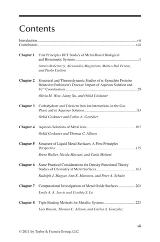
consult for a deeper discussion of this important topic.
The fundamental equation of quantum chemistry is, of course, the Schrödinger
equation
H E
Ψ Ψ
= (I.1)
where
H is the Hamiltonian operator
Ψ is the wavefunction
E is the energy
The main occupation of the quantum chemist, then, is to solve this equation for the
molecular system of interest. There is lengthy literature on the Schrödinger equa-
tion and methods for its solution. The unfamiliar reader may wish to consult an
introductory text [1–3] for a solid introduction to the theory and practice of quantum
chemistry calculations. For readers desiring a more in-depth treatment of the subject,
several advanced texts are available [4–6]. Several chapters in this book also contain
discussions of the theory.
Solution of the Schrödinger equation usually involves the Born–Oppenheimer
approximation that separates Equation I.1 into an electronic structure problem and
a dynamics problem. This approximation gives rise to the concept of the potential
energy surface in which the potential energy of a molecular system is expressed as
a function of the positions of the nuclei. The electronic structure problem is solved
under the assumption that the nuclei remain in a fixed position. The dynamics prob-
lem then considers the motion of the nuclei on the potential energy surface.
One may broadly divide the methods for solution of the electronic structure
problem into three categories: semiempirical methods, ab initio methods, and den-
sity functional theory (DFT) methods. Semiempirical methods include traditional
semiempirical methods in quantum chemistry (e.g., AM1, PM3, PM6) as well as
extended Hückel and tight-binding methods. These methods generally neglect some
terms in the Hamiltonian and approximate others to gain computational speed at the
expense of rigorous solution. The loss in accuracy is usually compensated through
parameterization to accurate data (either from experiment or higher-level theory).
Ab initio is taken to mean those methods based on the Hartree–Fock equations.
Examples include the Hartree–Fock method, Møller–Plesset perturbation theory
methods, coupled-cluster methods, and multi-reference methods [4,5]. Finally, DFT,
typically realized through the Kohn–Sham method [6], will form the basis for most
of the calculations presented in this book.
It is difficult to overstate the importance of the Kohn–Sham DFT method in the
treatment of metallic systems. There are many functionals available that permit the
efficient and accurate treatment of metallic systems. DFT methods are the driving
force behind the advances made in this area. Many chapters in this book emphasize
the importance of DFT and discuss the underlying theory. The interested reader is
directed to two recent reviews for a comprehensive review of many topics related to
the subject at this book [7,8].](https://image.slidesharecdn.com/2210950-250606103934-126f175f/85/Metallic-Systems-A-Quantum-Chemists-Perspective-Thomas-C-Allison-Ed-15-320.jpg)
![xi
Introduction
© 2011 by Taylor & Francis Group, LLC
Analogous to the importance of DFT in the solution of the electronic structure
problem, the Car–Parrinello method [9] for performing molecular dynamics calcula-
tions is one of the primary tools available to the quantum chemist. Car–Parrinello
molecular dynamics (CPMD) calculations, which are based on DFT calculations,
are efficient and accurate for a wide variety of chemical studies of metallic systems.
These methods are discussed in several chapters.
At the heart of many biological processes are molecules whose function depends
on the metal atoms they contain. Chapter 1 is concerned with understanding such
molecules with molecular detail. Topics in this chapter include first principles calcu-
lations of biomolecules and pharmaceuticals, the incorporation of solvation effects
via explicit solvent models, and studies of chemical reactivity. These topics are pre-
sented through several interesting applications.
Metal ions in solution can have significant effects on nearby molecules. In
Chapter 2, chemical dynamics calculations are used to investigate interactions
between metal ions and carbohydrates. The importance of using dynamics methods
that are based on ab initio calculations of the potential energy surface (as opposed
to classical force field methods) is emphasized. Results are presented both in the gas
phase and in solution, thus highlighting the role of the solvent.
The techniques introduced in Chapter 2 are used again in Chapter 3, this time
to explore the interactions of iron ions with α-synuclein proteins and the role that
this interaction may play in Parkinson’s disease. This chapter highlights the value
of chemical computation in obtaining molecular detail about important biological
processes that may ultimately lead to the development of more effective treatments.
Significant attention is given to classical molecular dynamics calculations, and the
value and limitations of these calculations versus their quantum counterparts are
discussed. Again, the role of the solvent is emphasized.
The discussion of metal ions in aqueous solutions, a central theme in the preced-
ing two chapters, culminates in Chapter 4. In this chapter, the effect of a metal ion
on surrounding water molecules is considered. Although on the surface this seems to
be simpler than the systems considered in the preceding chapters, the reader will dis-
cover the rich and detailed dynamics of these solutions. In particular, topics such as
the migration of a proton through aqueous solution and the formation of Zundel and
Eigen complexes are discussed. As before, the primary tool is chemical dynamics
calculations, but these are augmented with a rare event sampling technique known as
transition path sampling that is essential to the study of these systems.
The focus of the book shifts to liquid metal surfaces in Chapter 5. Such systems
have considerable industrial importance. In this chapter, dynamics simulations
based on DFT calculations are used to obtain molecular level detail of the layering
of various liquid metals. Attention is given to the comparison of simulation results
with experimental results. Liquid sodium surfaces are presented in detail with an
emphasis on the role played by geometrical confinement in the layering of these
systems.
Chapter 6 presents a wealth of practical advice on the calculation of solid metal
surfaces. This chapter contains a detailed discussion of the importance of DFT
in calculations on metals. The two main themes in this chapter are the choice of
the exchange-correlation potential and the choice of pseudopotential in these](https://image.slidesharecdn.com/2210950-250606103934-126f175f/85/Metallic-Systems-A-Quantum-Chemists-Perspective-Thomas-C-Allison-Ed-16-320.jpg)


![6 Metallic Systems: A Quantum Chemists’s Perspective
© 2011 by Taylor Francis Group, LLC
becomes a powerful predictive tool, ideal for the study of large biological systems
containing metal elements.
1.3 DISCUSSION
1.3.1
Solvent Effects on Nitrogen Fixation
Biomimetic Molybdenum Catalyst
Nitrogen fixation, i.e., the conversion of molecular nitrogen to ammonia, is a key
reaction in nature, performed by a number of different prokaryotes including bac-
teria, actinobacteria and certain types of anaerobic bacteria.24,114,115 The nitrogenase
enzyme catalyzes nitrogen fixation at room temperature through complex reactions
that are not fully understood.116 Artificial nitrogen fixation, instead, requires high
temperature and strong pressure and it is industrially achieved by the well-known
Haber–Bosch reaction.117 For years, coordination chemists have been searching for a
nonbiological catalyst that will render the nitrogen fixation under mild ambient con-
ditions feasible.118,119 Among many adopted strategies, those based on biomimetics
(i.e., compounds that perform a similar chemical reaction to that of natural enzymes)
of Fe- and Mo-containing nitrogenase enzyme seemed to be the most promising.120,121
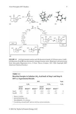
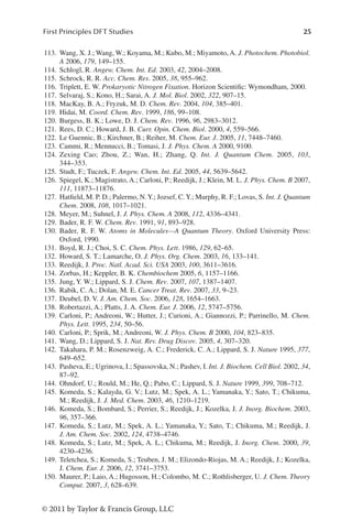
The search for the “Holy Grail of coordination chemistry”122 was over in 2002,30
when a molybdenum triamidoamine chelate complex ([Mo(hiptN3N)], where
hiptN3N=tris(hexaisopropylterphenyl)triamidoamine, Figure 1.1A and B)25–30 was
shown to catalyze the reduction of molecular nitrogen to ammonia with high effi-
ciency at room temperature. The catalytic process proceeds with the slow addi-
tion of a proton source (2,6-lutidiniumBAr4 where Ar is 3,5-(CF3)2C6H3, hereafter
referred to as LutH+ and Lut in the protonated and deprotonated forms respec-
tively, Figure 1.1C) and a reducing agent (decamethylcromocene) to a solution of
the catalyst in liquid heptane at ambient conditions (Figure 1.1D). Clearly, a full
description of the single steps of the whole process is extremely important not
only to comprehend the Yandulov and Schrock cycle in detail, but also to obtain
hints about the intricate functioning of the enzymes that naturally perform nitro-
gen fixation. While many intermediates of the cycle were experimentally char-
acterized,26,27,29 an exhaustive understanding of the molecular processes is still
lacking.24 To support the experimental research, DFT calculations, in vacuo and
corrected with implicit solvation models,123 were performed and provided prelimi-
nary insights into these reactions.122,124,125 While structural characterization of the
compounds was satisfactory, energetics of the catalytic cycle turned out to differ
remarkably from the available experimental data.26,27,125 In particular the exother-
mic character of the two protonation steps for which experimental data are avail-
able (step I=MoNNH+H+ → MoNNH2
+
and step II=MoN+H+ →MoNH+) was
overestimated (Figure 1.1D and Table 1.1), e.g., the free energy was predicted to
be too negative by these calculations.27,125 Such a discrepancy (that is well beyond
the standard DFT error, 3–5kcal mol−1)94 may be caused by many factors including
(i) the choice of the exchange and correlation functionals, (ii) the solvation model,
and (iii) the lack of a rigorous treatment of entropic effects. In this section we shall
describe an extensive study aimed at finding the reasons of the apparent failure of](https://image.slidesharecdn.com/2210950-250606103934-126f175f/85/Metallic-Systems-A-Quantum-Chemists-Perspective-Thomas-C-Allison-Ed-29-320.jpg)
![11
First Principles DFT Studies
© 2011 by Taylor Francis Group, LLC
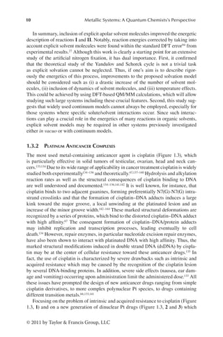
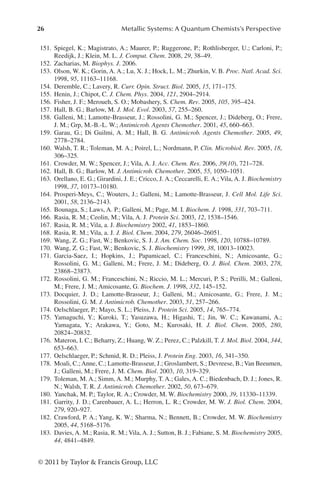
have been designed to bind DNA has led to a reduction in the structural distor-
tions induced by cisplatin, possibly reducing the risk of cross and intrinsic resis-
tance.133,145–147 Three hybrid QM/MM simulations (in which the Pt moiety and the
guanine are treated at the QM level, while the rest is treated at the MM level) were
performed: (i) starting from the x-ray structures of platinated DNA,142 (ii) the cis-
platin–DNA adduct in complex with High Mobility Group (HMG) protein,144 and
(iii) from cisplatin docked to the same oligomer in the canonical B-DNA confor-
mation.67 All simulations reproduced the relevant experimental and structural fea-
tures with good accuracy, even though the DNA duplex is rather flexible in all three
models. During the simulation, the helical parameters asymptotically approached
the values of the simulation based on the x-ray structure with the rise increasing
from 4 to 7 Å, the roll angle ranging from 28° to 61° and the global axis curva-
ture ranging from 48° to 57°.67 However, a complete structural agreement among
the three simulations is prevented by the puckering of the sugars: conformational
NH3
NH3
NH3
NH3
NH3
NH3
G G
G
G
DNA DNA
B: 3-DNA
C: 3'-DNA
NH3
NH3
NH3
NH3
CI
CI
Pt
Pt
G
G
Pt
Pt
Pt
Pt1 Pt1
Pt2
Pt2
Pt
H3N
DNA 1-DNA
2+
2+
1
2 3
H3N
N2–N1
N2–N1
O
H O
H
H3N
H3N
H3N
H3N
H3N
H3N
H3N
N3
N3
N2
N2
N1
N1
H3N
N3
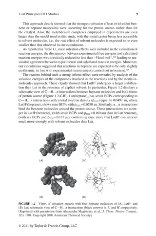
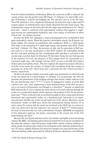
FIGURE 1.3 Cisplatin (1) and its adduct with dsDNA (1-DNA). The azole-bridged diplati-
num compounds [Pt2(í-OH)(í-pz)]2+ (2), [Pt2(í-OH)(í-1,2,3ta)]2+ (3), and the atom numbering
in the rings. Two different binding modes of 3 to DNA yield to adducts B and C. The 2-DNA
adduct (A) is equivalent to B. (Reprinted with permission from Katrin Spiegel et al., J. Phys.
Chem. B, 111(41), 11873. Copyright Oct 2007 American Chemical Society.)](https://image.slidesharecdn.com/2210950-250606103934-126f175f/85/Metallic-Systems-A-Quantum-Chemists-Perspective-Thomas-C-Allison-Ed-34-320.jpg)
2 (pz=Pyrazolate) (1) and
[{cis-Pt(NH3)2}2(μ-OH)(μ-1,2,3-ta-N1,N2)](NO3)2 (ta=1,2,3 Triazolate) (2)
Bound to 5′-d(CpTpCpTpG*pG*pTpCpTpCp)-3′, Resulting in Complexes A, B,
and C (Figure 1.3), Compared to NMR structure,149 and Reference
Simulation of the Unbound Decamer with the Same Sequence (DNA MD)
A B C A B C
QM/MM QM/MM QM/MM CMD CMD CMD DNA MD NMR
Rise 3.6±0.2 3.6±0.2 4.1±0.3 3.5±0.3 3.4±0.3 4.1±0.5 3.4±0.2 3.3
Roll 9±4 4±4 −5±5 6±7 6±5 −14±9 −3±7 5
Global axis
curvature
19±5 10±3 8±4 18±9 18±3 14±7 19±8 5
Rise, major and minor groove width (W) and depth (D) are given in [Å], Roll and global axis curvature
are in degrees. The minor and major groove parameters refer to the largest value measured at the plati-
nated site (G–G step). Structural parameters resulting from QM/MM MD and classical MD are labeled
as QM/MM and CMD, respectively.](https://image.slidesharecdn.com/2210950-250606103934-126f175f/85/Metallic-Systems-A-Quantum-Chemists-Perspective-Thomas-C-Allison-Ed-35-320.jpg)


![[48]
“What?”
“If we did collect this money we could donate it to the
college to have the swimming pool repaired.”
“That’s sweet of you and a good idea, Arden, but I don’t
believe we could do it,” objected Sim. “Besides, I don’t
exactly believe what it says on this poster. It seems very
silly for a young fellow to disappear just when he’s
coming into a lot of money—a fortune.”
“Perhaps he was made to disappear,” suggested Terry,
her eyes opening wide.
“Oh! You mean—kidnaped?” asked Arden.
“Yes.”
“Worse and more of it!” laughed Sim.
“Well, anyhow, we could try, couldn’t we?” Arden asked.
“You’d help, wouldn’t you, Terry?”
“Yes, indeed I’ll help. I’ve always fancied myself in the
rôle of a detective, spouting pithy Chinese philosophy
and generally getting underfoot.”
“Now, Terry, just be serious for once. And Sim, you also.
You know how disappointed you were when you found
out the swimming pool was——”
“Kapoot!” chuckled Sim, supplying Arden’s evident lack
of a word with the latest Russian expression. “Go on!”
“Well,” resumed Arden, pouting a little, “you never can
tell. Maybe we could do it. It isn’t impossible. Stranger
things have happened. And I just know I’ve seen that](https://image.slidesharecdn.com/2210950-250606103934-126f175f/85/Metallic-Systems-A-Quantum-Chemists-Perspective-Thomas-C-Allison-Ed-56-320.jpg)
![[49]
young man on the poster somewhere before. If I could
only remember where! Did either of you ever have that
feeling?”
“Lots of times. I’m for you, Arden!” declared Sim. “I’ll do
what I can and whatever you say. This mysterious Harry
Pangborn may very well be right around here.”
“Around Cedar Ridge!” shrilled Terry.
“Certainly! Why not? If the authorities didn’t think it
likely that he might be in this vicinity, why did they put
the poster up here in the post office? And they
mentioned Morrisville,” challenged Sim.
“There’s something in that,” Terry admitted.
“Oh, if he should be in hiding around here and we could
find him and claim the thousand dollars reward,”
breathed Arden, “wouldn’t it be just wonderful! And
what a sensation when we magnanimously turned the
money over to the college for the swimming pool. Oh,
oh!”
“Would you do that for dear old Alma Mater when you
don’t know her so very well?” asked Sim, who, with her
chums, was still gazing at the poster of the good-
looking but missing heir of the Pangborn estate of
millions.
“I’d do it for you, Sim, dear,” murmured Arden. “I want
you to be happy here, since I teased you so to come.”
“And you think I won’t be happy without the swimming
pool?”
“Will you?”](https://image.slidesharecdn.com/2210950-250606103934-126f175f/85/Metallic-Systems-A-Quantum-Chemists-Perspective-Thomas-C-Allison-Ed-57-320.jpg)
![[50]
“Not as happy as I would be with it.”
“But even admitting that this missing young man may
be around here,” suggested Terry, “what chance have
we of finding him? We have so much college work to
do. For, after all, we were sent here to learn something,”
she sighed.
“Granted,” laughed Arden. “But we may find time for a
little detective work on the side as well as for hazing.
Oh, it’s a wonderful prospect!” She swung around in a
few dance steps right there in the old post office.
“Well, we’d better be getting back,” suggested Sim after
this. “Oh, look at the clock!” she gasped. Then followed
a hurried sending of some picture postcards they had
bought; cards on which they marked with an X the
location of their room.
The three chums were bubbling with life, laughter, and
merriment as they turned to leave the little building, but
their mirth was turned to alarm as a stern voice assailed
them.
“Young ladies!”
They looked around to see Rev. Dr. Henry Bordmust
sternly regarding them from the doorway.
“Yes, Dr. Bordmust,” Sim almost whispered as the
chaplain appeared to be waiting for formal recognition.
“You are freshmen!” he accused, with a glance at their
mortarboards, the tassels of which told the tale. “You
know you are not permitted over here—in the post
office. It is against the college rules—for you freshmen.
Return at once! You must! You must!”](https://image.slidesharecdn.com/2210950-250606103934-126f175f/85/Metallic-Systems-A-Quantum-Chemists-Perspective-Thomas-C-Allison-Ed-58-320.jpg)
![[51]
[52]
He appeared strangely stirred and angry, and his dark
brows, shading his bright little eyes, bent into a frown.
But somehow, after that first booming and accusative
“young ladies,” the chaplain seemed exhausted, as
though the anger pent up in him had taken something
from his none too profuse vitality. He was an old man.
Now he essayed a wintry smile and added, as he gently
waved them out with motions of his thin white hands:
“That is to say, you shouldn’t have come here. You—er
—have no need to be—er—frightened at this first
infraction of the rules, but—er—another time you may
be—er—campused for such action.”
Then, having seen that the three were on their way out,
Dr. Bordmust turned to the window, evidently to buy
some stamps for the letters he held in one hand. He
murmured to himself in those queer, quavering,
meaningless tones:
“Too bad; too bad! I can’t always be watching! Dear
me!”
Wonderingly, Arden and her chums looked at the
shrinking figure in black as they passed out of the door.
But Dr. Bordmust gave them no further attention.](https://image.slidesharecdn.com/2210950-250606103934-126f175f/85/Metallic-Systems-A-Quantum-Chemists-Perspective-Thomas-C-Allison-Ed-59-320.jpg)
![[53]
[54]
“Well, don’t ask me what it all means,” begged Terry. “I
can’t even guess. But, oh! I do hope I’m not going to be
late for this first class.”
“So say we all of us,” chanted Sim.
“They can’t be too severe at the very beginning,”
murmured Arden.
Bordmust Hall, where most of the class sessions were
held, crowned with its classic architecture the summit of
the long slope which formed the eminence of the broad
acres about Cedar Ridge College. It was behind the
main, or dormitory, building in which were housed the
executive offices and the residence rooms of the faculty.
To the southwest of the hall, and easily viewed from the
steps, was the unused pool. To the northwest, and in
line with the main building, was the beautiful Gothic
chapel with its wonderful stained-glass windows. Near
the chapel was the unimposing home of the chaplain,
Rev. Dr. Bordmust; one of whose ancestors had partly
endowed Cedar Ridge. For this reason the hall was
named for him.
At the foot of the slope on which the hall stood were the
rambling fields and gardens where much of the farm
produce for the college tables was raised. The nearest
of the farm-lands, so called, was the orchard, part of
which could be seen from the southeast windows of the
dormitory. And it was this orchard that the taxi-man had
indicated in such a warning manner. It was this orchard
into which Tom Scott, the good-looking porter, had been
staring the night of the arrival of Arden Blake and her
chums. So much had been crowded into the
comparatively short time the three freshmen had been
at college that they had almost forgotten the strange](https://image.slidesharecdn.com/2210950-250606103934-126f175f/85/Metallic-Systems-A-Quantum-Chemists-Perspective-Thomas-C-Allison-Ed-61-320.jpg)
![[55]
orchard. Even now they had no chance to consider the
matter, for they, with many other girls, were hastening
to their first classes.
They gave a momentary glance toward the orchard,
with its quaint gnarled trees. The morning sun was
glinting on red, dark-green, and golden russet apples
which the gardener and his men had not yet started to
gather.
Arden, especially, gazed searchingly at the orchard.
Apple trees grow in such strange shapes and huddle so
closely to themselves, as if each one guarded a secret.
There was a puzzled look in Arden’s blue eyes as she
tried to guess what might be hidden by those trees and
the tall hedge surrounding them.
Sim was gazing rather sorrowfully at the pool building,
but Terry was smiling, perhaps because everything
seemed, for the moment, at least, to be so filled with
good and pleasant life.
“Go on in, kids!” Sim urged her two chums. “I’ll be
along in a minute or two. I just want to take a look at—I
just want to—oh, well, go on. Don’t wait for me.”
“But won’t you be late?” objected Arden.
“No, I have some time to my credit.”
As her surprised friends watched, Sim left them and
hurried down across a stretch of smooth lawn toward
the disused swimming pool.
“Too bad,” murmured Arden.
“What is?” asked Terry.](https://image.slidesharecdn.com/2210950-250606103934-126f175f/85/Metallic-Systems-A-Quantum-Chemists-Perspective-Thomas-C-Allison-Ed-62-320.jpg)
![[56]
“I really think Sim feels more keenly than we realize
about the pool. But she’s such a good sport. Look at
her! Going to view the ashes of her hopes or the
collapse of her dreams or something equally tragic.”
“Don’t let’s say anything about this,” proposed Terry. “If
Sim cares so much, I’m sure she’d rather not talk about
this little visit.”
Arden agreed and, taking Terry’s arm, they hurried into
the hall.
Sim reached the pool building and tried to get some
idea of the wreck within by peering through a window.
But the sill was too high to afford a view, even if the
window had not been made of heavily frosted glass,
quite opaque.
Then she stepped back and gazed up at the copper and
glass domed roof. Around the top of the building were
set at intervals glazed tiles depicting nautical scenes.
Dolphins were diving merrily as if to tantalize sea horses
with necks proudly arched, and mermaids flicked their
tails disdainfully at Father Neptune.
“I may as well try the door,” Sim murmured. “I’d like to
see what it’s like inside, though it will probably break my
heart!”
After several hard pushes to the extent of her strength,
she succeeded in swinging back the door. She found
herself in a sort of vestibule, but the inner door of this
opened easily, and then Sim stood almost on the edge
of the abandoned pool.
A peculiar smell assailed her, as of a place long shut up,
but at the same time it had something of out-of-doors](https://image.slidesharecdn.com/2210950-250606103934-126f175f/85/Metallic-Systems-A-Quantum-Chemists-Perspective-Thomas-C-Allison-Ed-63-320.jpg)
![[57]
about it, the odor of clean earth and ripe vegetables.
“It isn’t as bad as Toots said,” mused Sim. “At least, it
looks as though there isn’t so very much the matter. It
isn’t filled with vegetables, either; just a few bags as
yet, though they probably will bring in more when they
pick the apples. This must have been a beautiful pool
once.”
The bottom of the pool was tiled a pea green, a color
which must have given the water a most cooling tone
on a hot day. But the white tile sides no longer
gleamed, and in more than one place jagged dark
cracks ran crazily down the walls like streaks of black
lightning. Sim looked at the cracked tile and concrete
edge at her feet. The depth was still indicated, though
there was no water in the pool—5 feet.
“This is the shallow end, of course,” Sim thought, and
she walked slowly around the edge and toward the
melancholy spring-boards to which some strips of
cocoa-fiber matting still clung.
“How quiet it is in here,” Sim murmured. “Like a
museum after hours—or an Egyptian tomb.” She
shivered a little, though it was warm in the natatorium.
In the deep end several filled burlap bags were piled up,
and in each corner were barrels of cabbages leaning
against the walls.
“I thought, from what Toots said, the whole place would
be filled to the brim with cabbages and turnips,” Sim
said to herself, smiling a little ruefully. “I wonder how
long this pool is, or should I say was?”](https://image.slidesharecdn.com/2210950-250606103934-126f175f/85/Metallic-Systems-A-Quantum-Chemists-Perspective-Thomas-C-Allison-Ed-64-320.jpg)
![[58]
She began to measure the length with her eyes,
mentally swimming with long, smooth strokes while her
feet churned up and down.
“About seventy-five yards long, I guess,” she went on.
“And about twenty-five across. A lovely size. I could do
three lengths a day here and really enjoy it. Let’s see
how deep it is from the end of the board.”
She walked gingerly out on the diving plank, choosing
the center one for there were three at the deep end,
tiered at different heights. It was difficult to estimate,
without water in the pool and with the barrels and bags
of vegetables scattered about, how close the different
boards came to the surface of the filled space. Sim
decided that the plank she was standing on was the
lowest.
She permitted herself a little pre-diving, teetery bounce
on the very end, half fearful lest the dried wood should
crack beneath even her light weight. But it held, and
Sim gave a bolder jump.
“A straight dive—cutting the water about there!” With
her eyes Sim indicated to herself just the spot where
her finger tips should enter the water—had there been
any water there.
She jumped again and came down safely, with no
warning cracking of the dried plank. Then she balanced
herself on the very tip of the board before, mentally,
springing into the air. Now she performed a most
ambitious jump, but this time the stiffened wood
snapped back suddenly. Sim was thrown to one side,
and she swung her arms around and around like a child](https://image.slidesharecdn.com/2210950-250606103934-126f175f/85/Metallic-Systems-A-Quantum-Chemists-Perspective-Thomas-C-Allison-Ed-65-320.jpg)
![[59]
on its first roller skates, trying desperately not to topple
backward.
But her motions only caused the board to quiver more
violently, and in a split second Sim slipped off and clung,
with her finger tips only, to the edge of the plank, while
the hard-tiled bottom of the pool, seemingly miles
below, waited to receive her.
“Oh, gosh! What’ll I do?” poor Sim thought. “Those tiles
don’t look very soft, and I’ll drop in a minute!”
Her fingers ached from their stiff clinging grip, and her
arms were quickly tiring. She decided she must soon let
go for after a futile attempt to sling one leg up over the
side edge of the board it bent so alarmingly that she
feared it would snap. She began to swing to and fro like
a pendulum, hoping she might cast herself upon a bag
of vegetables which would serve to break her fall, when,
suddenly, she felt her wrists firmly gripped by two
hands, and she looked up to see Tom Scott, the porter-
gardener, smiling down at her. He was kneeling on the
end of the plank.
“Don’t jump!” he warned. “I’ll pull you up. It’s rather the
reverse of ‘don’t shoot, I’ll come down,’ isn’t it?” he said
lightly. He could not have taken better means to quiet
Sim’s excited nerves than with Mr. Crockett’s little coon
banter.
With what seemed no effort at all, Tom Scott lifted her
up and held her clear of the end of the board so her
legs did not scrape against it. Then he carefully walked
back with her toward the middle of the plank, where
there was no danger of its breaking, set her down, and](https://image.slidesharecdn.com/2210950-250606103934-126f175f/85/Metallic-Systems-A-Quantum-Chemists-Perspective-Thomas-C-Allison-Ed-66-320.jpg)
![[60]
stood grinning at her. A nice grin it was, too, Sim
thought later.
She managed to produce a weak, embarrassed smile.
“Thank you so much!” she said a bit stiffly. The man
must think her crazy. “I—I slipped! I—er—I was—that is,
I was trying——” To cover her confusion she looked at
her red finger tips.
“Hurt?” he inquired.
“Broke two or three nails,” Sim responded ruefully. “I’m
very glad you came along. I might have sprained an
ankle if I had let go, for this end must be nine feet
deep.”
“The water, when there is any, is over nine feet deep
nearest this wall,” said Tom Scott. “You certainly would
have been jarred a bit, to say the least.”
“Then I must thank you again. But please don’t mention
to anyone that you found me in such a silly fix, will
you?” Sim begged. She was quickly regaining her lost
composure. “I just wanted to get a look at the pool and
foolishly walked out on the board. I imagined myself
poising for a dive and I slipped off. You won’t tell?”
“Of course I won’t,” Tom agreed, somewhat gayly, it
seemed. “I came in to get a few of the early apples we
have stored here. One of the cooks asked me to. I
imagine there are going to be pies. But, honestly, I
won’t tell a soul.”
“Thank you,” Sim murmured.](https://image.slidesharecdn.com/2210950-250606103934-126f175f/85/Metallic-Systems-A-Quantum-Chemists-Perspective-Thomas-C-Allison-Ed-67-320.jpg)
![[61]
[62]
The young gardener walked up to the middle of the pool
and with athletic ease jumped down in it near several
bags of vegetables. He picked up one containing apples,
heaved it up on the edge and jumped up himself. Then,
slinging the sack up on his shoulder, he walked toward
the door, giving Sim a friendly backward glance as he
went out.
“What a nice young man!” said Sim to herself. “He
doesn’t seem like a gardener at all. No brogue and no
accent of any kind. I wish I could tell Arden and Terry,
but I’d rather die than have them know of this dizzy
adventure. I must have looked perfectly stupid hanging
there on the end of the plank!”
The clanging of a distant bell brought Sim back to
reality, and as she looked at her wrist watch she left all
thoughts of pools and good-looking rescuing gardeners
behind her. For it would need a swift dash to get her to
Bordmust Hall before she would be late for her class.](https://image.slidesharecdn.com/2210950-250606103934-126f175f/85/Metallic-Systems-A-Quantum-Chemists-Perspective-Thomas-C-Allison-Ed-68-320.jpg)
![[63]
[64]
The three found they were to be separated for the
morning session though the first class in the afternoon
would find them in the same room for English literature.
“And we must try to sit together,” called Arden to Sim
and Terry as they parted.
Inside the hall all was confusion. Girls were running
hither and yon. Stairways were crowded with students
going up or coming down, and all were excited. Doors
were suddenly pushed open by uncertain freshmen and
again by oversure sophomores. The latter, in a spirit of
fun, several times sent a poor “frosh” up to the top floor
when she should have remained on the first.
Another warning bell rang and, almost at once, the
corridors were empty and quiet. Inside their classrooms
the three girls from 513 looked, listened, and answered
somewhat in a daze. That first day always remained
more or less of a hazy recollection. Something of an
organization was arranged, the roll was checked and
corrected, names were asked and given, everyone was
on edge and nervous, even the instructors. Strange
faces, many of them timid, looked on other strange
faces, also somewhat timid.
Then came welcome noon, and the rush out of
Bordmust and some of the other study buildings to the
dining hall was comparable only to the New York
subway rush at five o’clock.
The afternoon classes were attended by all more
pleasantly and with less strain. To their delight, Arden,
Sim, and Terry managed to get into the same room and
sat near one another.](https://image.slidesharecdn.com/2210950-250606103934-126f175f/85/Metallic-Systems-A-Quantum-Chemists-Perspective-Thomas-C-Allison-Ed-70-320.jpg)
![[65]
As they were leaving Bordmust Hall, at the close of the
afternoon session, Arden heard someone say:
“Here come our three!”
Toots Everett, Jessica, and Pip were regarding the other
trio with sardonic smiles and, as Terry said later, “with
murder in their eyes.”
“Good afternoon, freshies! How about a little song for
my friends, here?” Jessica was mockingly speaking. “A
song befitting your talents. Arden Blake, come here!”
Arden stepped forward, blushing. “I can’t sing,” she
quavered.
“You shall learn. Your friend here, with the red hair,
looks like a singer. And while you two sing, Sim
Westover shall dance. On with the dance, freshies!”
The trio from 513 looked at one another in dismay, but
there was no help for it. Amused seniors and juniors
had gathered to see the fun. From the classmates of
Arden and her chums two kinds of advice was
forthcoming, the “don’t-you-do-it!” and “go-on-be-
sports!”
Finally, in a weak and uncertain voice, Arden and Terry,
after a moment of embarrassed consultation, sang one
verse from their prep-school song; something about
“Bring Me Violets for My Hair,” while Sim tapped about
more like a sparrow than a swan.
At last it was over.
“Not bad,” commented Toots.](https://image.slidesharecdn.com/2210950-250606103934-126f175f/85/Metallic-Systems-A-Quantum-Chemists-Perspective-Thomas-C-Allison-Ed-71-320.jpg)
![[66]
“I’ve seen worse,” said Pip.
“But not much,” was Jessica’s opinion.
Then the sophomores delivered a rhyming ultimatum.
They stood with their heads together and chanted:
“From yonder orchard, old and green,
Where, ’tis said, strange things are seen,
You three, upon this fatal day,
Must gather apples while ye may.
At once repair to that dread spot,
And in your quest dare pass it not.
Then bring, for our symbolic use,
Fair apples with but smallest bruise.
Ten perfect fruits, no less, must we
Your mentors have, in time for tea.”
There was a dramatic pause, following this delivery, and
then, as though they had rehearsed it, as, indeed, they
had, the three sophomores picked up the books they
had deposited on the ground in front of them while
singing, and marched away, leaving the trio from 513
the center of an excited and thrilled group.
“What does it all mean?” asked Sim.
“Is it part of the hazing?” asked Terry.
“Must we really go after the apples?” asked Arden in
astonishment.
“Yes,” said Mary Todd. “It’s just part of college life. And
you may as well go to the orchard now, while it is still
light and bright. I certainly hope I don’t have to do that
stunt. No orchard in mine.”](https://image.slidesharecdn.com/2210950-250606103934-126f175f/85/Metallic-Systems-A-Quantum-Chemists-Perspective-Thomas-C-Allison-Ed-72-320.jpg)
![[67]
“Some of us probably will have to gather the apples
later,” declared Jane Randall. “But a soph, who got a
little friendly with me, said that the best apples were at
the far side of the orchard. So you girls had better go
there at the start, as Toots and her crowd won’t accept
nubbins, and you don’t want to have to make two trips.”
“I should say not,” murmured Sim. “One is bad enough.”
Arden and Terry were still a bit bewildered, even after
this well-meant advice, and Sim declared she was
“dying from embarrassment.”
“I suppose we may as well go. What do you say, girls?”
asked Arden.
“Yes, let’s! Anything to get away from here!” Sim was
regarding the circle of amused girls.
“You take our books to our room, will you?” Terry asked
Mary Todd. “We’ll let you know later how we make out.”
The fated trio started down the southern slope of
Bordmust Hall hill toward the picturesque orchard
where, even now, though it was not very late, the
shadows were lengthening and the sun had lost some of
its brightness. They crossed a field, deep with grass,
crawled through the bars of a snake-rail fence, and
found themselves beneath the trees.
“I vote we pick up the first apples we can see,” voiced
Terry.
“Certainly!” agreed Arden.
“Apples are apples,” quoth Sim. “Why should we go to
the far end to gather fine fruit when windfalls may](https://image.slidesharecdn.com/2210950-250606103934-126f175f/85/Metallic-Systems-A-Quantum-Chemists-Perspective-Thomas-C-Allison-Ed-73-320.jpg)
![[68]
answer?”
“Why, indeed,” assented Arden. “But still I suppose we
had better not pick up these.” With her foot she kicked
out from amid the fallen leaves some withered,
wrinkled, and partly rotted specimens.
“No, they won’t do,” declared Sim.
“Then let’s separate a bit. We can cover more ground
that way,” suggested Arden. “Whoever first finds some
decent apples must give a shout, and we’ll gather
there.” She was quite businesslike.
“All right, Colonel!” laughed Terry. “‘You take the
highland and I’ll take the low,’” she sang softly. “Scatter,
my lassies!”
They separated and began the search in the growing
dusk.
Apples there were, but such poor things, windfalls and
rots, that even the enthusiastic Arden began to feel
discouraged. They might, after all, need to go to the far
end of the orchard. Still, it was delightful beneath the
old, gnarled trees. Their trunks were shaped like
dragons, their branches like Chinese letters, and the
roots, where they cropped out above the ground, like
intertwined serpents grim and black, seeming to writhe
in the shifting shadows. A little wind rustled the leaves,
swung the hanging fruit, and made the limbs squeak as
they rubbed one on the other.
Here and there they wandered, growing more and more
apprehensive and nervous as the darkness deepened.
There seemed to be something sinister about that
orchard, although it was so close to the life and joy of](https://image.slidesharecdn.com/2210950-250606103934-126f175f/85/Metallic-Systems-A-Quantum-Chemists-Perspective-Thomas-C-Allison-Ed-74-320.jpg)
![[69]
Cedar Ridge College. The taxi-man had surely warned
them—but of what? This was no time to think about
that.
“Ah!” Sim suddenly exclaimed. “A perfect apple, red and
round!” She picked it up from beneath a large gnarled
tree. “And there are others,” she called. “This way! Over
here, girls!” Her voice was joyous.
Arden and Terry ran toward Sim. But as Sim stooped to
pick up another apple she saw something in a pile of
leaves. It looked like—surely not the leg of blue overalls!
A last lingering gleam of the setting sun, shining
through a cleft in the hills, glinted upon that leg. Sim
glided closer. Could it be——?
It was part of an overall suit, and there, thrust out of
the lower end and twisted grotesquely to one side, was
a foot!
“Oh-h-h-h-ee!” screamed Sim, dropping her apples.
“Oh, girls, look here! Quick! Hurry!”
She stood in a panic of terror, rooted as firmly to the
spot, for the moment, as one of the black gnarled trees.
“What is it, Sim? What’s the matter?” gasped Terry, the
first to arrive.
“Look!” Sim pointed, breathless. She and the others, for
Arden was now one of the trio beneath the tree, saw
more than just the overall leg and the foot. They saw
the huddled form of a man partly buried in the fallen
leaves. And they could see—his face!
“Why, it’s Tom—the porter!” cried Arden. Instantly she
was down on her knees beside him. “His head is cut.](https://image.slidesharecdn.com/2210950-250606103934-126f175f/85/Metallic-Systems-A-Quantum-Chemists-Perspective-Thomas-C-Allison-Ed-75-320.jpg)


![[S.L._Kakani]_Material_Science_(New_Age_Pub.,_2006(BookSee.org).pdf](https://cdn.slidesharecdn.com/ss_thumbnails/s-230301071329-4f25d7e9-thumbnail.jpg?width=640&height=640&fit=bounds)


