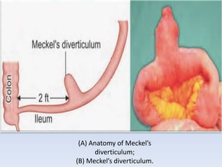
MECKEL’S DIVERTICULUM
• It is a congenital diverticulum arising from the
terminal ileum and is part of the unobliterated
proximal portion of the vitellointestinal duct.
It is:
• ™
. 2% common.
• ™
. 2 feet from the ileocaecal valve.
• ™
. 2 inch in length.
• ™
. 2% of Meckel’s diverticulum only will be
symptomatic.
• ™
. 50% of symptomatic are below 2 years of age.
• ™
. 20% heterotopic epithelium.
• ™
. 2:1 female preponderance is seen.
• It is congenital, results from incomplete
closure of vitellointestinal duct.
• It is the most common congenital anomaly of
small intestine.
• Arises from the antimesenteric border of the
ileum, containing all three layers of the bowel
with independent blood supply.
• In 20% of cases mucosa contains heterotopic
epithelium like gastric (commonest—50%),
colonic and pancreatic tissues (5%).
• It may be connected to or communicated with
the umbilicus through a band or fistula.
• It may be associated with oesophageal atresia,
exomphalos and anorectal malformations.
(A) Anatomy of Meckel’s
diverticulum;
(B) Meckel’s diverticulum.
Presentations in Meckel’s Diverticulum
• Asymptomatic—in majority cases.
• Severe haemorrhage most common, seen in children
aged 2 year or younger (Maroon-coloured blood).
• Intestinal obstruction due to bands/adhesions/
intussusception.
• Perforation.
• Intussusception, volvulus of small bowel.
• Peptic ulceration.
• Diverticulitis (20%)—features mimic acute appendicitis.
• Littre‘s hernia—it is presence of Meckel’s diverticulum
in hernial sac as content.
• It is observed in inguinal/femoral hernia
Diagnosis
• Technetium (Tc99) radioisotope scan is very useful (90–95%
accuracy). 90% of heterotrophic gastric mucosa can be
identified in Meckel’s diverticulum by radioisotope study. It
can detect
• Meckel’s diverticulum with minimal bleeding also (0.1 ml/
minute). So it is very useful investigation in children
presenting with bleeding.
• X-ray abdomen to see complications like obstruction,
perforation.
• Laparoscopy is very useful.
• Enteroclysis/small bowel enema under fluoroscopy may
show the
• Meckel’s diverticulum. It is probably the most accurate
investigation.
Treatment
• Asymptomatic Meckel’s diverticulum can be
left alone when identified during laparotomy.
• Resection of a short segment of ileum
containing Meckel’s diverticulum and end-to-
end anastomosis is done.
• Meckelian diverticulectomy with closure of
enterotomy also can be done, but chances of
retaining heterotopic tissues and stenosis are
higher.
THANK YOU

MECKEL’S DIVERTICULUM.pptx

  • 1.
  • 2.
    • It isa congenital diverticulum arising from the terminal ileum and is part of the unobliterated proximal portion of the vitellointestinal duct.
  • 3.
    It is: • ™ .2% common. • ™ . 2 feet from the ileocaecal valve. • ™ . 2 inch in length. • ™ . 2% of Meckel’s diverticulum only will be symptomatic. • ™ . 50% of symptomatic are below 2 years of age. • ™ . 20% heterotopic epithelium. • ™ . 2:1 female preponderance is seen.
  • 4.
    • It iscongenital, results from incomplete closure of vitellointestinal duct. • It is the most common congenital anomaly of small intestine. • Arises from the antimesenteric border of the ileum, containing all three layers of the bowel with independent blood supply.
  • 5.
    • In 20%of cases mucosa contains heterotopic epithelium like gastric (commonest—50%), colonic and pancreatic tissues (5%). • It may be connected to or communicated with the umbilicus through a band or fistula. • It may be associated with oesophageal atresia, exomphalos and anorectal malformations.
  • 6.
    (A) Anatomy ofMeckel’s diverticulum; (B) Meckel’s diverticulum.
  • 7.
    Presentations in Meckel’sDiverticulum • Asymptomatic—in majority cases. • Severe haemorrhage most common, seen in children aged 2 year or younger (Maroon-coloured blood). • Intestinal obstruction due to bands/adhesions/ intussusception. • Perforation. • Intussusception, volvulus of small bowel. • Peptic ulceration. • Diverticulitis (20%)—features mimic acute appendicitis. • Littre‘s hernia—it is presence of Meckel’s diverticulum in hernial sac as content. • It is observed in inguinal/femoral hernia
  • 8.
    Diagnosis • Technetium (Tc99)radioisotope scan is very useful (90–95% accuracy). 90% of heterotrophic gastric mucosa can be identified in Meckel’s diverticulum by radioisotope study. It can detect • Meckel’s diverticulum with minimal bleeding also (0.1 ml/ minute). So it is very useful investigation in children presenting with bleeding. • X-ray abdomen to see complications like obstruction, perforation. • Laparoscopy is very useful. • Enteroclysis/small bowel enema under fluoroscopy may show the • Meckel’s diverticulum. It is probably the most accurate investigation.
  • 9.
    Treatment • Asymptomatic Meckel’sdiverticulum can be left alone when identified during laparotomy. • Resection of a short segment of ileum containing Meckel’s diverticulum and end-to- end anastomosis is done. • Meckelian diverticulectomy with closure of enterotomy also can be done, but chances of retaining heterotopic tissues and stenosis are higher.
  • 10.