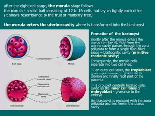
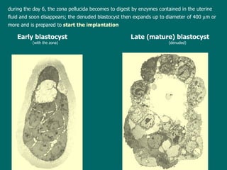
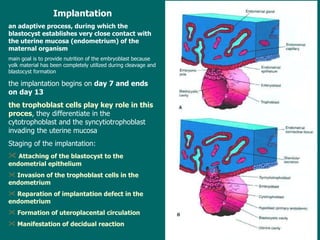
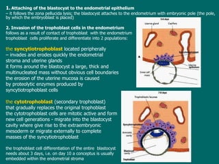
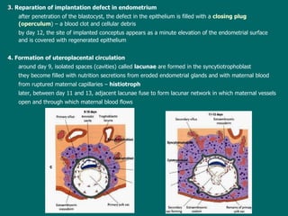
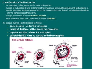
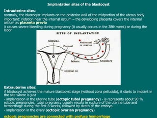
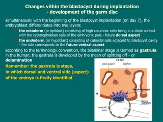
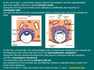
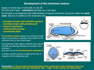
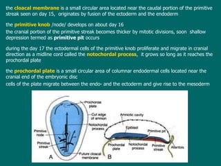
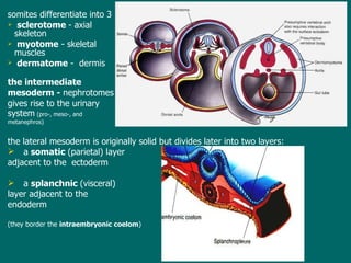
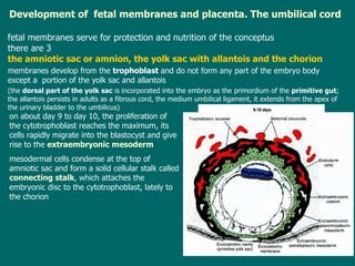
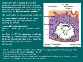
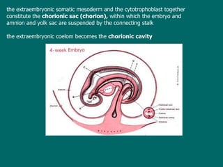
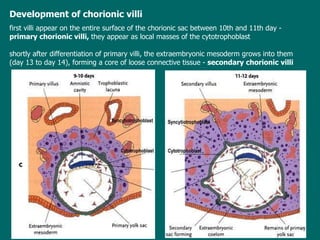
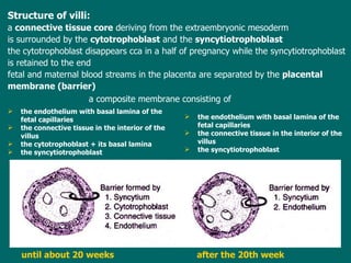
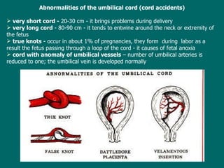
Lecture 4   ESS_2nd semester Outline of implantation  of the blastocyst . Abnormal sites of implantation (extrauterine pregnancies) D evelopment of the germ disc:  O rigin of the intraembryonic mesoderm . N otogenesis  -  development of the chorda dorsalis Somites and their derivatives Outline of development of fetal membranes:  The a mniotic sac,  the yolk sac and  chorion . P lacenta  - structure and f unction .  Anomalies of the placenta  The umbilical cord.  Anomalies of the  umbilical cord
Results of fertilization : restoration of diploid number of chromosomes (half from the mother and half from the father) determination of the sex of the new individual  (XX -female, XY-male) initiation of cleavage  (not  fertiliz ed  oocyte degenerates  during  12- 24 hours after ovulation )
Cleavage of the zygote the phase of development  characterized by  mitotic division s  of the zygote  resulting in  formation  of  the blastocyst cells are  called  blastomeres  and become smaller with each division the  cleavage  takes place  partly in the  uterine tube  (during the first three days), partly in the  uterus  (from day 4 to 6) the  first division  resulting in  2-cell stage  needs  about 24 to 30 hours , s ubsequent divisions follow in shorter intervals  because  blastomeres become progressively smaller
TEM : SEM:
af ter  the eight-cell stage,  the  morula  stage follows the morula -  a solid ball  consisting of  12 to 16 cells  that lay on tightly each other (it shows resemblance to the fruit of mulberry tree) the morula enters the uterine cavity  where  is  transform ed  into the blastocyst Formation of  the b lastocyst shortly after the morula   enters the uterus (on day 4), fluid from the uterine cavity passes through the zona pellucida to form  a single  fluid-filled  space  – blastocystic cavity  ( primitive chorionic cavity ) Consequently, the morula cells separate into two cell lines: an outer cell layer, the  trophoblast   ( g reek trophé  =  nutrition)  - gives rise to chorion and finally fetal part of the placenta a group of centrally located cells, called as the  inner cell mass  or  embryoblast  - gives rise to the embryo t he blastocyst is en closed  with the zona pellucida and lies free in the uterus cavity
Early blastocyst      Late (mature) blastocyst (with the zona) (denuded) d uring the day 6 ,  the zona pellucida becomes to digest by enzymes  contained in the uterine  fluid  and soon disappears ; t he denuded blastocyst then expands up to diameter of 400   m  or  more  and is prepared to  start the implantation
Implantation an adaptive process, during which the blastocyst establishes very close contact with  the  uterine mucosa  ( endometrium )   of the  maternal organism main goal  is to  provid e  nutrition of the embryoblast because yolk material has been completely utilized during cleavage and blastocyst formation t he implantation  begins  on  day 7 and ends on day 13 the  trophoblast cells play  key role in  this proces , they differentiate in the  cytotrophoblast  and the  syncytiotrophoblast invading the uterine mucosa Staging of the implantation: Attaching of the blastocyst to the endometrial epithelium  Invasion of the trophoblast cells in the endometrium Reparation of implantation defect in  the  endometrium Formation of uteroplacental circulation Manifestation of decidual reaction
1.   Attaching of the blastocyst to the endometrial epithelium   –  it follows the zona pellucida lysis; the blastocyst attaches to the endometrium with embryonic pole (the pole ,   by which  the embryoblast is placed) 2. Invasion of the trophoblast cells in the endometrium follows as a result of  contact  of trophoblast  with the endometrium trophoblast  cells proliferate and differentiate into 2  populations : the  syncytiotrophoblast  located peripherally   –  invades  and erodes  quick ly  the endometrial  stroma and uterine glands  it  forms  around the blastocyst  a large, thick and  multinucleated mass without obvious cell boundaries t he erosion of the uterine mucosa  is caused  by  proteolytic enzymes produced by  syncytiotrophoblast  cells the  cytotrophoblast  (secondary trophoblast)  that gradually replaces the original trophoblast t he  cyto trophoblast cells are mitotic active and form  new cell generations  -  migrate into the blastocyst cavity where give rise to the extraembryonic  mesoderm  or migrate externally to  complete  masses of the syncytiotrophoblast t he trophoblast cell differentiation of the  entire  blastocyst  needs about 3 days, i.e.  on  day  10  a conceptus is  usually  embedded within the endometrial stroma
3.   Reparation of implantation defect in endometrium after penetration of  the blastocyst, the defect in the epithelium is filled with a  closing plug (operculum )  –  a blood clot and cellular debris b y day 12, the site of implanted conceptus appears as a minute elevation of the endometrial surface  and is covered with  regenerated epithelium 4. Formation of uteroplacental circulation around  day 9, isolated spaces (cavities) called  lacunae   are formed  in the syncytiotrophoblast they  become filled with nutrition secretions from eroded endometrial glands and with maternal blood  from ruptured maternal capillaries  –   histiotroph later,   b etween day 11 and 13, adjacent lacunae fuse  to  form lacunar network in which maternal vessels open and through which maternal blood flows
5. Manifestation of decidual reaction the  conceptus evokes reaction of the  entire  endometrium consists in endometrial stromal cell changes  that  enlarge and accumulate glycogen and lipid droplets,  in  vascular alterations (capillary network around the conceptus becomes denser), and glandular alterations  – uterine  glands increase their activity changes are referred to as the  decidual reaction   and  the  decidual transformed endometrium to as  the   decidua The decidua involves 4 distinct regions as follows: basal decidua - under the conceptus marginal decidua - at the side of the conceptus capsular decidua - above the conceptus parietal decidua - has no contact with the conceptus
Implantation sites  of the blastocyst Intrauterine sites :  normally ,  the blastocyst implants on the posterior wall of the midportion of the uterus body important: nidation  near the internal ostium  – the  develop ing  placenta covers the internal ostium as  placenta previa it  cause s  severe bleeding during pregnancy ( it usually occurs  in the 28th week) or  during the labor Extrauterine sites :  i f blastocyst  achieves  the mature blastocyst  stage ( without zona pellucida) , it  starts to implant in the site where is just  -  implantation in the uterine tube ( ectopic tubal pregnancy ) - is represents about 90 % ectopic pregnancies; tubal pregnancy usually results in rupture of the uterine tube and hemorrhage during the first 8 weeks, followed by death of the embryo   - implantation in the ovary ( ectopic ovarian pregnancy ) ectopic pregnancies are connected with profuse hemorrhage
Changes vithin the blastocyst during implantation - d evelopment of the germ disc   simultaneously with  the  beginning of the  blastocyst implantation (on day 7), the embryoblast differentiates into two layers: the ectoderm  (or epiblast) consisting of high columnar cells being in a close contact with the cytotrophoblast cells of the embryonic pole  -  future  dorsal aspect the endoderm  (or hypoblast) consisting of cuboidal cells adjacent to blastocyst cavity  -  the side corresponds to the  future ventral aspect a ccording to the  terminology  convention, the bilaminar stage is termed as  gastrula   i n the human, the gastrula  is  develop ed  by the mean of splitting off  -  or  delamination   Remember: t he gastrula is stage , in which  dors al and  ventral  side (aspect) of the embryo is firstly identified
to  the end of day 7, small cavities appear between the ectoderm and the cytotrophoblast  they  fuse together and form a single   amniotic cavity d uring the next  days  the amniotic cavity enlarges by proliferation and migration of  a mnioblast  cells are cells that derive from the cytotrophoblast  that is in contact with  ends of the ectodermal plate at this time , c oncurrently, the cytotrophoblast cells of abembryonic blastocyst pole migrate into the blastocyst cavity where they give rise to the  extraembryonic mesoderm d uring the 10th day several extraembryonic cells become continuous with the endoderm  and form  exocoelomic membrane it  circumscribes cavity  termed  as  primary yolk sac d ue enlargement of the amniotic sac, the bilaminar embryo is migrat ing  step by step in the centre of the blastocyst to  the end of the  2 nd   week ,  the embryo has a form of a disc  ( 1   mm ) -  embryonic disc
Development  of the trilaminar embryo begins on about day 14 and ends on day 20 t he third germ layer  -   mesoderm  develops as a new  layer t he process is  accompanied with  differe n tiation of  special embryonic structures  called  the  axial  ones   that are  in midline on the embryonic disc primitive streak with primitive groove primitive knob with primitive pit cloacal membrane notochordal process prochordal plate t he   primitive streak  appears as a thickened linear band of the ectoderm caudally in the midline of the  embryonic disc on  the  dorsal aspect t he ectodermal cells rapidly proliferate and migrate  v entrally   and laterally between the ectoderm and  endoderm they  give rise to the   intraembryonic mesoderm a  shallow primitive groove is visible at the site of  migration of the ectodermal cells Re member:   as the primitive streak develops in the caudal part of the embryonic disc,  the craniocaudal polarity and side orientation /left – right/ of the embryo are determined
t he   cloacal membrane  is a small circular area located near the caudal portion of the primitive streak  seen  on day 15 ,  originates by  fusion of the ectoderm and the endoderm t he  primitive knob   /node/ develops  on about day 16 the cranial portion of the primitive streak  becomes thicker by mitotic divisions, soon  shallow depression  termed  as  primitive pit  occurs d uring the day 17 the ectodermal cells of the primitive knob proliferate and migrate in cranial direction as a midline cord called the  notochordal process ,  it  grows so long as it reaches the prochordal plate th e   prochordal plate  is a small circular area of columnar endodermal cells located near the cranial end of the embryonic disc c ells of th e  plate migrate between the endo- and the ectoderm and give rise to the mesoderm
Notogenesis = d evelopment of notochord ( = chorda dorsalis) notochord is a cellular rod that differentiates from the notochordal process and forms the primitive axis of the embryo a s  t he notochordal process elongates, the primitive pit extends into it to form a lumen known as the   notochordal canal   (canal of Lieberkuhn) f loor of the notochordal process fuses with underlying embryonic endoderm and cells of fused regions degenerate so that small multiplied openings arise in the floor of the notochordal process , v ia these openings the notochordal canal communicates with  a  yolk sac cavity later , the openings rapidly become confluent and the floor of the notochordal canal disappears completely a  rest of the notochordal process form s  a flattened grooved plate - the notochordal plate that differentiate as follows: - the central part of the plate gives rise to the  notochord   (chorda dorsalis) -  the paired peripheral parts of the plate give rise to the  embryonic mesoderm
Origin of the embryonic mesoderm – conclusion : it  derives from 3 different sources:  -  the  primitive streak   -  mesoderm of the caudal end of the embryo -  the  prochordal process   -  mesoderm of the middle part of the embryo -  the  prochordal plate   -  mesoderm of the cephalic region of the embryo Remember:   n otochord is the structure around which the vertebral column forms i n adult, the notochord persists only as the nuclei pulposi of intervertebral discs it always degenerates in vertebral bodies
Development of somites   a s  the  notochord  and the neural tube  form, the intraembryonic  mesoderm on each   side thickens to form longitudinal column of the  paraxial mesoderm each  paraxial column is con tinuous  laterally with the  intermediate   mesoderm   it  thins gradually  into a layer of the  lateral   mesoderm   (is continuous with the extraembryonic mesoderm covering the yolk sac and amnion)
by  days 20 to 21, the paraxial mesoderm  begins to divide into   paired  cuboidal bodies  -   somites first somites form in the future occipital region of the embryo, further ones follow caudally to the end of the 5th week,  42 to 44  pairs of somites  are costituted -  4 occipital, 8 cervical ,  12 thoracical, 5 lumbar, 5 sacral, and 8 to 10 coccygeal number of  somites  is  used to  determining   an  embryo´s age
somites differentiate into 3 sclerotome  - axial skeleton myotome  -  skeletal muscles  dermatome  -  dermis t he intermediate   mesoderm  -  nephrotomes gives rise to the urinary system   (pro-, meso-, and  metanephros) t he lateral mesoderm is original ly  solid but divides later into two layers: a  somatic  (parietal) layer  adjacent to the  ectoderm a  splanchnic  (visceral)  layer adjacent to the  endoderm  (they border  the  intraembryonic coelom )
D evelopment of  fetal membranes and placenta. The umbilical cord fetal membranes  serve for protection and nutrition of the conceptus there are 3   the amniotic sac or amnion, the yolk sac with allantois and the   chorion membranes develop from the  trophoblast  and  do not form  any  part of the embryo  body except a  portion   of the yolk sac and allantois   (t he  dorsal part of the yolk sac  is incorporated into the embryo as   the primordium of the  primitive gut ; the allantois  persists  in adults as a fibrous cord, the medium umbilical   ligament , it  extends from the apex of the urinary bladder to the umbilicus ) o n about day 9 to day 10, the proliferati on  of the cytotrophoblast reaches the maximum, its cells rapidly migrate into the blastocyst and give rise to the  extraembryonic mesoderm mesodermal cells condense at the top of amniotic sac and form a solid cellular stalk called   connecting stalk , which attaches the embryonic disc to the cytotrophoblast, lately to the chorion
i n remaining part of blastocyst cavity, the extraembryonic mesodermal cells are arranged loosely being separated with small cavities  these  ultimately fuse together and form a single large space called the  extraembryonic coelom The coelom splits the mesoderm into two layers: -  extraembryonic somatic  (somatopleuric) mesoderm, lining the  cyto trophoblast and covering the amnion, extraembryonic splanchnic  (splanchnopleuric) mesoderm around the yolk sac o n about day 13, the  secondary yolk sac  develops by proliferation of the endoderm of the embryonic disc that grows along the inner surface of the exocoelomic membrane f rom the caudal part of the secondary yolk sac a  f inger-like outpouching or diverticulum projects in the connecting stalk and is called the  allantois  -  day 16 i n  human embryos, the allantois is rudimentary  (partly is involved in early blood formation, partly is associated with development of the urinary bladder  - a s the bladder enlarges, the allantois becomes the urachus)
t he extraembryonic somatic mesoderm and the cytotrophoblast together  constitute   the  chorionic sac  (chorion),  within which the embryo and amnion and yolk sac   are suspended by the connecting stalk th e extraembryonic coelom becomes the  chorionic cavity
Development of chorionic villi first villi appear on the entire surface of the chorionic sac between 10th and   11th day  -  primary chorionic villi ,  they appear as  local masses  of the cytotrophoblast  s hortly after differentiation of primary villi, the extraembryonic mesoderm grows into them  (day 13 to day 14), forming a core of loose connective   tissue   -  secondary chorionic villi
Chorionic sac - 4th month Placenta - 6th month maternal aspect
Placenta a  membrane-like and  temporary organ that develops  between  the  3 - 8 weeks the site of  nutrient and gas  exchange between the mother and fetus t he  full term  human placenta  is a   discoid ,  a diameter  15 - 2 5  cm  and  2- 3 cm  thick it  weighs 500  to  600 g  the human placenta is   hemochorionic  -  the blood of mother enters the intervillous space and flows slowlyaround the villi, allowing an exchange metabolic and gaseous products with fetal  blood  shortly after birth a baby, the placenta and fetal membranes are expelled from the uterus as  the  afterbirth 2 parts close associated each other  of the placenta :  the fetal part or villous chorion   -  smooth with insertion of umbilical cord and outlines of umbilical vessels that are seen through the amnion the maternal part or decidua basalis   -  is divided into irregular convex areas -  cotyledons  seperated by  placental septa
Fetal part :  a chorionic plate  +   chorionic villi project into the intervillous space  (is  deriv ing  from the lacunae developed in the syncytiotrophoblast during the 2nd week )   chorionic villi may be either free or anchored to the decidua basalis   = main stem villi , o ne main stem villus  form s a unit of the fetal part of the placenta  known as  - the  cotyledon , they are separated each other by  septa  of placenta m aternal blood circulates through the intervillous space, bringing nutritive and other substances necessary for embryonic and fetal growth, and taking away the waste products of fetal metabolism
Structure of villi: a  connective tissue core  deriv ing  from the extraembryonic   mesoderm is surrounded by the  cytotrophoblast  and the   syncytiotrophoblast the cytotrophoblast disappears  cca in a  half of pregnancy  while  the   syncytiotrophoblast is  retain ed  to the end fetal and maternal blood stream s in the placenta  are  separated  by the  placental membrane ( barrier )   a composite membrane consisting   of   until about 20 weeks after the 20th week the endothelium  with  basal lamina   of the fetal capillaries t he connective tissue in the interior of the villus  the cytotrophoblast + its basal  lamina   the syncytiotrophoblast t he endothelium  with  basal lamina   of the fetal capillaries t he connective tissue in the interior of the villus  the syncytiotrophoblast
Placental circulation Fet o placental circulation : Deoxygenated blood leaves the fetus and passes through the 2  umbilical arteries to the placenta ,  arteries branch and ultimately give rise to  capillaries  of chorionic villi , oxygenated  fetal blood return s  to the fetus through  the  one umbilical vein Maternal - placental circulation:  80 to 100 spiral arteries  (are branches of  the uterine artery ) open in the middle of the placenta; blood flows into  the  intervillous space and passes over  villus   surfaces  toward the chorionic plate; t he maternal blood leaves the intervillous space through the  endometrial veins (located near the periphery of the placenta) Maternal part:  is decidua basalis that usually forms a compact layer   known as the  basal plate   p rotrudes  between  individual cotyledons as  placental septa
normally, no intermingling of fetal and maternal blood occurrs P lacental activities has three main  functions:   metabolic, trans port of gases and nutrients , and endocrine  secretion Placental metabolism  - placenta, in particular during early pregnancy, synthesizes glycogen, cholesterol, and fatty acids that all serve as a source of nutrients and energy for the embryo Placental trans port  -  is bidirectionally (between the placenta and maternal blood and vice versa) gases, nutrients, hormones, electrolytes, antibodies, wastes, and also several drugs are transported across the placental  membrane 4  main transport  mechanisms  are utilized:  simple cell diffusion, facilitated diffusion, active transport, and pinocytosis Placental endocrine secretion :  the syncytiotrophoblast  is endocrine active and  produces hormones of 2 categories: protein hormones : human chorionic gonadotropin (hCG), human chorionic somatomammotropin (hCS) or placental lactogen, human chorionic thyrotropin (hCT), and  human chorionic corticotropin (hCACTH) steroid hormones:  progesterone + estrogens
Abnormalities of the placenta Abnomalities in the size  and  shape : v ery small placenta -  is   found in women suffering from chronic hypertension v ery large placenta  – is found in fetal hydrops  -  a condition of the fetus with severe hemolytic disease resulting from serological incompatibility between the mother and baby p lacenta membranous  - extremely thin placenta, the chorionic villi persist on the smooth chorion ,  which takes part in formation of the placenta (together with villous chorion) p lacenta bipartita or tripartita  - a placenta with two or three incomplete lobes (after delivery, one or two lobes may be retained in the uterus and may cause postpartum uterine hemorrhage or uterine infection p lacenta duplex, triplex or multiplex  - a placenta that is divided in two, three or more completely separate lobes   p lacenta succenturiata   - a  placenta with one main lobe  and one or two small accessory lobes
b attledore placenta  (marginal insertion - insertio marginalis) - the umbilical cord is attached to the margin of the placenta v elamentous placenta  (insertio velamentosa) - the umbical cord is not attached to the placenta but to the amniochorionic sac (such placenta is hazardous to the fetus because vessels may be compressed during pregnancy or ruptured during the course of  labor p lacenta fenestrata  - a placenta with openings in the chorionic plate and absence of chorionic villi in sites of openings
Abnormalities in location of the placenta within the uterus: p lacenta praevia  - placental attachment may partially or completely obturate the cervix is hazardous to both mother and child  because w ith the expansion of the lower part of the  uterus, the placenta is stretched and then bleeding may occur from the 20th week -  most  frequently in 28th week d ue to the position of the placenta, spontaneous vaginal delivery is a great risk Abnormalities in the placenta attachment: p lacenta accreta  - a basal decidua or plate is hypoplastic (poorly developed) and chorionic villi are in direct contact with the myometrium of the uterus to which they are firmly attached p lacenta increta  - chorionic villi penetrate the myometrium!  s pontaneous expulsion of the placenta is not possible ( such placenta must be removed by surgery ).
The umbilical cord a t the end of the second month, the amniotic sac enlarges and sheathes the umbilical cord core  od the cord si  form ed by  the material of the connecting stalk  surface  of the cord  is covered with the amniotic ectoderm  t he umbilical cord contains  2 umbilical arteries ,  1  vein , rarely rests of the allantois Due an enlargement of the amniotic sac, the extraembryonic coelom disappears and amnion lies to close vicinity of the chorionic sac , b oth membranes ultimately fuse in one common layer known as  amniochorionic membrane
in the  full term  fetus,  the umbilical cord measures 1-2 cm in diameter and 30-90 cm in length (average 55 cm)  t he cord is usually attached near the center of the placenta   ( central insertion) other ways of insertion are:  marginal and  velamentous one
Abnormalities of the umbilical cord (cord accidents) v ery short cord -  20-30 cm - it brings problems during delivery v ery long cord  - 80-90 cm - it tends to entwine around the neck or extremity of the fetus true knots -  occur in about 1% of pregnancies, they form  during  labor as a result the fetus passing through a loop of the cord - it causes of fetal anoxia c ord with anomaly of umbilical vessels  – number of umbilical arteries is reduced to one; the umbilical vein is developed normally
 

Lecture4

  • 1.
    Lecture 4 ESS_2nd semester Outline of implantation of the blastocyst . Abnormal sites of implantation (extrauterine pregnancies) D evelopment of the germ disc: O rigin of the intraembryonic mesoderm . N otogenesis - development of the chorda dorsalis Somites and their derivatives Outline of development of fetal membranes: The a mniotic sac, the yolk sac and chorion . P lacenta - structure and f unction . Anomalies of the placenta The umbilical cord. Anomalies of the umbilical cord
  • 2.
    Results of fertilization: restoration of diploid number of chromosomes (half from the mother and half from the father) determination of the sex of the new individual (XX -female, XY-male) initiation of cleavage (not fertiliz ed oocyte degenerates during 12- 24 hours after ovulation )
  • 3.
    Cleavage of thezygote the phase of development characterized by mitotic division s of the zygote resulting in formation of the blastocyst cells are called blastomeres and become smaller with each division the cleavage takes place partly in the uterine tube (during the first three days), partly in the uterus (from day 4 to 6) the first division resulting in 2-cell stage needs about 24 to 30 hours , s ubsequent divisions follow in shorter intervals because blastomeres become progressively smaller
  • 4.
  • 5.
    af ter the eight-cell stage, the morula stage follows the morula - a solid ball consisting of 12 to 16 cells that lay on tightly each other (it shows resemblance to the fruit of mulberry tree) the morula enters the uterine cavity where is transform ed into the blastocyst Formation of the b lastocyst shortly after the morula enters the uterus (on day 4), fluid from the uterine cavity passes through the zona pellucida to form a single fluid-filled space – blastocystic cavity ( primitive chorionic cavity ) Consequently, the morula cells separate into two cell lines: an outer cell layer, the trophoblast ( g reek trophé = nutrition) - gives rise to chorion and finally fetal part of the placenta a group of centrally located cells, called as the inner cell mass or embryoblast - gives rise to the embryo t he blastocyst is en closed with the zona pellucida and lies free in the uterus cavity
  • 6.
    Early blastocyst Late (mature) blastocyst (with the zona) (denuded) d uring the day 6 , the zona pellucida becomes to digest by enzymes contained in the uterine fluid and soon disappears ; t he denuded blastocyst then expands up to diameter of 400  m or more and is prepared to start the implantation
  • 7.
    Implantation an adaptiveprocess, during which the blastocyst establishes very close contact with the uterine mucosa ( endometrium ) of the maternal organism main goal is to provid e nutrition of the embryoblast because yolk material has been completely utilized during cleavage and blastocyst formation t he implantation begins on day 7 and ends on day 13 the trophoblast cells play key role in this proces , they differentiate in the cytotrophoblast and the syncytiotrophoblast invading the uterine mucosa Staging of the implantation: Attaching of the blastocyst to the endometrial epithelium Invasion of the trophoblast cells in the endometrium Reparation of implantation defect in the endometrium Formation of uteroplacental circulation Manifestation of decidual reaction
  • 8.
    1. Attaching of the blastocyst to the endometrial epithelium – it follows the zona pellucida lysis; the blastocyst attaches to the endometrium with embryonic pole (the pole , by which the embryoblast is placed) 2. Invasion of the trophoblast cells in the endometrium follows as a result of contact of trophoblast with the endometrium trophoblast cells proliferate and differentiate into 2 populations : the syncytiotrophoblast located peripherally – invades and erodes quick ly the endometrial stroma and uterine glands it forms around the blastocyst a large, thick and multinucleated mass without obvious cell boundaries t he erosion of the uterine mucosa is caused by proteolytic enzymes produced by syncytiotrophoblast cells the cytotrophoblast (secondary trophoblast) that gradually replaces the original trophoblast t he cyto trophoblast cells are mitotic active and form new cell generations - migrate into the blastocyst cavity where give rise to the extraembryonic mesoderm or migrate externally to complete masses of the syncytiotrophoblast t he trophoblast cell differentiation of the entire blastocyst needs about 3 days, i.e. on day 10 a conceptus is usually embedded within the endometrial stroma
  • 9.
    3. Reparation of implantation defect in endometrium after penetration of the blastocyst, the defect in the epithelium is filled with a closing plug (operculum ) – a blood clot and cellular debris b y day 12, the site of implanted conceptus appears as a minute elevation of the endometrial surface and is covered with regenerated epithelium 4. Formation of uteroplacental circulation around day 9, isolated spaces (cavities) called lacunae are formed in the syncytiotrophoblast they become filled with nutrition secretions from eroded endometrial glands and with maternal blood from ruptured maternal capillaries – histiotroph later, b etween day 11 and 13, adjacent lacunae fuse to form lacunar network in which maternal vessels open and through which maternal blood flows
  • 10.
    5. Manifestation ofdecidual reaction the conceptus evokes reaction of the entire endometrium consists in endometrial stromal cell changes that enlarge and accumulate glycogen and lipid droplets, in vascular alterations (capillary network around the conceptus becomes denser), and glandular alterations – uterine glands increase their activity changes are referred to as the decidual reaction and the decidual transformed endometrium to as the decidua The decidua involves 4 distinct regions as follows: basal decidua - under the conceptus marginal decidua - at the side of the conceptus capsular decidua - above the conceptus parietal decidua - has no contact with the conceptus
  • 11.
    Implantation sites of the blastocyst Intrauterine sites : normally , the blastocyst implants on the posterior wall of the midportion of the uterus body important: nidation near the internal ostium – the develop ing placenta covers the internal ostium as placenta previa it cause s severe bleeding during pregnancy ( it usually occurs in the 28th week) or during the labor Extrauterine sites : i f blastocyst achieves the mature blastocyst stage ( without zona pellucida) , it starts to implant in the site where is just - implantation in the uterine tube ( ectopic tubal pregnancy ) - is represents about 90 % ectopic pregnancies; tubal pregnancy usually results in rupture of the uterine tube and hemorrhage during the first 8 weeks, followed by death of the embryo - implantation in the ovary ( ectopic ovarian pregnancy ) ectopic pregnancies are connected with profuse hemorrhage
  • 12.
    Changes vithin theblastocyst during implantation - d evelopment of the germ disc simultaneously with the beginning of the blastocyst implantation (on day 7), the embryoblast differentiates into two layers: the ectoderm (or epiblast) consisting of high columnar cells being in a close contact with the cytotrophoblast cells of the embryonic pole - future dorsal aspect the endoderm (or hypoblast) consisting of cuboidal cells adjacent to blastocyst cavity - the side corresponds to the future ventral aspect a ccording to the terminology convention, the bilaminar stage is termed as gastrula i n the human, the gastrula is develop ed by the mean of splitting off - or delamination Remember: t he gastrula is stage , in which dors al and ventral side (aspect) of the embryo is firstly identified
  • 13.
    to theend of day 7, small cavities appear between the ectoderm and the cytotrophoblast they fuse together and form a single amniotic cavity d uring the next days the amniotic cavity enlarges by proliferation and migration of a mnioblast cells are cells that derive from the cytotrophoblast that is in contact with ends of the ectodermal plate at this time , c oncurrently, the cytotrophoblast cells of abembryonic blastocyst pole migrate into the blastocyst cavity where they give rise to the extraembryonic mesoderm d uring the 10th day several extraembryonic cells become continuous with the endoderm and form exocoelomic membrane it circumscribes cavity termed as primary yolk sac d ue enlargement of the amniotic sac, the bilaminar embryo is migrat ing step by step in the centre of the blastocyst to the end of the 2 nd week , the embryo has a form of a disc ( 1 mm ) - embryonic disc
  • 14.
    Development ofthe trilaminar embryo begins on about day 14 and ends on day 20 t he third germ layer - mesoderm develops as a new layer t he process is accompanied with differe n tiation of special embryonic structures called the axial ones that are in midline on the embryonic disc primitive streak with primitive groove primitive knob with primitive pit cloacal membrane notochordal process prochordal plate t he primitive streak appears as a thickened linear band of the ectoderm caudally in the midline of the embryonic disc on the dorsal aspect t he ectodermal cells rapidly proliferate and migrate v entrally and laterally between the ectoderm and endoderm they give rise to the intraembryonic mesoderm a shallow primitive groove is visible at the site of migration of the ectodermal cells Re member: as the primitive streak develops in the caudal part of the embryonic disc, the craniocaudal polarity and side orientation /left – right/ of the embryo are determined
  • 15.
    t he cloacal membrane is a small circular area located near the caudal portion of the primitive streak seen on day 15 , originates by fusion of the ectoderm and the endoderm t he primitive knob /node/ develops on about day 16 the cranial portion of the primitive streak becomes thicker by mitotic divisions, soon shallow depression termed as primitive pit occurs d uring the day 17 the ectodermal cells of the primitive knob proliferate and migrate in cranial direction as a midline cord called the notochordal process , it grows so long as it reaches the prochordal plate th e prochordal plate is a small circular area of columnar endodermal cells located near the cranial end of the embryonic disc c ells of th e plate migrate between the endo- and the ectoderm and give rise to the mesoderm
  • 16.
    Notogenesis = development of notochord ( = chorda dorsalis) notochord is a cellular rod that differentiates from the notochordal process and forms the primitive axis of the embryo a s t he notochordal process elongates, the primitive pit extends into it to form a lumen known as the notochordal canal (canal of Lieberkuhn) f loor of the notochordal process fuses with underlying embryonic endoderm and cells of fused regions degenerate so that small multiplied openings arise in the floor of the notochordal process , v ia these openings the notochordal canal communicates with a yolk sac cavity later , the openings rapidly become confluent and the floor of the notochordal canal disappears completely a rest of the notochordal process form s a flattened grooved plate - the notochordal plate that differentiate as follows: - the central part of the plate gives rise to the notochord (chorda dorsalis) - the paired peripheral parts of the plate give rise to the embryonic mesoderm
  • 17.
    Origin of theembryonic mesoderm – conclusion : it derives from 3 different sources: - the primitive streak - mesoderm of the caudal end of the embryo - the prochordal process - mesoderm of the middle part of the embryo - the prochordal plate - mesoderm of the cephalic region of the embryo Remember: n otochord is the structure around which the vertebral column forms i n adult, the notochord persists only as the nuclei pulposi of intervertebral discs it always degenerates in vertebral bodies
  • 18.
    Development of somites a s the notochord and the neural tube form, the intraembryonic mesoderm on each side thickens to form longitudinal column of the paraxial mesoderm each paraxial column is con tinuous laterally with the intermediate mesoderm it thins gradually into a layer of the lateral mesoderm (is continuous with the extraembryonic mesoderm covering the yolk sac and amnion)
  • 19.
    by days20 to 21, the paraxial mesoderm begins to divide into paired cuboidal bodies - somites first somites form in the future occipital region of the embryo, further ones follow caudally to the end of the 5th week, 42 to 44 pairs of somites are costituted - 4 occipital, 8 cervical , 12 thoracical, 5 lumbar, 5 sacral, and 8 to 10 coccygeal number of somites is used to determining an embryo´s age
  • 20.
    somites differentiate into3 sclerotome - axial skeleton myotome - skeletal muscles dermatome - dermis t he intermediate mesoderm - nephrotomes gives rise to the urinary system (pro-, meso-, and metanephros) t he lateral mesoderm is original ly solid but divides later into two layers: a somatic (parietal) layer adjacent to the ectoderm a splanchnic (visceral) layer adjacent to the endoderm (they border the intraembryonic coelom )
  • 21.
    D evelopment of fetal membranes and placenta. The umbilical cord fetal membranes serve for protection and nutrition of the conceptus there are 3 the amniotic sac or amnion, the yolk sac with allantois and the chorion membranes develop from the trophoblast and do not form any part of the embryo body except a portion of the yolk sac and allantois (t he dorsal part of the yolk sac is incorporated into the embryo as the primordium of the primitive gut ; the allantois persists in adults as a fibrous cord, the medium umbilical ligament , it extends from the apex of the urinary bladder to the umbilicus ) o n about day 9 to day 10, the proliferati on of the cytotrophoblast reaches the maximum, its cells rapidly migrate into the blastocyst and give rise to the extraembryonic mesoderm mesodermal cells condense at the top of amniotic sac and form a solid cellular stalk called connecting stalk , which attaches the embryonic disc to the cytotrophoblast, lately to the chorion
  • 22.
    i n remainingpart of blastocyst cavity, the extraembryonic mesodermal cells are arranged loosely being separated with small cavities these ultimately fuse together and form a single large space called the extraembryonic coelom The coelom splits the mesoderm into two layers: - extraembryonic somatic (somatopleuric) mesoderm, lining the cyto trophoblast and covering the amnion, extraembryonic splanchnic (splanchnopleuric) mesoderm around the yolk sac o n about day 13, the secondary yolk sac develops by proliferation of the endoderm of the embryonic disc that grows along the inner surface of the exocoelomic membrane f rom the caudal part of the secondary yolk sac a f inger-like outpouching or diverticulum projects in the connecting stalk and is called the allantois - day 16 i n human embryos, the allantois is rudimentary (partly is involved in early blood formation, partly is associated with development of the urinary bladder - a s the bladder enlarges, the allantois becomes the urachus)
  • 23.
    t he extraembryonicsomatic mesoderm and the cytotrophoblast together constitute the chorionic sac (chorion), within which the embryo and amnion and yolk sac are suspended by the connecting stalk th e extraembryonic coelom becomes the chorionic cavity
  • 24.
    Development of chorionicvilli first villi appear on the entire surface of the chorionic sac between 10th and 11th day - primary chorionic villi , they appear as local masses of the cytotrophoblast s hortly after differentiation of primary villi, the extraembryonic mesoderm grows into them (day 13 to day 14), forming a core of loose connective tissue - secondary chorionic villi
  • 25.
    Chorionic sac -4th month Placenta - 6th month maternal aspect
  • 26.
    Placenta a membrane-like and temporary organ that develops between the 3 - 8 weeks the site of nutrient and gas exchange between the mother and fetus t he full term human placenta is a discoid , a diameter 15 - 2 5 cm and 2- 3 cm thick it weighs 500 to 600 g the human placenta is hemochorionic - the blood of mother enters the intervillous space and flows slowlyaround the villi, allowing an exchange metabolic and gaseous products with fetal blood shortly after birth a baby, the placenta and fetal membranes are expelled from the uterus as the afterbirth 2 parts close associated each other of the placenta : the fetal part or villous chorion - smooth with insertion of umbilical cord and outlines of umbilical vessels that are seen through the amnion the maternal part or decidua basalis - is divided into irregular convex areas - cotyledons seperated by placental septa
  • 27.
    Fetal part : a chorionic plate + chorionic villi project into the intervillous space (is deriv ing from the lacunae developed in the syncytiotrophoblast during the 2nd week ) chorionic villi may be either free or anchored to the decidua basalis = main stem villi , o ne main stem villus form s a unit of the fetal part of the placenta known as - the cotyledon , they are separated each other by septa of placenta m aternal blood circulates through the intervillous space, bringing nutritive and other substances necessary for embryonic and fetal growth, and taking away the waste products of fetal metabolism
  • 28.
    Structure of villi:a connective tissue core deriv ing from the extraembryonic mesoderm is surrounded by the cytotrophoblast and the syncytiotrophoblast the cytotrophoblast disappears cca in a half of pregnancy while the syncytiotrophoblast is retain ed to the end fetal and maternal blood stream s in the placenta are separated by the placental membrane ( barrier ) a composite membrane consisting of until about 20 weeks after the 20th week the endothelium with basal lamina of the fetal capillaries t he connective tissue in the interior of the villus the cytotrophoblast + its basal lamina the syncytiotrophoblast t he endothelium with basal lamina of the fetal capillaries t he connective tissue in the interior of the villus the syncytiotrophoblast
  • 29.
    Placental circulation Feto placental circulation : Deoxygenated blood leaves the fetus and passes through the 2 umbilical arteries to the placenta , arteries branch and ultimately give rise to capillaries of chorionic villi , oxygenated fetal blood return s to the fetus through the one umbilical vein Maternal - placental circulation: 80 to 100 spiral arteries (are branches of the uterine artery ) open in the middle of the placenta; blood flows into the intervillous space and passes over villus surfaces toward the chorionic plate; t he maternal blood leaves the intervillous space through the endometrial veins (located near the periphery of the placenta) Maternal part: is decidua basalis that usually forms a compact layer known as the basal plate p rotrudes between individual cotyledons as placental septa
  • 30.
    normally, no interminglingof fetal and maternal blood occurrs P lacental activities has three main functions: metabolic, trans port of gases and nutrients , and endocrine secretion Placental metabolism - placenta, in particular during early pregnancy, synthesizes glycogen, cholesterol, and fatty acids that all serve as a source of nutrients and energy for the embryo Placental trans port - is bidirectionally (between the placenta and maternal blood and vice versa) gases, nutrients, hormones, electrolytes, antibodies, wastes, and also several drugs are transported across the placental membrane 4 main transport mechanisms are utilized: simple cell diffusion, facilitated diffusion, active transport, and pinocytosis Placental endocrine secretion : the syncytiotrophoblast is endocrine active and produces hormones of 2 categories: protein hormones : human chorionic gonadotropin (hCG), human chorionic somatomammotropin (hCS) or placental lactogen, human chorionic thyrotropin (hCT), and human chorionic corticotropin (hCACTH) steroid hormones: progesterone + estrogens
  • 31.
    Abnormalities of theplacenta Abnomalities in the size and shape : v ery small placenta - is found in women suffering from chronic hypertension v ery large placenta – is found in fetal hydrops - a condition of the fetus with severe hemolytic disease resulting from serological incompatibility between the mother and baby p lacenta membranous - extremely thin placenta, the chorionic villi persist on the smooth chorion , which takes part in formation of the placenta (together with villous chorion) p lacenta bipartita or tripartita - a placenta with two or three incomplete lobes (after delivery, one or two lobes may be retained in the uterus and may cause postpartum uterine hemorrhage or uterine infection p lacenta duplex, triplex or multiplex - a placenta that is divided in two, three or more completely separate lobes p lacenta succenturiata - a placenta with one main lobe and one or two small accessory lobes
  • 32.
    b attledore placenta (marginal insertion - insertio marginalis) - the umbilical cord is attached to the margin of the placenta v elamentous placenta (insertio velamentosa) - the umbical cord is not attached to the placenta but to the amniochorionic sac (such placenta is hazardous to the fetus because vessels may be compressed during pregnancy or ruptured during the course of labor p lacenta fenestrata - a placenta with openings in the chorionic plate and absence of chorionic villi in sites of openings
  • 33.
    Abnormalities in locationof the placenta within the uterus: p lacenta praevia - placental attachment may partially or completely obturate the cervix is hazardous to both mother and child because w ith the expansion of the lower part of the uterus, the placenta is stretched and then bleeding may occur from the 20th week - most frequently in 28th week d ue to the position of the placenta, spontaneous vaginal delivery is a great risk Abnormalities in the placenta attachment: p lacenta accreta - a basal decidua or plate is hypoplastic (poorly developed) and chorionic villi are in direct contact with the myometrium of the uterus to which they are firmly attached p lacenta increta - chorionic villi penetrate the myometrium! s pontaneous expulsion of the placenta is not possible ( such placenta must be removed by surgery ).
  • 34.
    The umbilical corda t the end of the second month, the amniotic sac enlarges and sheathes the umbilical cord core od the cord si form ed by the material of the connecting stalk surface of the cord is covered with the amniotic ectoderm t he umbilical cord contains 2 umbilical arteries , 1 vein , rarely rests of the allantois Due an enlargement of the amniotic sac, the extraembryonic coelom disappears and amnion lies to close vicinity of the chorionic sac , b oth membranes ultimately fuse in one common layer known as amniochorionic membrane
  • 35.
    in the full term fetus, the umbilical cord measures 1-2 cm in diameter and 30-90 cm in length (average 55 cm) t he cord is usually attached near the center of the placenta ( central insertion) other ways of insertion are: marginal and velamentous one
  • 36.
    Abnormalities of theumbilical cord (cord accidents) v ery short cord - 20-30 cm - it brings problems during delivery v ery long cord - 80-90 cm - it tends to entwine around the neck or extremity of the fetus true knots - occur in about 1% of pregnancies, they form during labor as a result the fetus passing through a loop of the cord - it causes of fetal anoxia c ord with anomaly of umbilical vessels – number of umbilical arteries is reduced to one; the umbilical vein is developed normally
  • 37.

Editor's Notes

  • #24 According to the DEVELOPMENTING HUMAN; Chorion = Extraembryonic mesoderm + 2 layers of trophoblast cells (cytotrophoblast and syncitotrophoblast cells.