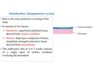
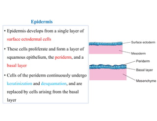
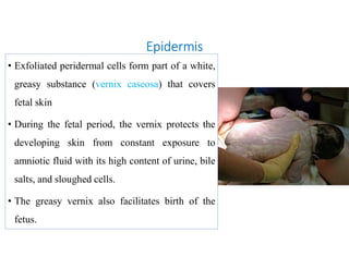
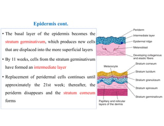
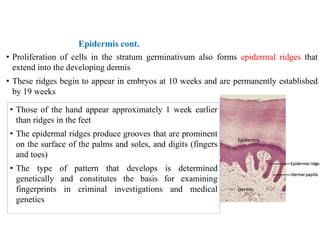
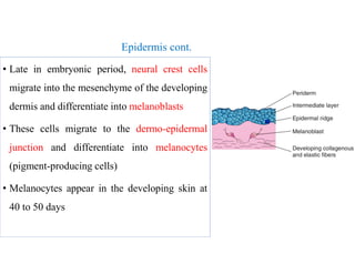
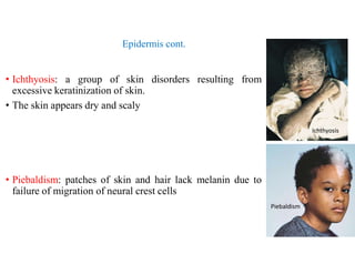
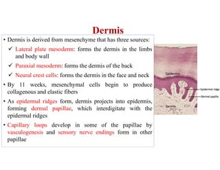
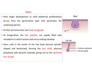
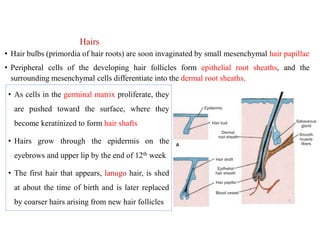
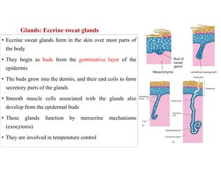
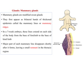
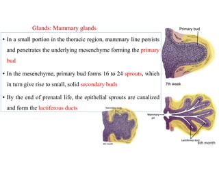
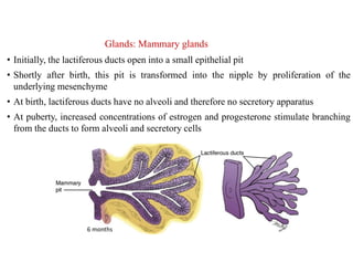
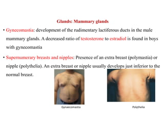
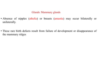
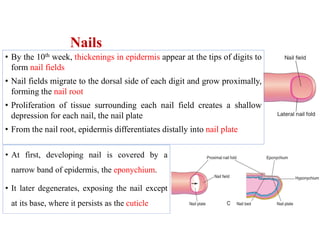
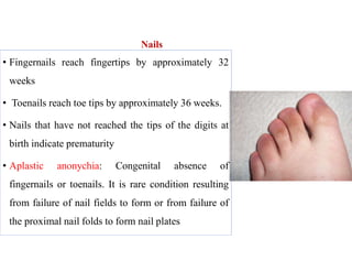
KILIMANJARO CHRISTIAN MMEDICAL UNIVERSITY COLLEGE
Faculty of Nursing and Rehabilitation Medicine
Department of Anatomy and Neuroscience
DEVELOPMENT OF INTEGUMENTARY SYSTEM (SESSION 17)
Date: Dec. 5th 2022, from 10:30 am – 12:30PM , GYM.
Lecturer: J. S. Kauki, BSc, MSc, on PhD, Email: jskauki@gmail.com.
Office ext. 69 IPH Block, 3RD Floor, Anatomy dept.
S16 Embryology lect.BSc.1 1
Objectives:
At the end of the session, students should be able to:
• Describe the development of the skin, accessory glands and nails.
• Identify important congenital malformations of the integumentary
system
Introduction
• Integumentary system consists of skin and its appendages:
 Epidermis
 Dermis
 Hairs
 Glands:
 Sebaceous glands
 Sweat glands
 Mammary glands
 Nails
Introduction: Integumentary system
• Skin is the outer protective covering of the
body
• It consists of two layers:
 Epidermis: superficial epithelial tissue
derived from surface ectoderm
 Dermis: deep layer composed of dense,
irregularly arranged connective tissue
derived from mesenchyme
• The embryonic skin at 4 to 5 weeks consists
of a single layer of surface ectoderm
overlying the mesoderm
Epidermis
• Epidermis develops from a single layer of
surface ectodermal cells
• These cells proliferate and form a layer of
squamous epithelium, the periderm, and a
basal layer
• Cells of the periderm continuously undergo
keratinization and desquamation, and are
replaced by cells arising from the basal
layer
Epidermis
• Exfoliated peridermal cells form part of a white,
greasy substance (vernix caseosa) that covers
fetal skin
• During the fetal period, the vernix protects the
developing skin from constant exposure to
amniotic fluid with its high content of urine, bile
salts, and sloughed cells.
• The greasy vernix also facilitates birth of the
fetus.
Epidermis cont.
• The basal layer of the epidermis becomes the
stratum germinativum, which produces new cells
that are displaced into the more superficial layers
• By 11 weeks, cells from the stratum germinativum
have formed an intermediate layer
• Replacement of peridermal cells continues until
approximately the 21st week; thereafter, the
periderm disappears and the stratum corneum
forms
Epidermis cont.
• Proliferation of cells in the stratum germinativum also forms epidermal ridges that
extend into the developing dermis
• These ridges begin to appear in embryos at 10 weeks and are permanently established
by 19 weeks
• Those of the hand appear approximately 1 week earlier
than ridges in the feet
• The epidermal ridges produce grooves that are prominent
on the surface of the palms and soles, and digits (fingers
and toes)
• The type of pattern that develops is determined
genetically and constitutes the basis for examining
fingerprints in criminal investigations and medical
genetics
Epidermis cont.
• Late in embryonic period, neural crest cells
migrate into the mesenchyme of the developing
dermis and differentiate into melanoblasts
• These cells migrate to the dermo-epidermal
junction and differentiate into melanocytes
(pigment-producing cells)
• Melanocytes appear in the developing skin at
40 to 50 days
Epidermis cont.
• Ichthyosis: a group of skin disorders resulting from
excessive keratinization of skin.
• The skin appears dry and scaly
• Piebaldism: patches of skin and hair lack melanin due to
failure of migration of neural crest cells
Ichthyosis
Piebaldism
Dermis
• Dermis is derived from mesenchyme that has three sources:
 Lateral plate mesoderm: forms the dermis in the limbs
and body wall
 Paraxial mesoderm: forms the dermis of the back
 Neural crest cells: forms the dermis in the face and neck
• By 11 weeks, mesenchymal cells begin to produce
collagenous and elastic fibers
• As epidermal ridges form, dermis projects into epidermis,
forming dermal papillae, which interdigitate with the
epidermal ridges
• Capillary loops develop in some of the papillae by
vasculogenesis and sensory nerve endings form in other
papillae
Hairs
• Hairs begin development as solid epidermal proliferations
(buds) from the germinative layer that penetrates the
underlying dermis
• At their terminal ends, hair buds invaginate
• At Invagination, the hair papillae, are rapidly filled with
mesoderm in which vessels and nerve endings develop
• Soon, cells in the center of the hair buds become spindle-
shaped and keratinized, forming the hair shaft, whereas
peripheral cells become cuboidal, giving rise to the epithelial
hair sheath
Hairs
• As cells in the germinal matrix proliferate, they
are pushed toward the surface, where they
become keratinized to form hair shafts
• Hairs grow through the epidermis on the
eyebrows and upper lip by the end of 12th week
• The first hair that appears, lanugo hair, is shed
at about the time of birth and is later replaced
by coarser hairs arising from new hair follicles
• Hair bulbs (primordia of hair roots) are soon invaginated by small mesenchymal hair papillae
• Peripheral cells of the developing hair follicles form epithelial root sheaths, and the
surrounding mesenchymal cells differentiate into the dermal root sheaths.
Hairs
• Melanoblasts migrate into the hair bulbs and differentiate into melanocytes
(pigment-producing cells)
• The melanin produced by these cells is transferred to the hair-forming cells in the
germinal matrix several weeks before birth
• The relative content of melanin accounts for different hair colors.
Glands: Sebaceous glands
• The epithelial wall of the hair follicle usually shows a small bud penetrating the
surrounding mesoderm
• Cells from these buds form the sebaceous glands
• Cells from the central region of the gland
degenerate, forming a fat-like substance (sebum)
secreted into the hair follicle, and from there,
they reach the skin
• Sebaceous glands, independent of hair follicles,
such as those of the glans penis and labia
minora, develop as cellular buds from the
epidermis that invade the dermis
Glands: Eccrine sweat glands
• Eccrine sweat glands form in the skin over most parts of
the body
• They begin as buds from the germinative layer of the
epidermis
• The buds grow into the dermis, and their end coils to form
secretory parts of the glands
• Smooth muscle cells associated with the glands also
develop from the epidermal buds
• These glands function by merocrine mechanisms
(exocytosis)
• They are involved in temperature control
Glands: Apocrine sweat glands
• Apocrine sweat glands develop along body hair
• They begin to develop during puberty
• They arise from the same epidermal buds that produce hair follicles
• These sweat glands open onto hair follicles instead of skin
• Sweat produced by these glands contains lipids, proteins, and pheromones, and
odor originating from this sweat is due to bacteria that break down these products
Glands: Mammary glands
• Mammary glands are modified sweat glands
• They first appear as bilateral bands of thickened
epidermis called the mammary lines or mammary
ridges
• In a 7-week embryo, these lines extend on each side
of the body from the base of forelimb to the base of
hind limb
• Major part of each mammary line disappears shortly
after it forms, leaving a small remnant in the thoracic
region
Glands: Mammary glands
• In a small portion in the thoracic region, mammary line persists
and penetrates the underlying mesenchyme forming the primary
bud
• In the mesenchyme, primary bud forms 16 to 24 sprouts, which
in turn give rise to small, solid secondary buds
• By the end of prenatal life, the epithelial sprouts are canalized
and form the lactiferous ducts
6th month
Glands: Mammary glands
• Initially, the lactiferous ducts open into a small epithelial pit
• Shortly after birth, this pit is transformed into the nipple by proliferation of the
underlying mesenchyme
• At birth, lactiferous ducts have no alveoli and therefore no secretory apparatus
• At puberty, increased concentrations of estrogen and progesterone stimulate branching
from the ducts to form alveoli and secretory cells
6 months
Glands: Mammary glands
• Gynecomastia: development of the rudimentary lactiferous ducts in the male
mammary glands. A decreased ratio of testosterone to estradiol is found in boys
with gynecomastia
• Supernumerary breasts and nipples: Presence of an extra breast (polymastia) or
nipple (polythelia). An extra breast or nipple usually develops just inferior to the
normal breast.
Polythelia
Gynaecomastia
Glands: Mammary glands
• Absence of nipples (athelia) or breasts (amastia) may occur bilaterally or
unilaterally.
• These rare birth defects result from failure of development or disappearance of
the mammary ridges
Nails
• By the 10th week, thickenings in epidermis appear at the tips of digits to
form nail fields
• Nail fields migrate to the dorsal side of each digit and grow proximally,
forming the nail root
• Proliferation of tissue surrounding each nail field creates a shallow
depression for each nail, the nail plate
• From the nail root, epidermis differentiates distally into nail plate
• At first, developing nail is covered by a
narrow band of epidermis, the eponychium.
• It later degenerates, exposing the nail except
at its base, where it persists as the cuticle
Nails
• Fingernails reach fingertips by approximately 32
weeks
• Toenails reach toe tips by approximately 36 weeks.
• Nails that have not reached the tips of the digits at
birth indicate prematurity
• Aplastic anonychia: Congenital absence of
fingernails or toenails. It is rare condition resulting
from failure of nail fields to form or from failure of
the proximal nail folds to form nail plates
Questions?
The coming Lecture will be on the skeletal system, therefore get
prepared and study:
• The appendicular skeleton
• The axial skeleton
THANK YOU

INTEGUMENTARY SYSTEM DEVELOPMENT.pdf

  • 1.
    KILIMANJARO CHRISTIAN MMEDICALUNIVERSITY COLLEGE Faculty of Nursing and Rehabilitation Medicine Department of Anatomy and Neuroscience DEVELOPMENT OF INTEGUMENTARY SYSTEM (SESSION 17) Date: Dec. 5th 2022, from 10:30 am – 12:30PM , GYM. Lecturer: J. S. Kauki, BSc, MSc, on PhD, Email: jskauki@gmail.com. Office ext. 69 IPH Block, 3RD Floor, Anatomy dept. S16 Embryology lect.BSc.1 1
  • 2.
    Objectives: At the endof the session, students should be able to: • Describe the development of the skin, accessory glands and nails. • Identify important congenital malformations of the integumentary system
  • 3.
    Introduction • Integumentary systemconsists of skin and its appendages:  Epidermis  Dermis  Hairs  Glands:  Sebaceous glands  Sweat glands  Mammary glands  Nails
  • 4.
    Introduction: Integumentary system •Skin is the outer protective covering of the body • It consists of two layers:  Epidermis: superficial epithelial tissue derived from surface ectoderm  Dermis: deep layer composed of dense, irregularly arranged connective tissue derived from mesenchyme • The embryonic skin at 4 to 5 weeks consists of a single layer of surface ectoderm overlying the mesoderm
  • 5.
    Epidermis • Epidermis developsfrom a single layer of surface ectodermal cells • These cells proliferate and form a layer of squamous epithelium, the periderm, and a basal layer • Cells of the periderm continuously undergo keratinization and desquamation, and are replaced by cells arising from the basal layer
  • 6.
    Epidermis • Exfoliated peridermalcells form part of a white, greasy substance (vernix caseosa) that covers fetal skin • During the fetal period, the vernix protects the developing skin from constant exposure to amniotic fluid with its high content of urine, bile salts, and sloughed cells. • The greasy vernix also facilitates birth of the fetus.
  • 7.
    Epidermis cont. • Thebasal layer of the epidermis becomes the stratum germinativum, which produces new cells that are displaced into the more superficial layers • By 11 weeks, cells from the stratum germinativum have formed an intermediate layer • Replacement of peridermal cells continues until approximately the 21st week; thereafter, the periderm disappears and the stratum corneum forms
  • 8.
    Epidermis cont. • Proliferationof cells in the stratum germinativum also forms epidermal ridges that extend into the developing dermis • These ridges begin to appear in embryos at 10 weeks and are permanently established by 19 weeks • Those of the hand appear approximately 1 week earlier than ridges in the feet • The epidermal ridges produce grooves that are prominent on the surface of the palms and soles, and digits (fingers and toes) • The type of pattern that develops is determined genetically and constitutes the basis for examining fingerprints in criminal investigations and medical genetics
  • 9.
    Epidermis cont. • Latein embryonic period, neural crest cells migrate into the mesenchyme of the developing dermis and differentiate into melanoblasts • These cells migrate to the dermo-epidermal junction and differentiate into melanocytes (pigment-producing cells) • Melanocytes appear in the developing skin at 40 to 50 days
  • 10.
    Epidermis cont. • Ichthyosis:a group of skin disorders resulting from excessive keratinization of skin. • The skin appears dry and scaly • Piebaldism: patches of skin and hair lack melanin due to failure of migration of neural crest cells Ichthyosis Piebaldism
  • 11.
    Dermis • Dermis isderived from mesenchyme that has three sources:  Lateral plate mesoderm: forms the dermis in the limbs and body wall  Paraxial mesoderm: forms the dermis of the back  Neural crest cells: forms the dermis in the face and neck • By 11 weeks, mesenchymal cells begin to produce collagenous and elastic fibers • As epidermal ridges form, dermis projects into epidermis, forming dermal papillae, which interdigitate with the epidermal ridges • Capillary loops develop in some of the papillae by vasculogenesis and sensory nerve endings form in other papillae
  • 12.
    Hairs • Hairs begindevelopment as solid epidermal proliferations (buds) from the germinative layer that penetrates the underlying dermis • At their terminal ends, hair buds invaginate • At Invagination, the hair papillae, are rapidly filled with mesoderm in which vessels and nerve endings develop • Soon, cells in the center of the hair buds become spindle- shaped and keratinized, forming the hair shaft, whereas peripheral cells become cuboidal, giving rise to the epithelial hair sheath
  • 13.
    Hairs • As cellsin the germinal matrix proliferate, they are pushed toward the surface, where they become keratinized to form hair shafts • Hairs grow through the epidermis on the eyebrows and upper lip by the end of 12th week • The first hair that appears, lanugo hair, is shed at about the time of birth and is later replaced by coarser hairs arising from new hair follicles • Hair bulbs (primordia of hair roots) are soon invaginated by small mesenchymal hair papillae • Peripheral cells of the developing hair follicles form epithelial root sheaths, and the surrounding mesenchymal cells differentiate into the dermal root sheaths.
  • 14.
    Hairs • Melanoblasts migrateinto the hair bulbs and differentiate into melanocytes (pigment-producing cells) • The melanin produced by these cells is transferred to the hair-forming cells in the germinal matrix several weeks before birth • The relative content of melanin accounts for different hair colors.
  • 15.
    Glands: Sebaceous glands •The epithelial wall of the hair follicle usually shows a small bud penetrating the surrounding mesoderm • Cells from these buds form the sebaceous glands • Cells from the central region of the gland degenerate, forming a fat-like substance (sebum) secreted into the hair follicle, and from there, they reach the skin • Sebaceous glands, independent of hair follicles, such as those of the glans penis and labia minora, develop as cellular buds from the epidermis that invade the dermis
  • 16.
    Glands: Eccrine sweatglands • Eccrine sweat glands form in the skin over most parts of the body • They begin as buds from the germinative layer of the epidermis • The buds grow into the dermis, and their end coils to form secretory parts of the glands • Smooth muscle cells associated with the glands also develop from the epidermal buds • These glands function by merocrine mechanisms (exocytosis) • They are involved in temperature control
  • 17.
    Glands: Apocrine sweatglands • Apocrine sweat glands develop along body hair • They begin to develop during puberty • They arise from the same epidermal buds that produce hair follicles • These sweat glands open onto hair follicles instead of skin • Sweat produced by these glands contains lipids, proteins, and pheromones, and odor originating from this sweat is due to bacteria that break down these products
  • 18.
    Glands: Mammary glands •Mammary glands are modified sweat glands • They first appear as bilateral bands of thickened epidermis called the mammary lines or mammary ridges • In a 7-week embryo, these lines extend on each side of the body from the base of forelimb to the base of hind limb • Major part of each mammary line disappears shortly after it forms, leaving a small remnant in the thoracic region
  • 19.
    Glands: Mammary glands •In a small portion in the thoracic region, mammary line persists and penetrates the underlying mesenchyme forming the primary bud • In the mesenchyme, primary bud forms 16 to 24 sprouts, which in turn give rise to small, solid secondary buds • By the end of prenatal life, the epithelial sprouts are canalized and form the lactiferous ducts 6th month
  • 20.
    Glands: Mammary glands •Initially, the lactiferous ducts open into a small epithelial pit • Shortly after birth, this pit is transformed into the nipple by proliferation of the underlying mesenchyme • At birth, lactiferous ducts have no alveoli and therefore no secretory apparatus • At puberty, increased concentrations of estrogen and progesterone stimulate branching from the ducts to form alveoli and secretory cells 6 months
  • 21.
    Glands: Mammary glands •Gynecomastia: development of the rudimentary lactiferous ducts in the male mammary glands. A decreased ratio of testosterone to estradiol is found in boys with gynecomastia • Supernumerary breasts and nipples: Presence of an extra breast (polymastia) or nipple (polythelia). An extra breast or nipple usually develops just inferior to the normal breast. Polythelia Gynaecomastia
  • 22.
    Glands: Mammary glands •Absence of nipples (athelia) or breasts (amastia) may occur bilaterally or unilaterally. • These rare birth defects result from failure of development or disappearance of the mammary ridges
  • 23.
    Nails • By the10th week, thickenings in epidermis appear at the tips of digits to form nail fields • Nail fields migrate to the dorsal side of each digit and grow proximally, forming the nail root • Proliferation of tissue surrounding each nail field creates a shallow depression for each nail, the nail plate • From the nail root, epidermis differentiates distally into nail plate • At first, developing nail is covered by a narrow band of epidermis, the eponychium. • It later degenerates, exposing the nail except at its base, where it persists as the cuticle
  • 24.
    Nails • Fingernails reachfingertips by approximately 32 weeks • Toenails reach toe tips by approximately 36 weeks. • Nails that have not reached the tips of the digits at birth indicate prematurity • Aplastic anonychia: Congenital absence of fingernails or toenails. It is rare condition resulting from failure of nail fields to form or from failure of the proximal nail folds to form nail plates
  • 25.
  • 26.
    The coming Lecturewill be on the skeletal system, therefore get prepared and study: • The appendicular skeleton • The axial skeleton
  • 27.