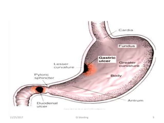
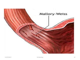
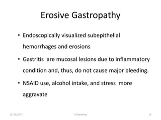
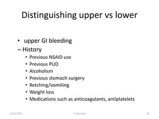
AMBO UNIVERSITY
COLLEGE MEDICINE AND HEALTH
SCIENCE
DEPARTMENT OF MEDICINE
SEMINAR PRESENTATION ON :-
GASTRO INTESTINAL BLEEDING
PREPARED BY: ABDISA GELETA
MODULATOR :DR SAMSON
2016 GC
11/25/2017 1GI bleeding
OUT LINE
• Introduction
• Ethiology of upper GI bleeding
• Ethiology of lower GI bleeding
• Approach to patient with GI bleeding
• Management course
11/25/2017 2GI bleeding
GI bleeding
• Gastrointestinal bleediing (GI bleeding), also
known as gastrointestinal hemorrhage, is all forms
of blood loss from the gastrointestinal tract.
• Bleeding is typically divided into two main
types: upper gastrointestinal bleeding and lower
gastrointestinal bleeding.
• Upper GI bleeding refers to bleeding that arises
from the GI tract proximal to the ligament of Treitz
and accounts for nearly 80% of significant blood
loss
• 20% LGIB- distal to ligament of Treitz
11/25/2017 3GI bleeding
Differentiation of UGIB and LGIB
4
•Hematemesis: vomiting bright red blood or blood that is
dark, like coffee grounds,Indicates upper GI bleeding
•Melena: passage of dark pitchy stools stained with blood
pigments Indicates upper GI bleeding
•Hematochezia: passage of bright red blood with stool.
Indicates lower GI bleeding
11/25/2017 GI bleeding
• Hematochezia usually represents a lower GI
source of bleeding, although an upper GI
lesion may bleed so briskly that blood does
not remain in the bowel long enough for
melena to develop.
• When hematochezia is the presenting symptom
of UGIB, it is associated with hemodynamic
instability and dropping hemoglobin.
11/25/2017 5GI bleeding
possible cause upper GI bleeding
11/25/2017 6GI bleeding
Peptic ulcer disease
• Develop due to an imbalance between aggressive
factors and protective factors, leading to an
interruption in the mucosal integrity
• Aggressive factors .Protective factor
-acid -bicarbonate
-h.pylori -mucus
-pepsin - prostaglandin
-NSAID
11/25/2017 7GI bleeding
Types PUD
• Gastric ulcer:
– Burning, gnawing
epigastric pain that
occurs with anything in
the stomach;
– Pain is worst after eating
(in contrast to duodenal
ulcer).
– Anorexia/weight loss,
vomiting.
– Associated with blood
type A.
• Duodenal ulcer:
– Burning, gnawing
epigastric pain that occurs
with an empty stomach
(hunger pain) and is
relieved by food or
antacids (in contrast to
gastric ulcers).
– Night time awakening
(when stomach empties).
– Nausea, vomiting.
– Associated with blood type
O.
11/25/2017 8GI bleeding
11/25/2017 9GI bleeding
Esophageal Varices
• The increased pressure in the esophageal plexus
produces dilated tortuous vessels called varices.
• Variceal rupture produces massive hemorrhage
into the lumen.
•present with hematemesis, melena, or
hematochezia
11/25/2017 10GI bleeding
Mallory–Weiss Syndrome
• is characterized by longitudinal mucosal lacerations
(intramural dissections) in the distal esophagus and
proximal stomach, which are usually associated with
forceful retching.
Common Alcoholic patient after binge drinking
-First :- vomit food and gastric contents
-Followed by :- forceful retching & bloody vomitus.
11/25/2017 11GI bleeding
Risk factor
• Retching,
• Alcoholism
• Increased intraabdominal pressure
symptom
• Epigastric pain,
• Thoracic substernal pain,
• Hematemesis
11/25/2017 12GI bleeding
11/25/2017 13GI bleeding
Erosive Gastropathy
• Endoscopically visualized subepithelial
hemorrhages and erosions
• Gastritis are mucosal lesions due to inflammatory
condition and, thus, do not cause major bleeding.
• NSAID use, alcohol intake, and stress more
aggravate
11/25/2017 14GI bleeding
Esophagitis
• Esophageal inflammation secondary to repeated
exposure of the esophageal mucosa to the acidic
gastric secretions in gastroesophageal reflux disease
(GERD) leads to an inflammatory response, which
can result in chronic blood loss.
11/25/2017 15GI bleeding
UGI TUMORS
• Acute bleeding represents a late stage of disease
when the neoplasm outgrows its blood supply and
causes mucosal ulceration.
• Bleeding can result from diffuse mucosal ulceration
or from erosion into an underlying vessel.
11/25/2017 16GI bleeding
11/25/2017 17GI bleeding
Lower GI bleeding
• Bleeding that originates from sources located distal
to the Ligament of Treitz.
• Accounts for 1% of acute hospital admissions each
year.
• Much less common reason for hospitalization, when
compared with upper GI hemorrhage.
1811/25/2017 GI bleeding
• GI bleeding that persists or recurs without a diagnosed
etiology after the initial routine work-up is known as
Obscure GI bleeding.
• Approximately 5% of pts will have obscure GIB.
• Obscure GIB is further categorized as either:-
• Obscure occult or
• Obscure overt bleeding
• Small intestinal bleeding usually is considered under
obscure causes of GI bleeding
1911/25/2017 GI bleeding
possible causes of lower
gastrointestinal bleeding (LGIB
• Anatomic (diverticulosis)
• Vascular (angiodysplasia, ischemic, radiation-
induced)
• Inflammatory (inflammatory bowel disease,
infectious)
• Neoplastic
11/25/2017 GI bleeding 20
Diverticular disease
• Presence of symptomatic diverticula.
• Most are asymptomatic, but 3–5% will develop massive
bleeding.
• 99% of patients with these bleeding require four or fewer
units of blood before they stabilize.
• Bleeding resolves without intervention 80% of the time.
• Rebleeding occurs in 25–30% of pts after the first episode.
• If bleeding ceases spontaneously a second time, the
recurrence rate is as high as 50%. 2111/25/2017 GI bleeding
Vascular cause
Bowel ischemia
• Occlusion of the inferior mesenteric artery can present
with abdominal colic and rectal bleeding.
• Risk groups are the elderly people who have evidence
of generalised atherosclerosis.
Angiodysplasia
• Angiodysplasia is a diseases of the elderly in which
vascular malformations develop in the proximal colon.
• Bleeding can be acute and profuse; it usually stops
spontaneously but commonly recurs. 2211/25/2017 GI bleeding
Inflammatory cuases
Infectious
• Bacterial (C. difficile , enterotoxic E. coli, Campylobacter
jejuni…),
• Viral (herpes simplex viruses, HIV, and CMV).
• Parasites (Amebiasis, Cryptosporidiosis, )
• Fungal (Candida species, Histoplasmosis)
2311/25/2017 GI bleeding
Noninfectious
• IBD:- UC, CD,
 GI bleeding may be the 1st presentation
 Bloody diarrhea (more in ulcerative colitis)
 acute hemorrhage (more likely in cd)
• Radiation colitis:
 After treatment for pelvic malignancies.
 Patients present with bright-red blood per rectum,
diarrhea, tenesmus, and crampy pelvic pain
2411/25/2017 GI bleeding
Neoplasia
• Colorectal carcinoma:
Not common but has to be ruled out in painless,
intermittent, and slow bleeding.
Frequently associated with iron deficiency anemia
• Polyps :-
more commonly, the bleeding occurs after a
polypectomy.
juvenile polyps are the second most common cause of
bleeding in patients younger than 20 years of age
• Others: lipoma, lymphoma, leiomyoma,
leiomyosarcoma
2511/25/2017 GI bleeding
Anorectal disease
• Haemorrhoids
• Anal fissures
• Anal fistula
• Proctitis: Actinomycosis israelii ,Treponema pallidum
• Rectal trauma
2611/25/2017 GI bleeding
Haemorrhoids and anal fissures
Dilated veins both from deep and superficial plexus
• Haemorrhoidal bleeding is bright red and occurs
during or after defecation.
• Types-internal and external
Anal fissure should be suspected when fresh rectal
bleeding and anal pain occur during defecation
11/25/2017 27GI bleeding
APPROACH TO THE PATIENT
GI BLEEDING
Goal of the evaluation:
• To assess the severity of the bleed,
• Identify potential sources of the bleed &
determine if there are conditions that may affect
subsequent management.
Initial evaluation
• Includes a history, physical examination& laboratory
tests
11/25/2017 28GI bleeding
History
• Abdominal pain
• Hematemesis
• Melana
• Hematochezia
• Features of blood loss: shock, anemia
• Features of underlying cause: dyspepsia, jaundice,
weight loss
• Drug history: - like
NSAIDs(Aspirin)
Anticoagulants,
•History of epistaxis or hemoptysis
11/25/2017 29GI bleeding
Bleeding manifestations:
• Hematemesis (red blood or coffee-ground emesis)-
UGIB
-frankly bloody emesis -moderate to severe bleeding
that may be ongoing,
-coffee-ground emesis-limited bleeding.
• melena (black, tarry stool)- UGIB(90%)
11/25/2017 30GI bleeding
• Hematochezia :- LGIB or if it is massive
UGIB(orthostatic hypotension)
-bright red:-left colon
-Maroon colour:-right side of colon
-Hematochezia with abdominal pain;may represent
ischemic colitis,
-Painless hematochezia:- diverticulosis and colonic
tumors
11/25/2017 31GI bleeding
patients can be
• Low risk patients
Self limited bleeding, young and otherwise healthy
patients ;Internal hemorrhoid
• High risk patients
hemodynamic instability, serious comorbid diseases,
persistent bleeding-promptly resuscitate and
hospitalize and need more evaluation
3211/25/2017 GI bleeding
Symptom assessment:
To assess the severity and potential source of bleeding,
orthostatic dizziness, confusion, angina, severe
palpitations, and cold extremities-severe bleeding
Epigastric or right upper quadrant pain
Odynophagia, dysphagia
3311/25/2017 GI bleeding
Emesis, retching, or coughing prior to hematemesis
Jaundice, weakness, fatigue, anorexia, abdominal
distention
 Dysphagia, early satiety, involuntary weight loss,
cachexia
GI bleeding 3411/25/2017
• Medication History
 Pay attention to:
aspirin and other NSAIDs
antiplatelet agents and anticoagulants
GI bleeding 3511/25/2017
• Assess comorbid illnesses
 illnesses may:
-Make patients more susceptible to hypoxemia eg CAD,
pulmonary disease
-Predispose patients to volume overload eg CHF, renal
disease ;need attention during resustation
3611/25/2017 GI bleeding
-Result in bleeding that is more difficult to control (eg,
coagulopathies, thrombocytopenia, significant hepatic
dysfunction)-may need fresh frozen plasma or
platelets.
-Predispose to aspiration (eg hepatic encephalopathy)—
needs intubation.
GI bleeding 3711/25/2017
Distinguishing upper vs lower
• upper GI bleeding
– History
• Previous NSAID use
• Previous PUD
• Alcoholism
• Previous stomach surgery
• Retching/vomiting
• Weight loss
• Medications such as anticoagulants, antiplatelets
11/25/2017 GI bleeding 38
– Symptoms
• Nausea/vomiting
• Hematemesis
• Melena
• Rarely hematochezia (massive bleed)
11/25/2017 GI bleeding 39
11/25/2017 GI bleeding 40
• Lower GI bleeding
-History
• Previous colon cancer
• Previous colon surgery
• Known diverticulosis
• Known hemorrhoids
– Symptoms
• Abdominal pain or can be painless
• Hematochezia
• Melena
11/25/2017 GI bleeding 41
Physical examination
• Signs of hypovolemia
Resting tachycardia- Mild to moderate
hypovolemia
Orthostatic hypotension-at least 15% blood loss
 Supine hypotension-at least 40% loss
• SKIN changes:
– Palmar erythema-Cirrhosis
– Purpura /Echymosis-Bleeding disorders
• Signs of dehydration (dry mucosa, sunken eyes)
• Digital rectal exam: fresh blood, occult blood may be
found.
11/25/2017 GI bleeding 42
Estimating Degree of Blood Loss RR, HR, and BP can be used
to estimate the degree of blood loss/hypovolumia
Class I Class II Class III Class IV
Volume Loss (ml)
Or %
0-750 or
Up to 15%
750-1500 or
15-30%
1500-2000 or
30-40%
>2000 or
>40%
RR 14-20 20-30 30-40 >40
HR <100 >100 >120 >140
BP unchanged unchanged reduced reduced
Urine Output
(ml/hr)
>30 20-30 5-15 Anuric
Mental State Restless Anxious Anxious/confuse
d
Confused/
lethargic
GI bleeding 4311/25/2017
Lab Diagnosis
• CBC, blood group
-(CBC, WBC, HCT/Hb, platelet count…)
-Hb may be normal initially
-CBC should be checked frequently(q4-6h) during
the first day.
-Patients with slow, chronic GIB may have very low
hemoglobin values despite normal blood pressure
and heart rate.
11/25/2017 GI bleeding 44
• Blood chemistry
-BUN/creatinine >20:1 UGIB
• NG tube lavage to exclude UGIB
• LFT
• Coagulation studies
• ECG and cardiac enzymes
• Stool examination for- parasites, blood cells, Occult
blood in chronic occult blood loss
• GI Endoscopy, sigmoidoscopy: Valuable for
visualization biopsy taking and endoscopic treatment
GI bleeding 4511/25/2017
Nasogastric Lavage
• This procedure may confirm:-
-Recent bleeding (coffee ground appearance)
-Active bleeding (red blood in the aspirate)
-nonbloody bilious fluid-pylorus is open and that
there is no active upper GI bleeding distal to the
pylorus.
-Negative lavage-bleeding may be distal to closed
pylorus
-Lack of blood in the stomach (active bleeding
less likely but does not exclude an upper GI
lesion11/25/2017 GI bleeding 46
GI bleeding 47
to remove particulate matter, fresh blood, and
clots from the stomach to facilitate endoscopy.
11/25/2017
Endoscopy
• Initial diagnostic examination for all patients
presumed to have UGIB.
• Endoscopy should be performed immediately
after:-
-Endotracheal intubation (if indicated)
-Hemodynamic stabilization, and
-Adequate monitoring in ICU is achieved
• Used: Diagnostic and Therapeutic(Hemostasis,
luminal restoration (dilation, ablation, stenting),
lesion removal (e.g., polypectomy), percutaneous
endoscopic gastrostomy)
11/25/2017 GI bleeding 48
Endoscopy cont…
• Urgent indication
-Shock
-Hct < 30
-Suspected variceal hemorrhage
-Recurrence of bleeding from unknown source
• Contra indication:- acute MI, , agitation,
circulatory imbalance
• Complication: aspiration, worsening of bleeding,
perforation,
11/25/2017 GI bleeding 49
Endoscopy cont..
11/25/2017 GI bleeding 50
Colonoscopy
-The gold standard for diagnosis of colonic mucosal disease
-Done for pts who don’t respond for resuscitation
-Gives good diagnosis yield if it is done with in 6-12hrs of
bleeding---Identifies lesion in 75 % or more
-Bowel preparation with purgatives 2-4hrs before the
procedure
-Can provide endoscopic therapy
-Complications 0.5-1 %
GI bleeding 5111/25/2017
Imaging
• Chest X-Ray-Chest radiographs should be ordered to
exclude aspiration pneumonia and effusion.
• Abdominal X-Ray- upright and supine films should be
ordered to exclude perforated viscus and ileus.
GI bleeding 5211/25/2017
Suggested algorithm for patients
with acute UGI bleeding
11/25/2017 GI bleeding 53
Suggested algorithm for patients with
acute lower GI bleeding
11/25/2017 GI bleeding 54
Management of GI bleeding
There are steps in GI bleeding Mgt
• Initial assessment
• Resuscitation measures
• Identifying source of bleeding
• Institution with specific therapy
GI bleeding 5511/25/2017
Management of GI bleeding
Patient Assessment
If the patient:
• Hemodynamically stable OPD
• No evidence of active bleeding
or
If the patient:
• Is hemodynamically unstable
• Is continuously bleeding ICU
• Hct drops by 6%
GI bleeding 5611/25/2017
Resuscitation
• Airways
Massive hematemesis +
mental obtundation
-Intubation
• Breathing
Administer oxygen with IN
cannula,or facemask or
endotracheal tube
• Circulation
-Fluid resuscitation
crystalloids (NS or RL)
• Blood transfusion
If Hb < 7 or actively
bleeding Hb goal =10
NB: cautions in high risk
patient
• Correct coagulopathy
GI bleeding 5711/25/2017
Specific Rx Measures- Upper GI
. Esophageal varices
-Endoscopy: Best therapy-band ligation & sclerotherapy
-Somatostatin, octereotide 50–100 µg/h IV infusion
-Vasopressin
-Propranolol
-Antibiotics
-Transjugular intrahepatic portosystemic shunt( TIPS)
GI bleeding 5811/25/2017
PUD
-PPI: pantoprazole 80mg iv bolus then 8mg/hr infusion,
if no bleeding in 24 hrs switch to PO Omeprazole
20mg/day
-Drugs enhancing mucosal defenses: prostaglandin
analogues (e.g misoprostol)
-Antacids –promote ulcer healing through stimulation
gastric defense mechanism
-Therapy of H.Pylori
(omeprazole20mg,amoxacillin1000mg,clarithromicin
500mg all po BID *10days)
GI bleeding 5911/25/2017
Surgical Mx GIB
UGIB:-indications are perforation, uncontolled
hemorrhage ,GOO and recurrent ulcer following gastric
surgery.
LGIB:-persitent or recurrent bleeding from wide variety
of colonic sources of GI bleeding that can not be
treated medically / endoscopically
GI bleeding 6011/25/2017
REFERENCES
-Harrison principles of internal medicine 18th edition.
-Up to date 21.6
-Cecil - Textbook of Medicine
GI bleeding 6111/25/2017
11/25/2017 GI bleeding 62
THANK YOU!

Gi bleeding abdi!

  • 1.
    AMBO UNIVERSITY COLLEGE MEDICINEAND HEALTH SCIENCE DEPARTMENT OF MEDICINE SEMINAR PRESENTATION ON :- GASTRO INTESTINAL BLEEDING PREPARED BY: ABDISA GELETA MODULATOR :DR SAMSON 2016 GC 11/25/2017 1GI bleeding
  • 2.
    OUT LINE • Introduction •Ethiology of upper GI bleeding • Ethiology of lower GI bleeding • Approach to patient with GI bleeding • Management course 11/25/2017 2GI bleeding
  • 3.
    GI bleeding • Gastrointestinalbleediing (GI bleeding), also known as gastrointestinal hemorrhage, is all forms of blood loss from the gastrointestinal tract. • Bleeding is typically divided into two main types: upper gastrointestinal bleeding and lower gastrointestinal bleeding. • Upper GI bleeding refers to bleeding that arises from the GI tract proximal to the ligament of Treitz and accounts for nearly 80% of significant blood loss • 20% LGIB- distal to ligament of Treitz 11/25/2017 3GI bleeding
  • 4.
    Differentiation of UGIBand LGIB 4 •Hematemesis: vomiting bright red blood or blood that is dark, like coffee grounds,Indicates upper GI bleeding •Melena: passage of dark pitchy stools stained with blood pigments Indicates upper GI bleeding •Hematochezia: passage of bright red blood with stool. Indicates lower GI bleeding 11/25/2017 GI bleeding
  • 5.
    • Hematochezia usuallyrepresents a lower GI source of bleeding, although an upper GI lesion may bleed so briskly that blood does not remain in the bowel long enough for melena to develop. • When hematochezia is the presenting symptom of UGIB, it is associated with hemodynamic instability and dropping hemoglobin. 11/25/2017 5GI bleeding
  • 6.
    possible cause upperGI bleeding 11/25/2017 6GI bleeding
  • 7.
    Peptic ulcer disease •Develop due to an imbalance between aggressive factors and protective factors, leading to an interruption in the mucosal integrity • Aggressive factors .Protective factor -acid -bicarbonate -h.pylori -mucus -pepsin - prostaglandin -NSAID 11/25/2017 7GI bleeding
  • 8.
    Types PUD • Gastriculcer: – Burning, gnawing epigastric pain that occurs with anything in the stomach; – Pain is worst after eating (in contrast to duodenal ulcer). – Anorexia/weight loss, vomiting. – Associated with blood type A. • Duodenal ulcer: – Burning, gnawing epigastric pain that occurs with an empty stomach (hunger pain) and is relieved by food or antacids (in contrast to gastric ulcers). – Night time awakening (when stomach empties). – Nausea, vomiting. – Associated with blood type O. 11/25/2017 8GI bleeding
  • 9.
  • 10.
    Esophageal Varices • Theincreased pressure in the esophageal plexus produces dilated tortuous vessels called varices. • Variceal rupture produces massive hemorrhage into the lumen. •present with hematemesis, melena, or hematochezia 11/25/2017 10GI bleeding
  • 11.
    Mallory–Weiss Syndrome • ischaracterized by longitudinal mucosal lacerations (intramural dissections) in the distal esophagus and proximal stomach, which are usually associated with forceful retching. Common Alcoholic patient after binge drinking -First :- vomit food and gastric contents -Followed by :- forceful retching & bloody vomitus. 11/25/2017 11GI bleeding
  • 12.
    Risk factor • Retching, •Alcoholism • Increased intraabdominal pressure symptom • Epigastric pain, • Thoracic substernal pain, • Hematemesis 11/25/2017 12GI bleeding
  • 13.
  • 14.
    Erosive Gastropathy • Endoscopicallyvisualized subepithelial hemorrhages and erosions • Gastritis are mucosal lesions due to inflammatory condition and, thus, do not cause major bleeding. • NSAID use, alcohol intake, and stress more aggravate 11/25/2017 14GI bleeding
  • 15.
    Esophagitis • Esophageal inflammationsecondary to repeated exposure of the esophageal mucosa to the acidic gastric secretions in gastroesophageal reflux disease (GERD) leads to an inflammatory response, which can result in chronic blood loss. 11/25/2017 15GI bleeding
  • 16.
    UGI TUMORS • Acutebleeding represents a late stage of disease when the neoplasm outgrows its blood supply and causes mucosal ulceration. • Bleeding can result from diffuse mucosal ulceration or from erosion into an underlying vessel. 11/25/2017 16GI bleeding
  • 17.
  • 18.
    Lower GI bleeding •Bleeding that originates from sources located distal to the Ligament of Treitz. • Accounts for 1% of acute hospital admissions each year. • Much less common reason for hospitalization, when compared with upper GI hemorrhage. 1811/25/2017 GI bleeding
  • 19.
    • GI bleedingthat persists or recurs without a diagnosed etiology after the initial routine work-up is known as Obscure GI bleeding. • Approximately 5% of pts will have obscure GIB. • Obscure GIB is further categorized as either:- • Obscure occult or • Obscure overt bleeding • Small intestinal bleeding usually is considered under obscure causes of GI bleeding 1911/25/2017 GI bleeding
  • 20.
    possible causes oflower gastrointestinal bleeding (LGIB • Anatomic (diverticulosis) • Vascular (angiodysplasia, ischemic, radiation- induced) • Inflammatory (inflammatory bowel disease, infectious) • Neoplastic 11/25/2017 GI bleeding 20
  • 21.
    Diverticular disease • Presenceof symptomatic diverticula. • Most are asymptomatic, but 3–5% will develop massive bleeding. • 99% of patients with these bleeding require four or fewer units of blood before they stabilize. • Bleeding resolves without intervention 80% of the time. • Rebleeding occurs in 25–30% of pts after the first episode. • If bleeding ceases spontaneously a second time, the recurrence rate is as high as 50%. 2111/25/2017 GI bleeding
  • 22.
    Vascular cause Bowel ischemia •Occlusion of the inferior mesenteric artery can present with abdominal colic and rectal bleeding. • Risk groups are the elderly people who have evidence of generalised atherosclerosis. Angiodysplasia • Angiodysplasia is a diseases of the elderly in which vascular malformations develop in the proximal colon. • Bleeding can be acute and profuse; it usually stops spontaneously but commonly recurs. 2211/25/2017 GI bleeding
  • 23.
    Inflammatory cuases Infectious • Bacterial(C. difficile , enterotoxic E. coli, Campylobacter jejuni…), • Viral (herpes simplex viruses, HIV, and CMV). • Parasites (Amebiasis, Cryptosporidiosis, ) • Fungal (Candida species, Histoplasmosis) 2311/25/2017 GI bleeding
  • 24.
    Noninfectious • IBD:- UC,CD,  GI bleeding may be the 1st presentation  Bloody diarrhea (more in ulcerative colitis)  acute hemorrhage (more likely in cd) • Radiation colitis:  After treatment for pelvic malignancies.  Patients present with bright-red blood per rectum, diarrhea, tenesmus, and crampy pelvic pain 2411/25/2017 GI bleeding
  • 25.
    Neoplasia • Colorectal carcinoma: Notcommon but has to be ruled out in painless, intermittent, and slow bleeding. Frequently associated with iron deficiency anemia • Polyps :- more commonly, the bleeding occurs after a polypectomy. juvenile polyps are the second most common cause of bleeding in patients younger than 20 years of age • Others: lipoma, lymphoma, leiomyoma, leiomyosarcoma 2511/25/2017 GI bleeding
  • 26.
    Anorectal disease • Haemorrhoids •Anal fissures • Anal fistula • Proctitis: Actinomycosis israelii ,Treponema pallidum • Rectal trauma 2611/25/2017 GI bleeding
  • 27.
    Haemorrhoids and analfissures Dilated veins both from deep and superficial plexus • Haemorrhoidal bleeding is bright red and occurs during or after defecation. • Types-internal and external Anal fissure should be suspected when fresh rectal bleeding and anal pain occur during defecation 11/25/2017 27GI bleeding
  • 28.
    APPROACH TO THEPATIENT GI BLEEDING Goal of the evaluation: • To assess the severity of the bleed, • Identify potential sources of the bleed & determine if there are conditions that may affect subsequent management. Initial evaluation • Includes a history, physical examination& laboratory tests 11/25/2017 28GI bleeding
  • 29.
    History • Abdominal pain •Hematemesis • Melana • Hematochezia • Features of blood loss: shock, anemia • Features of underlying cause: dyspepsia, jaundice, weight loss • Drug history: - like NSAIDs(Aspirin) Anticoagulants, •History of epistaxis or hemoptysis 11/25/2017 29GI bleeding
  • 30.
    Bleeding manifestations: • Hematemesis(red blood or coffee-ground emesis)- UGIB -frankly bloody emesis -moderate to severe bleeding that may be ongoing, -coffee-ground emesis-limited bleeding. • melena (black, tarry stool)- UGIB(90%) 11/25/2017 30GI bleeding
  • 31.
    • Hematochezia :-LGIB or if it is massive UGIB(orthostatic hypotension) -bright red:-left colon -Maroon colour:-right side of colon -Hematochezia with abdominal pain;may represent ischemic colitis, -Painless hematochezia:- diverticulosis and colonic tumors 11/25/2017 31GI bleeding
  • 32.
    patients can be •Low risk patients Self limited bleeding, young and otherwise healthy patients ;Internal hemorrhoid • High risk patients hemodynamic instability, serious comorbid diseases, persistent bleeding-promptly resuscitate and hospitalize and need more evaluation 3211/25/2017 GI bleeding
  • 33.
    Symptom assessment: To assessthe severity and potential source of bleeding, orthostatic dizziness, confusion, angina, severe palpitations, and cold extremities-severe bleeding Epigastric or right upper quadrant pain Odynophagia, dysphagia 3311/25/2017 GI bleeding
  • 34.
    Emesis, retching, orcoughing prior to hematemesis Jaundice, weakness, fatigue, anorexia, abdominal distention  Dysphagia, early satiety, involuntary weight loss, cachexia GI bleeding 3411/25/2017
  • 35.
    • Medication History Pay attention to: aspirin and other NSAIDs antiplatelet agents and anticoagulants GI bleeding 3511/25/2017
  • 36.
    • Assess comorbidillnesses  illnesses may: -Make patients more susceptible to hypoxemia eg CAD, pulmonary disease -Predispose patients to volume overload eg CHF, renal disease ;need attention during resustation 3611/25/2017 GI bleeding
  • 37.
    -Result in bleedingthat is more difficult to control (eg, coagulopathies, thrombocytopenia, significant hepatic dysfunction)-may need fresh frozen plasma or platelets. -Predispose to aspiration (eg hepatic encephalopathy)— needs intubation. GI bleeding 3711/25/2017
  • 38.
    Distinguishing upper vslower • upper GI bleeding – History • Previous NSAID use • Previous PUD • Alcoholism • Previous stomach surgery • Retching/vomiting • Weight loss • Medications such as anticoagulants, antiplatelets 11/25/2017 GI bleeding 38
  • 39.
    – Symptoms • Nausea/vomiting •Hematemesis • Melena • Rarely hematochezia (massive bleed) 11/25/2017 GI bleeding 39
  • 40.
    11/25/2017 GI bleeding40 • Lower GI bleeding -History • Previous colon cancer • Previous colon surgery • Known diverticulosis • Known hemorrhoids
  • 41.
    – Symptoms • Abdominalpain or can be painless • Hematochezia • Melena 11/25/2017 GI bleeding 41
  • 42.
    Physical examination • Signsof hypovolemia Resting tachycardia- Mild to moderate hypovolemia Orthostatic hypotension-at least 15% blood loss  Supine hypotension-at least 40% loss • SKIN changes: – Palmar erythema-Cirrhosis – Purpura /Echymosis-Bleeding disorders • Signs of dehydration (dry mucosa, sunken eyes) • Digital rectal exam: fresh blood, occult blood may be found. 11/25/2017 GI bleeding 42
  • 43.
    Estimating Degree ofBlood Loss RR, HR, and BP can be used to estimate the degree of blood loss/hypovolumia Class I Class II Class III Class IV Volume Loss (ml) Or % 0-750 or Up to 15% 750-1500 or 15-30% 1500-2000 or 30-40% >2000 or >40% RR 14-20 20-30 30-40 >40 HR <100 >100 >120 >140 BP unchanged unchanged reduced reduced Urine Output (ml/hr) >30 20-30 5-15 Anuric Mental State Restless Anxious Anxious/confuse d Confused/ lethargic GI bleeding 4311/25/2017
  • 44.
    Lab Diagnosis • CBC,blood group -(CBC, WBC, HCT/Hb, platelet count…) -Hb may be normal initially -CBC should be checked frequently(q4-6h) during the first day. -Patients with slow, chronic GIB may have very low hemoglobin values despite normal blood pressure and heart rate. 11/25/2017 GI bleeding 44
  • 45.
    • Blood chemistry -BUN/creatinine>20:1 UGIB • NG tube lavage to exclude UGIB • LFT • Coagulation studies • ECG and cardiac enzymes • Stool examination for- parasites, blood cells, Occult blood in chronic occult blood loss • GI Endoscopy, sigmoidoscopy: Valuable for visualization biopsy taking and endoscopic treatment GI bleeding 4511/25/2017
  • 46.
    Nasogastric Lavage • Thisprocedure may confirm:- -Recent bleeding (coffee ground appearance) -Active bleeding (red blood in the aspirate) -nonbloody bilious fluid-pylorus is open and that there is no active upper GI bleeding distal to the pylorus. -Negative lavage-bleeding may be distal to closed pylorus -Lack of blood in the stomach (active bleeding less likely but does not exclude an upper GI lesion11/25/2017 GI bleeding 46
  • 47.
    GI bleeding 47 toremove particulate matter, fresh blood, and clots from the stomach to facilitate endoscopy. 11/25/2017
  • 48.
    Endoscopy • Initial diagnosticexamination for all patients presumed to have UGIB. • Endoscopy should be performed immediately after:- -Endotracheal intubation (if indicated) -Hemodynamic stabilization, and -Adequate monitoring in ICU is achieved • Used: Diagnostic and Therapeutic(Hemostasis, luminal restoration (dilation, ablation, stenting), lesion removal (e.g., polypectomy), percutaneous endoscopic gastrostomy) 11/25/2017 GI bleeding 48
  • 49.
    Endoscopy cont… • Urgentindication -Shock -Hct < 30 -Suspected variceal hemorrhage -Recurrence of bleeding from unknown source • Contra indication:- acute MI, , agitation, circulatory imbalance • Complication: aspiration, worsening of bleeding, perforation, 11/25/2017 GI bleeding 49
  • 50.
  • 51.
    Colonoscopy -The gold standardfor diagnosis of colonic mucosal disease -Done for pts who don’t respond for resuscitation -Gives good diagnosis yield if it is done with in 6-12hrs of bleeding---Identifies lesion in 75 % or more -Bowel preparation with purgatives 2-4hrs before the procedure -Can provide endoscopic therapy -Complications 0.5-1 % GI bleeding 5111/25/2017
  • 52.
    Imaging • Chest X-Ray-Chestradiographs should be ordered to exclude aspiration pneumonia and effusion. • Abdominal X-Ray- upright and supine films should be ordered to exclude perforated viscus and ileus. GI bleeding 5211/25/2017
  • 53.
    Suggested algorithm forpatients with acute UGI bleeding 11/25/2017 GI bleeding 53
  • 54.
    Suggested algorithm forpatients with acute lower GI bleeding 11/25/2017 GI bleeding 54
  • 55.
    Management of GIbleeding There are steps in GI bleeding Mgt • Initial assessment • Resuscitation measures • Identifying source of bleeding • Institution with specific therapy GI bleeding 5511/25/2017
  • 56.
    Management of GIbleeding Patient Assessment If the patient: • Hemodynamically stable OPD • No evidence of active bleeding or If the patient: • Is hemodynamically unstable • Is continuously bleeding ICU • Hct drops by 6% GI bleeding 5611/25/2017
  • 57.
    Resuscitation • Airways Massive hematemesis+ mental obtundation -Intubation • Breathing Administer oxygen with IN cannula,or facemask or endotracheal tube • Circulation -Fluid resuscitation crystalloids (NS or RL) • Blood transfusion If Hb < 7 or actively bleeding Hb goal =10 NB: cautions in high risk patient • Correct coagulopathy GI bleeding 5711/25/2017
  • 58.
    Specific Rx Measures-Upper GI . Esophageal varices -Endoscopy: Best therapy-band ligation & sclerotherapy -Somatostatin, octereotide 50–100 µg/h IV infusion -Vasopressin -Propranolol -Antibiotics -Transjugular intrahepatic portosystemic shunt( TIPS) GI bleeding 5811/25/2017
  • 59.
    PUD -PPI: pantoprazole 80mgiv bolus then 8mg/hr infusion, if no bleeding in 24 hrs switch to PO Omeprazole 20mg/day -Drugs enhancing mucosal defenses: prostaglandin analogues (e.g misoprostol) -Antacids –promote ulcer healing through stimulation gastric defense mechanism -Therapy of H.Pylori (omeprazole20mg,amoxacillin1000mg,clarithromicin 500mg all po BID *10days) GI bleeding 5911/25/2017
  • 60.
    Surgical Mx GIB UGIB:-indicationsare perforation, uncontolled hemorrhage ,GOO and recurrent ulcer following gastric surgery. LGIB:-persitent or recurrent bleeding from wide variety of colonic sources of GI bleeding that can not be treated medically / endoscopically GI bleeding 6011/25/2017
  • 61.
    REFERENCES -Harrison principles ofinternal medicine 18th edition. -Up to date 21.6 -Cecil - Textbook of Medicine GI bleeding 6111/25/2017
  • 62.

Editor's Notes

  • #36 bismuth salts used to treat acid stomach and formerlyused in the treatment of syphilis
  • #38 Hepatic encephalopathy or portosystemic encephalopathy (PSE) represents a reversible impairment of neuropsychiatric function associated with impaired hepatic function. Despite the frequency of the condition, we still lack a clear understanding of pathogenesis. Nevertheless, decades of experience have suggested that an increase in ammonia concentration is implicated and that there may be a role for inhibitory neurotransmission through gamma-aminobutyric acid (GABA) receptors in the central nervous system (CNS) and changes in central neurotransmitters and circulating amino acids.
  • #57 Postural hypotention (15% of bood ) ,supine Hypotention (40%) ,shock
  • #58 Blood for cross match, BT should be indivdualized high risk px INR =international normalizing ratio are measures of the extrinsic pathway of coagulation. FFP= FRESH FROYHEN PLASMA
  • #59  TIPS percutaneous approach using an expandable metal stent, which is advanced under angiographic guidance to the hepatic veins and then through the substance of the liver to create a direct portocaval shunt. sclerotherapy /sklərəυθerəpi/ noun the treatment of a varicose vein by injecting a sclerosant agent(an irritating liquid injected into tissue to harden it) into the vein, and so encouraging the blood in the vein to clot Octreotide is a long-acting analog of somatostatin