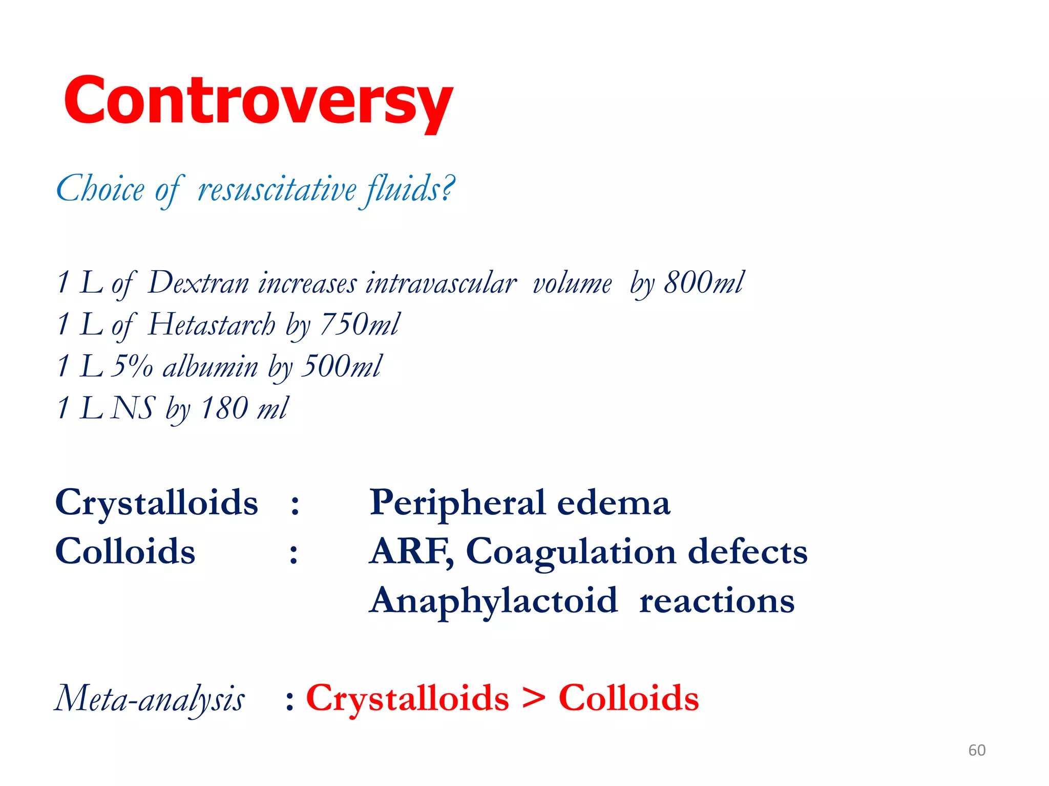
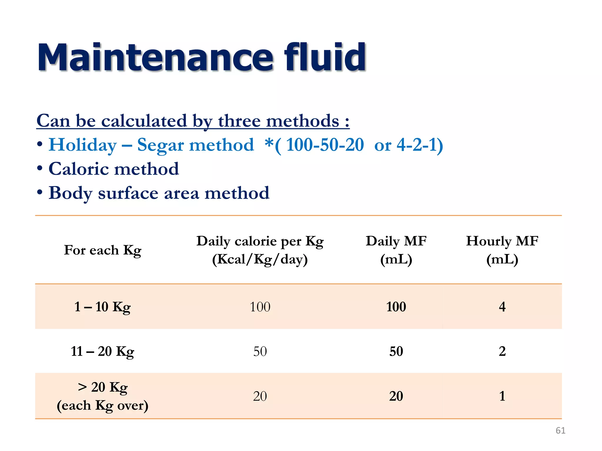
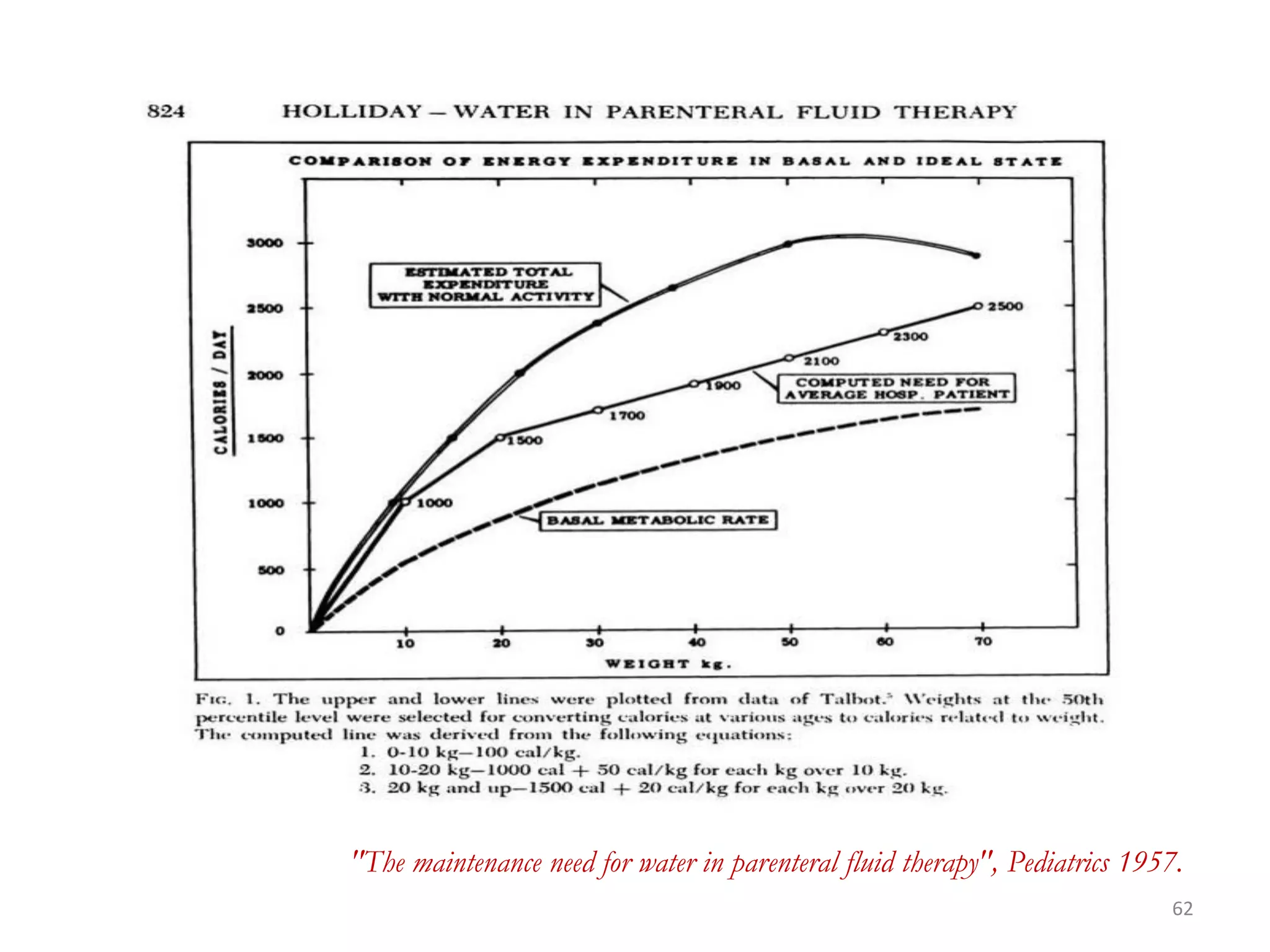
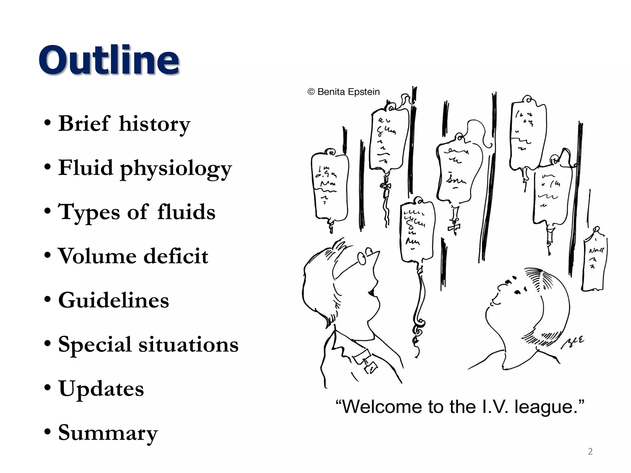
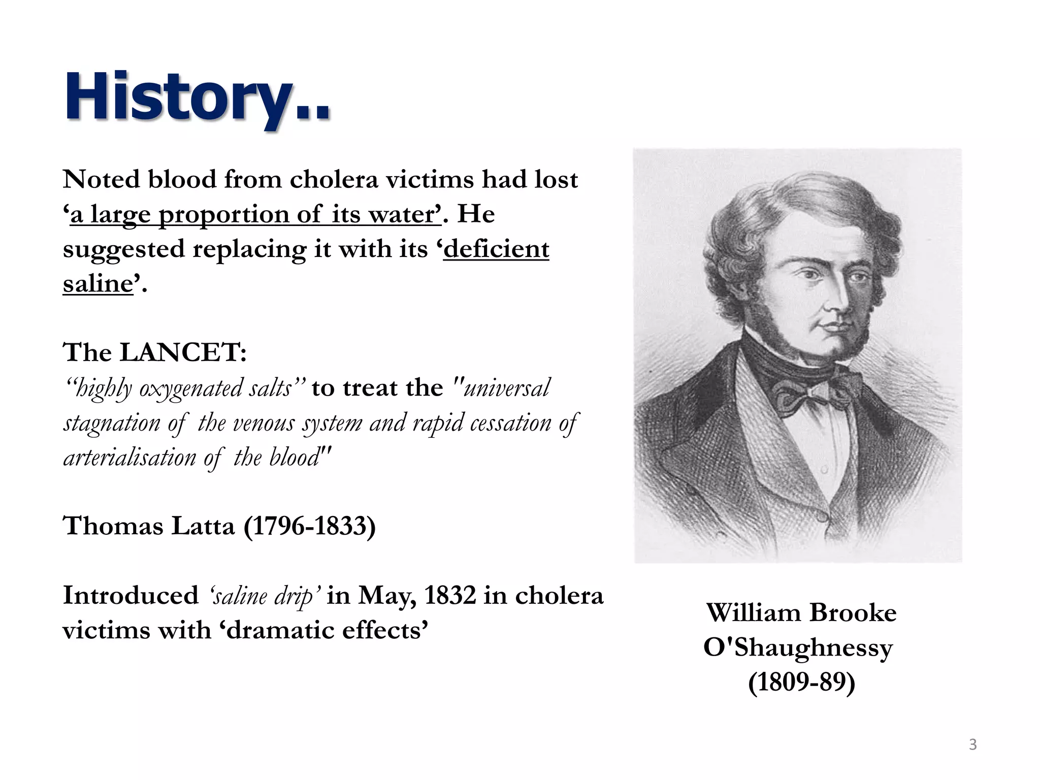
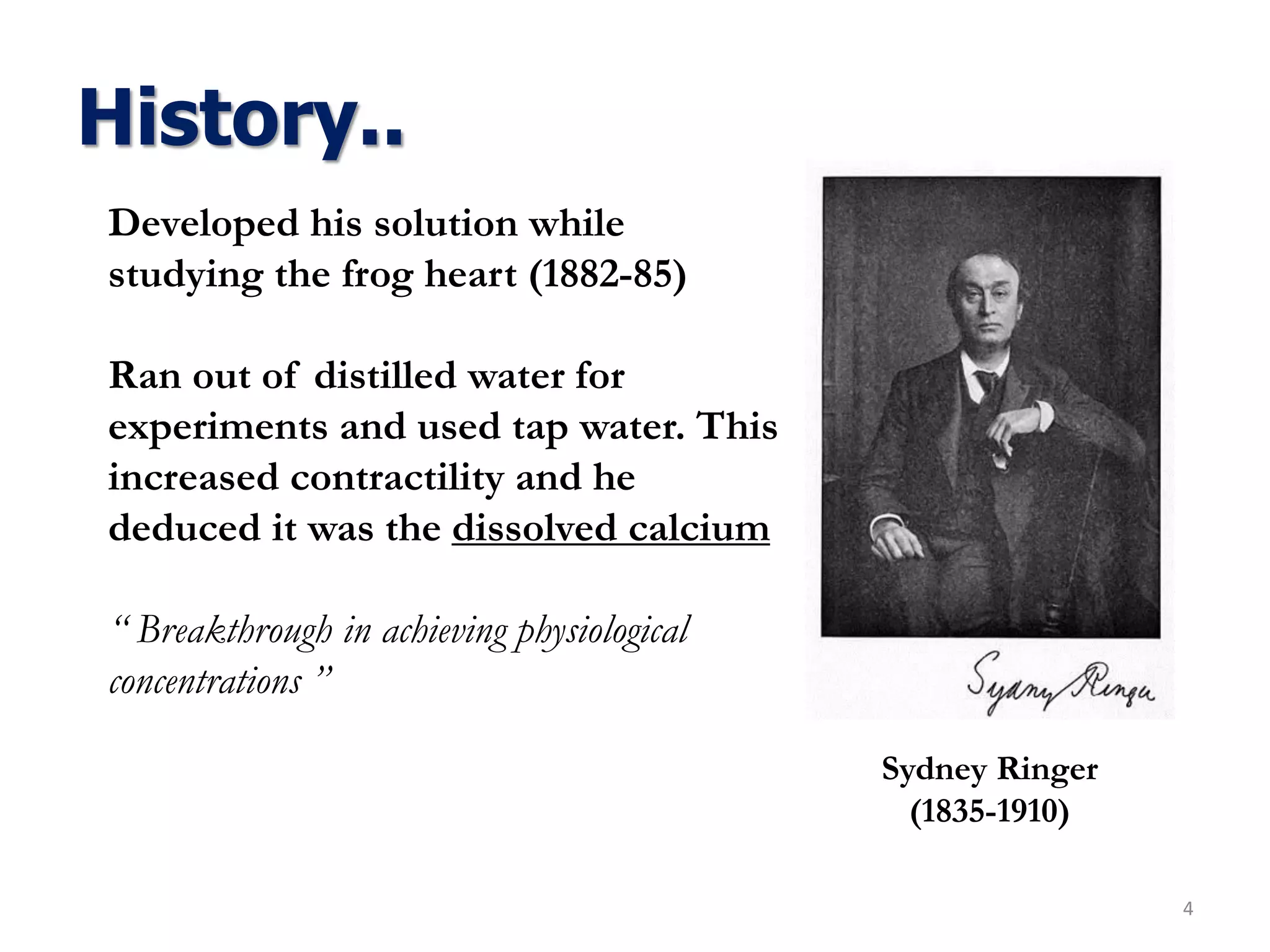
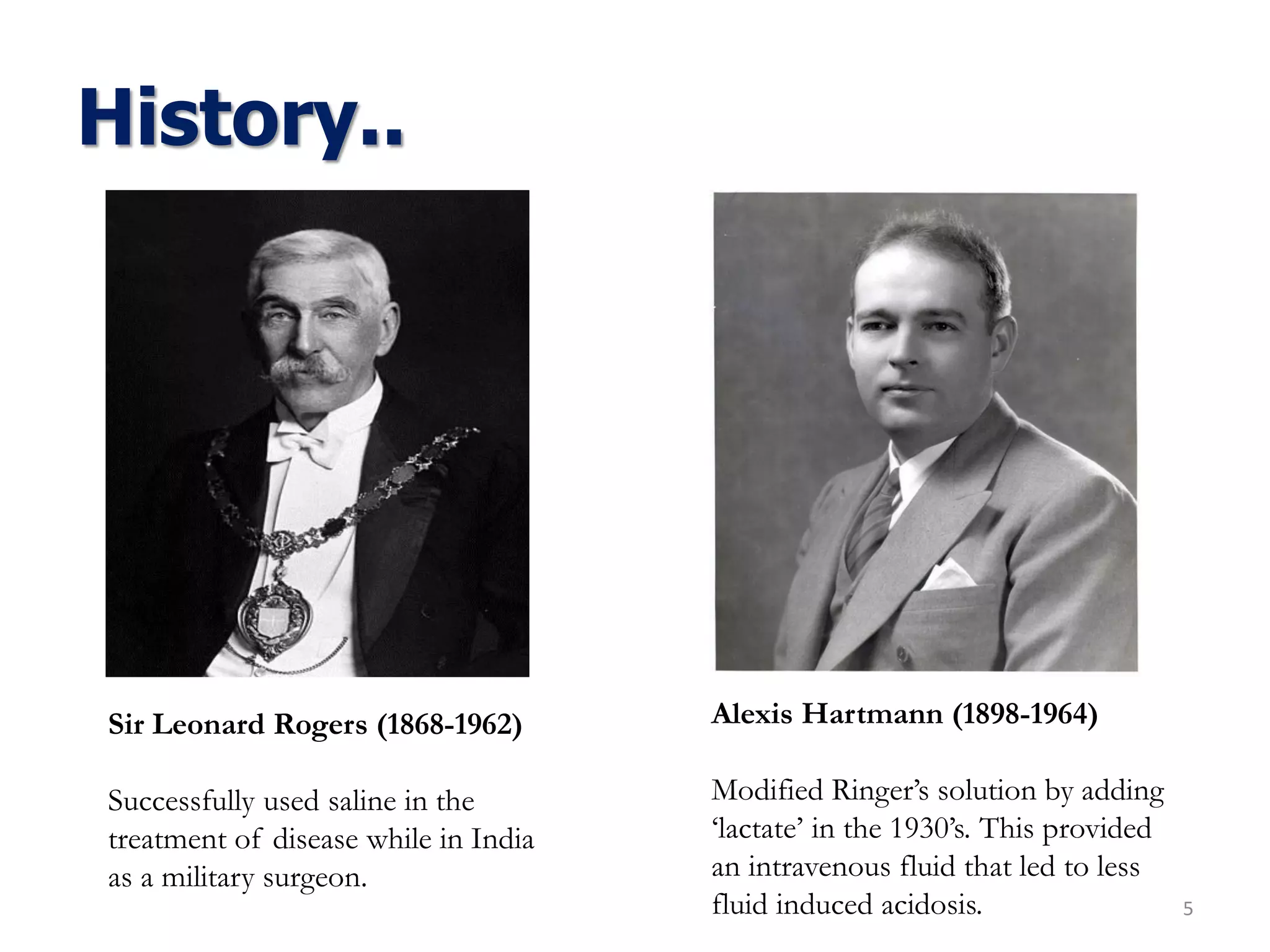
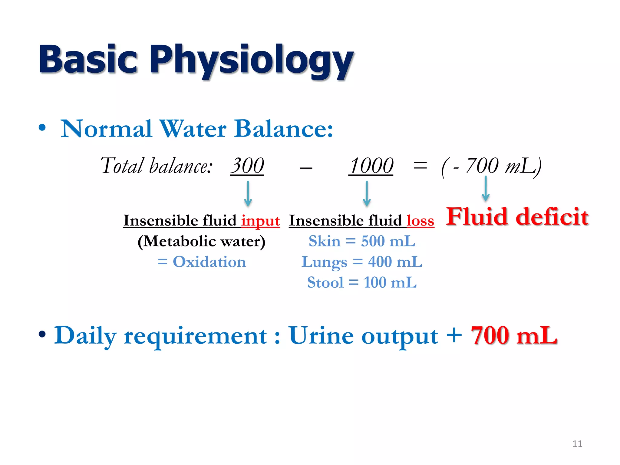
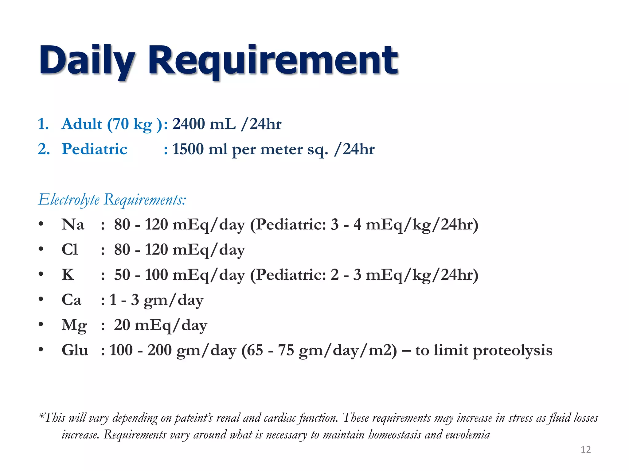
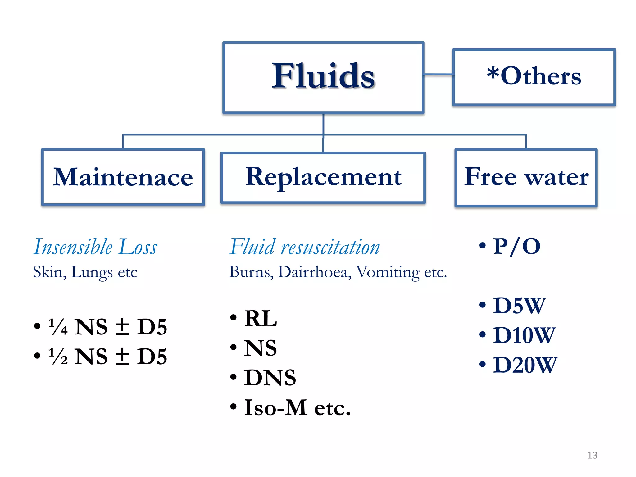
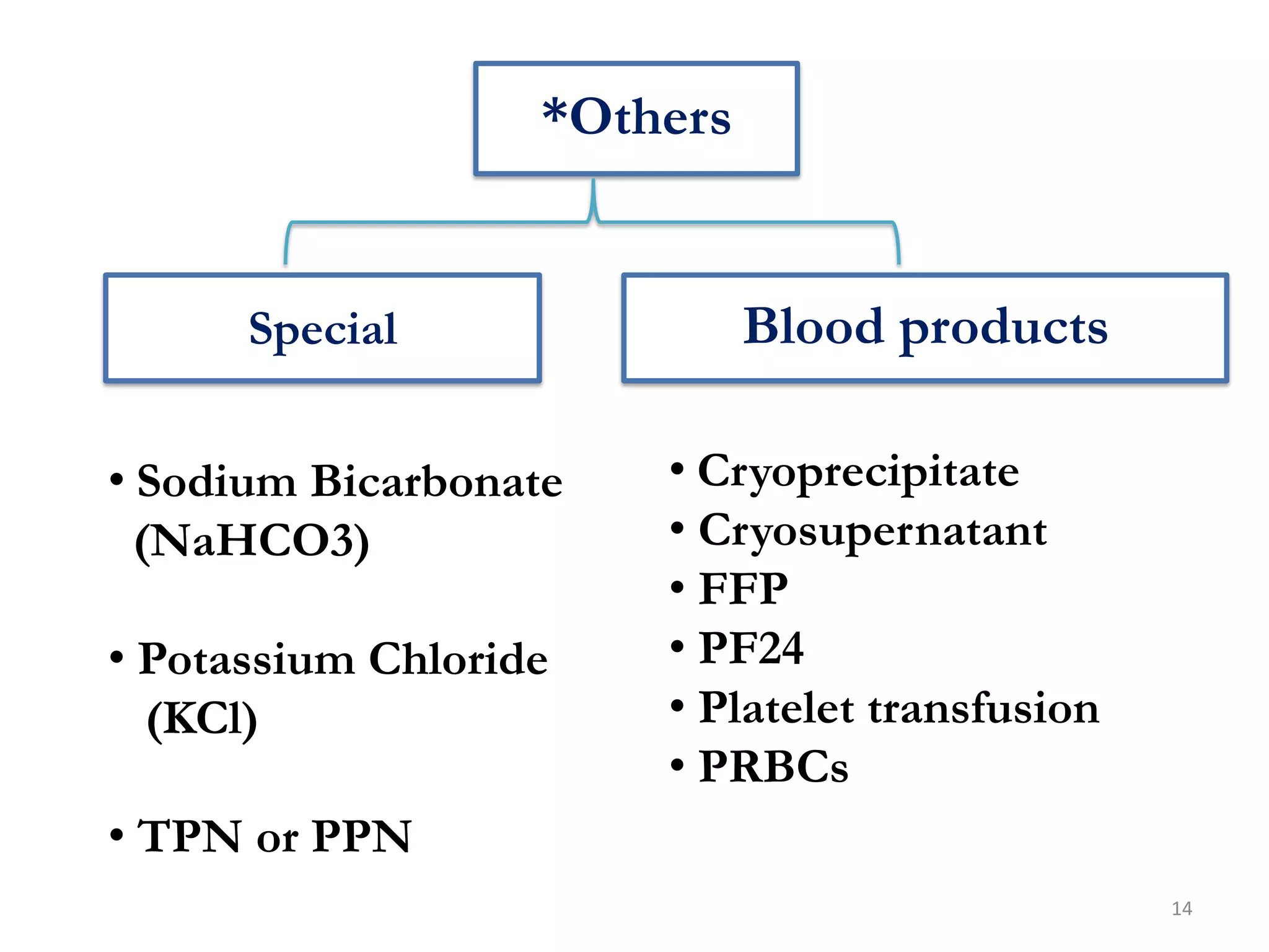
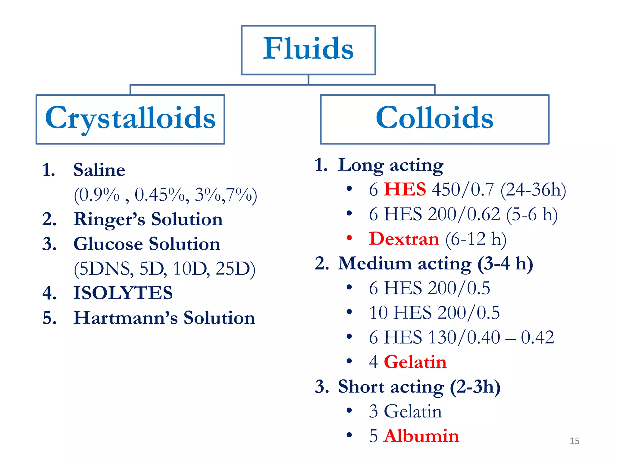
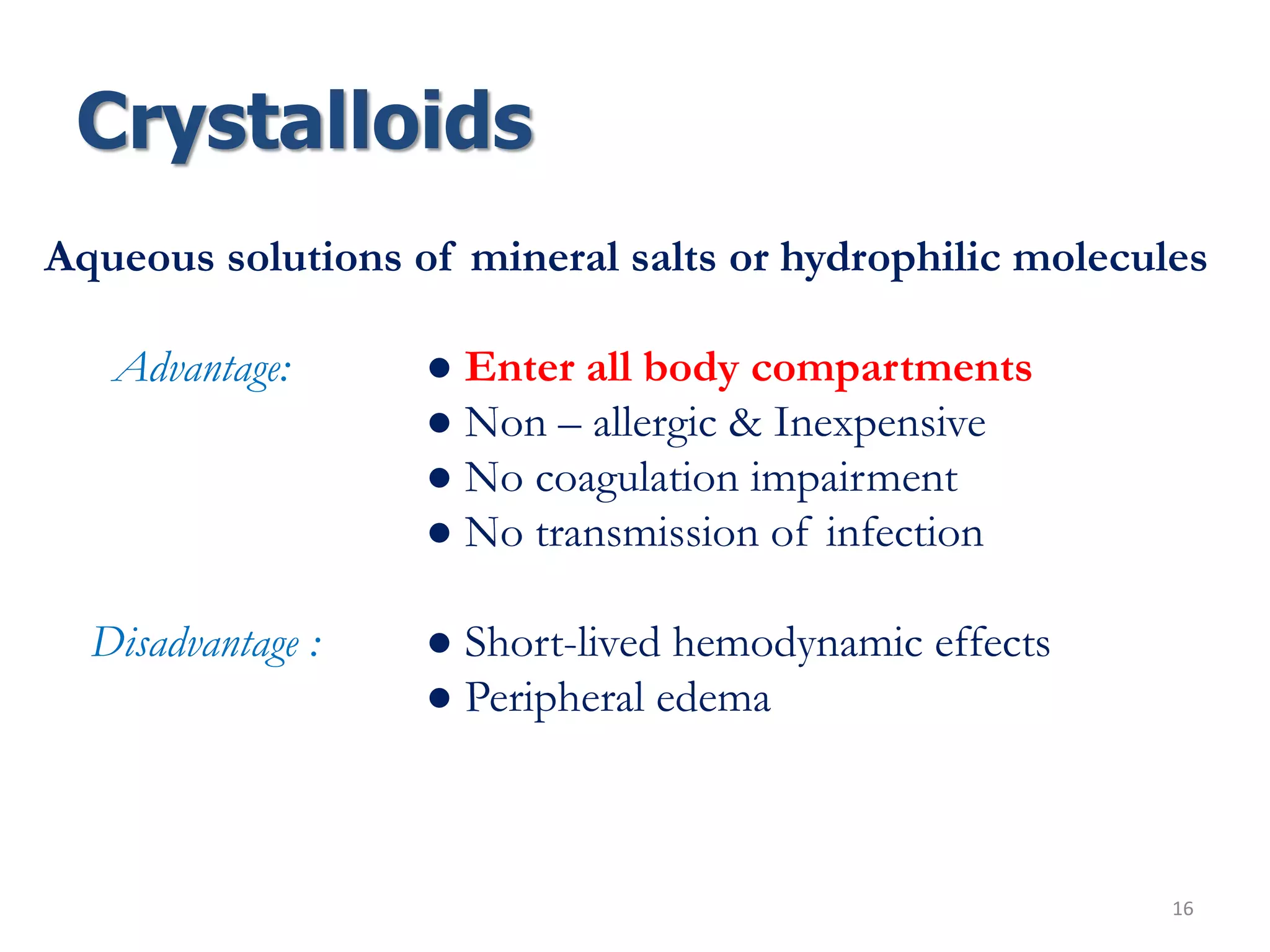
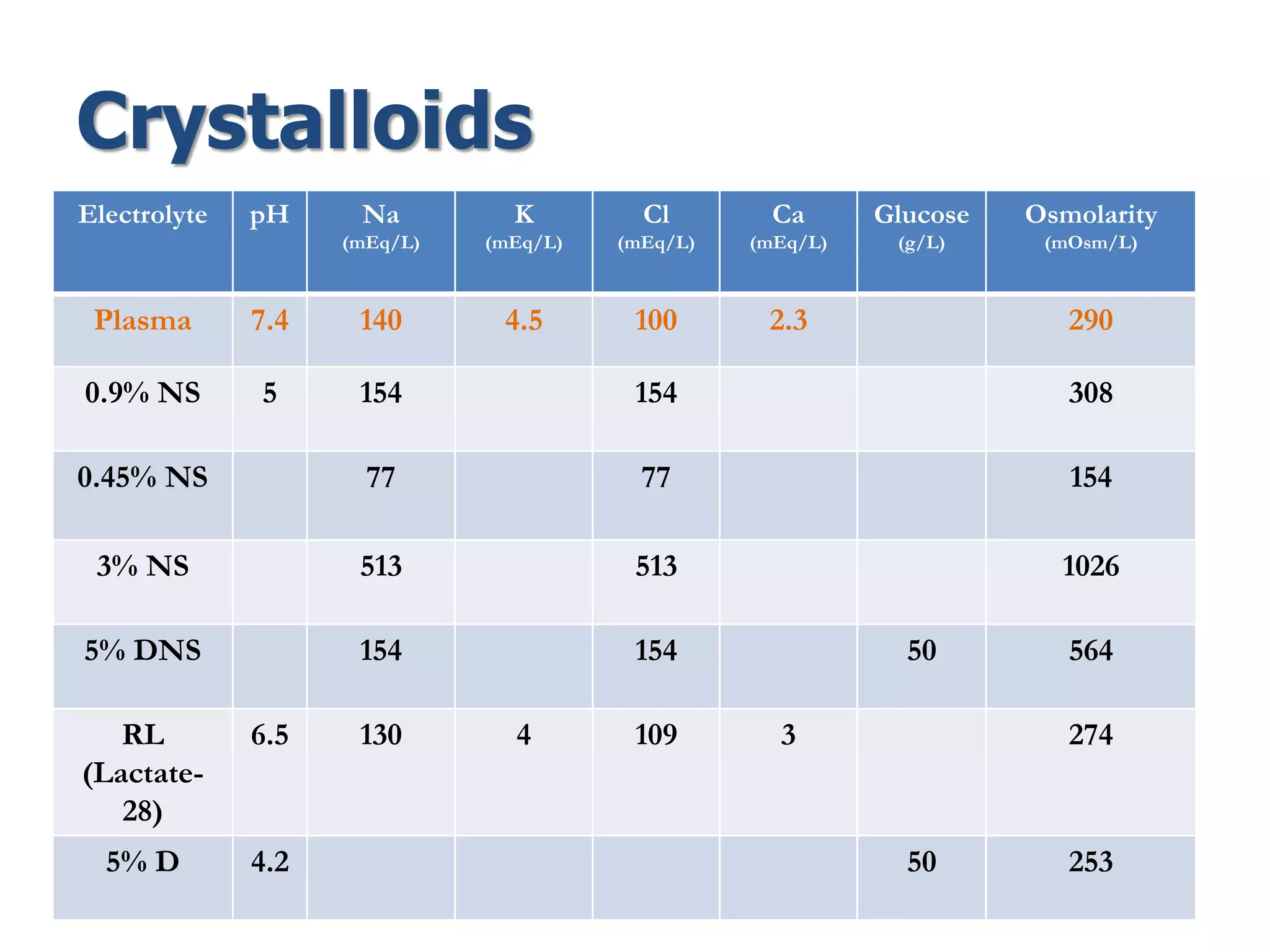
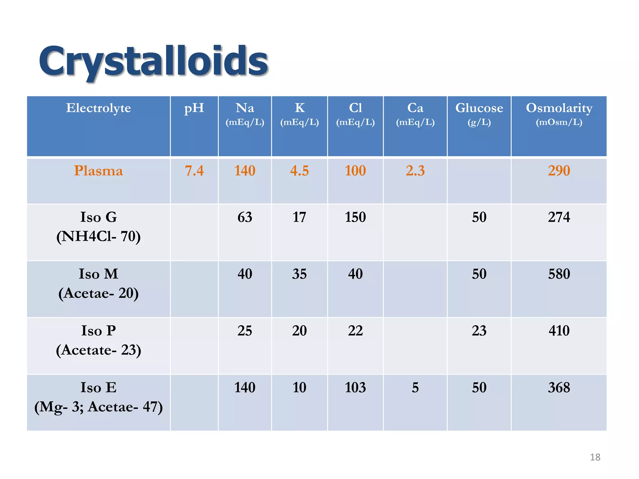
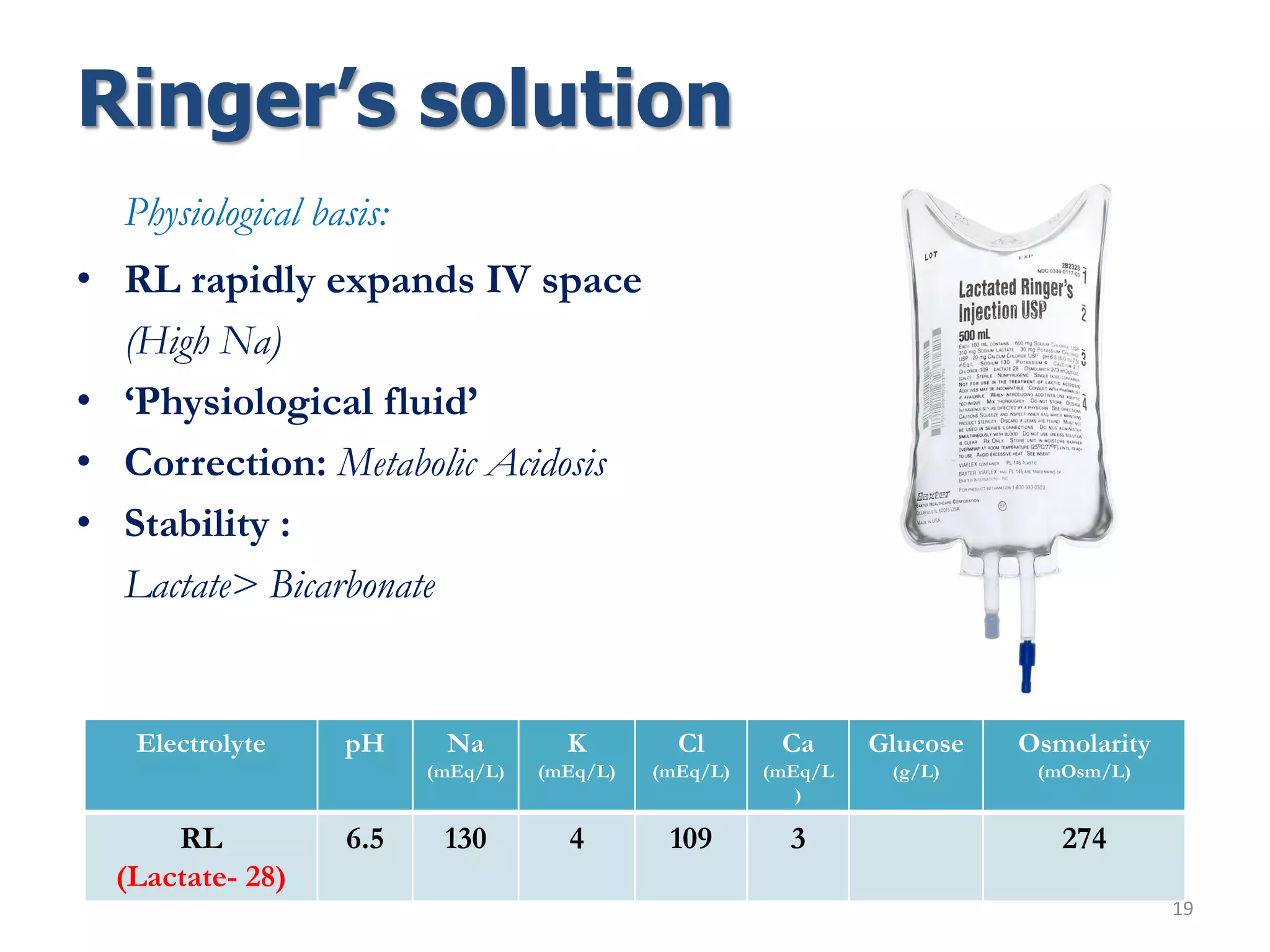
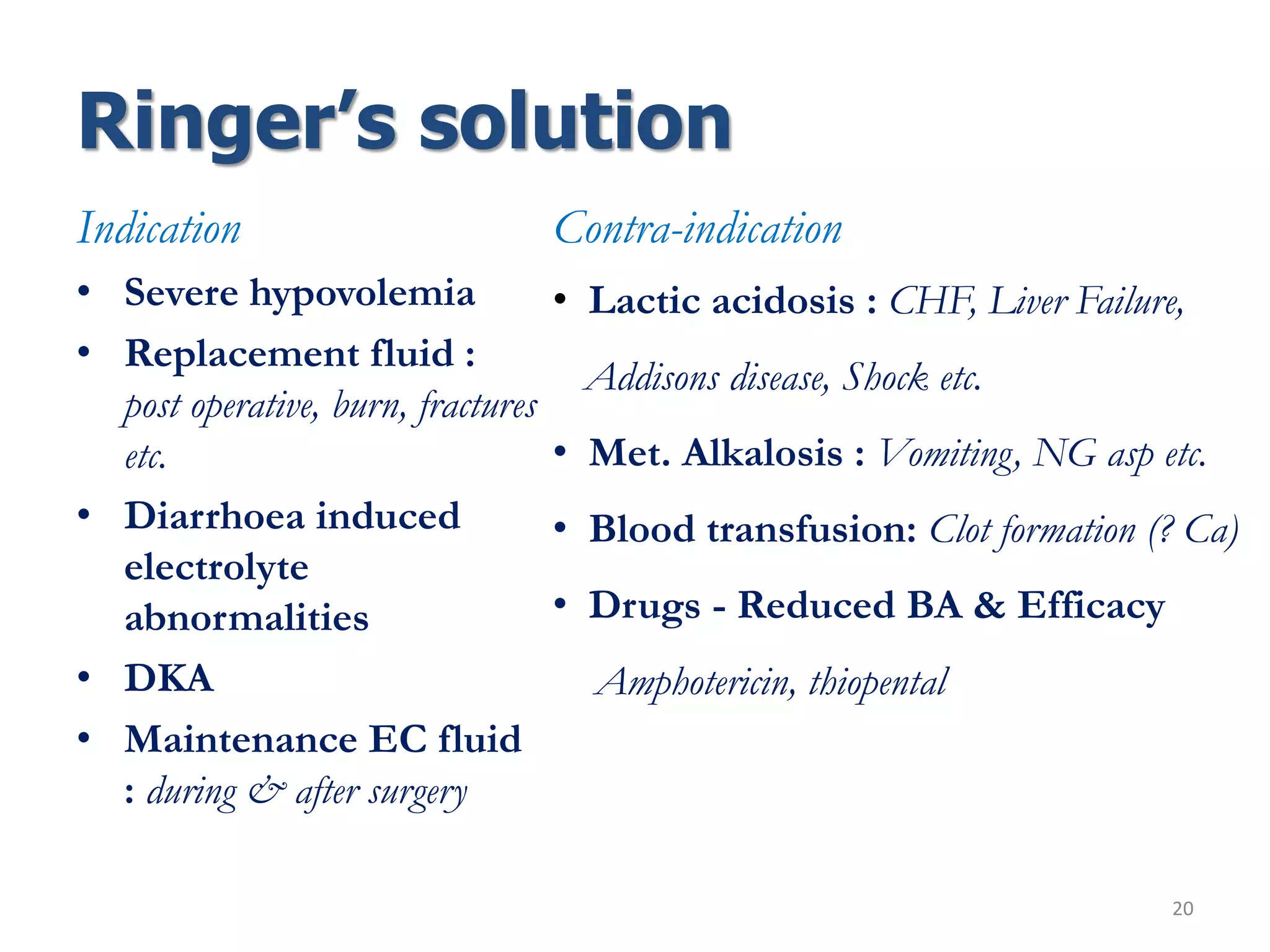
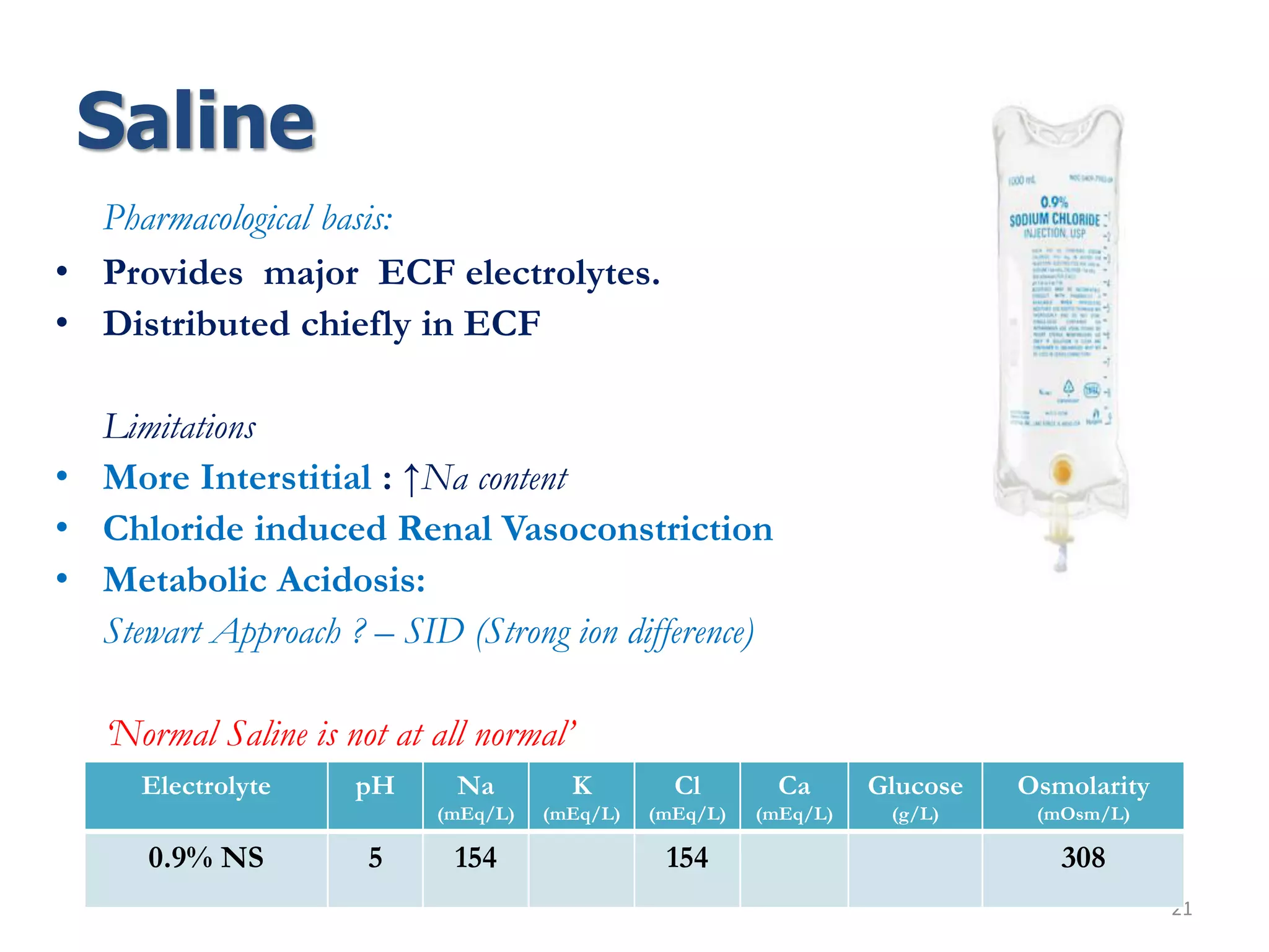
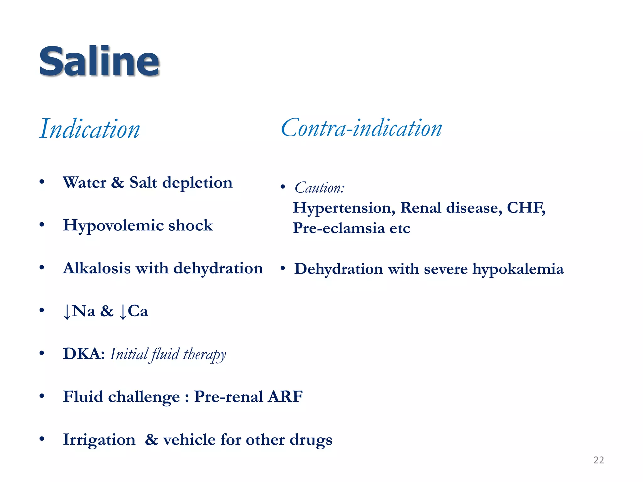
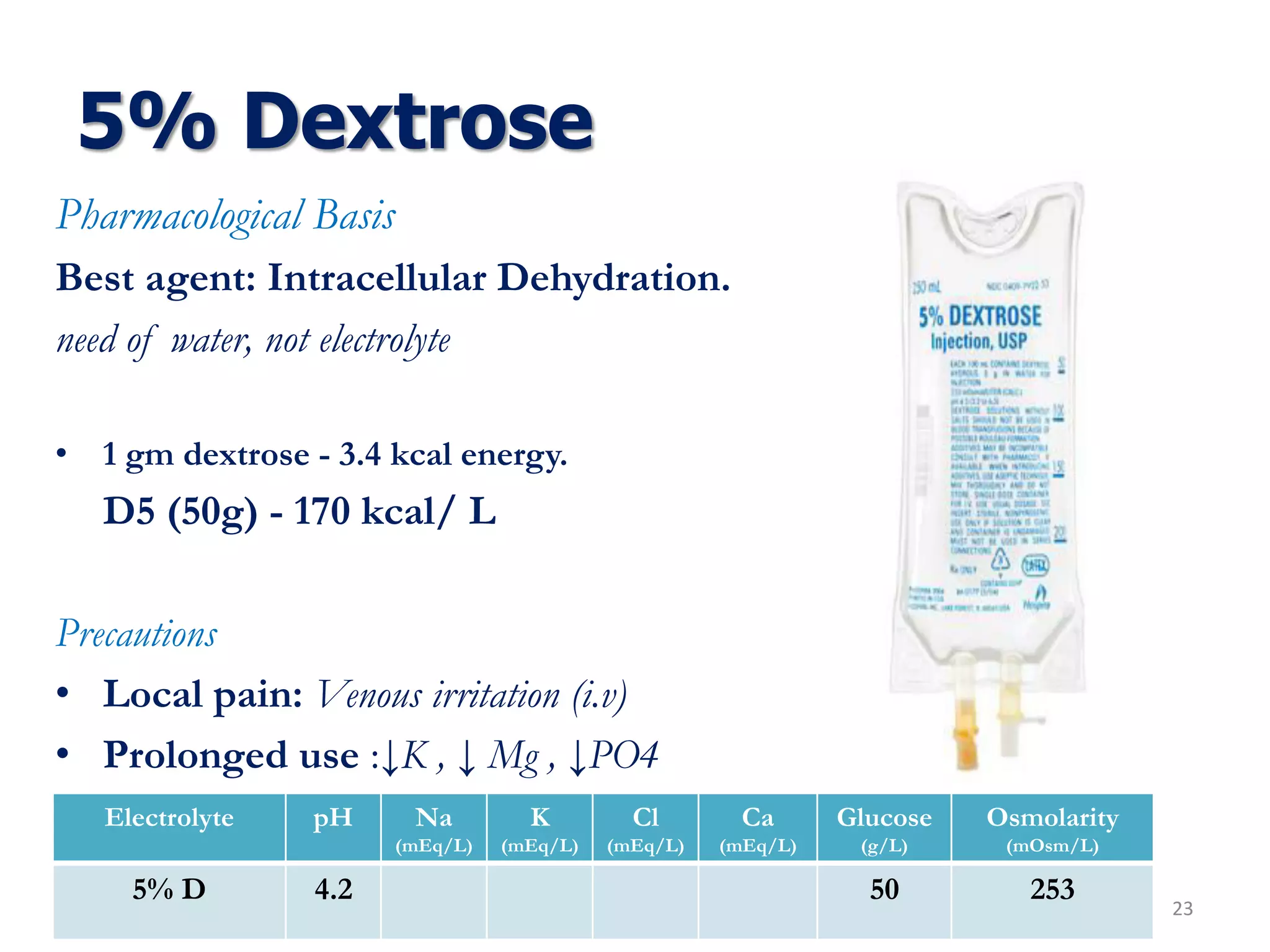
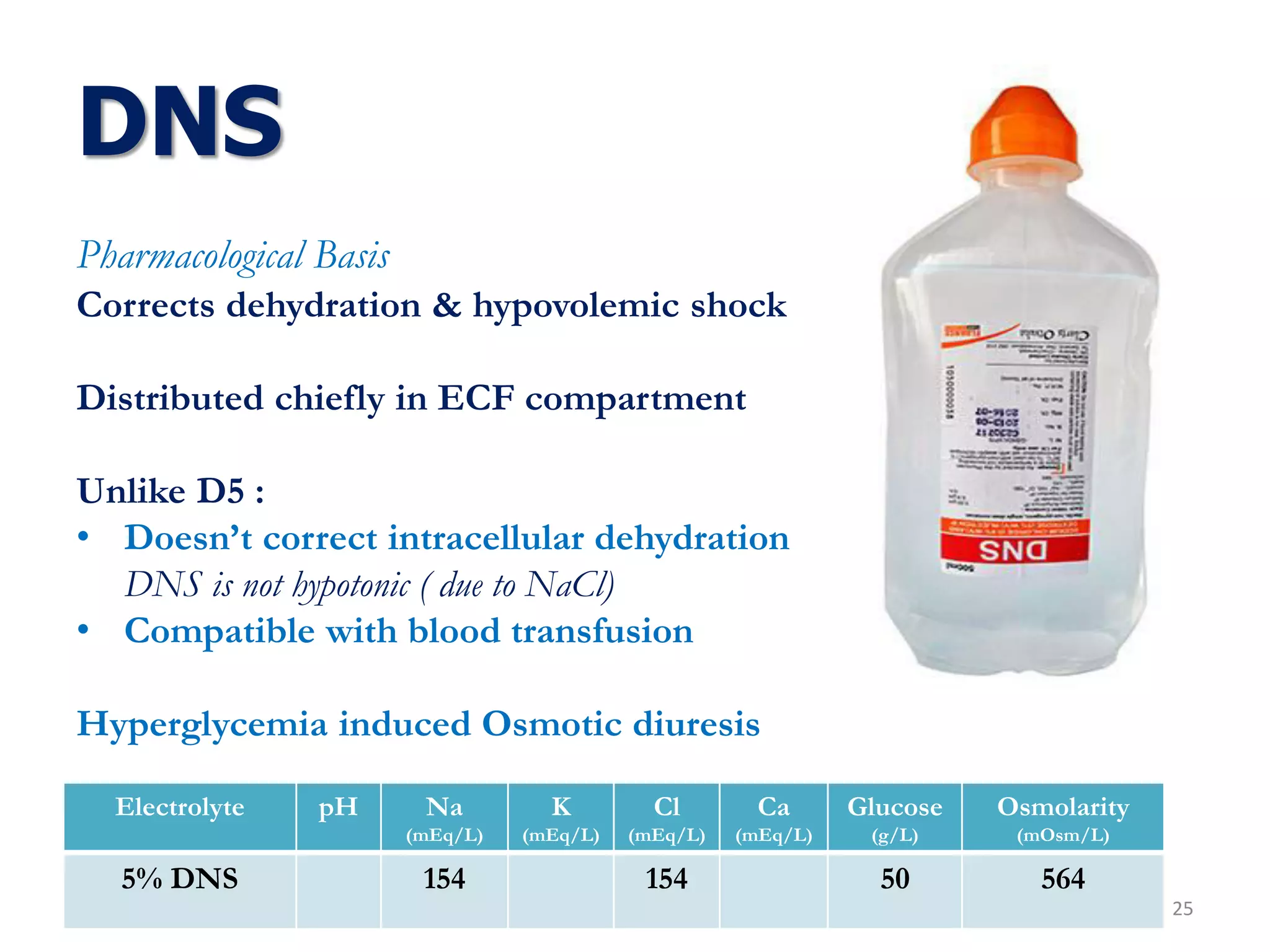
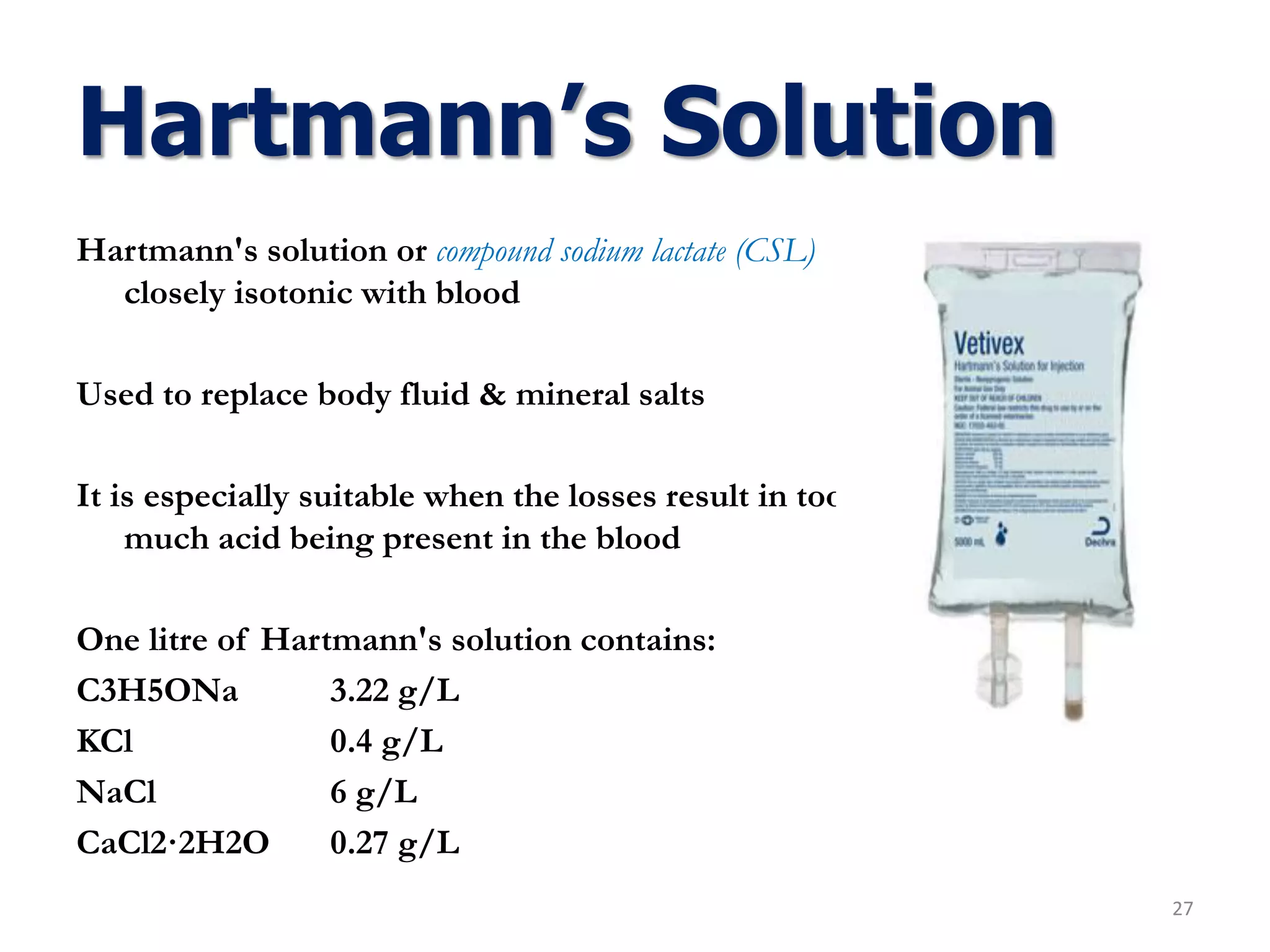
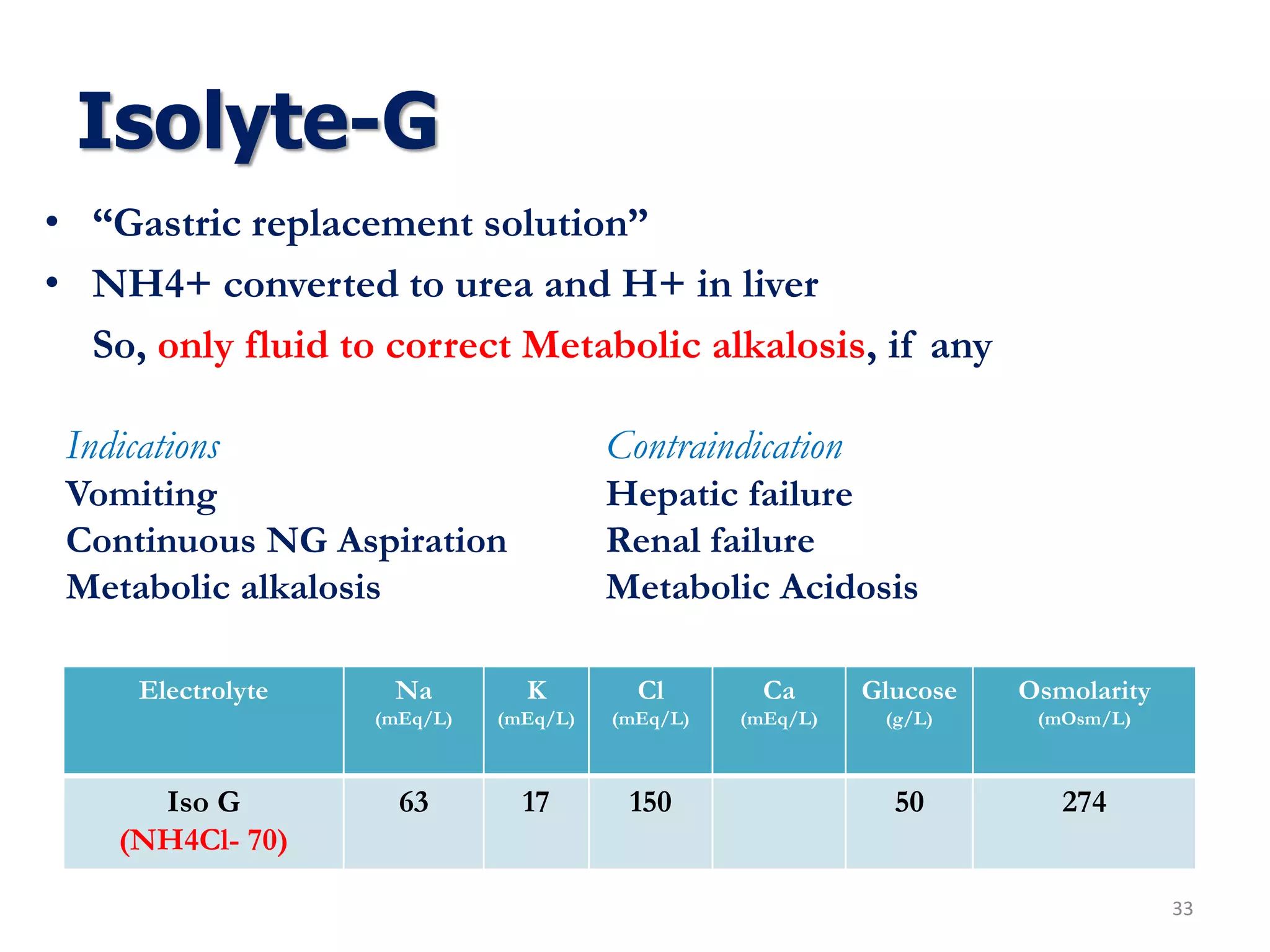
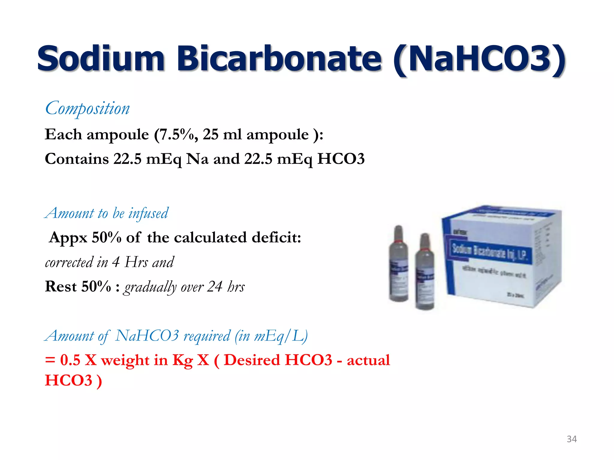
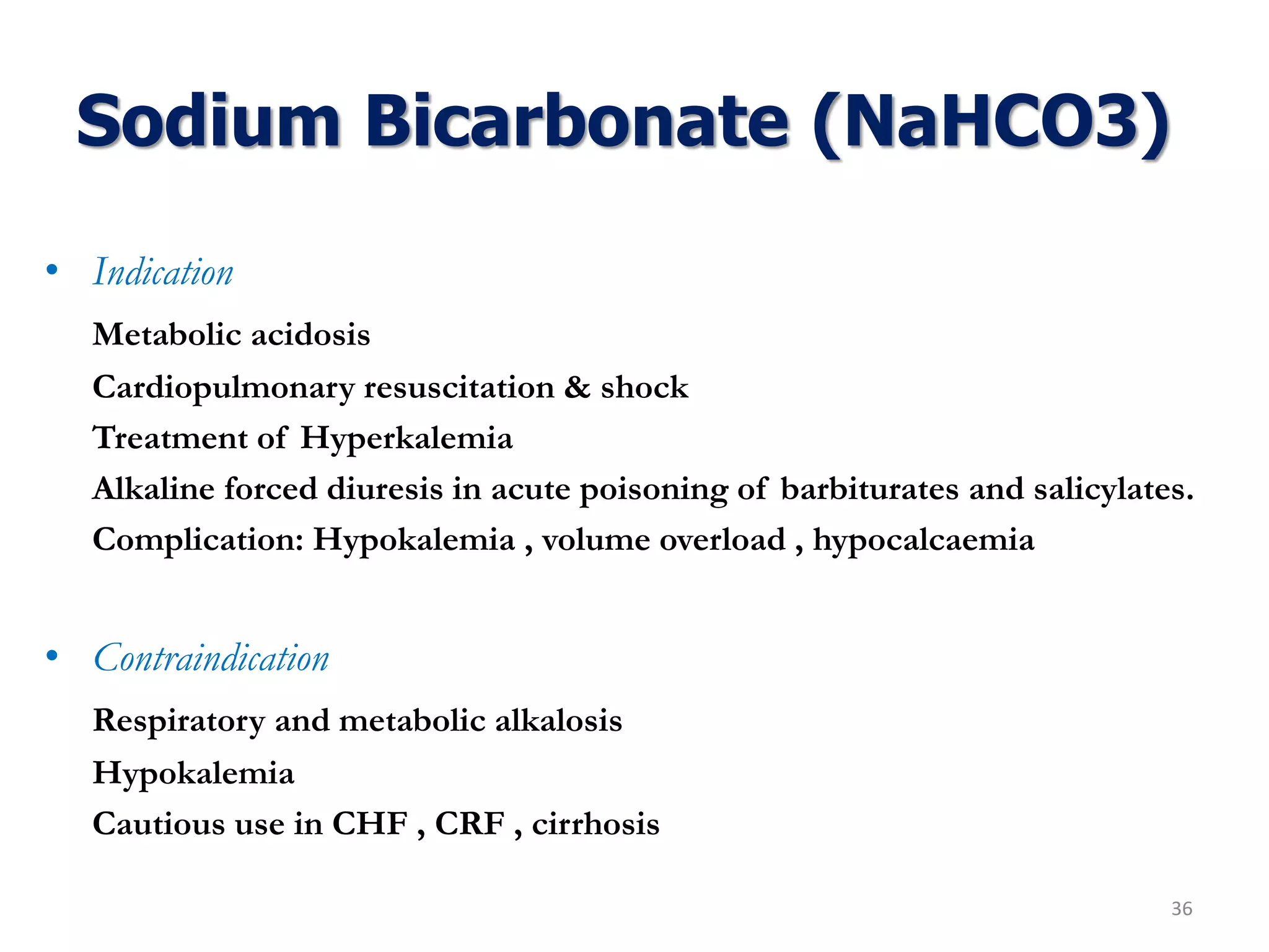
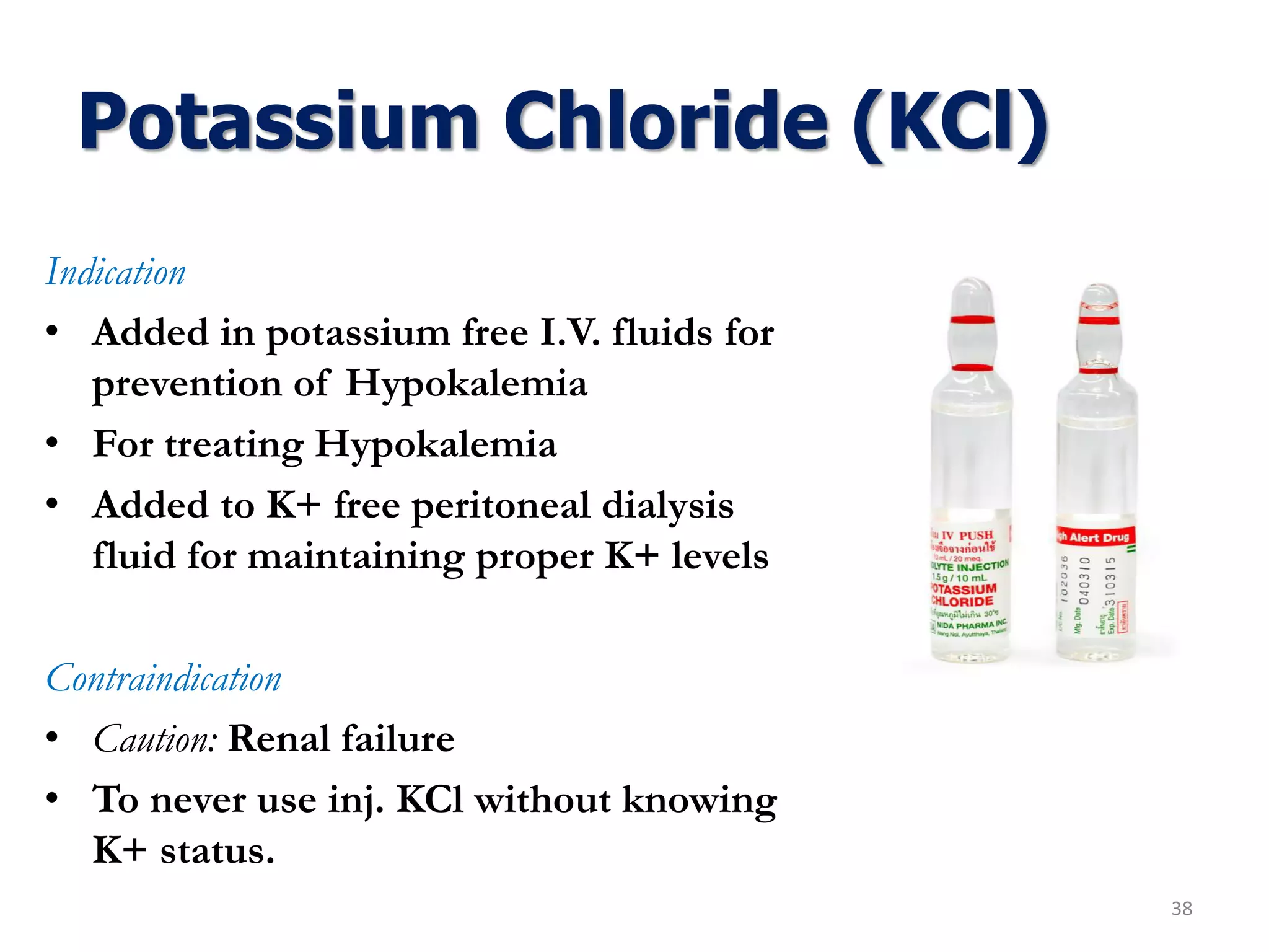
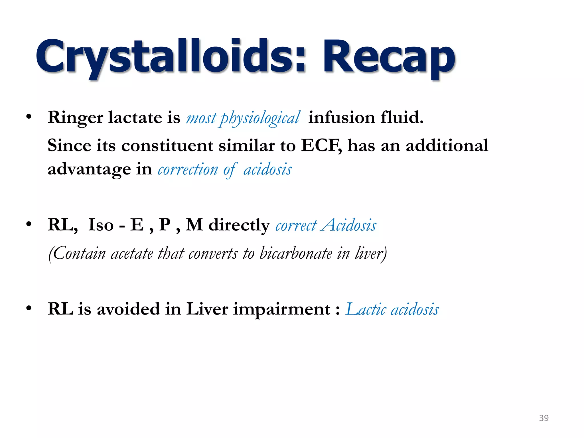
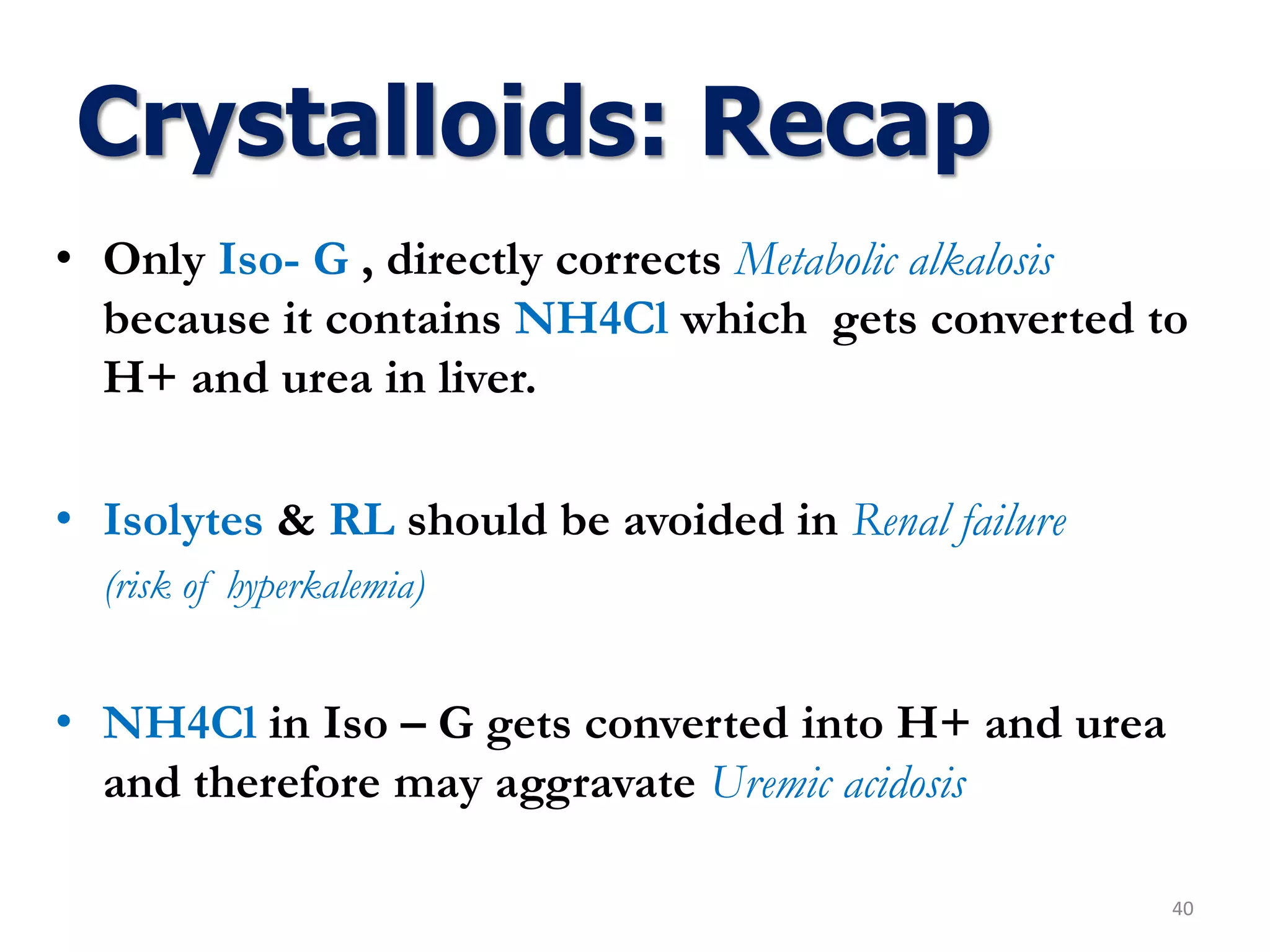
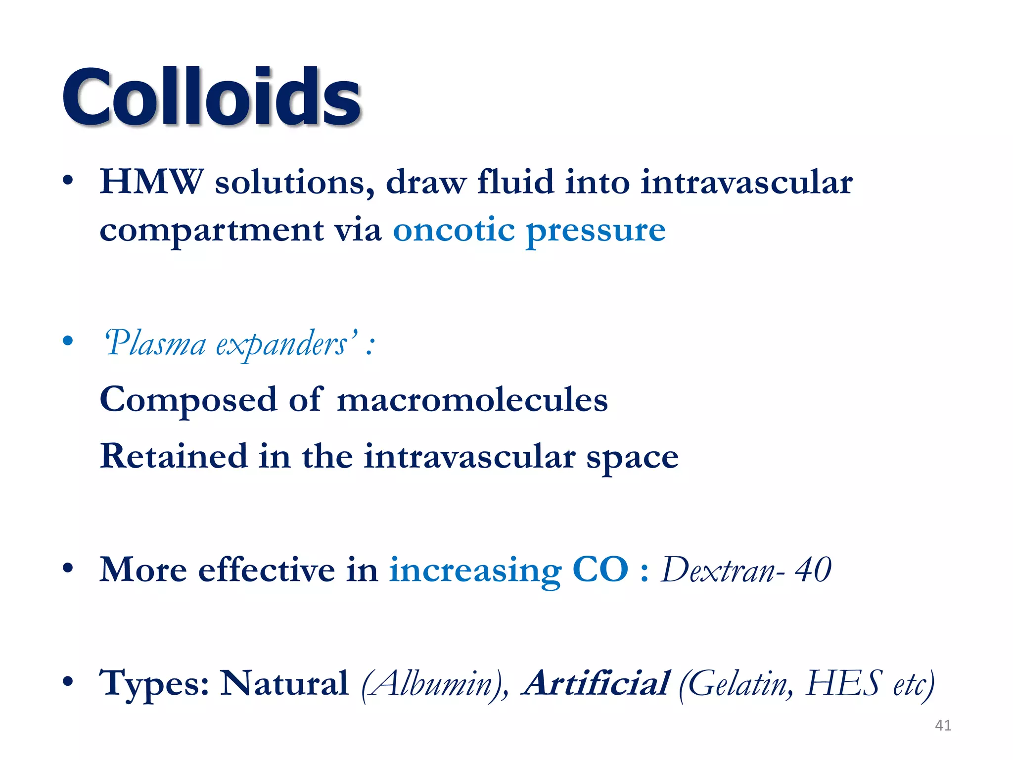
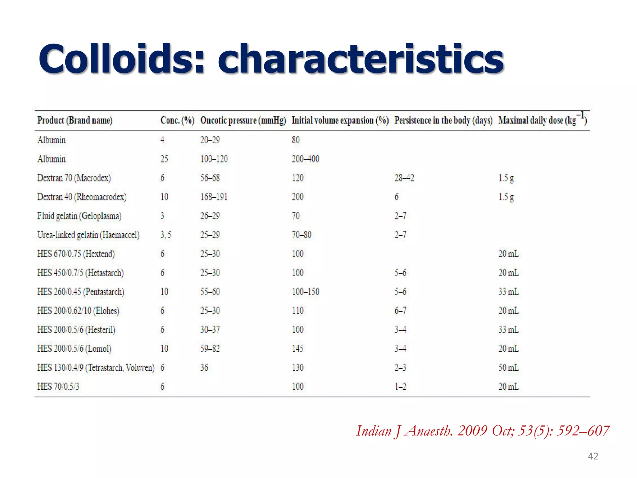
2. Outline
2
• Brief history
• Fluid physiology
• Types of fluids
• Volume deficit
• Guidelines
• Special situations
• Updates
• Summary
3. History..
3
William Brooke
O'Shaughnessy
(1809-89)
Noted blood from cholera victims had lost
‘a large proportion of its water’. He
suggested replacing it with its ‘deficient
saline’.
The LANCET:
‘‘highly oxygenated salts’’ to treat the "universal
stagnation of the venous system and rapid cessation of
arterialisation of the blood"
Thomas Latta (1796-1833)
Introduced ‘saline drip’ in May, 1832 in cholera
victims with ‘dramatic effects’
4. History..
4
Sydney Ringer
(1835-1910)
Developed his solution while
studying the frog heart (1882-85)
Ran out of distilled water for
experiments and used tap water. This
increased contractility and he
deduced it was the dissolved calcium
“ Breakthrough in achieving physiological
concentrations ”
5. History..
5
Sir Leonard Rogers (1868-1962)
Successfully used saline in the
treatment of disease while in India
as a military surgeon.
Alexis Hartmann (1898-1964)
Modified Ringer’s solution by adding
‘lactate’ in the 1930’s. This provided
an intravenous fluid that led to less
fluid induced acidosis.
6. Introduction
• Fluid therapy ?
“Accorded similar status as drug prescribing”
Myburgh JA, Mythen MG. Resuscitation fluids. N Engl J Med 2013;369:1243-51.
• Goals :
a. Normovolemia (osmolarity & cellular permeability)
b. Hemodynamic stability
c. Normal Electrolytemia or Electroneutrality
d. Stable tissue-perfusion
e. Adequate urine output
f. Avoidance of fluid overload & deficit
6
7. Basic Physiology
7
‘Third Space’ - Acute sequestration in body compartment not in
equilibrium with ECF . Eg: Intestinal obstruction – Severe pancreatitis – Peritonitis –
Major venous obstruction – Capillary leak syndrome – Burns
8. 8
Volume Homeostasis:
• Osmoregulation
• Volume regulation
• Thirst reflex
Cation+
(meq/L)
Anion-
(meq/L)
Na 10
K 150
Mg 40
PO4 10
SO4 30
HCO3 10
Protein 40
Na 142
K 4
Mg 2
Ca 4.5
Cl 102
PO4 2
SO4 1
HCO3 27
Protein 16
Organic acid 5
ICF ECF
200
153
153
200
9. Plasma Osmolality
Serum osmolality = 2Na + Glucose + BUN
9
18 2.8
• Normal : 285 (275 – 295) mOsm/L
Isotonic : 250-375 mOsm/L
Hypotonic : < 250 mOsm/L
Hypertonic : > 375 mOsm/L
Effective plasma osmolality is determined by those plasma solutes that do not freely permeate cell
membrane and act to hold water within the ECF.
Lipid soluble solutes such as urea which can cross the cell membrane hence do not contribute to
osmotic pressure gradient between ECF and ICF.
eOsm= 2 X Na (mEq/L) + Glucose (mg/dl) / 18 (mOsm / Kg)
10. Basic Physiology
Daily water balance :
Water Intake (2400 mL) = Water Loss (2400 mL)
Insensible Loss ↑ :
• Normal insensible loss 8 ml/kg (60 % respiration + 25 % skin)
• 500 ml : moderate sweating
• 1.0-1.5 lit : severe sweating / high fever
• 0.5-3.0 lit : exposed wound surface ( burns) & laprotomy
• Water needs 15% for every ºC rise in temp
10
Fluids : 1300 mL
Solid food : 800 mL
Metabolic water : 300 mL
Urine : 1300 mL
Faeces : 100 mL
Insensible loss : 1000 mL
11. Basic Physiology
• Normal Water Balance:
Total balance: 300 – 1000 = ( - 700 mL)
11
Fluid deficitInsensible fluid input
(Metabolic water)
= Oxidation
Insensible fluid loss
Skin = 500 mL
Lungs = 400 mL
Stool = 100 mL
• Daily requirement : Urine output + 700 mL
12. Daily Requirement
1. Adult (70 kg ): 2400 mL /24hr
2. Pediatric : 1500 ml per meter sq. /24hr
Electrolyte Requirements:
• Na : 80 - 120 mEq/day (Pediatric: 3 - 4 mEq/kg/24hr)
• Cl : 80 - 120 mEq/day
• K : 50 - 100 mEq/day (Pediatric: 2 - 3 mEq/kg/24hr)
• Ca : 1 - 3 gm/day
• Mg : 20 mEq/day
• Glu : 100 - 200 gm/day (65 - 75 gm/day/m2) – to limit proteolysis
*This will vary depending on pateint’s renal and cardiac function. These requirements may increase in stress as fluid losses
increase. Requirements vary around what is necessary to maintain homeostasis and euvolemia
12
13. Fluids
Maintenace Replacement Free water
*Others
13
Insensible Loss
Skin, Lungs etc
• ¼ NS ± D5
• ½ NS ± D5
Fluid resuscitation
Burns, Dairrhoea, Vomiting etc.
• RL
• NS
• DNS
• Iso-M etc.
• P/O
• D5W
• D10W
• D20W
16. Crystalloids
16
Aqueous solutions of mineral salts or hydrophilic molecules
Advantage: ● Enter all body compartments
● Non – allergic & Inexpensive
● No coagulation impairment
● No transmission of infection
Disadvantage : ● Short-lived hemodynamic effects
● Peripheral edema
19. Ringer’s solution
Physiological basis:
• RL rapidly expands IV space
(High Na)
• ‘Physiological fluid’
• Correction: Metabolic Acidosis
• Stability :
Lactate> Bicarbonate
19
Electrolyte pH Na
(mEq/L)
K
(mEq/L)
Cl
(mEq/L)
Ca
(mEq/L
)
Glucose
(g/L)
Osmolarity
(mOsm/L)
RL
(Lactate- 28)
6.5 130 4 109 3 274
20. Ringer’s solution
20
Indication
• Severe hypovolemia
• Replacement fluid :
post operative, burn, fractures
etc.
• Diarrhoea induced
electrolyte
abnormalities
• DKA
• Maintenance EC fluid
: during & after surgery
Contra-indication
• Lactic acidosis : CHF, Liver Failure,
Addisons disease, Shock etc.
• Met. Alkalosis : Vomiting, NG asp etc.
• Blood transfusion: Clot formation (? Ca)
• Drugs - Reduced BA & Efficacy
Amphotericin, thiopental
21. Saline
Pharmacological basis:
• Provides major ECF electrolytes.
• Distributed chiefly in ECF
Limitations
• More Interstitial : ↑Na content
• Chloride induced Renal Vasoconstriction
• Metabolic Acidosis:
Stewart Approach ? – SID (Strong ion difference)
‘Normal Saline is not at all normal’
21
Electrolyte pH Na
(mEq/L)
K
(mEq/L)
Cl
(mEq/L)
Ca
(mEq/L)
Glucose
(g/L)
Osmolarity
(mOsm/L)
0.9% NS 5 154 154 308
22. Saline
Indication
• Water & Salt depletion
• Hypovolemic shock
• Alkalosis with dehydration
• ↓Na & ↓Ca
• DKA: Initial fluid therapy
• Fluid challenge : Pre-renal ARF
• Irrigation & vehicle for other drugs
22
Contra-indication
• Caution:
Hypertension, Renal disease, CHF,
Pre-eclamsia etc
• Dehydration with severe hypokalemia
23. 5% Dextrose
Pharmacological Basis
Best agent: Intracellular Dehydration.
need of water, not electrolyte
• 1 gm dextrose - 3.4 kcal energy.
D5 (50g) - 170 kcal/ L
Precautions
• Local pain: Venous irritation (i.v)
• Prolonged use :↓K , ↓ Mg , ↓PO4
23
Electrolyte pH Na
(mEq/L)
K
(mEq/L)
Cl
(mEq/L)
Ca
(mEq/L)
Glucose
(g/L)
Osmolarity
(mOsm/L)
5% D 4.2 50 253
24. 5% Dextrose
Indications
• Dehydration
• i.v adm. of drugs
• Ketosis – Starvation,
diarrhoea, fever etc.
• Protects liver
• Correction of ↓Na in pure
water loss (DI)
24
Contraindications
• Cerebral edema
• Neurosurgical procedure
• Acute Ischaemic stroke
• Hypovolemic Shock
• ↓Na + Water intoxication
• Along with Oxytocin
• Blood transfusion
• Fetal distress
25. Pharmacological Basis
Corrects dehydration & hypovolemic shock
Distributed chiefly in ECF compartment
Unlike D5 :
• Doesn’t correct intracellular dehydration
DNS is not hypotonic ( due to NaCl)
• Compatible with blood transfusion
Hyperglycemia induced Osmotic diuresis
DNS
25
Electrolyte pH Na
(mEq/L)
K
(mEq/L)
Cl
(mEq/L)
Ca
(mEq/L)
Glucose
(g/L)
Osmolarity
(mOsm/L)
5% DNS 154 154 50 564
26. Indications
• Salt water depletion with supply of energy
• Vomitting or NG asp induced Hypocholeremic alkalosis
• Compatible with blood products
Contraindications
• Caution: Cardiac, Hepatic & Renal compromise
• Hypovolemic shock
DNS
26
27. Hartmann's solution or compound sodium lactate (CSL)
closely isotonic with blood
Used to replace body fluid & mineral salts
It is especially suitable when the losses result in too
much acid being present in the blood
One litre of Hartmann's solution contains:
C3H5ONa 3.22 g/L
KCl 0.4 g/L
NaCl 6 g/L
CaCl2·2H2O 0.27 g/L
Hartmann’s Solution
27
28. Contraindications
• Relatively contraindicated in patients with diabetes mellitus, as one of
the isomers of lactate is glucogenic
Side effects
• Fluid retention
• Symptoms: nausea, vomiting, headache, dizziness, drowsiness,
confusion
• Local inflammation
Hartmann’s Solution
28
Electrolyte pH Na K Cl Ca Glucose Osmolarity
Hartmann’s Sol
(Lactate- 29)
6.5 131 5 111 2 274
30. Isolyte-M
• Richest source of K+
• “Ideal maintenace fluid”
(corrects acidosis & supplies energy)
30
Indications
Maintenance fluid therapy
Hypokalemia (diarrhoeal loss)
Contraindication
Renal failure
↓Na & Water intoxication
Adrenocortical insufficiency
Burns
Electrolyte Na
(mEq/L)
K
(mEq/L)
Cl
(mEq/L)
Ca
(mEq/L)
Glucose
(g/L)
Osmolarity
(mOsm/L)
Iso M
(Acetate- 20)
40 35 40 50 580
31. Isolyte-P
Double water ;
Same electrolyte as ISO-M
Indications
Maintenance fluids : Infants
Adults: Excessive water loss (DI)
Contraindications
↓Na
Renal failure (↑K)
Hypovolemic shock
31
Electrolyte Na
(mEq/L)
K
(mEq/L)
Cl
(mEq/L)
Ca
(mEq/L)
Glucose
(g/L)
Osmolarity
(mOsm/L)
Iso P
(Acetate- 23)
25 20 22 23 410
32. Isolyte-E
“Extracellular replacement solution’’
– Corrects acidosis
– Supplies energy,
– Replaces water deficit.
Only fluid to correct Mg deficiency
(Double conc. Of K & acetate)
Indications
Diarrhoea
Metabolic acidosis
Maintenance of ECF volume
(pre-operatively)
Contraindication
Metabolic alkalosis:
Vomiting or continuous NG aspiration
Electrolyte Na
(mEq/L)
K
(mEq/L)
Cl
(mEq/L)
Ca
(mEq/L)
Glucose
(g/L)
Osmolarity
(mOsm/L)
Iso E
(Mg- 3; Acetae- 47)
140 10 103 5 50 368
33. Isolyte-G
• “Gastric replacement solution”
• NH4+ converted to urea and H+ in liver
So, only fluid to correct Metabolic alkalosis, if any
33
Indications
Vomiting
Continuous NG Aspiration
Metabolic alkalosis
Contraindication
Hepatic failure
Renal failure
Metabolic Acidosis
Electrolyte Na
(mEq/L)
K
(mEq/L)
Cl
(mEq/L)
Ca
(mEq/L)
Glucose
(g/L)
Osmolarity
(mOsm/L)
Iso G
(NH4Cl- 70)
63 17 150 50 274
34. Sodium Bicarbonate (NaHCO3)
Composition
Each ampoule (7.5%, 25 ml ampoule ):
Contains 22.5 mEq Na and 22.5 mEq HCO3
Amount to be infused
Appx 50% of the calculated deficit:
corrected in 4 Hrs and
Rest 50% : gradually over 24 hrs
Amount of NaHCO3 required (in mEq/L)
= 0.5 X weight in Kg X ( Desired HCO3 - actual
HCO3 )
34
35. Sodium Bicarbonate (NaHCO3)
Special precautions
• Should not be given as bolus except in emergency
• Avoid overdose & alkalosis by giving repeated small doses & monitoring pH
• Never to treat Acidosis without treating the etiology
• Renal failure: NaCO3 may cause tetany & pulmonary edema
• To never correct acidosis without correcting the associated hypokalemia
(NaHCO3 will shift K+ from ECF to ICF , this will aggravate hypokalemia)
• Never mix inj. Calcium with inj. NaHCO3 in same syringe as it may
precipitate calcium carbonate
• Avoid mixing of inj. NaHCO3 with inotropes
35
36. Sodium Bicarbonate (NaHCO3)
• Indication
Metabolic acidosis
Cardiopulmonary resuscitation & shock
Treatment of Hyperkalemia
Alkaline forced diuresis in acute poisoning of barbiturates and salicylates.
Complication: Hypokalemia , volume overload , hypocalcaemia
• Contraindication
Respiratory and metabolic alkalosis
Hypokalemia
Cautious use in CHF , CRF , cirrhosis
36
37. Potassium Chloride (KCl)
• Injectable KCl Composition
Commonly used preparation is 15% KCl 10ml
ampoule
1 ml = 150 mg KCl = 2mEq Potassium
So 1 ampoule = 10 ml = 20 mEq K+
• Basic rules for using Inj. KCl
To never give direct I.V. KCl injection
To never add >40 mEq / litre
To never infuse >10 mEq / hr
To never add KCl in Isolyte – M
To always use injection KCl diluted in infusion
To monitor serum K+ levels closely
37
38. Potassium Chloride (KCl)
Indication
• Added in potassium free I.V. fluids for
prevention of Hypokalemia
• For treating Hypokalemia
• Added to K+ free peritoneal dialysis
fluid for maintaining proper K+ levels
Contraindication
• Caution: Renal failure
• To never use inj. KCl without knowing
K+ status.
38
39. • Ringer lactate is most physiological infusion fluid.
Since its constituent similar to ECF, has an additional
advantage in correction of acidosis
• RL, Iso - E , P , M directly correct Acidosis
(Contain acetate that converts to bicarbonate in liver)
• RL is avoided in Liver impairment : Lactic acidosis
39
Crystalloids: Recap
40. • Only Iso- G , directly corrects Metabolic alkalosis
because it contains NH4Cl which gets converted to
H+ and urea in liver.
• Isolytes & RL should be avoided in Renal failure
(risk of hyperkalemia)
• NH4Cl in Iso – G gets converted into H+ and urea
and therefore may aggravate Uremic acidosis
40
Crystalloids: Recap
41. Colloids
• HMW solutions, draw fluid into intravascular
compartment via oncotic pressure
• ‘Plasma expanders’ :
Composed of macromolecules
Retained in the intravascular space
• More effective in increasing CO : Dextran- 40
• Types: Natural (Albumin), Artificial (Gelatin, HES etc)
41
44. Albumin
• Composition
Physiological plasma protein
Contributes 80% of normal oncotic pressure
Single polypeptide chain (585 a.a.); Mol. Wt. – 69,000 Dalton
Heat treated preparations commercially available :
5% solution (50 gm/dl), 20% (200gm/dL), 25% (250gm/dl)
As Na+ load is small, 25% albumin is also called :
Salt Poor Albumin
• Degree of volume expansion
5% solution is isooncotic (80%)
25% solution is hyperoncotic (200 – 400%) within 30 minutes.
The effect persists for 16 - 24 h
44
Dubois MJ, Vincent JL. Colloid Fluids. In: Hahn RG, Prough DS, Svensen CH, editors. Perioperative Fluid Therapy. 1st edition. New York: Wiley; 2007. pp.
153–611.
45. Albumin
Indication
• Plasma volume expansion: Acute hypovulemic shock, burns etc.
• Correction of hypoproteinemia as in Hepatic & Renal impairment
• Following Paracentesis, Liver transplantation, Acute Lung injury etc
• As an exchange fluid to replace removed plasma in Therapeutic plasmapheresis
Advantage
• Lesser side effects as it’s a natural colloid
• 25% Albumin has a greater degree of volume expansion than other colloids
• Albumin acts as a principal binding protein of endogenous and exogenous
substances.
• It also possesses antioxidant and scavenging effects.
• Albumin being negatively charged protein contributes to the formation of
normal anion gap, influencing the acid-base status
45
Dubois MJ, Vincent JL. Colloid Fluids. In: Hahn RG, Prough DS, Svensen CH, editors. Perioperative Fluid Therapy. 1st edition. New York: Wiley;
2007. pp. 153–611.
46. Albumin
Precautions and contraindications
• Fast infusion will rapidly increase circulatory volume with resultant
overload and pulmonary edema
• Severe anemia or Cardiac failure or low cardiac reserves
• Not to be used for parenteral nutrition
Volume overload:
• In septic shock the release of inflammatory mediators has been
implicated in increasing the ‘leakiness’ of the vascular endothelium
• The administration of exogenous albumin may compound the
problem by adding to the interstitial edema
46
Dubois MJ, Vincent JL. Colloid Fluids. In: Hahn RG, Prough DS, Svensen CH, editors. Perioperative Fluid Therapy. 1st edition. New York: Wiley;
2007. pp. 153–611.
47. Dextran
• Highly branched polysaccharide molecules which are available for use as an
‘artificial colloid’
• They are produced by synthesis using the bacterial enzyme dextran sucrase
from the bacterium Leuconostoc mesenteroides (B512 strain) which is growing in a
sucrose medium
Physiochemical properties
Two dextran solutions are now most widely used:
6% solution with an average molecular weight of 70,000 (dextran 70)
10% solution with an average weight of 40,000 (dextran 40, LMW-dextran)
Metabolism & Excretion
Kidneys primarily excrete dextran solutions.
Smaller molecules (14000–18000 kDa) are excreted in 15minutes, whereas
larger molecules stay in circulation for several days
Up to 40% of dextran-40 and 70% of dextran-70 remain in circulation at 12 h
47
Dextran 40 and dextran 70. A review.Atik M Arch Surg. 1967 May; 94(5):664-72.
48. Dextran
Degree of volume expansion
Both dextran-40 and dextran-70 lead to a higher volume expansion as
compared to HES and 5% albumin. The duration lasts for 6–12
hours
Indications
Dextran-40 is used mainly to improve micro-circulatory flow in
microsurgical re-implantations
Extracorporeal circulation: It has been used in extracorporeal
circulation during cardio-pulmonary bypass.
Advantages
Volume expansion: Dextrans leads to 100–150% ↑ in intravascular volume
Microcirculation: Dextran 40 helps in improving microcirculatory flow by
two mechanisms, i.e.,
1. by decreasing the viscosity of blood by haemodilution and
2. by inhibiting erythrocytic aggregation.
48Dextran 40 and dextran 70. A review.Atik M Arch Surg. 1967 May; 94(5):664-72.
49. Gelatin
Gelatin solutions were first used as colloids in man in 1915
Formed when the connective tissues of animals are boiled
They have the property of dissolving in hot water & forming a
jelly when cooled
It is a large MW protein formed from hydrolysis of collagen
New-generation Gelatins:
• Succinylated or modified fluid gelatins
(e.g., Gelofusine, Plasmagel, Plasmion)
• Urea cross-linked gelatins (e.g., Polygeline )
• Oxypolygelatins (e.g., Gelifundol )
• Polygeline ( Haemaccel, Hoechst )
49Roberts J, Nightingale P. Properties and use of gelatins. In: Webb AR, editor. Therapeutics. Germany: Braun; 2003. pp. 45–52.
50. Gelatin
Degree of volume expansion
Gelatins lead to 70 to 80% of volume expansion. But duration of action is
shorter in comparison to both albumin and starches
Indications
Hypovolemia due to acute blood loss.
Acute normovolaemic haemodilution
Extracorporeal circulation – cardiopulmonary by-pass
Volume pre-loading prior to regional anaesthesia
Advantages
Cost effective
No limit of infusion
No effect of renal impairment
50Roberts J, Nightingale P. Properties and use of gelatins. In: Webb AR, editor. Therapeutics. Germany: Braun; 2003. pp. 45–52.
51. HES
• HES are derivatives of Amylopectin
• First developed in United States in
the 1970s
• Since then, further generations of
HES have been developed, differing
in their mean molecular weight
(MW), molar substitution (MS), and
C2/C6 ratio
• Hydroxyethyl starches are identified
by three numbers, e.g., 10% HES
200/0.5 or 6% HES 130/0.4
51
Pharmacokinetics of hydroxyethyl starch in normal subjects.Yacobi A, Stoll RG, Sum CY, Lai CM, Gupta SD, Hulse JD
J Clin Pharmacol. 1982 Apr; 22(4):206-12.
52. HES
HES preparations are characterized by the following properties:
1. Concentration: low or iso-oncotic (6%) or high or hyperoncotic (10%)
2. Average Molecular Weight (MW):
low (≃70 kDa), medium ( 200 kDa), or high ( ≃ 450 kDa)
3. Molar substitution (MS): low (0.45–0.58) or high (0.62–0.70)
Higher the MS (hydroxyethyl groups) , the greater the resistance to degradation,
and consequently, the longer its intravascular persistence
4. C2/C6 ratio: low (<8) or high (>8).
The ratio refers to the site where substitution has occurred on the initial glucose
molecule. The higher the C2/C6 ratio, longer the half-life and hence, longer
persistence in the blood
52
Pharmacokinetics of hydroxyethyl starch in normal subjects.Yacobi A, Stoll RG, Sum CY, Lai CM, Gupta SD, Hulse JD
J Clin Pharmacol. 1982 Apr; 22(4):206-12.
53. HES
Degree of volume expansion
The increase in colloid osmotic pressure obtained with HES is equivalent to albumin.
HES results in 100% volume expansion similar to 5% albumin.
It results in greater volume expansion as compared to gelatins. Duration of volume
expansion is usually 8-12 h
Indications
Stabilization of systemic haemodynamics
Anti-inflammatory properties: HES has been shown to preserve intestinal
microvascular perfusion in endotoxaemia due to their anti inflammatory properties
Advantages
Cost effectiveness
Maximum allowable transfusable volume: 50 ml/kg
53
Pharmacokinetics of hydroxyethyl starch in normal subjects.Yacobi A, Stoll RG, Sum CY, Lai CM, Gupta SD, Hulse JD
J Clin Pharmacol. 1982 Apr; 22(4):206-12.
54. HES
Metabolism
Following the infusion of HES there is initially a rapid amylase-dependent
breakdown and renal excretion.
Plasma half life is 5 days and 90% is eliminated in 42 days.
Smaller HES molecules (<50,000 to 60,000 Dalton) are eliminated rapidly
by glomerular filtration.
Medium sized molecules get excreted into the bile and faeces. Another
fraction is taken up by the reticulo-endothelial system (RES) where the
starch is slowly broken down.
Thus, trace amounts of the preparations can be detected for several weeks
after administration
54
Pharmacokinetics of hydroxyethyl starch in normal subjects.Yacobi A, Stoll RG, Sum CY, Lai CM, Gupta SD, Hulse JD
J Clin Pharmacol. 1982 Apr; 22(4):206-12.
56. Severity of dehydration?
56
Acute volume loss triggers two compensatory responses:
1. Transcapillary Refill
(shift if interstitial fluids to bloodstream)
2. Activation of RAAS
Compensate for 15-20% of volume loss
Cheuvront, Samuel N., and Robert W. Kenefick. "Dehydration: physiology, assessment, and performance effects."
Comprehensive Physiology (2014).
57. Volume deficit ?
57
Volume deficits are best estimated by acute changes in Weight
Deficit may be Pure water deficit or Combined water & electrolyte deficit
Pure water deficit is reflected biochemically by hypernatremia, increase in plasma, osmolality,
concentrated urine, and low urine [Na+] (<15mEq/L)
Treatment involves replacement of enough water to restore plasma [Na+] to normal
The excess Na+ for which water must be provided can be estimated from the
following equation:
ΔNa = (140 Na – plasma Na) × TBW
ΔNa : represents the total Na+ (mEq) in excess of water
*Divide ΔNa by 140 to obtain the amount of water required to return serum Na+ to
140mEq/L
58. Volume deficit ?
58
This fluid deficit must be corrected in addition to giving maintenance fluids
for ongoing obligatory losses.
Combined water and electrolyte deficit is commonly associated with “Third
spacing’
Urine Na+ is often < 10mEq/L as a result of Na+conservation from RAAS
system
Decreased blood volume diminishes renal perfusion and often produces
Pre-renal azotemia (BUN: Creatinine = 20-25: 1)
The composition of the correction fluid should take into account the plasma
[Na+]. If it is normal, fluid and electrolyte losses are probably isotonic, and
the replacement fluid should be isotonic NS or its equivalent.
59. Volume deficit ?
59
Thus replacement therapy should be planned in 2 steps:
1. Na+ deficit ?
2. ECF Volume deficit ?
a. clinical findings and b. changes in body wt
Signs : ↓ Skin turgor, dry mucous membrane, ↑Pulse rate, ↑SBP
Thirst : An early symptom;
Serum Osmolarity > 300mOsm/L : Dehydration
Urine output : < 30mL/hr or 720mL/day;
Narrow pulse pressure: <20 mmHg ? Severe
Fluid loss : BUN & Hct
Thomas, David R., et al. "Understanding clinical dehydration and its treatment." Journal of the American Medical Directors Association 9.5 (2008): 292-301.
60. Controversy
60
Choice of resuscitative fluids?
1 L of Dextran increases intravascular volume by 800ml
1 L of Hetastarch by 750ml
1 L 5% albumin by 500ml
1 L NS by 180 ml
Crystalloids : Peripheral edema
Colloids : ARF, Coagulation defects
Anaphylactoid reactions
Meta-analysis : Crystalloids > Colloids
61. Maintenance fluid
61
Can be calculated by three methods :
• Holiday – Segar method *( 100-50-20 or 4-2-1)
• Caloric method
• Body surface area method
For each Kg
Daily calorie per Kg
(Kcal/Kg/day)
Daily MF
(mL)
Hourly MF
(mL)
1 – 10 Kg 100 100 4
11 – 20 Kg 50 50 2
> 20 Kg
(each Kg over)
20 20 1
63. Monitoring end-points
63
1. Urine Output:
>0.5ml/kg/hr (1ml/kg/hr for a child)
2. Restoration of normal mental status and lack of
clinical signs of deficit
3. Other endpoints:
Normalization of laboratory parameters.
(BUN: Creatinine ratio and electrolyte values)
Gaieski, David F. et al. "Early goal-directed hemodynamic optimization combined with therapeutic hypothermia in comatose survivors of out-of-hospital
cardiac arrest." Resuscitation 80.4 (2009): 418-424.
65. Update: Diagnosis of Hypovolemia
65
Dehydration (ICF) ≠ Volume depletion (ECF)
Fluid deficit ?
Posture related syncope
Reflex tachycardia while standing or sleeping
Clinical examination & Lab tests
Poor sensitivity: Capillary refill time (CRT); Skin turgidity
Intravascular volume therapy in Adults: Guidelines from the association of Scientific Medical Societies in
Germany, April 1; 2016
66. Diagnosis of Hypovolemia?
66
Clinical examination
• Tongue, Jugular vein, Nail blanch test
• Pulmonary auscultation(Pleural effusion?)
• Abdominal palpation (Ascites?)
• Dependent edema
Lab tests
• Urea- creatinine ratio Advanced volume loss?
• Hypernatremia
• Base excess : tissue hypoxia; underperfusion
• Lactate : anaerobic metabolism; ischemic damage
• SCVO2 : desaturation of venous RBCs
Intravascular volume therapy in Adults: Guidelines from the association of Scientific Medical Societies in
Germany, April 1; 2016
67. Diagnosis of Hypovolemia?
67
CVP ?
“poor correlation between CVP and volume responsiveness; must not be used to
diagnose volume deficit in spontaneously breathing or ventilated patient”
Intravascular volume therapy in Adults: Guidelines from the association of Scientific Medical Societies in Germany, April 1; 2016
Marik, Paul E., Michael Baram, and Bobbak Vahid. "Does central venous pressure predict fluid responsiveness?: a systematic review of the literature and the tale of
seven mares." CHEST Journal 134.1 (2008): 172-178.
Existing guidelines : ( those including CVP)
1. S3 Guidelines: Post Infarction, Cardiogenic Shock: Diagnosis Monitoring & Therapy (2012)
2. Surviving Sepsis Campaign Guidelines For Managememt Of Severe Sepsis And Septic Shock (2012)
3. The German ‘S2K’ Sepsis Guideline (2010)
68. Diagnosis of Hypovolemia?
68
Indicators of Volume responsiveness: Latest recommendation
1. Fluid Challenge
2. Positional maneuvers facilitating Autotransfusion
(Passive Leg Rising / Trendenlenburg position: 300-450 mL)
Contraindication: Raised ICP, Visceral surgeries, Spontaneous breathing etc ? Risk of Aspiration
3. Volume preload indices
Intrathoracic Blood volume & Global EDV
Intravascular volume therapy in Adults: Guidelines from the association of Scientific Medical Societies in Germany, April 1; 2016
69. Diagnosis of Hypovolemia?
69
Indicators of Volume responsiveness: Latest recommendation
4. Dynamic preload indices
SVV; PPV, SPV
5. Echocardiography (TTE / TEE)
• Compulsory in ICUs and suspected cardiac events
• IVC diameter :
< 10mm : Hypovolemia likely
> 22mm : Hypervolemia
Intravascular volume therapy in Adults: Guidelines from the association of Scientific Medical Societies in Germany, April 1; 2016
70. Special Indications
70
• Post – operative
• Renal & Hepatic failure
• Trauma ± Head injury
• Burns
• Diabetic Ketoacidosis
71. Post – operative
71
For maintenance fluids : 4/2/1 rule
Req/day: 1-2 mmol/kg of Na + 0.5-1 mmol/kg of K+
So for a 55-60kg, euvolemic person :
A rate of approx 125 ml per hour of 0.45%DNS + 20 mEq of
KCl per litre per day will give : apx. 100 meq of Na+ and 60
meq of K+ per day.
Provides free water for insensible losses
Dextrose prevents catabolism and proteolysis
72. Renal & Hepatic failure
72
• Prevention of Contrast Induced Nephropathy :
Hydration with NS at 100-150ml/hr
• Reduce intake in patients who are oliguric
• Avoid K+ containing solution. Monitor electrolytes
• Use hypertonic solutions with caution
• In polyuric phase – replacement with NS (0.9%).
Rate equal to previous hours urine output
• Liver failure - use Dextrose, avoid RL
73. Trauma with Head injury
73
Resuscitate with isotonic fluids preferably NS.
Avoid synthetic colloid in patients with head injury or ICH
ESICM task force guidelines. Intensive Care Med 2012;38:368-83
Avoid dextrose containing solutions
Avoid hypotonic solutions
‘There is no advantage in hypertonic fluids with brain injury’
JAMA 2004 Mar 17; 291(11):1350-7
Mannitol : Effective only if BBB intact
74. Burns
74
Loss of Na and water in first 6-8hrs and continues for 48hrs
• Initial resuscitation – Parkland formula
First 24 h: RL at 4ml/kg/% TBSA; give half in first 8 h & the
remaining over next 16 h
Second 24 h: Colloid at 20-60% of calculated plasma volume to
maintain adequate urinary output
• After initial resuscitation use a combination fluid : Infusion of
albumin and 5%D.
• From 3rd day reduce intake as there is sodium and water
reabsorption.
75. Diabetic Ketoacidosis
75
Water and electrolyte loss due to osmotic diuresis
Hyperkalemia due to acidosis. However there is total body
potassium deficit
Initial resuscitation with 1sotonic saline or RL along with i.v
Insulin 0.1units/kg/hr
After 3-4L of NS give 0.45%NS to avoid Hyperchloremic Acidosis
Give 0.45%DNS when blood sugar is <200mg/dl
20-30mEq/L of K+ to be added if <5.3 and patient has good
urine output
77. Summary
• Accurate fluid balance is a myth
• ‘NS is normal at all’
• RL is most ‘physiological’ fluid
• Never to treat Acidosis without treating the etiology
• CVP or Hct : Unreliable indicators to determine fluid loss
• Volume deficit:
– Arterial base deficit, Lactate, Osmolarity
– Volume responsiveness: PLR, Fluid challenge, Echo Indexes
• Four phases of intravenous fluid therapy: A conceptual model
• Volume resuscitation: Crystalloids > Colloids (metanalysis)
• Monitoring endpoints: Urine output, clinical signs, BUN & electrolytes
77