Восточноевропейское научное журнал, зарегистрированное и публикуемое в Польше, охватывает все области науки и публикуется на нескольких языках. Каждое из 12 ежегодных изданий включает рецензируемые статьи, и авторы получают бесплатный экземпляр журнала. Открытый доступ к электронной версии журнала также предоставляется.
![-6-
WschodnioeuropejskieCzasopismoNaukowe(EastEuropeanScientificJournal)|NAUKIMEDYCZNEINAUKIOZDROWIU#IІІ,2015
NAUKI MEDYCZNE I NAUKI O ZDROWIU - МЕДИЦИНСКИЕ
НАУКИ
REHABILITATION OF MYOFASCIAL PAIN SYNDROM IN OVERTENSION RELATED DISEASES
Petya Kasnakova, PhD
Medical University Medicl college Plovdiv
Bulgaria
aBSTRACT
Myofascial pain syndrome (MPS) is connected with the presence of pain points /trigger points/, combined with trophic changes
and functional complaints. MPS includes several diseases, varied in their etiology and clinical symptoms - insertionitis, bursitis,
periarthritis, tendovaginitis, etc., characterized by pain and damaged joint function, without affecting the joint cavity. Rehabilitation
therapy is structured depending on the locality and gravity of the process.
Key words: myofascial pain syndrome, rehabilitation therapy.
Introduction:Myofascialpainsyndromeisdefinedasachronic
localized pain syndrome. The main feature determining MPS
are the myofascial trigger points in the muscles, having a typical
pattern in which the pain spreads. Trigger points are described
as hyperirritable points in taut bands of miscle fibres. The pain
in these areas is described as dull, deep and constant. In additio,
trigger points contribute to increasing the pain,reducing
flexibility in stretching the contracted muscle, and weakening
the muscle strength. Periarthritis of the shoulder joint is a
common clinical syndrome with polyetiological genesis. Strain
in the shoulder joint usually results in the development of
degenerative processes[5,6].Of 1700 cases, 38% were cases of
insertionitis of the shoulder (W.Becker 1978). Minor ruptures
occur mostly in the area of the muscle-tendon attachment to
the bone, called insertions. First, insertionosis is observed,
and later, in the event of an aseptic inflammation, insertionitis
develops. This condition is common in cases of chronic
strain, usually work-related. Degenerative changes result. The
condition may be complicated and become chronic with the so
called frozen shoulder syndrome. Bursitis is an inflammation
of the synovial sacs. Most often it affects the shoulder bursa, the
prepatellar bursa and the bursa at the Achilles tendon. When all
the soft tissues in the area of the joint are affected, the condition
is called periarthritis. Periarthritis of the shoulder joint is the
most common [3,5].The syndrome is characterized by pain
and restricted movements in the shoulder joint.
The rehabilitation therapy of the forms of the myofascial
pain syndrome is a complex one – medicamentous treatment,
physiotherapy – electro therapy, cryotherapy, medicinal
massage –myofascial relief and drainage, kinesitherapy,
ergotherapy and mechanic therapy, depending on the progress
of the condition, the stage and the functional insufficiency.
In general, the treatment is not surgical, and in most forms it
requires a lot of effort on behalf of the medical rehabilitation
team. Physical therapy constitutes the main form of treatment;
it consists of a set of exercises, specially selected and adapted
to the stage of the condition. These exercises are to be done
persistently, competently and regularly. Persistence and
cooperation on the part of the patient are essential. The
classical methods of therapy include rest, changes to the way
of movement, non-steroid anti-inflammatory products and
physiotherapy. The last may involve warming up, cooling,
ultra sound, electrostimulation, massage, etc., but therapeutic
exercise is the distinctive feature of the effective remedial
scheme.
The aim of the observation is to monitor the recovery of
the function of the shoulder joint in patients with myofascial
pain syndrome who have applied our combined rehabilitation
methods, in terms of alleviating the pain, improving the
flexibility of the shoulder joint, overcoming the muscle
imbalance, overall strengthening of the affected upper limb.
Subjects and methods: The subjects of this research are 20
patients with myofascial pain syndrome of the shoulder joint
in the period 2014-2015, treated by students during their
practical training at the university training facilities of the
Department of Physical and Rehabilitation Therapy at ‘St.
Panteleymon’Hospital, ‘Plovdiv’ Hospital, Medical Centre 1,
Medical Centre 2 and Medical Centre 5 in Plovdiv. The said
patients underwent complex rehabilitation therapy including
kinesitherapy, massage, physical therapy. 11 of the subjects
are men, and 9 – women, at an average age of 57.9. The
rehabilitation schedule was implemented within 7-10 days,
and at the beginning and at the end of the therapy periods
the necessary tests and measurements were performed. The
procedures were administered once a day, each lasting for 40-
50 minutes.
Methodological instructions:Myofascial pain syndrome
is connected with the presence of typical trigger points in
combination with vegetative-vascular and vegetative trophic
changes, as well as functional complains. The physical therapy
complex is structured depending on the location and gravity of
the process[5].
Soft tissue disorders are often diagnosed as scapulohumeral
periarthritis. This, however, is not enough to develop the
appropriatetreatmentandrehabilitationplan.Thediagnosishas
to be detailed and clearly specified, the rehabilitation potential
has to be evaluated, a functional assessment – diagnosis
of the dysfunction, has to be made, and kinesiological and
pathokinesiological analyses have to be performed, through:
- Examining the flexibility of the shoulder girdle – The
overall flexibility and mobility in the shoulder girdle are to be
examined, as well as the flexibility of the scapulohumeral joint](https://image.slidesharecdn.com/eesj33-160202073457/85/Eesj-3-3-6-320.jpg)
![-7-
WschodnioeuropejskieCzasopismoNaukowe (EastEuropeanScientificJournal)| NAUKIMEDYCZNEINAUKIOZDROWIU #I,2015
itself, with a fixed scapula [8].For the purpose of assessing the
contractures, it is important to differentiate the motions in
the shoulder girdle from those in the shoulder joint, since the
restrictions in the scapulohumeral joint can be compensated
through increased movements in the scapulothoracic joint;
- Goniometry of the shoulder joint: extension and flexion;
abduction and adduction; external and internal rotation;
- Manual muscle testing in the Lovett method (0-5) for a
quantitative assessment of any motor deficit present. In case of
suspected peripheral nerve damage, MMT is to be performed
to establish if the muscle function is impaired. In addition to
examining the motions and flexibility of the shoulder joint, the
motions of the shoulder blade are also to be examined. [1];
- Local status – presence of pain; the pain arc syndrome -
surmountable pain during motion – the so called syndrome of
Cyriax, is often observed;
- Hand grip strength test – for measuring muscle strength.
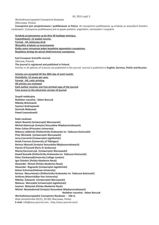
Results and discussion:
The objectives and tasks of the rehabilitation therapy were
focused towards overcoming or curbing the dysfunctions and
disabilities of the patients.
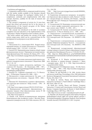
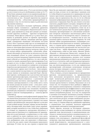
Fig.1 Shows a break-down of the examined patients by gender.
We performed the first measurements and examinations at
the beginning of the treatment(х1), and repeated them at the
end of the therapy (х2
)of the twenty patients – n=20.
Table 1 Shows the results from the active range of motion
during goniometry of the affected limb at the shoulder girdle.
Table1
Data on the active range of motion
Motion Number - n Х1 Х2 d=Х2-Х1
Flexion 20 50,60 160,00 100,50
Extension 20 20,00 40,00 20,00
Abduction 20 40,50 140,50 100,00
Adduction 20 10,00 25,00 15,00
Internal rotation 20 35,50 65,50 30,00
External rotation 20 25,00 75,00 50,00
The analysis of the obtained results shows that at the end of
the remedial course of therapy the range of motion increased,
although it is still not within the physiological normal range
of motion. This shows that the therapeutical complex provides
a good initial basis for functional recovery, but the time
is insufficient and the shortened period is limited by pain
syndromes.
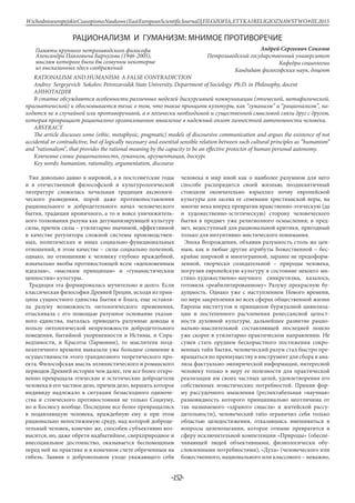
The results from the manual muscle testing are presented in
Table 2.
Table2
Changes in ММТ of the shoulder girdle
Motion Number– n Х1 Х2 d=Х2-Х1
Flexion 20 2,00 4,00 2,00
Extension 20 2,00 4,50 2,50
Abduction 20 1,50 3,50 2,00
Adduction 20 2,50 4,50 2,00
Internal rotation 20 3,00 4,50 1,50
External rotation 20 2,00 4,00 2,00
The analysis of the obtained results shows that the pain is
considerable and obstructs movements; the muscles are weak,
and at the end of the treatment the patients did not regain their
muscle strength to the extent they had expected. They have to](https://image.slidesharecdn.com/eesj33-160202073457/85/Eesj-3-3-7-320.jpg)
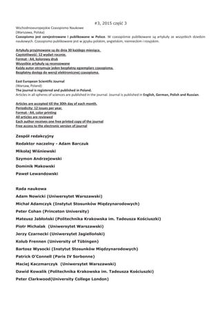
![-8-
WschodnioeuropejskieCzasopismoNaukowe(EastEuropeanScientificJournal)|NAUKIMEDYCZNEINAUKIOZDROWIU#IІІ,2015
continue their rehabilitation therapy in order to achieve good
results.
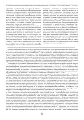
The results from the hand dynamometry are presented in
Table 3.
Table 3
Changes in muscle strength
Number – n =20 Х1 Х2 d=Х2-Х1
Affected limb 3,50 14,50 11,00
Unaffected limb 34,00 41,00 7,00
The analysis of the obtained results shows that muscle strength
was not restored to its normal values. Continuous rehabilitation
therapy is needed so that the limb could be used adequately. At
the beginning of the therapy, the grip strength is reduced – a
mere 3,50 kg, whereas that of the healthy limb is 34,00 kg. The
strength of the muscles stabilizing the shoulder is inadequate.
Table 4 shows a subjective assessment of the pain in motion.
Table 4
Pain assessment (0-3)
Number
– n
Examinations Degree of pain
20 Х1
0 1 2 3
Number - % Number - % Number - % Number - %
- - - - 2 10 18 90
Х2
1 5 3 15 16 80 -
The data show that at the beginning of the therapy the patients
had 90% of strong pain when moving. At the end of the
procedures the level of pain decreased to moderate and mild.
Physical therapy yielded good effect. A combination of
cryotherapy, remedial gymnastics and slight myofascial and
drainage massage, with IC and LFPEF, is recommended.
Intraarticular aplication of corticosteroids, as well as topical
application of corticosteroids, NSAIDs and Procaine
(Lidocaine) [5,10], also yields good results.
The remedial results in conditions of the shoulder joint
depend on the applied methods and their correct combination
in an efficient therapeutical complex: for example, cryotherapy
with kinesitherapy, underwater gymnastics with postisometric
relaxation, electrostimulation with active muscle contractions,
etc. Orthopedic means ensure the functional rest of the
kinematic chain, when needed.
In the moderate to severe stage, physiotherapeutic procedures,
such as the listed below, are recommended:
Cryotherapy- widely used in shoulder joint conditions,
especially those of traumatic or inflammatory nature. It
is applied in the form of: cryomassage with an ice cube,
diadynamic cruo-electrophoresis [10].
The local application of MF current within inhibiting
parameters is also recommended. Ultra sound segmentary
application paravertebrally at the level of C4 – C7. Pulse
mode, labile methods with small doses - 0,2- 0,4 W per square
centimeter, 3-4 minutes per field [5].
When developing individual therapeutic schedules, what
should be kept in mind is the functional disturbances of
the limb and the location of the problem. Kinesitherapy,
with its wide variety and therapeutic methods (analytical
gymnastics, postisometric relaxation in the Levit method,
therapy from a position, stretching, muscle-inhibiting
techniques, proprioceptive neuromuscular facilitation –
diagonal and reciprocal inhibition, etc.) has to be included
in the rehabilitation process from the very beginning of the
therapy to its completion. Rehabilitation methods have to be
applied below the pain threshold, regardless of whether they
are active or passive. Kinesitherapy should be preceded by the
administration of pain-relieving or myorelaxing procedures,
and in a chronic stage – by underwater gymnastics.
Kinesitherapy in the mild to severe stage includes:
- Therapy from position – a slight abduction of the shoulder,
the wrist – higher than the elbow, the elbow – higher than the
shoulder. Passive kinesitherapeutic techniques are contra-
indicated, with the exception of some manual soft-tissue
mobilizing techniques. It should be proceeded straight to
muscle relaxation:
- Relaxing swinging exercises: fixed shoulder blade, hanging
arm – the exercise of the pendulum, taking the arm to the side
for swinging and relaxing exercises.
- PIR (postisometric relaxation) for the contracted muscles;
PIR for trapezius; PIR for levator scapulae; PIR for the long
head of biceps brahii.
- Relaxation techniques of PNF in the method of Cabat [4,7].
- Analytical active exercises are applied against gravity to pain
for 10-20 min, twice a day, for the purpose of strengthening the
muscles that are prone to inhibiting and extending.
The following are contra-indicated:1. Pain-causing
kinesitherapy; 2. Redressing exercises; 3. Massage on the joint;
4. Local application of an irritating action; 5. Endogenic and
exogenic thermal action simultaneously.
Kinesitherapy is allowed when the pain lessens. Exercises
are strictly analytical. The patient is to be trained in relaxation
techniques.
A good alternative in the treatment of myofascial pain
syndrome is reflexive massage. At the beginning of the therapy,
transversal massage in the J. Cyrax method is applied (1978)
on the tendond of the affected muscles – transversely to the
muscle fibres and the tendon. With the tip of his/her thumb
and forefinger, the rehabilitation therapist applies pressure
to the place 3-4 times, for 2-3 seconds until bearable pain is](https://image.slidesharecdn.com/eesj33-160202073457/85/Eesj-3-3-8-320.jpg)
![-9-
WschodnioeuropejskieCzasopismoNaukowe(EastEuropeanScientificJournal)|NAUKIMEDYCZNEINAUKIOZDROWIU#IІІ,2015
experienced [12,13,14].
Findings and conclusions:
As a result of the complex rehabilitation therapy and the
efficiency of the treatment program applied by the students, the
active mobility and motor habits of the treated patients were
restored, though not to their full range to enable the patients to
be fit to work. The treatment should continue with subsequent
courses of therapy or in home conditions, provided the patients
are given clear instructions on the methods and techniques.
An excessively aggressive physio-rehabilitation therapy or an
unadequate one are equally unsuitable, and pose an equal risk
of consequential complications [2].
Any uncontrolled rehabilitation therapy – which is not
under the supervision of a doctor specialized in physical
and rehabilitation medicine – is always a precondition for
deterioration of the condition of the patient; it may become
chronic and lead to complications, regardless of how common
or banal the condition of the patient may seem at first sight [2].
In cases of ‘humeroscapular periarthritis’, refractory to
conservative treatment, a rupture of the rotator cuff is to be
suspected, and the diagnostics is to be more comprehensive [9],
which calls for surgical methods of treatment.
Inconclusion,itcanbeclaimedthatthecomplexrehabilitation
applied by us yielded good results in accordance with the
severity of the motor dysfunctions.
Bibliography:
1. Bank St. V. Krasteva, Y. Vajarov. Manual muscle testing with
the fundamentals of kinesiology and patokineziologiyata. II
edition. Medicine and Sports, Sofia, 1991, 120-125
2. Veselinova L. Rehabilitation problems late in the recovery
period after reconstruction at the event on the occasion of
«PASTA», in the journal. Physical medicine, rehabilitation,
health, number 3/2012, p. 22-25
3. Zhelev C. Guide to practical exercises on medical
gymnastics, Sofia, and Sports Medicine, 1991, 86-88
5. Koleva I. Algorithms for physical prevention, treatment
and rehabilitation of some common and socially significant
diseases, Sofia, 2007, p. 50-51
6. Stavrev P., A. Atanasov. Orthopedics and Traumatology,
Plovdiv, 2004, p. 129-130
7. Popov N. Introduction to kinesitherapy. Basic tools and
methods, NSA Press, 9-10,78,159, 164-165
8. Popov N. Therapy orthopedic diseases and injuries of upper
limb, Sofia, NSA Press, 2009, 180-182
9. Rusimov C. Totally passive and rotator cuff - arthroscopic
diagnosis and recovery magazine. Physical medicine,
rehabilitation, health, number 3/2012, p. 11-15
10.RiazkovaM.Practicalclinicalphysiotherapy,«Knowledge»
Ltd. 1999, p. 69-72
11. Yakovidis P, V. Zhelev. Massage Therapy in syndrome m.
Supraspinatus, Мag. Therapy and Rehabilitation, number 3-4,
2009, 36-38
12.Bentley E. The Essential Massage Book, Gaia Books, 2005
13.Yuksel I. Masaj Teknikleri, Alp Yayinevi, 2007
14.Maranki E., A. Maranki. Profilaktik masajla mucizevi
tedaviler, Nozaik, 2007](https://image.slidesharecdn.com/eesj33-160202073457/85/Eesj-3-3-9-320.jpg)
![-10-
WschodnioeuropejskieCzasopismoNaukowe(EastEuropeanScientificJournal)|NAUKIMEDYCZNEINAUKIOZDROWIU#IІІ,2015
SAFETY OF MESENHYMAL STEM CELLS THERAY IN INFLAMATORY BOWEL DEASES
Knyazev Oleg Vladimirovich,
Doctor of science, Head of department of treatment inflammatory bowel diseases,
Parfenov Asfold Ivanovich,
Doctor of Science, professor, Head of intestinal pathology department, asfold@mail.ru
Konoplaynnikov Anatoliy Georgievich
Doctor of science, professor, head of experimental radiotherapy
Kagramanova Anna Valeryevna,
Candidate of science, scientist researcher
Ruchkina Irina Nikolaevna,
Doctor of Science, Head scientist of department of inflammatory bowel diseases,
Fadeeva Nina Aleksandrovna,
junior scientist researcher of department of inflammatory bowel diseases
aBSTRACT
Aim: To compare safety profile of therapy in patients with ulcerative colitis (UC) and Crohn disease (СD), receiving anti-
inflammatory therapy, using bone marrow-derived mesenchymal stromal cells (MSC) and standart therapy with 5-aminosalicylic
acid (5-ASA), glucocorticosteroids (GCS) and immunosuppressive agents (IS)
Materials and methods. Adverse events were analyzed in 103 patients with inflammatory bowel disease (IBD) after administration
MSCs (56 patients UC and 47 patients CD). The findings were compared with data obtained in 208 patients with UC and CD,
receiving standard anti-inflammatory therapy. All analyzed patients were similar in demographic characteristics, the duration of
disease, the extent of disease, course of disease, phenotype and degree of disease.
The analysed groups did not include patients, treated with anti-TNF therapy. The safety of therapy was evaluated by presense of
complications, developed during follow-up period.
Results. We conducted analysis of side effects in 103 IBD patients, treated with mesenchymal stem cells, comparing with 208 UC
and CD patients, treated with standard anti-inflammatory therapy and finally we did not reveal any differences in developing acute
posttransfusional toxicity, infectious complications, exacerbation of chronic inflammatory diseases, serious infectious complications,
malignancy and death in UC and CD patients, besides transitive febrile.
Conclusion. Results of our study show that innovative method of cell therapy is safe in clinical practice.
Key words: safety of cell therapy, mesenchymal stromal cells, Crohn disease, ulcerative colitis, inflammatory bowel diseases
Mesenchymal stromal cells (mesenchymal stem cells; MSC)
are a heterogeneous group of cells, that can be isolated from
many tissues (bone marrow, adipose tissue, dental pulpe). First
described in 1960-years of XX century [1], MSC have recently
received attention in a number of different clinical fields for
their potential therapeutic effects.
Although often described as «adult stem cells», MSC’s have
limited cellular differentiation ability. Instead, pre-clinical
evidence suggests that MSCs exert their beneficial effects largely
through immunomodulatory and paracrine mechanisms.
MSCs home to sites of inflammation and secrete bioactive
molecules, and thus may be especially effective in different
proinflammatory diseases. [2].
Thereisagrowingbodyofliteraturedemonstratingtheefficacy
of MSC therapy in a variety of pre-clinical models, including
acute lung injury [3,4], septic shock [5], acute myocardial
infarction [6]. Several small clinical trials have investigated
efficacy and safety of MSCs in diseases including chronic
heart failure, acute myocardial infarction, hematological
malignancies, Crohn disease [7] and graft-versus-host disease.
However, safety concerns represent a significant barrier to
the successful translation of MSCs into an acceptable clinical
therapeutic. Potential risk is associated with its proliferative
capacity, susceptibility to infectious complications given their
immunosupressive effects, embolism of the cells, zoonoses
associated with cell culture reagents, and acute or chronic
immunogenicity of the cells themselves [8].
Therefore, we conducted a systematic review of randomized
and non-randomized controlled trials as well as uncontrolled
clinical trials in foreign literature, that examined the safety and
efficacy of intravascularly delivered MSCs, and revealed their
most frequent adverse events [9]. Adverse events were grouped
according to the immediacy of the event -acute infusional
toxicity, fever, the occurrence of organ system complications
(neurological, pulmonary, cardiovascular, gastrointestinal and
renal, and hematologic systems), infection, and the occurrence
of longer term adverse effects (death, malignancy).
Included studies were conducted in 14 different countries
from Asia, the Middle East, Europe, and North America. There
were eight RCTs (n = 369 patients) [10-17], 10 non–RCTs (n
= 466 patients) [18-27] and and 18 uncontrolled clinical trials
(n = 252 patients) [28-45]. Six of 36 studies were multi-centre
[12,13,20,23,32,33]. One non-controlled study had a mixed
adult-pediatric population [39], all other studies included only
adult participants. The follow-up period was reported in all
studies and the duration ranged from 0.5 to 60 months.
There were following diseases analyzed: eight randomized
controlled studies included patient populations with
cardiovascular diseases-acute myocardial infarction [11,12],
chronic heart failure [10,16], with neurological disease either
ischemic stroke [13], spinal cord injury [17], following stem
cell transplantation for hematological malignancies [15]. The
10 non-RCTs included patient populations with old myocardial
infarctions [25], stem-cell transplant post renal transplant[27],
tem cell transplant for hematological malignancy [18,19,23],
graft-versus-host disease [20,26], or healthy volunteers [24].
Sixteen studies used autological MSC [10,11,13,14,16,17
,22,24,25,27,29,31,32,37,43,45], eight used allogenic MSC](https://image.slidesharecdn.com/eesj33-160202073457/85/Eesj-3-3-10-320.jpg)
![-11-
WschodnioeuropejskieCzasopismoNaukowe(EastEuropeanScientificJournal)|NAUKIMEDYCZNEINAUKIOZDROWIU#IІІ,2015
[12,18,20,34,35,39,40,41]. Nine of the 36 studies cryopreserved
MSCs prior to administration [12,18,20,21,23,29,31,32,44],
and one study used both fresh and cryopreserved MSC [33],
while the remainder of studies used only fresh MSCs. A meta-
analysis revealed no significant differences in the occurrence of
acute infusional toxicity, infectious complications, reccurence
of chronic inflammatory diseases, serious infectious
complications, malignancy and death between patients treated
with MSC and control group. Significant association was
demonstrated between MSC injection and transient fever.
Further we demonstrate our data for safety profile of allogenic
mesenchymal stromal bone marrow cells in patients with
inflammatory bowel diseases over a 5-year follow-up period.
Aim of study: to compare safety profile of therapy in patients
with ulcerative colitis (UC) and Crohn disease (CD), received
combined antiinflammatory therapy including MSC and
standart therapy, including 5-ASA, Glucocorticosteroids
(GCS) and immunosupressive therapy.
Materials and methods. Systemic transplantation of allogenic
bone marrow MSC was perfomed in 74 UC and 64 CD patients
ranging from 2008 to 2014 years.
First analysed group included 56 UC patients, follow-up
period comprised in median 62±4 months. This group consists
of 29 (51,78%) man and 27 (48,22%) women (Table 1). Mean
age was 35,4±1,42 years. The second, control group included
84 UC patients, receiving standart anti-inflammatory therapy
with 5-ASA and GCS. This group consists of 46 (54,8%) man
and 38 (45,2%) women. Mean age - 34,98±1,23 years.
Third group included 47 CD patients, mean follow-up period
was 64±4 months. Nineteen (40,4%) man and twenty-eight
(59,26%) women were included in the third group. Mean age
was 30,4±1,2 years. Fourth control group consisted of 124
CD patients, receiving standart anti-inflammatory therapy
including 5-ASA, GCS and IS. In this group were 56 (45,2%)
man and 68 (54,8%) women. Mean age was- 36,8±1,5 years.
We did not include patients, receiving anti-TNF therapy, in
analysed groups.
Technique of receiving and cultivation MSC in a apropriate
for systemic transplantation amount (150-200 millions of
cells) was published [46]. This method is validated by Federal
Supervisory Agency for Health Care and Social Development
Ministry of Healthcare and Social Development of the Russian
Federation (License 2006/206). Bone marrow cells were
isolated by means of flushing the sternum or iliac crest of
healthy donor under local anesthesia and aseptic conditions.
All donors signed informed consent for using bone marrow
samples for scientific purposes. MSC culture was injected
intravenous drip-feed in dosage 1,5-2 mln by 1 kg body weight.
For systemic transplantation 130-160 mln allogenic MSC,
cultivated, were suspended in 200 ml steril isotonic solution,
consisting of heparin in concentration 50 U/ml. MSC culture
was injected during 40-60 minutes by means of intravenous
drip-feed infusion. Mathematical modeling of MSC treatment
was performed to assess maximal efficacy and minimal side
effects of MSCs. We analysed several trials, in which regimen
of MSC administration, frequency and the rationale for the cell
dose were examined [13,14,44,45]. All patients signed inform
consent for participating in study before MSC injection. Thus,
procedure of MSC cultivation was perfomed according GMP.
Safety of therapy was assesed by presense of complications,
occured during follow-up period, for example acute infusional
toxicity, fever; complications (neurological, pulmonary,
cardiovascular (arrhythmias e.t.c), urinary, gastrointestinal
tract and blood system), infection complications, exacerbation
of chronic inflammatory diseases, serious infectious
complications (pneumonia, sepsis, abscess), malignancy,
death. All persons, monitoring the complications were blinded
with the treatment.
Results and discussion. In the first group 3/56 UC patients
(5,4%) have acute infusional toxicity–looks like hives
immediately or after MSC injection, in the second group
allergic reaction like papulear urticaria was noticed in 1/84
(1,2%) patient, treated with sulfasalazin. Allergic reaction like
hives in first group patients had no statistically significance in
compare with second group of patients (x2-0,35; p=0.87). In
16/56 (28,6%) patients of first group were noted increasing of
temperature around 37,2-37,40 С during 12 hours after MSC
injection or fever around 38,00 С, in 1/84 (1,2%) patients of
second group was reported increasing temperature above 37,70,
caused by intravenous injection of prednisolon. Fever and
temperature increasing after MSC injection were statistically
significant compared to control group – relative risk (RR)
was 24,0 (95% CI 3.27 - 175.89); x2-21,12; p=0.0000043. In
the first UC group non-serious infectious complications and
exacerbation of chronic inflammatory diseases were revealed
in 7/56 (12,5%) patients, in second group - in 14/84 (16,7%)
patients. There was no significant difference in risk of infectious
complications and exacerbation of chronic inflammatory
diseases between two groups of UC patient, receiving
standart antiinflammatory therapy and MSC (RR-0,75;
95% CI 1.5-23.58; x2-0,16; р=0.66). In the first group serious
infectious complications (pneumonia, pleurisy, activation of
latent tuberculosis) were detected in 1/56 patients (1,8%), in
the second – in 5/84 (5,9%). There was no difference in this
complications between two groups (RR-0,3; 95%CI 0.04-2.5;
x2-0,59; р=0.44). Colorectal cancer was documented only in
1/56 (1,8%) patient in the first group. Diagnosis of colon cancer
was established in 10 days after MSC injection.
During five-year follow-up period malignancy was found in
4/84 (4,8%) in the second group (RR-0,5, 95%CI 0.05-4.96; x2-
0,01; р=0.97). In the first and in the second groups during five-
yearfollow-uponelethalcasefromeachgroupwasdocumented
and it was 1,8% и 1,2%, respectively (RR-1,5; 95%CI 0.1-23.49;
x2-0,19; р=0.66).
In the third group of CD patients acute infusional toxicity
like hives and Quincke’s edema were detected in 2/47 patients
(4,25%) immediately after MSC injection, in the fourth group
there were no complications during antiinflammatory therapy,
but these manifestations have no statistically significance
between groups (x2-2,3, p=0.07). Increase in body temperature
up to 37,2-37,40 С during 12 hours after MSC injection or fever
up to 38,00 С was noticed in 22 patients of third group (46,8%),
in the fourth group of patients there was no fever, associated
with intravenous interventions (medication injection) or per
os administration was found in 0/124 (0%). Fever and mild
increase of temperature after MSC injection were statistically
significant compare to control group – RR – 58,5 (95% CI 8.1 -
422.0), x2-58,5, p<0.001. Non-serious infectious complications](https://image.slidesharecdn.com/eesj33-160202073457/85/Eesj-3-3-11-320.jpg)
![-12-
WschodnioeuropejskieCzasopismoNaukowe(EastEuropeanScientificJournal)|NAUKIMEDYCZNEINAUKIOZDROWIU#IІІ,2015
and exacerbation of chronic inflammatory diseases during
therapy observed in 12 patients of 47 in the third group, that
accounts 25,5%, in the fourth group-in 48 (38,7%) patients of
124, that had no significant difference: RR – 0,67 (95% CI 0.39
- 1.15), x2-1,86, p=0.17.
There was no differences between third and fourth groups
in risk of serious infectious complications (pneumonia,
peurisy, activation of latent tuberculosis) during standart
antiinflammatory CD therapy and therapy with MSC. In the
third group one patient developed pneumonia 1/47 (2,1%), in
the fourth group two cases of pneumonia and one case of latent
tuberculosis activation were detected – 3/124 (2,4%) (RR-0,88,
95%CI 0.09-1.85; x2-0,21; р=0.7).
In the third group of CD patients no cases of colorectal cancer
were found. In the third group during five-year follow-up
period no lethal outcomes were documented, in the fourth
group one lethal case (0,8%), unlinked to underlying disease
was found (x2-0,26; р=0.61). In the fourth group malignant
transformation was noted in 2 patients (1,6%) from 124 (x2-
0,01; р=0.93).
In patients with UC and CD, receiving MSC treatment,
no cardiovascular, pulmonary, neurological, renal, and
hematologic systems complications were detected.
Conclusion
Our study includes comparative analysis of adverse events,
associated with MSC treatment and standart antiinflammatory
therapy in UC and CD patients. We analysed advanced
outcomes in 103 IBD patients, receiving MSC therapy and
compared this data with 208 UC and CD patients, who had the
same demographic charachteristics, disease duration, extent
of disease, course of disease, phenotype of disease, type of
severity. Thus we did not observe any significant differences in
MSC safety, aside from transient fever.
This analysis did not reveal any differences in acute
posttransfisional toxicity, infectious complications,
exacerbation of chronic inflammatory diseases, serious
infectious complications, malignancy and lethal cases in UC
and CD patients, treated with standart antiinflammatory
therapy.
We have detected significant association between MSC
injection and fever. However, fever was transient and not
associated with long term sequelae. The mechanisms for
fever are not clear but could be related to acute inflammatory
reactions by a subset of patients to particular preparations of
MSCs, not unlike similar reactions occasionally observed with
red blood cell and fresh frozen plasma administration [47].
Although malignant transformation is a theoretical risk, our
own experience and literature analysis, presented in this review
found no association between MSCs and tumour formation.
Concerns related to tumourgenicity of MSCs were raised by
preclinical studies demonstrating increased tumour burden in
vivo. [48]. Although recent position papers have suggested low
probability of malignant transformation and tumour formation
with MSCs [8]. Malignancy occurred only in studies involving
participants with ongoing or previous malignancies; no de
novo malignancies were observed.
Although MSC immunomodulatory effects may be beneficial
in pro-inflammatory diseases, these same effects may leave a
patient susceptible to infection. [49]. The question arised-
whether immunosupressive therapy could increase risk of
infections? This review did not demonstrate any evidence of
increased susceptibility to infections with MSC administration.
In our review, infections were common in already
immunosuppressed patients (e.g. following hematopoietic
stem cell transplant), however the infection rates were similar
to those in control group of patients [47].
Currently obtained data show that despite of strong
immunosupressive effect due to autoimmune agression, MSC
did not hinder the activity of immunocompetent cells, directed
against infectious agents [50-56].
Absense of posttransfusional reaction may be explained by
low MSC immunogenicity, due to absense HLA class II and
low level of expression HLA I class at their surface [57]. The
use of fetal bovine serum for culturing MSCs could be one of
the reasons for above mentioned posttransfusional toxicity,
and another potential concern with MSC therapy application
is the use of dimethylsulfoxide as criopreservative, which has
toxic side effects and could cause hypersensitivity reactions
[58,59]. Thus, greater vigilance may be needed in future studies
for reporting cellular viability and monitoring for potential
dimethylsulfoxide related adverse events. Results from our
study should provide some assurance to investigators and
health regulators that, with the present evidence, this innovative
therapy appears safe.
Table 1
Demographic characteristics of patients with CD and UC
Male-to–female ratio (%) Mean Age, years
1 group (n=56) 29:27 (51:48) 35,4±1,42
2 group (n=84) 46:38 (54:45) 34,98±1,23
3 group (n=47) 19:28 (40:59) 30,4±1,2
4 group (n=124) 56:68 (45:54) 36,8±1,5
Table 2
Summary аnalysis of side effects in UC patients, receiving MSC and in control group
Side effects in all
analysed clinical trials
Frequency in 1 group
UC patients
Frequence in 2 group
UC patients
95% CI p
Acute infusion
reaction
3/56 1/84 0.48-42.18 0,87
Fever 16/56 1/84 3.27 - 175.89 0.0000043](https://image.slidesharecdn.com/eesj33-160202073457/85/Eesj-3-3-12-320.jpg)
![-15-
WschodnioeuropejskieCzasopismoNaukowe(EastEuropeanScientificJournal)|NAUKIMEDYCZNEINAUKIOZDROWIU#IІІ,2015
43. Yang Z, Zhang F, Ma W, Chen B, Zhou F, et al. A novel
approach to transplanting bone marrow stem cells to repair
human myocardial infarction: delivery via a noninfarct-relative
artery. Cardiovasc Ther 2010, 28: 380–385.
44. Zhang X, Li JY, Cao K, Lu H, Hong M, et al.
Cotransplantation of HLA-identical mesenchymal stem
cells and hematopoietic stem cells in Chinese patients with
hematologic diseases. Int J Lab Hematol 2010, 32: 256–264.
45.ZhangZX,GuanLX,ZhangK,ZhangQ,DaiLJ.Acombined
procedure to deliver autologous mesenchymal stromal cells to
patients with traumatic brain injury. Cytotherapy 2008, 10:
134–139.
46. Zib A.F., Konoplyannikov A.G., Kolesnikova A.I.,
Pavlov V.V. Application of cell cultures in medicine from
mesemnchymal stem cells of human bone marrow. News of
Russian Academy of science 2004; 59(9): 71-76.
47. Hendrickson JE, Hillyer CD. Noninfectious serious
hazards of transfusion. Anesth Analg 2009, 108: 759–769.
48. Djouad F, Plence P, Bony C, Tropel P, Apparailly F, et al.
Immunosuppressive effect of mesenchymal stem cells favors
tumor growth in allogeneic animals. Blood 2003, 102: 3837–
3844.
49. Uccelli A, Moretta L, Pistoia V Mesenchymal stem cells in
health and disease. Nat Rev Immunol 2008, 8: 726–736.
50. Karlsson, H.; Samarasinghe, S.; Ball, L.M.; Sundberg, B.;
Lankester, A.C.; Dazzi, F.; Uzunel, M.; Rao, K.; Veys, P.; Le
Blanc, K.; Ringden, O.; Amrolia, P.J. Mesenchymal stem cells
exert differential effects on alloantigen and virus-specific T-cell
responses. Blood, 2008, 112, 532-541.
51. Majumdar M.X., Keane-Мооrе M., Buyaner D. et al.
Characterization and functionality of cells surface molecules
on human mesenchymal stem cell. Journal of Biomedical
Science, vol. 10, no. 2, pp. 228–241, 2003.
CHILD AND GADGET. INFLUENCE ON THE SPEECH AND COMMUNICATION
Belousova M.V.
MD, PhD, associate professor, department of pediatric neurology
Kazan State Medical Academy
Utkuzova M.A.
MD, PhD, associate professor, department of pediatric neurology
Kazan State Medical Academy
DAS KIND UND DIE ELEKTRONISCHEN MEDIEN. AUSWIRKUNGEN AUF SPRACHE UND KOMMUNIKATION
Belousova Marina Dr. med., Dozentin der Kasaner staatliche medizinische Akademie des Gesundheitsministeriums der Russischen
Föderation.
Utkuzova Marina Chefexpertin in Fragen der Kinderrehabilitation des Gesundheitsministeriums der Republik Tatarstan,
Kinderneurologe, Dr. med., Dozentin der Kasaner staatliche medizinische Akademie des Gesundheitsministeriums der Russischen
Föderation.
ABSTRACT
In research 130 children (1-5 years), which parents too early began to use electronic devices (smartphones, tablets) were examined.
Influence on the child parental relations, communication, socialization and development of the speech was revealed.
Zusammenfassung
Die Studie untersuchte 130 Kinder (1-5 Jahre), deren Eltern zu früh sind, um elektronische Geräte (Smartphones, Tabletten)
verwendet werden. Es zeigte die Auswirkungen auf die Kind-Eltern-Beziehungen, Kommunikation, Sozialisation und
Sprachentwicklung.
Schlüsselwörter: Kind-Eltern-Beziehungen, Kommunikation, Sozialisation Sprachentwicklung, autistischen Störung, die
elektronischen Medien
Key words: speech, verbal communication, autistic disorders, gadgets.
In den letzten Jahren hat die Zahl der Autismus-
Spektrum-Störungen (ASS) unter den Kindern in Russland
bedeutend zugenommen, diese entwickeln sich nach und
nach in die „Epidemie“ des XXI [1]. Jahrhunderts. Die
Autismus-Spektrum-Störungen stellen eine heterogene
Gruppe der Krankheiten mit unterschiedlicher Ätiologie und
pathogenetischem Mechanismus dar. ASS sind komplexe
psychischeEntwicklungsstörungen,diedurchdiefehlendeoder
gestörte Fähigkeit zur sozialen Interaktion, Kommunikation
und durch Verhaltensstereotypie gekennzeichnet sind [2].
Als einer der Gründe, die die Bildung der kommunikativen
Kompetenzen und die Sozialisierung von Kindern negativ
beeinflussen, sollen die Verbreitung und unbegründet
frühe Bekanntschaft des Kindes mit elektronischen Medien
(Tabletten, Spielkonsolen, Handys, MP3-Playern) in
Betracht gezogen werden, was zu einem ernsthaften Problem
wird. Die Eltern, die modische Geräte für beste Mittel zur
Tröstung, Ablenkung und Entwicklung halten, erkennen
keine Gefahr des Ersetzens und der Auswechslung von der
menschlichen Kommunikationsart durch die elektronischen
Geräte. Allmählich verlieren die Erwachsenen ihre führende
Rolle in Bezug auf die psychische, sprachliche, emotionelle,
kommunikative, soziale Entwicklung des Kindes und auch ihre
Kraft, auf das Kind Einfluss auszuüben.
In den Jahren 2010-2013 wurde von uns eine Befragung von
130 Familien durchgeführt, die die Klein- und Vorschulkinder
(im Alter von 1 bis 5 Jahren) haben. Alle Kinder wurden in 2
Gruppen geteilt: in der 1. Gruppe (n=80) sind Kinder ohne
Sprachstörungen, in der 2. Gruppe (n=50) sind Kinder mit
Zeichen der systemischen Sprachhemmung in Verbindung
mit Autismus-Syndrom. Die statistische Datenverarbeitung
wurde mit Verwendung des Softwarepakets STATISTICA
6.0.durchgeführt. Zur Beurteilung der in Form von
Kontingenztafeln dargestellten Attributmerkmale wurden für](https://image.slidesharecdn.com/eesj33-160202073457/85/Eesj-3-3-15-320.jpg)
![-16-
WschodnioeuropejskieCzasopismoNaukowe(EastEuropeanScientificJournal)|NAUKIMEDYCZNEINAUKIOZDROWIU#IІІ,2015
die Analyse Chiquadrattest (χ2), Student-t-Test für Bruchteile
verwendet. Die Unterschiede galten als sichere bei Р<0,05.
„Die Kinder des 21. Jahrhunderts und ihre Eltern, die
nach dem Jahr 1980 geboren sind, sind die Generation der
Multimediarevolution…“ [3]. Nach den Befragungsangaben
haben 96 (74%) der befragten Familien regelmäßig Gadgets
(Computer, Tablett-Geräte, Handys) für die Frühentwicklung
des Kindes (Computerspiele für Kinder, Präsentationen,
Graphikprogramme) benutzt, sowie auch zur Ablenkung
und Tröstung des Kindes, im Falle der Verhaltensstörungen
(Marotten, Hysterien), um das lange Warten wegen der Fahrt,
der Schlange oder in der Sprechstunde zu versüßen, damit
das Kind dem Gespräch mit dem Facharzt nicht stört; zum
Aufkommen der eigenen Zeit (Zeit für Telefongespräche, fürs
Erledigen dringender Arbeit, für Einzelstellung), wobei die
Gadgets sicher häufiger in der zweiten Gruppe benutzt wurden
(1.Gr. 67,5%; 2. Gr. 84%; Р=0,03).
Nach den Befragungsangaben von Eltern beginnt das
Kind mit der Bekanntschaft und aktiver Wechselwirkung mit
Gadgets schon im ersten Lebensjahr, so haben den Zugang
zum Handy:
im ersten Lebensjahr - 4 (15,3%) Kinder – alle aus der
Gruppe №2;
im zweiten Lebensjahr - 6 (23,1%) Kinder – (1. Gr. 2 (2,5%)
und 2. Gr. 4(8%));
im dritten Lebensjahr – 16 (61,5%) Kinder – (1. Gr. 12(15%)
und 2. Gr. 4(8%)).
Das Fernsehen ist ständig im Leben der 125 (96,2%)
Familien dabei. 110 (84,6%) der Kinder wurden bekannt
mit Fernsehsendungen und schauen sich sie regelmäßig seit
6. Lebensmonat bis 3 Jahre an, wobei 85 (65,4%) der Kinder
ohne Eltern fernsehen und 18,5% von ihnen selbstständig oft
zappen. Das Gesehene besprechen 81(62,3%) Kinder (1. Gr. -
60(75%); 2. Gr. - 21(42%); Р=0,000) mit den Eltern.
75 (57,7%) Kinder benutzen Gadgets selbstständig (sie
können Spiele abzulaufen starten, Musik und Zeichentrickfilme
abspielen lassen, Fotos anschauen). 35 (40,7%) der Kinder
spielen jeden Tag am Computer (1. Gr.- 21 (26,3%) und 2. Gr.
– 14 (28%); Р=0,1). Die Mehrzahl der Kinder (88,4%) wenden
für Spiele von 30 Minuten bis zur 1 Stunde Zeit auf, 4,7% der
Kinder spielten mehr als 1 Stunde pro Tag (1. Gr. - 2(2,5%) und
2. Gr. 2(4%); Р=0,014).
72% der Eltern machten sich darüber Sorgen, dass es
kompliziert war, die Aufmerksamkeit der Kinder vom Spiel
abzulenken (1. Gr. 33(41,3%), 2. Gr. 29(58%); Р=0,074); 36
Kinder (41,2%) weisen während des Spiels viele Emotionen auf
und können reizbar, zornig, aggressiv sein, fühlen sich beleidigt
und zeigen Wut, wenn etwas im Spiel ihnen nicht gelingt (1. Gr.
19(23,8%), 2. Gr.17(34%), Р=0,230).
Das Alter von 0 bis 3 Jahren ist die Periode der
intensiven Sprachbildung und des Mutterspracherwerbs, der
Gedächtnisentwicklung,derAufmerksamkeit,desanschaulich-
handelnden Denkens[4, 5]. Das ist die Zeit, wenn das
Selbstbewusstsein entsteht und die primäre Selbsteinschätzung
auftritt. In diesem Alter entwickeln sich besondere emotionelle
Beziehungen mit den näheren Leuten. Das Kind macht
sich mit Regeln und Grenzen der Umwelt bekannt und
versucht, sein Verhalten ihnen zu unterwerfen. Das ist die
Zeit, wenn die Rolle der liebevollen Eltern unersetzbar ist
und wenn jede „elektronische Ersatzbabysitter“ dem Kind
unrückgängig schaden kann, während sie autistisch-ähnliche
Verhaltensmuster prägt.
Die dominierende seelische Höchstleistung des Kindes
unter 3 Jahren ist die Wahrnehmung – visuelle, akustische,
kinästhetische. Die Kinder lockt multimodale Sinneserfahrung
an, die sie mühelos von technogenen Geräten bekommen
(lebendige, unter den Fingern verwandelte Bilder, Vielfältigkeit
von Farben, Formen, graphischen Gestaltungen, von
Illusion der Dreidimensionalität und der Tiefe des Raumes,
voller Begleitton, Vibrationsgefühl). Die Tastenbetätigung,
das Berühren des Tablett-Bildschirms wird durch eine
Inhaltsänderung begleitet, die bei den Kleinkindern oft
als gewünschter Spielausgang auftritt. Also, entstehen im
Leben des Kindes stereotyp eingebaute Bewegungs- und
Sprachprogramme, repetitive primitive musikalische
Fragmente statt der interaktiven Kommunikation, des
Rollenspiels. Die Manipulationen mit dem Tablet ersetzen
emotional reiche Erkenntnis der Welt und der menschlichen
Beziehungen. Deswegen verwandeln sich die Kleinkinder, die
zur leichten Beute von Tablets und Handys werden, in Homo
Ludens - den spielenden Menschen (Paskal Weyl).
Der Computer und die Kommunikation.
Eines der häufigsten und der frühesten Zeichen von
ASS ist die Sprachentwicklungsstörung. Die Sprach- und
Kommunikationsstörungen haben negative Auswirkungen
auf die Bildung von Denken, auf den Erwerb der sozialen
Kompetenzen, auf die Erkenntnisaktivität des Kindes und auf
sein Verhalten.
Dank der Evolution ist der Sprachbildungsapparat des
Kindes von der Geburt zur Aussprache bereit, aber für die
Sprecherziehung (d.h. für die Fähigkeit, mittels der Wörter
seine Gedanken mündlich und schriftlich auszudrücken)
braucht man Zeit. Die Sprecherziehung, die in den ersten drei
Lebensjahren aktiv ist, ist ohne Eltern-Kind-Kommunikation
unmöglich. Ausschließlich die Motivation, die Welt der
Erwachsenen zu verstehen, regt das Kind zur Erweiterung
aktiven Wortschatzes, zum Verschleifen richtiger Aussprache
an. Der Wunsch, Verständnis und Gehör zu finden, über
seine Bedürfnisse zu berichten, Phantasien und Überlegungen
zu teilen, also, eine sinnvolle Kommunikation mit einem
bedeutsamen Erwachsenen, ist die treibende Kraft der
Sprecherziehung [6]. Positiv gefärbte Reaktion der Eltern,
ihre interessierte Aufmerksamkeit zur Rede des Kindes,
tägliche gefühlsvolle verbale Kommunikation, Lesen der
Bücher, Auswendiglernen von Gedichten und Liedern,
Bemerkungen, die von den Eltern an das Kind adressiert sind
darüber, was draußen oder zu Hause passiert, - das alles sind
unentbehrliche Komponente für eine hohe Motivation zur
verbalen Kommunikation bei dem Kind und zur Entwicklung
seiner Rede.
Nach den Befragungsangaben von Eltern
wurden folgende familiäre Risikofaktoren der
Kommunikationsverhaltensstörung festgestellt:
1) Sprechdeprivation in der Familie (müde Eltern, die nach
der Arbeit kommen, einander ein paar Phrasen werfen und
danach den Fernseher anschalten und wortkarg, mit einzelnen
Kommentaren anschauen) in 78 (60%);
2) in 74 Familien (56,9%) ist ständige Hintergrundbelastung](https://image.slidesharecdn.com/eesj33-160202073457/85/Eesj-3-3-16-320.jpg)
![-17-
WschodnioeuropejskieCzasopismoNaukowe(EastEuropeanScientificJournal)|NAUKIMEDYCZNEINAUKIOZDROWIU#IІІ,2015
auf den akustischen Analysator zu vermerken (in dem Zimmer,
wo sich das Kind befindet, arbeitet Radio oder Fernseher als
Begleitton für sein Essen oder Spiel);
3) in 67 Familien (51,5%) ist die Funktion der
Sprachkommunikation an technogene Mittel delegiert, die kein
Erstellen interaktiver Kommunikation erfordern (man schaltet
für das Kind einen Zeichentrickfilm an, mit der Hoffnung, dass
es die Satzrede („sprachliche Klischees“) übernimmt, während
es sie von den Lieblingstrickfiguren hört);
4) in 59 Familien (45,4%) schlägt man dem Kind jene
Spieltätigkeit vor, für die man keine Rede braucht (Gadgets).
Tablet- und Handyspiele erfordern vom Kind keine Mühe,
die mit der Notwendigkeit der Sprachbeherrschung
verbunden sind. Stereotype motorische Grundfähigkeiten und
Reaktionsvermögen genügen vollauf.
Nach der Ansicht der meisten Eltern förderte der Umgang
mit Gadgets die dialogische Sprachentwicklung nicht, sondern
erschwerte nur die Kommunikationsprobleme des Kindes. So
bemerkt man Kommunikationsschwierigkeiten bei 62 (47,7%)
Kindern (20(25%) und 42(84%)), darunter auch beim Viertel
der Gesamtzahl der gesunden Kinder.
Die Einführung von Gadgets in den Lebensraum hat nicht
nur eine besondere Wirkung auf die Bildung der mündlichen
Sprache und des Denkens von einem modernen Kind, sondern
führte auch zum Entstehen einer besonderen Art schriftlicher
Sprache(SMS)ohneRechtschreibregelundHöflichkeitsformen,
deren Hauptmerkmale Kürze und Mindesttastendruck sind.
Die Kinder von heute hören zunehmend „mittels der Augen“
beim Lesen von SMS-Nachrichten und beim Briefverkehr im
Chat-Room und „sprechen mittels der Finger“.
Der Computer und das Sozium.
Die unbedingte Voraussetzung der normativen
frühkindlichen Entwicklung ist die Bildung eines
Befestigungssystems im ersten Lebensjahr, das durch eine
zuverlässige stabile Beziehung zwischen dem Kind und dem
betreuenden Erwachsenen gekennzeichnet ist [7]. Dieses
System wird aktiviert, wenn das Kind auf etwas Neues
und Unbekanntes stößt, und beinhaltet zwei gegenseitige
Verhaltenstendenzen: Bestrebung zum Neuen und Suche
nach der Unterstützung. Die Erschließung des Hausraumes,
die Begegnung der Welt menschlicher Beziehungen und
dem Gegenstandsreich geschieht im Hintergrund der
aktivierten Kind-Mutter-Bindung. Die Rolle des bedeutenden
Erwachsenen ist es, die Unterstützung und emotionale
Akzeptanz des Kindes zu gewähren, was die Entwicklung vom
basalen Vertrauen des Kindes zur Welt ermöglichen wird.
Nach den Befragungsergebnissen mögen 95 (73,1%)
Kinder (68(85%) und 27(54%))(Р=0,000) auf das Lesen
von Büchern hören. Jedoch gelingt es nur den Eltern aus
53(40,8%) Familien (28(35%) und 25(50%))(Р=0,084) täglich
die Zeit dem gemeinsamen Lesen zu widmen, für 25 (19,2%)
Kinder (10(12,5%) und 15(30%)) (Р=0,014) schaltet man die
Hörbücher an, während der gemeinsame Zeitvertreib ersetzt
wird.
Das Verbringen der Mußestunden von einem Kleinkind
mit dem Tablet oder Computerspiel setzt keine emotionale
Beteiligung der Eltern an diesem Prozess voraus, keine Hilfe
oder Unterstützung, was sich auf die Qualität der Eltern-Kind-
Bindung auswirkt.
Die Klein- und Vorschulkinder neigen dazu, die
Welt durch Tätigkeitsimitation, diese des Verhaltens von
Familienmitgliedern und von Mitgliedern der mikrosozialen
Umwelt zu entdecken [8, 9]. Nach den Befragungsangaben
verbringen die Eltern aus 86 (66,2%) Familien ihre Freizeit
vor dem Computer oder mit einem anderen elektronischen
Gerät. Die Lebensweise der Eltern, die den ganzen Tagesrest
mit dem Tablet oder vor dem Computer verbringen (während
sie die am Tage angefangene Arbeit erledigen, Bücher lesen,
im virtuellen Raum kämpfen oder den Film genießen),
kann die Überzeugung der Kinder in der Bedeutung und
Wichtigkeit dieser Tätigkeitsart und auch die Nachahmung
auswirken. Vor dem Hintergrund der mangelnden Reife
willkürlicher Verhaltens- und Handlungssteuerung (deren
Reifung auf jüngeres Schulalter fällt) macht die Annahme
der ähnlichen Verhaltensmuster später für das Vorschulkind
die Tagesregimeplanung und die Freizeitgestaltung schwierig
(„der Vater ist nach der Arbeit gekommen und spielt, ich bin
aus der Schule gekommen und spiele“).
In der „Vor-Computer-Epoche“ konnte das Kind mit den
Eltern nicht gleichstehen, während er körperlich und geistig
arbeitete. Er wusste, um „wie Vater“ zu werden, musste
er aufwachsen, Kenntnisse, Fähigkeiten und Fertigkeiten
erwerben. In den modernen Bedingungen wird für das Kind,
das sich im Computer ebenso gut wie Familienmitglieder
auskennt, das Elternrecht auf Dominanz nichtoffensichtlich.
„Ich spiele in diesem Spiel besser, als mein Vater, so bin ich
genauso klug wie er, warum befiehlt er mir?“. Gesetzmäßig sind
Probleme der Verweigerung von Befehlen der Erwachsenen
und die Verringerung von der Autorität der Eltern, was sich in
der Veränderung der Familiengliederung widerspiegelt.
Aber welche Rolle spielt der Computer in der modernen
Familie? Nur solche, die ihm die Familie zudiktiert, nur solche
Funktionen wird er übernehmen, die die Eltern bereit sind an
ihn zu delegieren.
Wenn ein Klein- oder Vorschulkind unkontrolliert und
dauerhaft mit dem Tablett „hängen bleibt“, wenn der Computer
für ihn zum besten Freund wird, zum Lieblings- oder einzigen
Weg, sich zu beschäftigen oder die Freizeit zu füllen, wenn die
Lebensweise der Familie dem Computer ermöglicht, in sie
als ständiger Begleiter des Familiensystems integriert zu sein,
wenn die Zeit am Computer für das Kind größeren Wert hat,
als die gemeinsame Kommunikation mit den Eltern, Lesen der
Bücher, Spaziergänge, Sporttreiben, dann müssen wir zugeben,
dass Computer-Freizeit nur diese Leere gefüllt hat, die schon
früher in der Familie existierte. “Die Kinder des Prozessors”.
Das sind die Kinder, die eben wir mit dem Computer bekannt
gemacht und ihm in Pflege gegeben haben.
Die Liste der Literatur
1) The site materials www.autisminrussia.ru (in Russ.)
2) Autism spectrum disorders in practice pediatrician.
Belousova M.V., Prusakov V.F., Utkuzova M.A. «Practical
Medicine» №6 (38) / 2009, p.36-41, http://mfvt.ru (in Russ.)
3) Kerdellan K. Greziyon G. Children- processor as the
Internet and video games form tomorrow’s adults. Lane. with
fr. A.Luschanova. - Yekaterinburg: U-Factors 2006 – 272p. (in
Russ.)
4) Razvitie lichnosti rebenka. Pod red. Golovej L.A. –
Ekaterinburg: Rama Pablishing, 2010. – 576s.](https://image.slidesharecdn.com/eesj33-160202073457/85/Eesj-3-3-17-320.jpg)
![-18-
WschodnioeuropejskieCzasopismoNaukowe(EastEuropeanScientificJournal)|NAUKIMEDYCZNEINAUKIOZDROWIU#IІІ,2015
5) Vygotskij L. S. Psihologija razvitija rebenka. — M: Izd-
vo Smysl, Izd-vo Jeksmo, 2004. — 512s.
6) Samohvalova A.G. Kommunikativnye trudnosti rebenka:
problemy, diagnostika, korrekcija. – SPb.: Rech’,2011. – 432s.
7) Mikirtumov B.E., Koshhavcev A.G., Grechanyj S.V.
Klinicheskaja psihiatrija rannego detskogo vozrasta. - SPb:
Piter, 2001. 256 s.
8) Jel’konin D.B. Psihologija igry. 2 izd. M.: Gumanit. Izd.
Centr VLADOS, 1999. – 360s.
9)Jel’koninD.B.Detskajapsihologija:posobiedljastudentov
vyssh.ucheb. zavedenij.-4-e izd., ster., - M. : Izdatel’skij centr
«Akademija», 2007. - 384 s.
ИЗУЧЕНИЕ ДЕЙСТВИЯ ЭКСТРАКОРПОРАЛЬНОГО ЛАЗЕРНОГО ОБЛУЧЕНИЯ КРОВИ
НА ИММУНОЛОГИЧЕСКИЕ ПОКАЗАТЕЛИ У БОЛЬНЫХ С ПОРАЖЕНИЕМ НЕРВНОЙ
СИСТЕМЫ ВИРУСНОЙ ЭТИОЛОГИИ
Березина Лариса Вячеславовна
кандидат медицинских наук, старший научный сотрудник отдела интенсивной терапии и детоксикации ГУ «Инсти-
тут эпидемиологии и инфекционных болезней им. Л.В. Громашевского НАМН Украины», г. Киев
Матяш Виктор Иванович
доктор медицинских наук, заведующий отделом интенсивной терапии и детоксикации ГУ «Институт эпидемиологии
и инфекционных болезней им. Л.В. Громашевского НАМН Украины», г. Киев
Холин Владимир Викторович
Директор ПМВП «Фотоника Плюс», г. Харьков
STUDY OF IN VITRO LASER IRRADIATION OF BLOOD ON IMMUNOLOGICAL PARAMETERS IN PATIENTS WITH
NERVOUS SYSTEM VIRAL
Berezina L.V. Candidate of Medical Sciences, senior fellow of the department of intensive care and detoxication, SI «The Lev
Gromashevsky Institute of Epidemiology and Infectious Disease NAMS of Ukraine», Kiev
Мatyash V.I. Doctor of Medical Sciences, head of the department of intensive care and detoxication, SI «The Lev Gromashevsky
Institute of Epidemiology and Infectious Disease NAMS of Ukraine», Kiev
Choline V.V. Director PMVP «Photonics Plus», Kharkiv
АННОТАЦИЯ
Представлена сравнительная характеристика вариантов лечения с использованием экстракорпорального лазерного
облучения крови длиной волны 405нм и 635нм для оценки их влияния на иммунологические показатели в комплексной
терапии больных с поражениями нервной системы вирусной этиологии.
ABSTRACT
The comparative characteristic of treatment options using the extracorporeal blood irradiation laser wavelength of 405nm and
635nm to assess their impact on immunological parameters in the treatment of patients with lesions of the nervous system viral
etiology.
Ключевые слова: экстракорпоральное лазерное облучение крови, вирусные поражения нервной системы, иммунологи-
ческие показатели.
Key words: vitro laser irradiation of blood, viral infection of the nervous system, immunological parameters.
Поражения нервной системы – нейроинфекции отно-
сятся к наиболее тяжелой патологии. В последние годы в
мире наблюдается увеличение случаев инфекционных по-
ражений нервной системы у иммунокомпетентных паци-
ентов. Учитывая, что инфекционные поражения нервной
системы чаще наблюдаются у больных молодого и средне-
го возраста, данную патологию можно отнести не только к
медицинской проблеме, а и к социальной.
Клинические проявления нейроинфекций варьируют
от легких, субклинических форм, до тяжелых, хронически
рецидивирующих энцефалитов, менингитов, миелитов, с
развитием полиорганной недостаточности, сопровождаю-
щихся высокой летальностью и инвалидизацией больных
[6, с. 448]. Патогенез нейроинфекционного процесса поли-
системный з особенностями характерными для каждого
возбудителя, но общими патофизиологическими меха-
низмами являются: прямое действие вирусов на нервные
клетки, подавление иммунного ответа, индукция аутоим-
мунных реакций, патогенное действие на клетки крови,
на факторы свертывания крови [3, с. 160], на сосудистую
стенку [1, с. 36]. Все вышеперечисленные особенности
отображают сложность и актуальность проблемы их ле-
чения.
Терапия больных с нейроинфекциями предусматрива-
ет комплексный подход, целью которого является не толь-
ко подавление репликативной активности инфекционных
агентов, а и коррекцию различных вирус индуцированных
патофизиологических и иммунологических нарушений.
В этом аспекте, по нашему мнению, перспективным
методом лечения, доступным для большого количества
больных, может стать использование в лечении фотонов
света [7, с. 26-30], которые имеют многогранное действие
[5, с. 608], как на вирусы [2, с. 256], так и на организм че-
ловека в целом [4, с. 52].
В связи с развитием полупроводниковых лазеров, ла-
зерные установки стали компактными, удобными для
использования. В результате использования импульсных
режимов работы, а также лазеров ультрафиолетового диа-
пазона было установлено, что низкоинтенсивное лазерное
облучение имеет значительный дезинтоксикационный
эффект, бактериостатическое и бактерицидное действие
за счет активации перекисного окисления липидов, кото-](https://image.slidesharecdn.com/eesj33-160202073457/85/Eesj-3-3-18-320.jpg)
![-19-
WschodnioeuropejskieCzasopismoNaukowe(EastEuropeanScientificJournal)|NAUKIMEDYCZNEINAUKIOZDROWIU#IІІ,2015
рое приводит к разрыву и деструкции оболочек инфекци-
онных агентов [8, с. 80-87].Это открыло путь к изучению
возможности использования лазерной терапии в инфек-
тологии. Проведение таких исследований на сегодняшний
день является актуальным.
Цель исследования. Сравнение трех вариантов лечения
с использованием экстракорпорального лазерного облуче-
ния крови длинной волны 405нм и 635нм для сравнитель-
ной оценки их влияния на иммунологические показатели
в комплексной терапии больных с вирусными поражения-
ми нервной системы.
В исследовании использовали гелий-неоновый лазер
«Лика-терапевт» «ПМВП «Фотоника Плюс»» Украина
(длинна волны: 405нм и 635нм). Методика: облучение кро-
ви в магистрали системы ПК интенсивностью 25-40 мВт
при заборе крови в пакет и при реинфузии; длительность
процедуры 55 минут. Курс: 6-8 сеансов в течении 3-х не-
дель.
Исследование проведено на базе отделения интенсив-
ной терапии и детоксикации ГУ «Институт эпидемиоло-
гии и инфекционных болезней им. Л.В. Громашевского
НАМН Украины».
Под наблюдением находились 120 больных с вирусны-
ми поражениями нервной системы разной степени тяже-
сти и течения. Больные были разделены на три группы
методом случайной выборки. Пациенты первой группы
(1-я группа исследования, n=30) в качестве дополнитель-
ной к базисной терапии получали екстракорпоральное
облучение крови длиной волны 405нм, второй (2-я груп-
па исследования, n=30) - в качестве дополнительной к ба-
зисной терапии получали екстракорпоральное облучение
крови длиной волны 635нм, третьей (группа сравнения,
n=60) – базисную терапию. Длительность лечения в груп-
пах составила 21 день (3 недели). Базисная терапия вклю-
чала этиотропные препараты, 25% раствор магния суль-
фата, глюкокортикоиды, дезинтоксикационные средства
(растворы 5% глюкозы и 0,9% NaCl, Рингера, реособилакт
и др.), спазмалитики, нестероидные противовоспалитель-
ные, другие препараты – по показаниям.
Больные трех групп существенно не отличались по
возрасту, полу, тяжестью заболевания. Возраст пациентов
– от 18 до 55 лет.
У пациентов всех групп по результатам ИФА, ПЦР об-
следования крови и ликвора преимущественно выявлены
поражения нервной системы EBV этиологии (53,3%, 43,%
и 36,6% случаев соответственно), на втором месте по ча-
стоте выявлен HSV I – у 20%, 23,4% и 20% больных соот-
ветственно, в незначительном количестве выявлена CMV,
HHV 6, VZV, коревая, краснушная, ассоциированная эти-
ология.
По поражению нервной системы во всех группах пре-
обладали арахноэнцефалиты (53,3%, 56,7% и 60% случаев
соответственно).
Критериями эффективности лечения были: динами-
ка параметров клеточного и гуморального иммунитета,
уровней циркулирующих иммунных комплексов и ауто-
иммунных антител к органам и тканям, показателей функ-
циональной активности нейтрофильных гранулоцитов и
моноцитов до и после лечения в группах исследования и в
группе сравнения.
У пациентов трех групп для оценки динамики пара-
метров клеточного звена иммунной системы определя-
лись абсолютные показатели Т-лимфоцитов, Т-хелпе-
ров, Т-цитотоксических лимфоцитов, NK-лимфоцитов,
В-лимфоцитов в крови. Динамика лабораторных показа-
телей клеточного звена иммунитета, таких как Т-хелпе-
ры, Т-цитотоксические лимфоциты, В-лимфоциты у всех
обследованных пациентов (до и после лечения) показала,
что у больных трех групп существенных изменений не
выявлено, после лечения абсолютные показатели увели-
чились. Что касается Т-лимфоцитов и NK-лимфоцитов,
то увеличение абсолютных показателей в группах иссле-
дования достоверно больше, чем в группе сравнения, в
динамике до и после окончания лечения эти показатели
составили в первой группе 1280±90,5 и 1680±79,6 (р<0,01),
во второй - 1420±85,5 и 1660±59,2 (р<0,01), в группе срав-
нения 1360±88,6 и 1366±89,8; в первой группе 125,5±15,2
и 190,5±21,6 (р<0,01), во второй - 125,5±15,3 и 185,0±20,5
(р<0,01), в группе сравнения 130,5±25,2 и 154,3±30,1 соот-
ветственно. При сравнении полученных абсолютных по-
казателей клеточного звена иммунитета в группах иссле-
дования, достоверных изменений не выявлено.
При исследовании уровней показателей гуморального
звена иммунитета в крови больных до и после лечения, та-
ких как IgG, IgM, IgA, изменений и разницы во всех груп-
пах не выявлено.
Больше показательной была динамика уровней цирку-
лирующих иммунных комплексов в группах исследования
по сравнению с группой сравнения: до лечения в первой
группе 54,5±6,5, во второй - 55,5±5,5, в группе сравнения
55,5±5,5 единиц оптической плотности, а в динамике по-
сле лечения эти показатели составили в первой группе
35,0±1,5, во второй - 40,0±1,5, что достоверно (р<0,01)
меньше, чем в группе сравнения 50,5±5,5. При этом в пер-
вой группе исследования уменьшение уровня циркулиру-
ющих иммунных комплексов было достоверно (р<0,01)
значительней, чем у пациентов другой группы.
У пациентов всех групп в крови определялись ауто-
иммунные антитела к суставам, щитовидной железе, ми-
окарду, почкам, печени, общему белку миелина разной
степени выраженности, наиболее высокие уровни от-
мечены к суставам и общему белку миелина. Динамика
уровней аутоиммунных антител в крови больных к таким
органам и системам, как: суставы, щитовидная железа,
миокард, печень, почки, общий белок миелина, у обследо-
ванных пациентов (до и после лечения) показала, что во
всех группах уровни уменьшались, но достоверно значи-
тельней (р<0,01) у больных первой группы исследования.
Во второй группе исследования не выявлено достоверных
изменений в снижении уровней аутоиммунных антител к
миокарду и почкам, при этом показатели уровней аутоим-
мунных антител к общему белку миелина до и после лече-
ния составили 30,0±1,5 и 20,5±0,5 (р<0,01) условных еди-
ниц соответственно, а показатели уровней аутоиммунных
антител к суставам, щитовидной железе, печени достовер-
но (р<0,05) снижались после окончания лечения. В группе
сравнения выявлено достоверное (р<0,05) снижение уров-
ней аутоиммунных антител ко всем исследуемым органам](https://image.slidesharecdn.com/eesj33-160202073457/85/Eesj-3-3-19-320.jpg)
![-20-
WschodnioeuropejskieCzasopismoNaukowe (EastEuropeanScientificJournal)| NAUKIMEDYCZNEINAUKIOZDROWIU #I,2015
ОСОБЛИВОСТІ ПОЄДНАНОГО ПЕРЕБІГУ ГАСТРОЕЗОФАГЕАЛЬНОЇ РЕФЛЮКСНОЇ І
ГІПЕРТОНІЧНОЇ ХВОРОБ В ЗАЛЕЖНОСТІ ВІД ФОРМИ ГАСТРОЕЗОФАГЕАЛЬНОЇ
РЕФЛЮКСНОЇ ХВОРОБИ
Фадєєнко Г.Д., Гріднєв О.Є.
Доктор медичних наук, професор
кандидат медичних наук, старший науковий співробітник,
ДУ «Національний інститут терапії ім. Л.Т. Малої НАМН України»
FEATURES COMBINED FLOW GASTROESOPHAGEAL REFLUX DISEASE AND HYPERTENSION DEPENDING ON THE
FORM GASTROESOPHAGEAL REFLUX DISEASE
Fadieienko G., Gridniev O., Doctor medical science, professor PhD, Senior Researcher, SE «National Institute for Therapy them.
LT Malaya NAMS of Ukraine»
Аннотация
В статье рассмотрены особенности биохимических показателей, суточного профиля артериального давления, эхо-
кардиоскопии, а также внутрипищеводной рН-метрии у пациентов с сочетанием гастроэзофагеальной рефлюксной
болезни и гипертонической болезни в зависимости от формы гастроэзофагеальной рефлюксной болезни. Приведены ре-
зультаты суточного мониторирования артериального давления, а также представлены данные о степени ремодели-
рования левого желудочка в группах больных с сочетанной патологией. Произведена оценка выраженности нарушений
моторной функции пищевода в группе лиц с неэрозивой и эрозивной формой гастроэзофагеальной рефлюксной болезни по
данным суточной внутрипищеводной рН-метрии. Представлено описание особенностей липидного профиля, а также
концентрации апелина в крови при сочетанном течении гипертонической болезни и гастроэзофагеальной рефлюксной
болезни. Уделено внимание дисбалансу в системе «перекисное окисление липидов-антиоксидантная защита» у лиц с ко-
морбидной патологией.
Abstract
The article describes the features of biochemical parameters, a daily profile of blood pressure, echocardiography and intraesophageal
pH-metry in patients with a combination of gastroesophageal reflux disease and arterial hypertension depending on the form of
gastroesophageal reflux disease. The results of daily monitoring of blood pressure and information about the degree of left ventricular
remodeling in patients with comorbidity are represented. The estimation of the severity of disorders of oesophageal motor function
in the group with nonerosive and erosive form of gastroesophageal reflux disease according to the daily intraesophageal pH meter
is demonstrated. The description of features of the lipid profile and apelin concentration in the blood in patients with combination
of arterial hypertension and gastroesophageal reflux disease is presented. Attention is paid to an imbalance in the system of «lipid
peroxidation-antioxidant protection» in individuals with comorbid disorders.
Ключевые слова: гастроэзофагеальная рефлюксная болезнь, гипертоническая болезнь, суточное мониторирование ар-
териального давления, эхокардиоскопия, рН-метрия пищевода, апелин, липидный профиль, перекисное окисление липи-
дов.
Key words: gastroesophageal reflux disease, arterial hypertension, ambulatory blood pressure monitoring, echocardioscopy,
esophageal pH-monitoring, apelin, lipid profile, lipid peroxidation.
Сочетание нескольких хронических неинфекционных
заболеваний у одного больного приобретает все боль-
шую актуальность в виду высокой частоты встречаемо-
сти и возникающих сложностей диагностики и лечения.
Многообразие клинических проявлений как патологии со
стороны сердечно-сосудистой системы, так и заболеваний
желудочно-кишечного тракта, в большинстве случаев тре-
бует особого внимания с целью дифференцировки имею-
щихся нарушений, оценки возможных патогенетических
механизмов и правильного подбора терапевтического
воздействия.
Гипертоническая болезнь (ГБ) рассматривается в каче-
стве одного из наиболее распространенных неинфекци-
онных заболеваний в странах с высоким экономическим
уровнем и одним из основных факторов риска сердеч-
но-сосудистой патологии [3]. Уровень адекватного кон-
троля АД не превышает 25-27 % даже в высокоразвитых
странах, что подтверждают отсутствие адекватного кон-
троля артериального давления (АД) в популяции.
Разнообразие клинических проявлений, в том числе
включающих экстраэзофагеальные признаки, обуславли-
вает особое внимание к гастроэзофагеальной рефлюксной
болезни (ГЭРБ). В 1998 году ГЭРБ была включена в «пятер-
ку» заболеваний, в наибольшей степени снижающих каче-
ство жизни больных. Частота симптомов ГЭРБ стреми-
тельно возрастает [4]. По недавним данным El-Serag H.B.
(2014) от основного симптома ГЭРБ – изжоги – страдают
до 40% взрослого населения США и до 10-25% - Европы
[9]. Согласно многочисленным популяционным иссле-
дованиям, ГЭРБ охватывает широкие масштабы с преоб-
ладанием в развитых странах [10]. Так, в США ежегодно
количество больных с частотой проявлений ГЭРБ один
раз в неделю ежегодно возрастает на 4% [8]. Более того,
увеличением массы тела, влиянием триггерных факторов
образа жизни, как в популяции европейских стран, так и
в Северной Америке, особенно в урбанизированных реги-
онах, непосредственно влияет на тяжесть эзофагеального
рефлюкса.
Общие факторы риска: психоэмоциональный стресс, ку-
рение, злоупотребление алкоголем, нерациональное пита-
ние с увеличением потребления насыщенных жиров, ра-
финированных углеводов, недостаточным употреблением](https://image.slidesharecdn.com/eesj33-160202073457/85/Eesj-3-3-20-320.jpg)
![-21-
WschodnioeuropejskieCzasopismoNaukowe(EastEuropeanScientificJournal)|NAUKIMEDYCZNEINAUKIOZDROWIU#IІІ,2015
микронутриентов, ожирение, гиподинамию и др. обуслав-
ливают частое сочетанное течение ГБ и ГЭРБ [2,7]. Так, по
данным Moraes-Filho J.P. (2014) артериальная гипертензия
выявляется у 29% лиц группы неэрозивной рефлюксной
болезни и у 20,6% пациентов группы ГЭРБ [11]. У пациен-
тов с сочетанием ГБ и ГЭРБ чаще наблюдались отклонения
при оценке суточного профиля АД (О.В. Хлынова и соавт.)
[6]. Так, у данной группы больных установлены частые на-
рушения суточного биоритма АД, а величина утреннего
подъёма систолического и диастолического АД оказалась
выше, чем при изолированной ГБ. С другой стороны, у
лиц с ГБ на фоне ГЭРБ наблюдались более низкие значе-
ния индекса гипертензивной нагрузки и вариабельности
АД в течение суток. Сочетание ГБ с неэрозивной ГЭРБ
ассоциировалось с более тяжёлым течением артериальной
гипертензии за счет большей частоты аритмий и повыше-
ния величины гипертонической нагрузки. Наличие эндо-
скопически позитивной ГЭРБ у лиц с ГБ способствовало
более благоприятному течению ГБ из-за значимо низкой
гипертонической нагрузки.
Еще одним важным аспектом практически любого пато-
логического процесса является дисбаланс в системе «пе-
рекисное окисление липидов-антиоксидантная защита»
(ПОЛ-АОЗ), сопровождающийся избыточной активацией
ПОЛ, на фоне недостаточной активации АОЗ или даже
ее снижении. На сегодняшний день роль окислительного
стресса не вызывает сомнений как в патогенезе ГБ, так и
ГЭРБ. Накопленные данные свидетельствуют о роли из-
быточного свободнорадикального окисления липидов
в механизмах развития и становления ГБ. Доказано уча-
стие АОЗ в поддержании функции нижнего пищеводного
сфинктера у животных и повышение продуктов перекис-
ного окисления липидов у пациентов с рефлюкс-эзофаги-
том.
Таким образом, полученные в результате проведенных
исследований данные являются противоречивыми, что
только усиливает интерес к изучению данной проблемы.
Безусловно, коморбидность заболеваний не может не от-
разится на их течение и требует индивидуального подхода
в каждом отдельном случае.
Цель исследования – изучить особенности суточного
профиля АД, данных эхокардиоскопии, рН-метрии пи-
щевода, показателей липидного спектра, уровня апелина
и состояния системы «перекисное окисление липидов-ан-
тиоксидантная защита» у лиц с сочетанием ГБ и ГЭРБ в
зависимости от формы ГЭРБ.
Работа выполнена в ГУ «Национальный институт тера-
пии им. Л.Т. Малой Национальной академии медицинских
наук Украины» и является фрагментом НИР «Разработать
способы прогнозирования течения и развития осложне-
ний гастроэзофагеальной рефлюксной болезни на основе
изучения уровня гормонов адипоцитарного происхожде-
ния», № гос.регистрации 0111U001127.
Методы исследования
В исследование были включены 126 пациентов с ГЭРБ и
гипертонической болезнью ІІ стадии 1-3 степеней. Среди
них пациенты мужского пола составили 55,56 %, женско-
го - 44,44 %, средний возраст больных - (56,84±1,17) лет,
средняя длительность ГБ – (11,42±0,73) лет. Контрольную
группу составили 20 практически здоровых лиц.
Определение стадии и степени артериальной гипертен-
зии и стратификация риска для оценки прогноза прово-
дились согласно клиническим рекомендациям по артери-
альной гипертензии Европейского общества гипертензии
(ESH) и Европейского общества кардиологов (ESC) 2013
года [3]. Диагноз ГЭРБ устанавливался согласно Монре-
альскому консенсусу (2006 г.) [12]. В исследование отби-
рались пациенты, имеющие проявления изжоги 2 раза в
неделю и более.
Антропометрические исследования включали измере-
ние роста и массы тела пациента с последующим расче-
том индекса массы тела (ИМТ) по общепринятой методи-
ке. Согласно рекомендациям международной группы по
ожирению ВОЗ (IOTF WHO, 1997) при значениях ИМТ
18,5 - 24,9 кг/м² массу тела оценивали как нормальную,
при ИМТ 16,0 - 18,4 кг/м² - дефицит массы тела, при ИМТ
меньше 16,0 кг/м² - выраженный дефицит массы тела. По-
казатели ИМТ 25,0 - 29,9 кг/м² соответствуют повышен-
ной массе тела (предожирение), при ИМТ 30,0 - 34,9 кг/
м² диагностировали ожирение I-ой степени, при ИМТ 35,0
- 39,9 кг/м² - ожирение II-й степени и при ИМТ более 40,0
кг/м² - ожирение III степени.
Для оценки процессов липоперокидации и антиокси-
дантной системы крови использовали определение в сы-
воротке крови малонового диальдегида и SH-групп, в ге-
молизате крови - глютатионпероксидазы по стандартным
методикам.
Для изучения метаболизма оксида азота определяли со-
держание его стабильных метаболитов – нитритов и ни-
тратов – в плазме крови и суточную экскрецию с мочой
(дневная и ночная) с использованием спектрофотометри-
ческого метода с помощью реакции Гриса после восста-
новления нитрата до нитрита цинковой пылью.
Содержание апелина-12 в плазме крови определяли им-
муноферментным методом с использованием набора ре-
активов «Apelin-12 (Human, Rat, Mouse, Bovine) EIA Kit»
производства Phoenix Pharmaceuticals (США).
Для оценки липидограммы использовали определение
в сыворотке крови общего холестерина, холестерина ли-
попротеидов высокой плотности, триглицеридов по стан-
дартным методикам, с последующим расчетом холестери-
на липопротеидов низкой и очень низкой плотности.
Суточное мониторирование АД проводили с использо-
ванием портативного аппарата АВРМ-04 (Meditech, Вен-
грия) по стандартной методике. Средняя продолжитель-
ность исследования составила (24,2±1,6) часа. Измерение
АД выполнялись с интервалом 15 мин в период бодрство-
вания и 30 мин во время сна [5]. Критерием верификации
повышения АД при СМАД было среднесуточное АД ≥
130/80 мм рт.ст. (днем - ≥ 135/85 мм рт.ст., в ночное вре-
мя ≥ 120/70 мм рт.ст.) согласно рекомендациям ESC/ESH
(2013).
Эхокардиографическое исследование проводилось по
стандартной методике на аппарате Vivid 3. По результатам
ультразвукового исследования оценивались конечно-ди-
астолический и конечно-систолический размеры левого
желудочка (КДР и КСР), толщина его задней стенки (ТЗ-
СЛЖ) и межжелудочковой перегородки (ТМЖП) в систо-](https://image.slidesharecdn.com/eesj33-160202073457/85/Eesj-3-3-21-320.jpg)
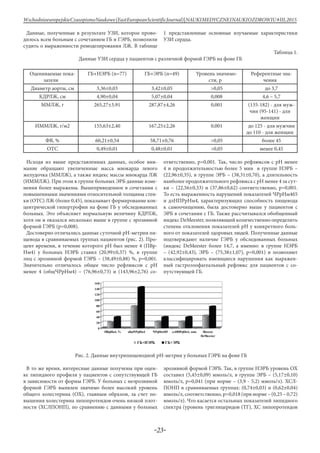
![-25-
WschodnioeuropejskieCzasopismoNaukowe(EastEuropeanScientificJournal)|NAUKIMEDYCZNEINAUKIOZDROWIU#IІІ,2015
Таблица 2.
Показатели активности перекисного окисления липидов и антиоксидантной защиты у больных ГБ в сочетании с ГЭРБ
Показатель НЭРБ +ГБ (n=77) ЭРБ+ГБ (n=49) Контроль (n=20)
МДА, мкмоль/л 4,40±0,10 4,40±0,13 2,71±0,06**
SH-группы, мкмоль/л 476,09±6,78 525,24±7,14* 676,92±21,75**
ГПО, мкмоль/мин/гHb 160,52±6,15 164,19±6,95 261,88±5,79**
* - достоверные отличия при сравнении с пациентами
НЭРБ +ГБ (р=0,001)
** - достоверные отличия группы контроля (р<0,05)
Полученные данные подтверждают усиление проокси-
дантных процессов на фоне сочетания ГБ и ГЭРБ, о чем
свидетельствует увеличение концентрации МДА. В то
же время наблюдается снижение уровеня SH-групп в сы-
воротке крови и концентрации глютатионпероксидазы
(ГПО) в гемолизате – маркеров антиоксидантной защиты.
Обращают внимание значимо более высокие уровни SH-
групп у пациентов с эрозивной формой ГЭРБ по сравне-
нию с группой НЭРБ (р=0,001), что может обуславливать
напряжение компенсаторных механизмов системы анти-
оксидации на фоне интенсивного ПОЛ.
Одним из биохимических факторов, представляющих
определенный интерес при проведении исследования,
был уровень апелина в крови. Являясь лигандом для ан-
гиотензинподобных рецепторов-1 (APJ), он секретиру-
ется эндотелиальными клетками и адипоцитами. Наряду
с большим разнообразием изоформ апелину присущи
множество точек приложения: стимуляция пролифера-
ции клеток слизистой оболочки желудка, секреции холе-
цистокинина, снижение секреции инсулина, гистамина,
соляной кислоты париетальными клетками и др. Все выше
перечисленное подтверждает ценность апелина в качестве
регулятора в желудочно-кишечном тракте. Апелин спо-
собствует восстановлению слизистой оболочки, участвует
в регуляции работы гладкой мускулатуры, а также обмене
веществ. Установлен вазодилатирующий эффект апели-
на посредством NO-зависимых механизмов стимуляции
APJ-рецепторов [1]. Получены данные о том, что апелин
является функциональным антагонистом перифериче-
ских эффектов ангиотензина ІІ. У пациентов с хрониче-
ской сердечной недостаточностью циркулирующий уро-
вень апелина достоверно ниже, чем у здоровых лиц, что
позволяет использовать содержание апелина в качестве
маркера дисфункции миокарда. Предотвращая деграда-
цию супероксиддисмутазы, апелин способен подавлять
продукцию свободных радикалов.
У обследованных пациентов с сочетанной патологией
(ГБ+ГЭРБ) наблюдалось значимое снижение концентра-
ции апелина в крови по сравнении с группой контроля,
в которой апелин крови составил (1133,42±17,85) пг/мл
(р<0,001). При этом в группе больных эрозивной формой
ГЭРБ на фоне ГБ концентрация апелина была достоверно
ниже ((700,91±22,86) пг/мл), чем в группе с неэрозивной
ГЭРБ - (789,87±19,81) пг/мл, р=0,005.
Выводы
1. У пациентов с сочетанием ГЭРБ и ГБ среднесуточные
показатели САД/ДАД и вариабельности АД не имели свя-
зи с формой ГЭРБ. Однако, у лиц с эрозивной формой
ГЭРБ по сравнению с неэрозивной формой чаще выявлял-
ся суточного биоритма САД и ДАД по типу «non-dipper»
и «night-peaker».
2. Для пациентов с сочетанием ЭРБ и ГБ характерна зна-
чимо более выраженная степень ремоделирования ЛЖ по
сравнению с группой ГБ+НЭРБ (р=0,001), с преобладани-
ем концентрической гипертрофии, о чем свидетельствуют
увеличенные индекс массы миокарда ЛЖ и относительная
толщина стенки ЛЖ.
3. У пациентов с сочетанием ЭРБ и ГБ, в сравнении с па-
циентами с сочетанием НЭРБ и ГБ, выявлены более вы-
раженные отклонения показателей суточной внутрипи-
щеводной рН-метрии, свидетельствующие о повышении
агрессивности рефлюксата и его более длительной экспо-
зиции на слизистой пищевода, наряду со снижением спо-
собности пищевода к самоочищению.
4. У пациентов с неэрозивной формой ГЭРБ и ГБ выяв-
лен значимо более высокий уровень ОХ (р=0,04), главным
образом, за счет повышения ХСЛПОНП (р=0,018), в срав-
нении с данными пациентов с эрозивной формой ГЭРБ и
ГБ
5. Наличие сочетанной патологии у обследованных боль-
ных сопровождалось достоверным (р<0,05) повышением
концентрации нитритов как в плазме крови, так и в в су-
точной моче по сравнению с контрольной группой. При
этом у пациентов с сочетанием НЭРБ и ГБ уровень ни-
тритов моче за сутки был достоверно ниже сравнительно с
пациентами с эрозивной формой ГЭРБ и ГБ (р<0,05).
6. На фоне сочетания ГБ и ГЭРБ выявлено усиление про-
цессов ПОЛ, о чем свидетельствует увеличение концен-
трации МДА, и снижение активности антиоксидансной
защиты (снижение уровеня SH-групп в сыворотке крови и
концентрации ГПО в гемолизате). Значимо более высокие
уровни SH-групп у пациентов с эрозивной формой ГЭРБ
по сравнению с группой НЭРБ (р=0,001) обуславливают
напряжение компенсаторных механизмов системы анти-
оксидации на фоне интенсивного ПОЛ.
7. В группе больных эрозивной формой ГЭРБ на фоне ГБ
выявлена достоверно боле низкая концентрация апелина
((700,91±22,86) пг/мл), чем в группе с неэрозивной ГЭРБ
- (789,87±19,81) пг/мл, р=0,005, что потверждает данные о
протективном воздействии апелина на слизистую оболоч-
ку пищеварительного тракта.
Перспективы исследования:
В виду высокой частоты встречаемости сочетания ГБ и
ГЭРБ, выраженного влияния данных расстройств на ка-
чество жизни пациентов, общности этиологических три-
ггерных факторов и патогенеза необходимо дальнейшее
изучение особенностей взаимного влияния данных забо-
леваний на характер клинических проявлений и процес-
сы прогрессирования. Проблема разработки адекватных](https://image.slidesharecdn.com/eesj33-160202073457/85/Eesj-3-3-25-320.jpg)
![-26-
WschodnioeuropejskieCzasopismoNaukowe(EastEuropeanScientificJournal)|NAUKIMEDYCZNEINAUKIOZDROWIU#IІІ,2015
методов коррекции и эффективных профилактических
мероприятий ГБ и ГЭРБ является актуальной и должна
послужить темой для последующих работ.
Список литературы
1. Березин А.Е. Апелин – новый биологический маркер
сердечно-сосудистого и метаболического риска // Ураїнсь-
кий кардіологічний журнал. – 2012. - № 5. – С. 105 – 110.
2. Біловол О.М., Боброннікова Л.Р. Патогенетичні взає-
мозв’язки метаболічних розладів та вільно радикального
окиснення ліпідів при поєднаному перебігу хронічного
холециститу та гіпертонічної хвороби // Сучасна гастро-
ентерологія. -2011. - №1 (57). - С. 12 - 17.
3. Клінічні рекомендації з артеріальної гіпертензії Євро-
пейського товариства гіпертензії (ESH) та Європейського
товариства кардіологів (ESC) 2013 року (Переклад Сіренко
Ю.М.) // «Артериальная гипертензия». - 2013. - №4(30).
4. Ситникова, У.Б. Клинико-патогенетическое значение
гастроэзофагеального рефлюкса у больных ИБС / У.Б.
Ситникова, Н.М. Балабина // Сибирский мед. журн. –2010.
–№5. – C. 20–23.
5. Суточное мониторирование артериального давления /
Пособие для врачей – СПб. : Питер, 2010 г. – 17c.
6. Хлынова О.В. Проблема коморбидности с учетом со-
стояния сердечно-сосудистой системы у пациентов с ар-
териальной гипертензией и кислотозависимыми забо-
леваниями / О.В. Хлынова, А.В. Туев, Л.Н. Береснева //
Казанский медицинский журнал. — 2013. — Т. 94, №1. —
С. 80–85.
7.ChouP.H.,LinCC2,LinCH.Prevalenceofgastroesophageal
reflux disease in major depressive disorder: a population-based
study / P.H. Chou, C.C. Lin, C.H. Lin [et al.] // Psychosomatics.
– 2014. – Vol. 55(2). – P. 155-162.
8. El-Serag H.B. Update on the epidemiology of gastro-
oesophageal reflux disease: a systematic review / H.B. El-Serag,
S. Sweet, C.C. Winchester // Gut. – 2014. – Vol. 63(6). – P.
871–880.
9. Ford AC. The natural history of gastro-oesophageal reflux
symptoms in the community and its effects on survival: a
longitudinal 10-year follow-up study/ A.C. Ford, D. Forman,
A.G. Bailey [et al.] // Aliment Pharmacol Ther. – 2013. - №
37(3). – Р. 323-31.
10. Hunt R. WGO Global Guideline – Coping with common
GI symptoms in the community / R. Hunt, E. Quigley [et al.] //
World Gastroenterology Organization. - 2013. – P. 1-37.
11. Mendes-Filho A.M. Influence of exercise testing in
gastroesophageal reflux in patients with gastroesophageal
reflux disease / A.M. Mendes-Filho, J.P. Moraes-Filho, А. Nasi
[et al.] // Arq Bras Cir Dig. – 2014. – Vol. 27(1) - P. 3-8.
12. Vakil N. Montreal definition and classification of
gastroesophageal reflux disease: a global evidence–based
consensus / N. Vakil, S.V. van Zanten, P. Kahrilas [et al.] // Am.
J.Gastroenterol. – 2006. – V. 101. – P. 1900–1920.](https://image.slidesharecdn.com/eesj33-160202073457/85/Eesj-3-3-26-320.jpg)
![-27-
WschodnioeuropejskieCzasopismoNaukowe(EastEuropeanScientificJournal)|NAUKIMEDYCZNEINAUKIOZDROWIU#IІІ,2015
ОЦЕНКА УДОВЛЕТВОРЕННОСТИ МЕДИЦИНСКОЙ ПОМОЩЬЮ ПАЦИЕНТАМИ, ПЕ-
РЕНЕСШИМИ ОПЕРАЦИЮ НА СЕРДЦЕ В ФГБУ «ФЕДЕРАЛЬНЫЙ ЦЕНТР
СЕРДЕЧНО-СОСУДИСТОЙ ХИРУРГИИ» МИНИСТЕРСТВА ЗДРАВООХРАНЕНИЯ
РОССИЙСКОЙ ФЕДЕРАЦИИ (Г.ПЕРМЬ) И ПУТИ СОВЕРШЕНСТВОВАНИЯ
МЕДИЦИНСКОЙ ПОМОЩИ
Иванова Ольга Вячеславовна
главная медицинская сестра,
ФГБУ «Федеральный центр сердечно-сосудистой хирургии» Министерства здравоохранения Российской Федерации (г.
Пермь)
Касимовская Наталия Алексеевна
кандидат медицинских наук, доцент кафедры общественного здоровья и здравоохранения им. Н.А.Семашко, декан
факультета ВСО и ПСР,
ГБОУ ВПО «Первый Московский государственный медицинский университет имени И.М. Сеченова» Министерства
здравоохранения Российской Федерации
ASSESSMENT OF SATISFACTION WITH MEDICAL CARE BY THE PATIENTS WHO UNDERWENT A OPERATION ON
HEART IN THE FGBU “FEDERAL CENTRE FOR CARDIOVASCULAR SURGERY” OF THE MINISTRY OF HEALTH OF THE
RUSSIAN FEDERATION (PERM) AND WAYS OF IMPROVEMENT OF MEDICAL CARE
Ivanova O. V., home nurse, FGBU «Federal centre for cardiovascular surgery» of the Ministry of health of the Russian Federation
(Perm)
Kasimovskaya N.A., сandidate of мedical sciences, reader First Moscow state medical University named after I. M. Sechenov of
Ministry of healthcare of the Russian Federation
Аннотация
Эффективная организация высокотехнологичной медицинской помощи по профилю сердечно-сосудистая хирургия
играет важную роль в увеличении качества и продолжительности жизни больных с болезнями системы кровообращения.
Кардиологическая реабилитация в послеоперационном периоде позволяет более быстро и качественно адаптировать
пациента к физическим нагрузкам и определить максимально возможный уровень физической активности, уменьшить
вероятность рецидива сердечно-сосудистых событий, а также снизить риск ухудшения функциональных показате-
лей сердечной деятельности. В статье проанализировано современное состояние организации ухода и реабилитации
в ФГБУ «ФЦССХ» Минздрава России (г. Пермь), в рамках оказания высокотехнологичной медицинской помощи. Анализ
представленных данных свидетельствует о необходимости оптимизации реабилитации пациентов после высокотех-
нологичных операций на сердце и сосудах в Пермском крае с целью увеличения доступности всем категориям граждан.
ABSTRACT
Effective organization of high-tech medical care on the profile of cardiovascular surgery plays an important role in increasing the
quality and length of life of patients with diseases of the circulatory system. Cardiac Rehabilitation in the postoperative period allows
more quickly and efficiently adapt to the patient’s physical activity and to determine the maximum possible level of physical activity,
to reduce the likelihood of recurrence of cardiovascular events, as well as reduce the risk of deterioration of the functional parameters
of the heart. The article analyzes the current state of the care and rehabilitation FGBI «FTSSSKH» the Ministry of Health of Russia
(Perm), in the provision of high-tech medical care. Analysis of the data indicates the need to optimize the rehabilitation of patients
after high-tech operations on the heart and blood vessels in the Perm region in order to increase the availability of all categories of
citizens.
Ключевые слова: кардиологическая реабилитация, оценка качества медицинской помощи, сердечно-сосудистая хи-
рургия.
Key words: cardiac rehabilitation, assessment of quality of care, cardiovascular surgery.
Постановка проблемы. Высокая летальность, инвалиди-
зация, большие трудопотери при болезнях системы кро-
вообращения и рост финансовых затрат на их лечение
требуют разработки системы, основанной на современ-
ных технологиях, обеспечивающей высокое качество и
эффективность медицинской реабилитации [3, с. 2-7; 13, с.
33-37; 14, с. 128-134]. Организация высокотехнологичной
медицинской помощи по профилю сердечно-сосудистая
хирургия играет важную роль в увеличении качества и
продолжительности жизни больных с болезнями системы
кровообращения. Кардиологическая реабилитация в по-
слеоперационном периоде позволяет более быстро и каче-
ственно адаптировать пациента к физическим нагрузкам и
определить максимально возможный уровень физической
активности, уменьшить вероятность рецидива сердеч-
но-сосудистых событий, а также снизить риск ухудшения
функциональных показателей сердечной деятельности. В
статье будет проанализировано современное состояние
организации ухода и реабилитации в ФГБУ «ФЦССХ»
Минздрава России (г. Пермь), в рамках оказания высоко-
технологичной медицинской помощи.
Анализ последних исследований и публикаций. Ввод в
эксплуатацию ряда кардиохирургических центров в раз-
личных регионах Российской Федерации позволило сде-
лать специализированную, в том числе высокотехнологич-
ную медицинскую помощь (ВМП) по профилю сердечно](https://image.slidesharecdn.com/eesj33-160202073457/85/Eesj-3-3-27-320.jpg)
![-28-
WschodnioeuropejskieCzasopismoNaukowe(EastEuropeanScientificJournal)|NAUKIMEDYCZNEINAUKIOZDROWIU#IІІ,2015
- сосудистая хирургия более доступной населению [1, с.
21-26; 10, с. 5-8]. Важным этапом оказания ВМП являют-
ся реабилитационные мероприятия в послеоперационном
периоде. (Гусев Е.И., Скворуба В.И., Чекнева Н.С. и др.,
1997, Бураковский В.И. с соавт., 1988, Погосова Г.В., 1998,
Князева Т.А. с соавт., 2002, Оганов Р.Г., Гольдблат Ю.В.,
2006, Ковальчук В.В., Скоромец А.А., 2007, Погосова Г.В.,
2007, Щегольков А.М., 2007, Аронов Д.М, Бубнова М.Г.,
2009, Акчурин Р.С., Власова Э.Е., 2010, Аверин Е.Е., 2010,
Суслова Г.И., Королев А.А., 2010; Вишняков Н.И., Рейн-
штейн Д.И., 2010, Гусев О.А., 2014, Попов В.С., 2014).
В настоящее время процесс восстановительного лечения
пациентов после кардиохирургических вмешательств в
организационном плане представляет собой многоэтап-
ную систему (Данилов, Ю. А., 2002., Кремнев Ю. А., 2002.,
M.Higgins, 2000, W.Vlasic, 2001, A.VomOrde, 2002, H.K.Yip,
2003, Будко А.А., Белякин Ю. А., 2004, Щегольков А.М.,
2005, Макарова А. Г., 2008, Суджаева С. Г., Губич Т. С.,
КазаеваН. А., 2010). В связи с этим особую актуальность
приобретает вопрос преемственности этапов реабилита-
ции, индивидуальный подход к восстановлению каждого
больного, с использованием методов психокоррекции и
применения обучающих программ [7;9, с. 66–78].
Реабилитация – это комплекс скоординированных меро-
приятий медицинского, обучающего, социального и про-
фессионального характера, направленный на адаптацию
пациента к новым условиям жизни путем выявления и
развития его резервных возможностей.
Кардиологическая реабилитация позволяет более бы-
стро и качественно адаптировать пациента к физическим
нагрузкам и определить максимально возможный уровень
физической активности, уменьшить вероятность рециди-
ва сердечно-сосудистых событий, а также снизить риск
ухудшения функциональных показателей сердечной дея-
тельности [12, с. 5 - 6, с. 49 - 51].
Эффективность кардиологической реабилитации обу-
словлена, прежде всего, тем, что операция рассматривает-
ся в качестве этапа лечения, направленного на устранение
анатомической основы патологии, что ведет к нормали-
зации или существенному улучшению кровообращения.
Однако длительное существование болезни приводит к
нарушениям функций сердечно-сосудистой системы, де-
тренированности вследствие гиподинамии и изменениям
психологического статуса пациента. На устранение этих
нарушений, мобилизацию резервов и поддержание опти-
мального функционального уровня направлен комплекс
мер, формирующий программу реабилитации кардиохи-
рургических больных[4,5].
В настоящее время медициной накоплен опыт реабилита-
ции пациентов после хирургического лечения различной
патологии, в том числе при ишемической болезни сердца,
осложненной инфарктом миокарда. Но потребность в реа-
билитации кардиологических пациентов, которым произ-
ведена операция на сердце, неуклонно растет.
Известно, что в странах с развитой экономикой, в те-
чение последних трех десятков лет, отмечается положи-
тельная динамика в виде снижения смертности от сердеч-
но-сосудистых заболеваний на 2,5-3% в год или на 50-60%
за весь указанный период. Внедрение среди населения
комплексных программ реабилитации, первичной и вто-
ричной профилактики позволили достичь такого успеха.
По данным R.S. Teyloretal., применение программ восста-
новительного лечения привело к статистически значимо-
му снижению показателей общей смертности на 20%, а от
сердечно-сосудистых заболеваний – на 26% [15, с. 682-692].
Накоплен опыт проведения реабилитационных меро-
приятий пациентам после кардиохирургических вмеша-
тельств [2, с. 176-241; 6, с. 152-175; 8, с. 31-33]. Согласно
результатам исследования, проведенного в Российском
научном центре хирургии РАМН, 74% пациентов, перенес-
ших операцию аорто-коронарного шунтирования, могут
вернуться к труду, в том числе за счет проведенной реа-
билитации. Кроме того, уменьшаются расходы бюджета за
счет снижения инвалидизации.
Показано [2;8, с. 31-33], что операции по реваскуляриза-
ции миокарда с последующей реабилитацией, в том числе
у пациентов, перенесших острый инфаркт миокарда, не
только улучшают качество их жизни, но и дают экономи-
ческий эффект. Исходя из выполненных авторами расче-
тов, средний прирост годового национального дохода в
результате восстановления трудоспособности больных
составляет 50 млн. рублей, причем 40% из них оценены как
следствие реабилитации.
Совершенствование системы реабилитации больных по-
сле кардиохирургических операций, развитие стациона-
ров восстановительного лечения, как необходимого звена
в системе стационарной медицинской помощи населению,
является крайне важной медико-социальной задачей.
Клиническими исследованиями установлено, что ранняя
активная реабилитация больных в условиях стационара,
мультидисциплинарный подход дают возможность суще-
ственно улучшить результаты лечения, снизить количе-
ство осложнений, инвалидизацию и летальность, улуч-
шить качество жизни больных и их семей.
Выделение нерешенных ранее частей общей проблемы.
В соответствии с программой Пермского края «Развитие
здравоохранения», утвержденной Постановлением Пра-
вительства Пермского края от 3 октября 2013 г. № 1319-п
«Об утверждении государственной программы Пермского
края «Развитие здравоохранения» (в ред. Постановлений
Правительства Пермского края от 22.05.2014 № 381-п, от
25.07.2014 № 684-п, от 30.09.2014 № 1094-п, от 11.12.2014 №
1446-п, от 10.04.2015 № 194-п, с изм., внесенными Поста-
новлением Правительства Пермского края от 18.12.2014 №
1475-п) больным после операций на сердце и магистраль-
ных сосудах предоставляются бесплатные путевки сроком
до 24 дней в санатории, расположенном на территории
Пермского края, обеспечивающем наиболее полный ком-
плекс лечебных мероприятий, а также соответствующие
условия проживания, питания [11, с. 2-7]. Однако, дей-
ствие данного Постановления распространяется только на
работающих граждан и проживающих в Пермском крае. В
тоже время 40% пациентов оперированных в ФГБУ «ФЦС-
СХ» Минздрава России (г.Пермь) проживают в других ре-
гионах Российской Федерации.
Кроме того, порядок перевода пациентов на реабилита-
ционный этап в медицинские организации общегород-
ской сети нуждается в дополнительной проработке. В](https://image.slidesharecdn.com/eesj33-160202073457/85/Eesj-3-3-28-320.jpg)
![-31-
WschodnioeuropejskieCzasopismoNaukowe(EastEuropeanScientificJournal)|NAUKIMEDYCZNEINAUKIOZDROWIU#IІІ,2015
МЕДИКО-СОЦІОЛОГІЧНИЙ МОНІТОРИНГ ФАКТОРІВ РИЗИКУ ХРОНІЧНИХ
НЕІНФЕКЦІЙНИХ ЗАХВОРЮВАНЬ В УМОВАХ ВИКО-РИСТАННЯ МЕДИЧНОЇ
ІНФОРМАЦІЙНОЇ СИСТЕМИ
Кондратюк Наталія Юріївна
науковий співробітник наукового відділу організації медичної допомоги
Державна наукова установа «Науково-практичний фактор профілактичної та клінічної медицини»
Державного управління справами
Шевченко Марина Вікторівна
кандидат медичних наук, старший науковий співробітник
старший науковий співробітник наукового
відділу організації медичної допомоги
Державна наукова установа «Науково-практичний фактор профілактичної та клінічної медицини»
Державного управління справами
Шестак Наталія Вікторівна
науковий співробітник наукового відділу організації медичної допомоги
Державна наукова установа «Науково-практичний фактор профілактичної та клінічної медицини»
Державного управління справами
Ященко Юрій Борисович
доктор медичних наук, професор,
завідувач наукового відділу організації медичної допомоги
Державна наукова установа «Науково-практичний фактор профілактичної та клінічної медицини»
Державного управління справами
MEDICAL-SOCIOLOGICAL MONITORING OF RISK FACTORS CHRONIC NONCOMMUNICABLE DISEASES IN
CONDITIONS OF USE MEDICAL INFORMATION SYSTEM
Kondratiuk N.Iu Researcher scientific department of medical care State Institu-tion of Science «Research and Practical Center of
Preventive and Clinical Medi-cine» State Administrative Department
Shevchenko M. V. PhD, Senior Researcher Senior Researcher scientific department of medical care State Institution of Science
«Research and Practical Center of Preventive and Clinical Medicine» State Administrative Department
Shestak N.V. Researcher scientific department of medical care State Institution of Science «Research and Practical Center of
Preventive and Clinical Medicine» State Administrative Department
Iashchenko Iu. B. MD, Professor, Head of the scientific department of medical care
State Institution of Science «Research and Practical Center of Preventive and Clinical Medicine» State Administrative Department
Аннотация
У статті висвітлено основні принципи технології прийняття діагностичних і управлінських рішень з профілак-
тики найпоширеніших хронічних неінфек-ційних захворювань серед населення на основі даних моніторингу факторів
ризику їх розвитку в умовах запровадження медичної інформаційної систе-ми.
ABSTRACT
In the article the basic principles of diagnostic technology decision-making and the prevention of the most common chronic non-
communicable diseases among the population on the basis of the monitoring of risk factors for their development in terms of the
introduction of medical information system.
Ключевые слова: медико-соціологічний моніторинг, хронічні неінфекційні захворювання (ХНІЗ), фактори ризику (ФР),
медична інформаційна система.
Key words: medical-sociological monitoring, chronic non-communicable diseases (NCD), risk factors (RF), medical information
system.
В основу написання статті покладено результати вико-
нання одного з етапів науково-дослідної роботи «Роз-
робка моделі організації багатофакто-рної профілактики
та управління якістю медичної допомоги при окремих
хронічних неінфекційних захворюваннях прикріплено-
го населення» (термін виконання – 2014-2018 рр. № дер-
жреєстрації 011U002118), яка виконується на базі ДНУ
«Науково-практичний центр профілактичної та клінічної
меди-цини» ДУС.
Постановка проблеми. ХНІЗ в світовому масштабі викли-
кають понад 35 млн випадків смертей в рік, що становить
більше ніж половина випадків смерті в кожному з регіонів
світу. Смертність і захворюваність від цих хво-роб має не-
гативний вплив як на розвиток суспільства, так і наванта-
ження на системи охорони здоров’я, а також несе значний
економічний тягар [4,11].
Особливістю, яка об’єднує хронічні неінфекційні захво-
рювання (ХНІЗ), є їх залежність від факторів ризику (ФР),
на які можна впливати з метою ко-рекції та зменшення їх
негативного впливу на організм людини. З точки зору про-
філактики ХНІЗ, інструментом запобігання їх розвитку
може стати ком-плексна система виявлення та моніторин-
гу основних ФЗ серед населення. Перспективним напрям-
ком у вирішенні проблеми виявлення і моніторингу ФР
є створення у закладі охорони здоров’я автоматизованої
системи моніто-рингу ФР ХНІЗ.](https://image.slidesharecdn.com/eesj33-160202073457/85/Eesj-3-3-31-320.jpg)
![-32-
WschodnioeuropejskieCzasopismoNaukowe(EastEuropeanScientificJournal)|NAUKIMEDYCZNEINAUKIOZDROWIU#IІІ,2015
Моніторинг та аналіз основних ФР, на частку яких при-
падає значна ча-стка майбутнього тягаря ХНІЗ, дає мож-
ливість визначити ефективність прийнятих рішень. На-
приклад, щонайменше 75% випадків серцево-судинних
захворювань пов’язано з неправильним харчуванням і
відсутністю фізичної активності, що веде до вищого індек-
су маси тіла, підвищеного тиску і не-сприятливим показ-
никами ліпідів крові, а також з тютюнокурінням.
Аналіз останніх досліджень і публікацій. Важливість
впливу на ФР виникнення ХНІЗ підтверджується даними
низки досліджень. Якщо впливати на ФР, то ймовірність
померти від ішемічної хвороби серця (ІХС) у віці від 30
років до 69 років може скоротитися на 22% у чоловіків і
19% у жінок в період між 2010–2025 рр, тобто можна по-
передити від 5 до 6,2 млн смертей від ІХС; 1,5 млн смер-
тей від інсульту; 2 млн смертей від діабету. В цілому вплив
на ФР може запобігти на глобальному рівні більш ніж 37
млн смертей від основних ХНІЗ (у 16 млн осіб віком 30–69
років та 21 млн осіб віком 70 років і старших) [5-10].
Як зазначають автори, основою профілактики ХНІЗ
є визначення най-більш істотних ФР, їх профілактика,
моніторинг і контроль на основі поета-пного здійснення
моніторингу (STEPS) рекомендований ВООЗ як інстру-
мент контролю за поширенням неінфекційних захворю-
вань [1-3].
Визначення невирішених частин загальної пробле-
ми. Перспектив-ним напрямком у вирішенні проведен-
ні моніторингу є створення автомати-зованої системи
моніторингу ФР ХНІЗ. Однак на шляху його створення
для широкого класу захворювань виникає ряд питань, од-
ним з яких є створення універсальної медичної інформа-
ційної системи.
Мета: полягає у розробці єдиного підходу до вибору ба-
зових змінних для вивчення при проведенні досліджень,
аналізу, моніторингу та контроля поширеності факторів
ризику розвитку та поширення ХНІЗ.
Методи дослідження, що використано в роботі відповіда-
ли поставле-ним завданням, а саме: метод бібліосемантич-
ного аналізу, контент-аналіз, спостереження, організацій-
ного проектування, структурно-логічний аналіз.
Результати дослідження та їх обговорення.
ВООЗ рекомендовано обирати для моніторингу і кон-
тролю наступні ФР, які:
• мають найбільший вплив на показники захворюваності
та смертності;
• піддаються впливу ефективних заходів первинної про-
філактики;
• дали позитивний досвід їх моніторингу та контролю.
Створення інформаційного простору вимагає прове-
дення певної робо-ти з кадрами, а саме внесення змін
до функціональних обов’язків медичного персоналу
підрозділів усіх рівнів допомоги, проведення хронометра-
жу тру-дових операцій і робочого часу, перегляду норма-
тивів навантаження. Велике значення в роботі з кадрами
відводиться підбору і навчанню персоналу. До-свід запро-
вадження такої системи є у Державній науковій установі
«Науково-практичний центр профілактичної та клінічної
медицини» Державного уп-равління справами.
Першим кроком у наповненні даними медичної інфор-
маційної системи стало створення електронного реєстру
пацієнтів. Дані про пацієнта, що вхо-дять до реєстру,
складають основу паспортної частини його електронної
ме-дичної картки. Електронна медична картка пацієн-
та є «ядром» інформацій-ної системи, яка містить повну
інформацію про його здоров’я та історію зве-рнень за
медичною допомогою. Дані про здоров’я всіх осіб із ам-
булаторної карти на паперовому носії були перенесені
медичним персоналом в елект-ронну медичну картку па-
цієнта, після їх оформлення у вигляді етапного епі-кризу
і сформульованого комплексного діагнозу. Це дозволило
перейти на пріоритетне заповнення медичної документа-
ції в електронному вигляді. При цьому обіг амбулаторних
карт пацієнта на паперових носіях, відповідно до чинного
законодавства відбувається паралельно не дивлячись на
впрова-дження цифрового підпису.
Що стосується цифрового підпису, то на сьогодні біль-
ше 500 співробі-тників пройшли реєстрацію у відповід-
них інстанціях і отримали електронні ключі з правом на
використання свого цифрового підпису. Впровадження
цифрового підпису здійснюється поетапно, відповідно до
певного переліку документів, які підлягають переведенню
на електронний документообіг і планується завершити
одночасно з процедурою сертифікації медичної інфо-рма-
ційної системи на безпеку.
Медична інформаційна система – автоматизована систе-
ма комплексно-го моніторингу ФР ХНІЗ яка призначена
для безперервного їх обліку у елек-тронній медичній карті
пацієнта, моніторингу та оцінки серед прикріпленого на-
селення (на індивідуальному та популяційному рівнях);
аналізу та підгото-вки стратегічних, організаційних, так-
тичних і оперативних заходів, прийнят-тя управлінських
рішень, спрямованих на усунення або зменшення нега-
тив-них наслідків їх впливу з метою мінімізації необхідних
ресурсних затрат.
Об’єктом медико-соціологічного моніторингу є пацієнти,
серед яких проводиться вивчення стану здоров’я за таких
умов: під час прикріплення до закладу охорони здоров’я
для медичного обслуговування, проходження планового
профілактичного медичного огляду, звернення до лікаря
первин-ної ланки за довідкою про стан здоров’я.
На етапі звернення пацієнта до закладу охорони здоров’я
(кабіне-ту/відділення профілактики) фахівцями з базовою
та неповною вищою меди-чною освітою (далі – середній
медичний персонал) проводиться:
- долікарське опитування щодо наявності найпоширені-
ших ФР роз-витку ХНІЗ, пов’язаних зі способом життя,
нездоровим харчуванням, низь-кою фізичною активністю,
тютюнопалінням тощо;
- долікарське обстеження: антропометричні вимірюван-
ня (визначення маси тіла, росту, окружності талії), розра-
хунок індексу маси тіла, вимірю-вання артеріального ти-
ску.
Забезпечення якості процесу виявлення та реєстрації ві-
домостей про пацієнта досягається шляхом внутрішнього
і зовнішнього контролю техно-логії збору даних. Підви-
щення компетентності середнього медичного персо-налу
досягається шляхом навчання і постійного підвищення
кваліфікації (пе-реважно шляхом дистанційної освіти).](https://image.slidesharecdn.com/eesj33-160202073457/85/Eesj-3-3-32-320.jpg)
![-34-
WschodnioeuropejskieCzasopismoNaukowe(EastEuropeanScientificJournal)|NAUKIMEDYCZNEINAUKIOZDROWIU#IІІ,2015
нання рекомендацій для пацієнтів та медичного персоналу
закладу охорони здоров’я.
Висновок.
Визначено основні аспекти медико-соціологічного
моніторингу факто-рів ризику хронічних неінфекційних
захворювань в умовах використання медичної інформа-
ційної системи.
Запропоновано примірні етапи моніторингу для вирі-
шення завдань з раннього виявлення, обліку, прогнозу
факторів ризику ХНІЗ як на індивіду-альному рівні, так
і серед прикріпленого населення в цілому та формування
звітності.
Список літератури:
1. Блум Дэйвид И. Состояние здравоохранения в миро-
вом масштабе / Д. И. Блум // Мировое здравоохранение.
– 2014. – №12. – С. 6–11
2. Глобальный план борьбы с диабетом 2011–2021. Меж-
дународ-ная Федерация диабета. – Брюссель, 2010. – С.7
3. Мониторинг факторов риска неинфекционных забо-
леваний. Принцип поэтапной реализации, предложенный
ВОЗ. Краткий обзор / Р. Бо-нита, М. де Куэртен,Т. Дуайер
и др. – Женева: ВОЗ, 2001
4. Contribution of six risk factors to achieving the 25×25 non-
communicable disease mortality reduction target: a modelling
study / V. Kontis, C. D. Mathers, J. Rehm [et al.] // The Lancet.
– 2014. – Vol. 384. – Р. 427–437
5. Di Cesare M. The contributions of risk factor trends to
cardiometabolicmortalitydeclinein26industrializedcountries
/ M. Di Cesare, J.E. Bennett, N. Best [et al.] // International
Journal of Epidemiology. – 2013. – № 42. – Р. 838–848
6. Ezzati M. Behavioral and dietary risk factors for non-
communicable diseases / M. Ezzati, E. Riboli // New England
Journal of Medicine: Research & Review. – 2013. – Vol. 369. – Р.
954–964
7. Ezzati M. Can non-communicable diseases be prevented?
Lessons from studies of populations and individuals / M.
Ezzati, E. Riboli // Science. – 2012. – Vol. 337. – Р. 1482–1487
8. Ford E. S. Proportion of the decline in cardiovascular
mortality disease due to prevention versus treatment: public
health versus clinical care / E. S. Ford, S. Capewell // Annual
Review of Public Health. – 2011. – № 32. – Р. 5–22
9. Global and regional mortality from 235 causes of death for
20 age groups in 1990 and 2010: a systematic analysis for the
Global Burden of Disease. Study 2010 / R. Lozano, M. Naghavi,
K. Foreman [et al.] // Lancet. – 2012. – Vol. 380. – Р. 2095–128
10. Inequalities in non-communicable diseases and effective
responses / M. Di Cesare, Y. H. Khang, P. Asaria [et al.] //
Lancet. – 2013. – № 381. – Р. 585–597
11. Policy dialogue on China’s changing burden of disease / L.
Yuanli , Y. Gonghuan , Z. Yixin [et al.] // The Lancet. – 2013. –
Vol. 381 (9882). – Р. 1961–1962](https://image.slidesharecdn.com/eesj33-160202073457/85/Eesj-3-3-34-320.jpg)
![-35-
WschodnioeuropejskieCzasopismoNaukowe(EastEuropeanScientificJournal)|NAUKIMEDYCZNEINAUKIOZDROWIU#IІІ,2015
ВПЛИВ КОМУНІКАЦІЇ АЕРОКОКІВ З ПАТОГЕННИМИ ТА УМОВНО-ПАТОГЕННИМИ
МІКРООРГАНІЗМАМИ IN VITRO НА ЇХ БІОЛОГІЧНІ ВЛАСТИВОСТІ
Степанський Дмитро Олександрович
ДЗ «Дніпропетровська медична академія
МОЗ України», завідувач кафедри мікробіології,
вірусології, імунології та епідеміології
Кошова Ірина Петрівна,
ДЗ «Дніпропетровська медична академія
МОЗ України» викладач
кафедри загальної та клінічної фармації
INFLUENCE OF COMBINATION OF AEROCOCCUSES WITH PATHOGENIC AND CONDITIONALLY PATHOGENIC
MICROORGANISMS IN VITRO ON THEIR BIOLOGICAL PROPERTIES
Stepanskyi Dmytro Oleksandrovitch SE «Dnepropetrovsk Medical Academy оf Ministry of Health of Ukraine», associate manager
by a Department Microbiology, Virology, Immunology and Epidemiology
Koshova Irina Petrivna SE «Dnepropetrovsk Medical Academy оf Ministry of Health of Ukraine», teacher General and Clinical
Pharmacy Department
Анотація
Нормальна мікрофлора організму людини являє собою сукупність різних біоценозів, що займають численні екологічні
ніші на шкірі і слизових оболонках всіх порожнин, сполучених із зовнішнім середовищем. Aerococcus viridans володіють ці-
лим рядом корисних властивостей. Була проведена серія експериментів з вивчення впливу аутоштамів аерококів на Гр +
і Гр - бактерії. При вивченні відстроченого антагонізму показано, що тест-мікроби по-різному реагують на присутність
у середовищі вирощування продуктів метаболізму аутоштамів аерококів. Крім того, навіть усередині одного і того ж
виду спостерігаються коливання. Аутоштами аерококів на відміну від стафілококів, кишкової палички та вібріона не
проявляють чутливості до дії активного початку псевдомонад. Аерококи володіють вираженими антагоністичними
властивостями по відношенню до псевдомонад. Колонії псевдомонад які виростають, як правило, не мають пігменту.
ABSTRACT
Normal microflora of the human body is a combination of different biocenoses, occupying numerous ecological niches on the skin
and mucous membranes of cavities connected with the environment. Aerococcus viridans has a number of useful properties. Series of
experiments on the effects on autostrains aerococcuses Gr + and Gr - bacteria was conducted. In studying of deferred antagonism is
shown that test microbes react differently to the presence metabolic products of aerococcuses autostrains in the environment of growing.
Moreover, even within the same species there are differences. Autostrains of aerococcuses unlike Staphylococci, E. coli and vibrio show
no sensitivity to the action of Pseudomonas. Aerococcuses have expressed antagonistic properties to Pseudomonas. Growing colonies
of Pseudomonas typically do not have pigment.
Ключові слова: нормальна флора,аерококи, псевдомонади, антагоністичні властивості.
Keywords: normal flora, aerococcuses, Pseudomonas, antagonistic properties.
Постановка проблеми. Розлад захисної функції мікро-
флори під впливом різних чинників призводить до шлун-
ково-кишкових захворювань інфекційної і аліментарної
етіології, знижується здатність нормальної мікрофлори
кишечнику ефективно пригнічувати ріст патогенних мі-
кроорганізмів.
Аналіз останніх досліджень і публікацій. Нормальна мі-
крофлора організму людини являє собою сукупність різ-
них біоценозів, що займають численні екологічні ніші на
шкірі і слизових оболонках всіх порожнин, сполучених із
зовнішнім середовищем [6,8]. Зменшення кількості нор-
мальної кишкової мікрофлори веде до ослаблення кон-
куренції з патогенами за рецептори слизової оболонки
кишечнику, знижується місцевий імунітет - продукція лі-
зоциму, імуноглобуліну А [2].
Сформована ситуація, вимагає пошуку нових анти-
бактеріальних засобів і підходів до лікування. Одним
з найбільш обговорюваних в останні роки підходів до
профілактики та лікування кишкових інфекцій є застосу-
вання пробіотиків [2,5,6]
Виділення невирішених раніше частин загальної про-
блеми. Багатьма дослідниками встановлено важливу роль
нормальної мікрофлори організму людини у підтримці
його фізіологічного стану, забезпеченні гомеостазу і жит-
тєдіяльності. Особливу цікавість викликають представни-
ки нормальної мікрофлори людини - пробіотичні мікро-
організми[7].
Однак є невелика кількість фундаментальних дослід-
жень, присвячених Aerococcus, Acetovibrio, Butyrivibrio,
Centipeda, Eutactarrium, Leptotrichia, Ruminobacteria,
Solenomonas, Succinivibrio. Aerococcus viridans володіють
цілим рядом корисних властивостей, вироблених у про-
цесі динамічної взаємодії як з макроорганізмом, так і з
мікробіоасоціантами [2]. Наприклад, виявлена здатність
аерококів окисляти молочну кислоту призводить до зни-
ження рівня гіпоксії в кишечнику; виділення пероксиду
водню, який розщеплюється каталазою епітелію кишеч-
ника і також призводить до оксигенації тканин [1,5].
Глутатіонредуктазна активність, утворення кон’югатів
глутатіону з ксенобіотиками та інші корисні властивості
становлять інтерес для подальшого вивчення їх впливу на
гомеостаз макроорганізму[4].
Метою дослідження було розширене вивчення взаємин
аерококів з](https://image.slidesharecdn.com/eesj33-160202073457/85/Eesj-3-3-35-320.jpg)
![-36-
WschodnioeuropejskieCzasopismoNaukowe(EastEuropeanScientificJournal)|NAUKIMEDYCZNEINAUKIOZDROWIU#IІІ,2015
Гр + і Гр - бактеріями, що мають значення у визначенні
біоценотичних взаємин між патогенними бактеріями і
нормальною мікрофлорою шкіри і слизових поверхонь
організму людини.
Виклад основного матеріалу.
У 1 -й серії експериментів вивчені взаємини аерококів з
Гр + і Гр – бактеріями. Для цього використовувався метод
відстроченого антагонізму[3]. Тест - культурою служив
аутоштам A. viridans №15, виділений від людини.
На МПА засівалась штрихом звісь добової культури ауто
штаму
A. viridans в ізотонічному розчині натрію хлориду (2x10⁸
мікробних клітин).
Для визначення оптичної щільності мікробної звісі вико-
ристовувався прилад Densi-La-Meter. Оптична щільність
за шкалою Мс Farlanda дорівнювала 0,7 ОД. Посів прово-
дився по діаметру чашки Петрі.
Посіви інкубували 24 години при 37°С. Після інкубації
до культури
A. viridans, що виросла на чашці, перпендикулярно штри-
хом підсівали взвісь свіжовиділених добових культур тест
- штамів. У наших дослідженнях був розширений спектр
умовно-патогенних і патогенних бактерій. Оптична щіль-
ність вимірювалась для кожної культури окремо і була
ідентичною до щільності ауто штаму A. viridans.
У табл.1 представлені результати проведеного дослід-
ження.
Отримані дані (табл.1) свідчать про те, що при вивченні
відстроченого антагонізму тест-мікроби по різному реагу-
ють на присутність у середовищі вирощування продуктів
метаболізму аутоштамів аерококів. Крім того, навіть усе-
редині одного і того ж виду спостерігаються коливання. Це
особливо чітко видно при вивченні як тест-мікроба холер-
ного вібріона. Причому, чим більше вірулентний вібріон,
тим більше він стійкий до антагоністичній дії аерококів.
Відзначені значні зони затримки росту відносно вібріонів,
стафілококів, дифтерійної палички, псевдомонад, бактерій
групи кишкової палички, бацил.
Таблиця 1
Спектр антагоністичної активності ауто штамів A. viridans
Види тест-культур Кількість культур Діаметр зон затримки росту (мм)
тест-культур аерококами; M ±m
S. aureus ( № 209 )
Госпітальні штами
1
50
15,0 ± 1,0
12,3 ± 0,9
V. eltor ( № 5879 ), вірулентний, холе-
рогенний
V. nag ( № 5459 ), вірулентний, холе-
рогенний
V. nag ( № 5454 ), вірулентний, енте-
ропатогенний
V. nag ( Р-6078 ), авірулентний
НАГ- вібріони, виділені з водоймищ
1
1
1
1
19
7,2 ± 0,4
10,0 ± 0,6
12,3 ± 0,9
37,0 ± 4,0
21,6 ± 1,4
P. vulgaris (OX 19 221)
Госпітальні штами
1
4
7,0 ± 1,0
5,1 ± 0,4
P. mirabilis (“H” № 2091)
P. rettgeri (Титов № 1)
1
1
6,0 ± 1,0
7,0 ± 1,0
P. аeruginosa (№ 501)
Госпітальні штами
1
26
5,5 ± 0,3
5,22 ± 0,25
E. faecalis (№ 6783) 1 4,0 ± 1,0
Госпітальні штами E. coli 8 5,7 ± 0,8
P. аlcalifaciens (№ 26240 ) 1 11,0 ± 2,0
B. subtilis (CCM 2216 ATCC 6051 ) 1 4,0 ± 1,0
C. diphteriae (штамм PW-8), віру-
лентний
1 25,0 ± 0,6
C. рerfringens (музейний)
С. sporogenes (музейний)
1
2
12,0 ± 2,0
13,0 ± 2,0
S. flexneri (музейний) 1 9,0 ± 1,0
S. typhimurium (№ 5710 )
S. sonnei (№ 4997 )
1
1
8,0 ± 1,0
9,0 ± 1,0
Наступна серія дослідів показала вплив аерококів на 26
госпітальних штамів P. aeruginosa, виділених від хворих
Дніпропетровського обласного опікового центру, що охо-
плюють усі серогрупові і серотипові варіанти цього виду
мікроорганізмів. Облік результатів експерименту прово-
дився шляхом вимірювання діаметру зони пригнічення
росту в мм. Антагоністичні властивості аутоштамів аеро-
коків проти псевдомонад було досліджено такожв іншому
варіанті експериментів.
На м’ясо-пептони агар в чашках Петрі засівали газоном](https://image.slidesharecdn.com/eesj33-160202073457/85/Eesj-3-3-36-320.jpg)
![-38-
WschodnioeuropejskieCzasopismoNaukowe(EastEuropeanScientificJournal)|NAUKIMEDYCZNEINAUKIOZDROWIU#IІІ,2015
СПИСОК ЛІТЕРАТУРИ:
1. Кременчуцкий Г.Н., Рыженко С.А., Вальчук С.И. Роль
микроэкологииорганизмачеловека и принципыеекоррек-
ции. - Днепропетровск, 2003. - С. 58-59.
2. Коррекциядисбиотическихнарушений при заболева-
нияхжелудочно-кишечноготракта и печенибиологически-
активными добавками с пробиотическимдействием / Со-
ловьева Н.В., Лейхтер С.Н., Бажукова Т.А. [и др.] // Обзоры
по клиническойфармакологии и лекарственнойтерапии.
— 2013. — Т. 8. – С. 48—57.
3. Об унификациимикробиологическихметодовисследо-
вания, применяемых в клинико-диагностическихлабора-
торияхлечебно-профилактическихучреждений. Приказ№
535 МЗ СССР от 22.04.1985. М., 1985. 56 с.
4. Риженко С.А., Вальчук С.І. Вплив живої культури
Aerococcusviridans на фактори імунологічної реактивності
організму людини invitro // Одес. мед. журн. – 2003. - № 5
(79). – С.108-111.
5. Рыженко С.А. ВлияниеА-бактерина на гомеостаз орга-
низмачеловека при различныхпатологическихсостояниях
// Сб. материаловмеждунар. науч.-практ. конф. памяти Г.И.
Гончаровой «Пробиотическиемикроорганизмы – совре-
менноесостояниевопроса и перспективыиспользования» /
Под ред. В.А. Алешкина. – М., 2002. – С. 41-42.
6. Clinicalefficacyofprobiotics: reviewoftheevidencewithfocus
onchildren / S. Michail, F. Sylvester, G. Fuchs, R. Issenman //
J. Pediatr. Gastroenterol. Nutr. – 2006. - Vol. 43, N4. – P. 550
–557.
7. Michail S, Sylvester F, Fuchs G, Issenman R. Clinical efficacy
of probiotics: review of the evidence with focus on children. J.
Pediatr. Gastroenterol.Nutr. 2006;43(4):550-557.
8. Steinhoff U. 2005. Who controls the crowd? New
findings and old questions about the intestinal microflora.
ImmunologyLetters, Volume 99, Issue 1, 15 June, Pages 12—16.
BIOTIPES OF AEROCOCCUS VIRIDANS
Stepanskyi Dmytro Oleksandrovitch
SE «Dnepropetrovsk Medical Academy
оf Ministry of Health of Ukraine»,
associate manager by a Department Microbiology,
Virology, Immunology and Epidemiology
Koshova Irina Petrivna
SE «Dnepropetrovsk Medical Academy
оf Ministry of Health of Ukraine»,
teacher General and Clinical
Pharmacy Department
Kremenchutskyi Gennady Mykolayovitc,
SE «Dnepropetrovsk Medical Academy of Ministry
of Health of Ukraine» professor Department
Microbiology, Virology,
Immunology and Epidemiology
Xeniya Fedoryak
Medical University of Lodz, Poland
English division, 2nd year
ABSTRACT
A. viridans microorganisms are widespread in nature, they are representatives of the macro-microbiocenosis. There is evidence
in the literature on the allocation of A. viridans during various diseases without an analysis of the immune status and the study of
other possible pathogens that are the main cause of the disease. The presented in the literature genetic analysis of A. viridans took
from the pathological focus does not gives us an opportunity to associate it with certain characteristics aerococcus. We conducted the
research of excretion A. viridans from healthy macroorganisms and from the environment to study their biological characteristics (for
antibacterial compounds, the ability to produce reactive oxygen species, antioxidant system activity and antagonist activity). Based on
these characteristics A. viridans were divided into three biotypes with strict compliance scientists features.
Key words: aerococcus, biotypes, biochemical activity, аntioxidant protection.
Formulation of the problem. Despite the fact that it is long
time since the discovery and identification of aerococcus there
is still no information about dissemination and characteristics
of aerococcus takes part in the composition of microbial
associations of endothermal animals. For the first time
aerococcus was sorted out in 1953 [42, 43], by the inoculation
of the air, the air from the hospitals [33], soil [34].
Analysis of recent research and publications. Right after it
was reported that aerococcus found in pathological material in
the infectious process. A. viridans were isolated from the blood
of patients with endocarditis [25, 28], from the urine of patients
with urinary tract infection [31], from the blood of patients
with hypogranulocytosis [24, 40].
Aerococcus is prevalent in the animal microflora. The
hemocultures of aerococcus were obtained from mice with
bacterieamia [29]. It has been found that A. viridans is sensitive
to penicilline, macrolide and chloramphenicol and resistant to
aminoglycoside [26]. Were described the incidents of diseases
in animals: pigs [36], cows’ mastitis [39], amphibious – turtles
[40].
The discovery of A. viridans in the focus of inflammation
may not be indicative of their aetiological role without detailed](https://image.slidesharecdn.com/eesj33-160202073457/85/Eesj-3-3-38-320.jpg)
![-39-
WschodnioeuropejskieCzasopismoNaukowe(EastEuropeanScientificJournal)|NAUKIMEDYCZNEINAUKIOZDROWIU#IІІ,2015
bacteriological and virologic research of pathological material
(disclosure or absence of Chlamydia or viruses). No study
indicated the discovery of pathogenicity factors in aerococcus
and attempts to simulate an experimental animal model of
infection has not been successful [32].
At the same time it was shown the wide representation of
aerococcus in microbiocenosis of macroorganisms [18].
Biological treatment and prevention drugs, developed on
the basis of A. viridans 167 [2, 7, 11, 19, 20] were separated
from the human milk by the professor Gorbunova M. L. (1965)
have demonstrated clinical efficacy in various pathological
processes [21].
It has shown effectiveness for bacterial infection of the
nasopharynx [4], increased immunoresponsiveness body
[3, 15, 20] for preventing and treating salmonellosis [21],
staphylococcal infection [13, 14, 16, 19] for control and
normalization of the vaginal flora [ 18], the correction of
dysbiosis and homeostasis of the organism [12], pulmonary
tuberculosis [5], urologic complications of pathological
processes [8], an antagonistic effect on the meningococcus [1].
Bold unsolved aspects of the problem. It has showed the
intraspecificdifferencebetweenaerococcus[17].Duringstudies
at the molecular level in aerococcus (genetic characteristics,
distribution of cell wall proteins during electrophoresis) have
emerged a large number of aerococcus cultures with slight
differences in characteristics assigned. This distribution is
separated cultures do not allow to make the correlation between
the molecular differences and their biological characteristics.
The aim of the study. The object of our work is to try to
spread the culture divided into two fundamental characteristics
of A. viridans (the ability to oxidize lactate production of
hydrogen peroxide and selenium reduction of its salt).
Presenting main material. We studied gram-positive,
catalase-negative cocci taken from biological material from
healthy people, animals and birds, and delivered a comparative
study to their taxonomic characteristics.
It has been studied 118 aerococcus crops; taken from the
human body, 16 cultural objects taken from the environment
(water, wash vegetables), 21 cultures taken from animals (mice
- 10 aerococcus crops, pigs - 3 cultures, cows- 5 crops, chicken
- 6 of Culture), a strain of sample preparation and bacterial-
bacterine N and 4 links aerococcus cultures. As reference
cultures were used strains A. viridans from the collection SSM:
№1911 [28, 32, and 41].
Separation and identification of aerococcus cultures was
held in the agreements methods offered by GN Kremenchutskiy
et al (2009) [7].
For separation (gram+, gram- ) cocci was used fluid from
the oral cavity of healthy people and faeces of animals and
birds as biological material. Intake and held crop material and
identifying microbial culture were made in accordance with
[10].
To distinguish aerococcus forms on biochemical activity
was developed an indicated medium, that includes sodium
selenite. The following composition: 1 l of distilled water:
potassium iodide - 26 g, soluble starch - 10 g, sodium selenite
- 0, 4 g, dry nutrient medium - 32 g. The incorporation into
the potassium iodide and soluble starch in medium makes it
possible to evaluate the activity of oxidase aerococcus resulting
color of the colonies in the culture medium as deep blue
color. Aerococcus colonies that reduce sodium selenite in the
selenium initial stain appear red.
Sensitivity to antibiotics are evaluated by agar diffusion
method using discs [10]. Aerococcus antagonistic action to
the test cultures of microorganisms has been studied by the
methods of deferred antagonism.
Glutathione peroxidase activity was determined by the
method of [9]. The protein was identified using the method of
[34]. Superoxide was determined by the method of [36]. The
activity of superoxide was identified by the method of [6]. SOD
activity was expressed in micromoles / min. mg protein.
During sowing crops aerococcus cultures separated on
indicated media was noticed another color of the colonies
and of the medium depending on the biochemical activity
aerococcus. Culture, which oxidize potassium iodide sprouted
colony with an intense dark color with dark coloring of the
growth medium, the cultures were evaluated among 1 biotype
aerococcus.Culturesthatreduceseleniumfromsodiumselenite
colony sprouted, painted in red. These cultures were assessed
as two biotypes aerococcus. Furthermore, it was observed that
the growth of the colonies colored red with fade out within the
area around the medium indicates the oxidation of potassium
iodide and at the same time reducing selenium from sodium
selenite about.
These cultures were rated as 3 biotype of aerococcus.( Pic.1)
Distribution of separated cultures from different sources by
biotypes is shown in Tab. 1
Pic. 1. Three biotypes Aerococcus viridans:
a)Black colonies – 1 biotype, red – 2 biotype of aerococcus;
b)Red colonies with black zones – 3 biotype.](https://image.slidesharecdn.com/eesj33-160202073457/85/Eesj-3-3-39-320.jpg)
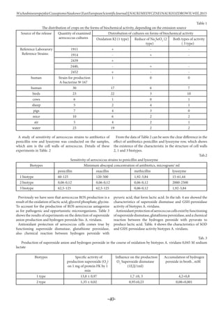
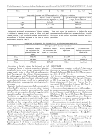
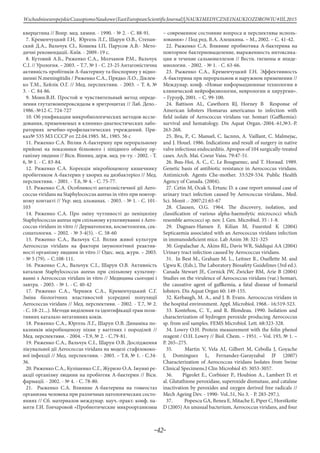
![-43-
WschodnioeuropejskieCzasopismoNaukowe(EastEuropeanScientificJournal)|NAUKIMEDYCZNEINAUKIOZDROWIU#IІІ,2015
cases of infective endocarditis. J Heart Valve Dis 14: 317-319.
38. Spakovaґ T., Elecko J., Vasil M., Legaґth J., Pristas P.,
Javorskyґ P. Limited genetic diversity of Aerococcus viridans
strains isolated from clinical and subclinical cases of bovine
mastitis in Slovakia Polish Journal of Veterinary Sciences Vol.
15, No. 2 (2012), 329-335
39. Torrent A, Deniz S, Ruiz A, Calabuig P, Sicilia J, Oros
J (2002) Esophageal diverticulum associated with Aerococcus
viridans infection in a loggerhead sea turtle (Caretta caretta). J
Wildl Dis 38: 221-223.
40. Uh, Y., Son, J. S., Jang, I. H., Yoon, K. J., & Hong,
S. K. Penicillin-resistant Aerococcus viridans bacteremia
associated with granulocytopenia. Journal of Korean Medical
Science.-2002.- 17(1), 113-115
41. Williams, R.E.O., Hirch, A., and Cowan, S.T.
Aerococcus, a new bacterial genus. J.Gen. Microbiol., 1953,
Vol. 79,№8, 475-480
ОСОБЕННОСТИ АНАТОМИИ МОЗОЛИСТОГО ТЕЛА МОЗГА (ПО ДАННЫМ
НЕЙРОСОНОГРАФИИ) И НЕРВНО-ПСИХИЧЕСКОЕ РАЗВИТИЕ У ДЕТЕЙ С
ГИПОКСИЧЕСКИ-ИШЕМИЧЕСКОЙ ЭН-ЦЕФАЛОПАТИЕЙ
Кощавцев А.Г.
доцент кафедры психиатрии,
Санкт-Петербургский Педиатрический Медицинский Университет
Краснощекова Е.И.
доктор биологических наук, профессор кафедры психофизиологии
Санкт-Петербургский Университет
ANATOMYOFTHECORPUSCALLOSUMOFTHEBRAIN(AC-CORDINGNEUROSONOGRAPHY)ANDDEVELOPMENT
OF CHIL-DREN WITH HYPOXIC-ISCHEMIC ENCEPHALOPATHY
Koshchavtcev A.G., docent of the department of psychiatry Saint-Petersburg Pediatric Medical University
Krasnoshchekova E.I., doctor of biology, professor Saint-Petersburg University
Аннотация
Исследование на протяжении первого года жизни младенцев указывает, что на неврозоподобные и резидуальные нару-
шения у детей в возрасте 12-14 месяцев влияют как особенности течения беременности, так и родов. Для доношенных
и переношенных детей большее значение имеет течение родов. Для недоношенных в возрасте до 33 недель и в возрасте
34-35 недель гестации решающая роль принадлежит сосудистым нарушениям и уровню морфологической зрелости моз-
га. А у недоношенных в возрасте 36-37 недель исходы зависят от большего количества факторов, кроме пе-речисленных
для других гестационых групп, в том числе и от показателей качества брачных отношений, возраста матери. Резуль-
таты срезового ис-следования большой группы детей до месяца находятся в контексте мень-шего по количеству детей
исследования детей первого года жизни. Вместе тем, «косой» размер клюва мозолистого тела органично «включается»
в данную систему координат, являясь, наряду с другими показателями, мар-кером гестации и гипоксического поражения
мозга.
ABSTRACT
Research during the first year of infant data confirms that the residual and neu-rotic disorders in children aged 12-14 months are
associated with hypoxia dur-ing pregnancy and childbirth. During the act of birth is important for full-term infants. For premature
babies under the age of 33-35 weeks of gestation deci-sive role belongs to vascular disorders and morphological level of maturity of
the brain. Premature infants aged 36-37 weeks, the outcome depends on many factors, including the quality indicators of marital
relations, maternal age. Size «beak» of the corpus callosum is a marker of gestation and hypoxic brain dam-age.
Ключевые слова: Нервно-психическое развитие младенца, недоношенные дети, доношенные дети, гипоксически-ише-
мическая энцефалопатия, (ГИЭ), мозолистое тело головного мозга (МТ).
Key words: infant development, premature babies, full-term infants, hypoxia, hypoxic-ischemic encephalopathy, corpus callosum
of brain.
Интегративную работу головного мозга выполняют ас-
социативные тракты, связывающие различные области
коры. Мозолистое тело (МТ) осуществляет взаимодей-
ствие полушарий, объединяя их ассоциативные си-сте-
мы. Топографическая приуроченность межполушарных
проекций к от-делам МТ позволяет судить о состоянии
областей коры. Известно, что по-ражение коры головного
мозга присутствует у 35-85% новорожденных с гипокси-
чески-ишемической энцефалопатией (ГИЭ)[6]. Парциаль-
ные ин-сульты как следствие ГИЭ в коре отмечаются чаще
в области погранично-го кровоснабжения из бассейнов
различных сосудов.
Гипоксия и ишемия приводят к нейрональным потерям
и появлению неорганизованных миелиновых волокон бе-
лого вещества, что сопровож-дается появлением гребней
и борозд, известных как «грибовидные» изви-лины [6]. В
лобных долях двусторонние острые инфаркты «водораз-
дель-ного» типа между передней и средней мозговой ар-
териями являются след-ствием нарушения мозгового кро-
вообращения в первые 24 часа жизни. Результатом потерь
нейронов является гипермиелинизация и замена па-рен-
химы мозга спиномозговой жидкостью [5].
Задачей исследования было сравнить нервно-психиче-
ское развитие недоношенных и доношенных младенцев
с гипоксически-ишемической эн-цефалопатией со стан-
дартными нейросонографическими данными и дан-ными](https://image.slidesharecdn.com/eesj33-160202073457/85/Eesj-3-3-43-320.jpg)
![-44-
WschodnioeuropejskieCzasopismoNaukowe(EastEuropeanScientificJournal)|NAUKIMEDYCZNEINAUKIOZDROWIU#IІІ,2015
строения мозолистого тела.
В обследовании приняли участие 350 детей с ГИЭ пер-
вого месяца жизни, оказавшихся на отделении патоло-
гии новорожденных Детской го-родской больницы № 22
Санкт-Петербурга вследствие гипоксии и асфик-сии из
которых 6 детей имели гестацию менее 29 недель, 14 детей
- 29-31 недель, 87 детей – 32-34 недель, 113 младенцев были
недоношенными на 35-37 неделе, 118 детей были доношен-
ными (38-40 недель) и 12 детей были переношенными (41-
43 недели).
Топографически мозолистое тело разделяют на «клюв»
(СС1), «ко-лено» (СС2), ростральное тело (СС3), среднее
тело в составе переднего (СС4) и заднего (СС5), «переше-
ек» (СС6) и «валик» (СС7) [3]. Проводи-лась нейросоно-
графия на приборе LOGIQ S8 в саггитальной плоскости и
определялся «косой» размер клюва (Скриншот 1 НСГ ре-
бенка на 32 неделе гестации, Скриншот 2 НСГ ребенка на
40 неделе гестации) в см. Очевидно, что «клюв» мозоли-
стого тела топографически «привязан» к ассоциативным
связям лобной доли, которые влияют на мотивацию к
деятельности, ха-рактерологические особенности, сукцес-
сию, математические способности ребенка.
Учитывались степень тяжести гипоксически-ишеми-
ческой энцефало-патии, определяемая при клиническом
осмотре по Якунину, Ямпольской [4], данные рутинной
нейросонографии (дилатация желудочков, дилатация
межполушарной щели и субарахноидальных пространств,
наличие повы-шенной эхогенности перивентрикулярных
зон и перивентрикулярная лей-комаляция, перивентри-
кулярные кровоизлияния по степеням, наличие субэпин-
димальных кист, нарушение визуализации борозд и изви-
лин).
Скриншот НСГ 1
Скриншот НСГ 2
Эмпирически мы выделили некую иерархию в размерах
клюва МТ от самого малого размера (0,09-0,19 см), через
средние его размеры (0,2-0,29 см; 0,3-0,39 см), к большо-
му (свыше 0,4 см). Данная последователь-ность понятна
при наличии глубокой недоношенности и органического
поражения мозга (например, гидроцефалия), при которых
мозолистое те-ло, по-видимому, истончено.
Показателями исхода ГИЭ мы считали степень тяжести
перинаталь-ной энцефалопатии (показано, что средняя и
тяжелые степени тяжести все-гда отражаются на последу-
ющем развитии), данные нейросонографии (вентрикуло-
мегалия и внутрижелудочковые кровоизлияния приводят
к развитию Минимальной мозговой дисфункции и более
тяжелым наруше-ниям ЦНС) и мы оценивали наличие
невротических, неврозоподобных и резидуальных нару-
шений в возрасте 12-14 месяцев (нарушения сна; ги-пер-
кинезы в виде тиков, яктации; дефицит внимания; дорече-
вые нарушения в данном возрасте; детский церебральный
паралич).
При однофакторном дисперсионном анализе отмечают-
ся различия с высокой степенью достоверности между
группами детей с минимальными значениями величины
«клюва» МТ и его значениями - 0,2-0,29 см по большинству
показателей с четкой приуроченностью к гестации. То есть
при «косом» размере «клюва» МТ 0,09-0,19 см чаще отме-
чается макси-мальная недоношенность (33,9+2,96 неделя](https://image.slidesharecdn.com/eesj33-160202073457/85/Eesj-3-3-44-320.jpg)
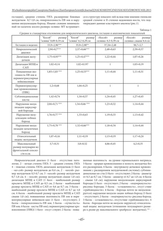
![-46-
WschodnioeuropejskieCzasopismoNaukowe(EastEuropeanScientificJournal)|NAUKIMEDYCZNEINAUKIOZDROWIU#IІІ,2015
статистические различия при р<0,05 при однофакторном
дисперсионном анализе программы STATISTICA 10; ***
- статистиче-ские различия при р<0,005 при однофактор-
ном дисперсионном анализе программы STA-TISTICA 10.
Вместе с тем, кластерный анализ показывает общее отда-
ленное влия-ние гестации на все показатели, как бы «пре-
ломляясь» под влиянием гипо-ксического фактора.
Рис.1 Кластерный анализ выборки
Это также иллюстрирует иерархический анализ при
факторном ана-лизе (Таблица 2), где, однако, показатель
размера клюва МТ располагает-ся в одной группе с геста-
цией и показателями визуализации борозд, а по-казатель
неврологического диагноза расположен в одной группе с
показа-телями расширения ликворопроводящих путей,
гиперэхогенностью ПВ зон и показателем внутрижелудоч-
ковых кровоизлияний.
Таблица 2
Факторный анализ выборки
Следовательно, срезовое исследование большой группы
детей до ме-сяца подтверждает «традиционные» представ-
ления о влиянии гипоксии, асфиксии, недоношенности на
созревание мозга. С другой стороны, осо-бенности анато-
мии мозолистого тела органично «включаются» в данную
систему координат, являясь существенным фактором для
анализа.
Нами ранее было проведено лонгитюдное исследова-
ние исходов ГИЭ у недоношенных и доношенных на той
же базе. В работе оценивались ис-ходы ГИЭ у 39 детей к
12-14 месячному возрасту в виде невротических, неврозо-
подобных и резидуальных нарушений. К ним мы относи-
ли: нару-шения сна; гиперкинезы в виде тиков, яктации;
дефицит внимания; дорече-вые нарушения в данном воз-
расте; детский церебральный паралич. Психо-логический
профиль младенцев в лонгитюдном исследовании харак-
теризо-вался показателями теста развития детей первого
года жизни О.В.Баженовой (1986)[1], показателями теста
«Оценка уровня психомо-торного развития ребенка» ,
разработанного Л.Т.Журбой, Е.М.Мастюковой совместно
с Е.Д.Айнгорн (1981)[2]. Кроме того, мы учи-тывали неко-
торые анамнестические показатели: возраст матери; ее се-](https://image.slidesharecdn.com/eesj33-160202073457/85/Eesj-3-3-46-320.jpg)
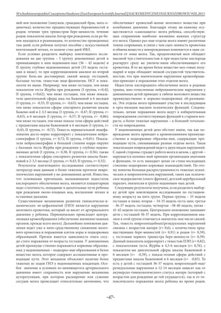
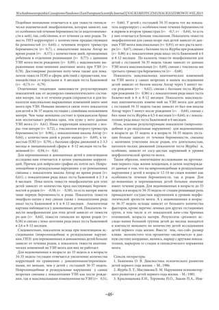
![-49-
WschodnioeuropejskieCzasopismoNaukowe(EastEuropeanScientificJournal)|NAUKIMEDYCZNEINAUKIOZDROWIU#IІІ,2015
лева Н.Н., Александров Т.А., Ялфимов А.Н., Кощавцев А.Г.
Особенности меж-полушарных связей и биоэлектриче-
ской активности мозга доношен-ных и недоношенных де-
тей. / Пятая международная конференция по когнитивной
науке. – Калининград, 2012 – С.467-468.
4. Якунин Ю.А., Ямпольская Э.И., Кипнис С.Л., Сысоева
И.М. Болезни нервной системы у новорожденных детей и
детей раннего возраста. – М.: Медицина, 1979.
5. Fenichel GM. Clinical Pediatric Neurology: A Signs and
Symptoms Ap-proach. 5th ed. Philadelphia, Pa: Elsevier
Saunders; 2005.
6. Zanelli SA, Naylor M, Dobbins N, et al. Implementation
of a ‘Hypo-thermia for HIE’ program: 2-year experience in a
single NICU. J Perinatol. 2008;28(3):171-175
ВЗАЄМОЗВ’ЯЗОК ЕЛЕКТРИЧНОГО ОПОРУ ТА МОРФОЛОГІЧНИХ ЗМІН СТЕГНОВОЇ
КІСТКИ ЩУРІВ ПІСЛЯ МОДЕЛЮВАННЯ ВІДКРИТОГО ПЕРЕЛОМА
(експериментальне дослідження)
Павлова Тетяна Михайлівна
аспірант кафедри екстреної та невідкладної медичної допомоги, ортопедії і травматології
Харківського національного медичного університету
Березка Микола Іванович
доктор медичних наук, професор завідувач кафедри екстреної та невідкладної медичної допомоги,
ортопедії і травматології
Харківського національного медичного університету
RELATIONSHIP OF ELECTRICAL RESISTANCE END MORPHOLOGICAL CHANGES THIGH RATS AFTER MODELING
OF OPEN FRACTURE (experimental study)
Pavlova T.M. graduate student emergency and emergency care, orthopedics and traumatology of Kharkiv National Medical
University
Berezka N.I. MD, Professor Head of the Department of Emergency and emergency medicine, orthopedics and traumatology of
Kharkiv National Medical University
Анотація
Проведено гістологічне, електронно-мікроскопічне дослідження та вивчений електричний опір відламків стегнової
кістки на білих щурах після моделювання відкритого перелому різної тяжкості. Виявлені гістологічні, ультраструктур-
ні зміни в трьох досліджених точках дистального і проксимального відділу уламків кістки. Встановлено кореляційний
взаємозв’язок електрогенеза кісткової тканини і станом остеоцитів, з використанням приладу мультиметра цифро-
вого UT70B. Метод електричного опору може бути використаний для оцінки життєздатності кісткової тканини в
клінічних умовах при відкритих діафізарних переломах кісток кінцівок при госпіталізації постраждалих.
ABSTRACT
A histological, electron microscopic examination and study of electrical resistance femur fracture in white rats after an open
fracture simulations of varying severity. Identified histological, ultrastructural changes in the three studied locations proximal and
distal bone fragments. Established correlation between electrogenesis osteocytes and bone, using a digital multimeter device UT70B.
Electrical resistance method can be used to assess the viability of bone in a clinical setting with open diaphyseal fractures of limbs
affected during hospitalization.
Ключові слова: перелом кістки, електричний опір, остеоцити, гістологічний аналіз
Key words: bone fracture, electric resistance, osteocytes, histological analysis.
Постановка проблеми. У всіх країнах світу у зв’язку з
розвитком промисловості, збільшення кількості транс-
портних засобів на шляхах спостерігається зріст травма-
тизму дорослого населення внаслідок ДТП, виробничого
травматизму, техногенних катастроф та кататравми, який
досягає 50 – 70 % основних причин інвалідності та смерт-
ності. Щорічно в Україні травмується приблизно від 1,5
до 2 мільйонів людей, смертність від травм складає 8,14
% (132,8 випадків на 100 тис. населення та має тенденцію
до збільшення. Кількість травмованих внаслідок високо-
енергетичної травми складає контингент постраждалих у
віковій групі від 20 до 55 років [1,2,3].
Серед усіх переломів довгих кісток кінцівок питома вага
відкритих діафізарних переломів становить від 28% до
53% [4]. При цьому великою проблемою в лікуванні та-
ких потерпілих являються різного роду ускладнення, такі
як порушення процесу репаративної регенерації - до 20%,
гнійні ускладнення - 57,4 [5]. Важливе значення для кон-
солідації перелому має життєздатність кісткової тканини
після впливу травми різної інтенсивності [6,7].
Аналіз останніх досліджень і публікацій. В даний час під
електрогенезом кісткової тканини розуміють здатність
генерувати електричні потенціали, що визначається її ак-
тивними біоелектричними властивостями. В основі кон-
цепції полягає мембранно-іонна теорія, а саме: місцем
електрогенеза є поверхнева мембрана клітини, різниця
потенціалів, що виникає має іонну природу і обумовлена
асиметрією розподілу електричних зарядів по обидві сто-
рони мембрани [8].
При переломі на кісткову тканину впливає напруга, що
виникає у кістці, та залишкова кінетична енергія, яка по-
ширюється вздовж тканин сегмента у вигляді хвильових
коливань. Руйнування мембрани клітин відбувається в
момент, коли напруги, що у кістки, перевищують допу-
стимі для мембрани значення. При пошкодженні кістко-
вої тканини, викликаному травмою різної інтенсивності](https://image.slidesharecdn.com/eesj33-160202073457/85/Eesj-3-3-49-320.jpg)
![-50-
WschodnioeuropejskieCzasopismoNaukowe(EastEuropeanScientificJournal)|NAUKIMEDYCZNEINAUKIOZDROWIU#IІІ,2015
відбувається зміна електричних потенціалів, порушується
їх стереометрія, утворюється електронегативний та елек-
тропозитивний заряд, порушується організація кісткової
тканини [6,9,10].
Виділення невирішених раніше частин загальної пробле-
ми. При проведенні ретроспективного аналізу існуючих
методів дослідження жіттєздатності кісткової тканини
та м’яких тканин при відкритих переломах кісток та їх
ускладненнях ми прийшли до висновку, що, безумовно, всі
методи діагностики хороші, проте базуються на віявлені
вторинно структурних або анатомічних змінах в кістковій
тканині, або певною мірою є запізнілими і не можуть дати
оцінку структурно-функціонального стану кісткової тка-
нини при відкритих переломах кісток кінцівок під впли-
вом травми різної інтенсивності на ранніх термінах (1-6
годин з моменту травми) госпіталізації.
У зв’язку з цим нами був запропонований метод оцінки
структурно-функціонального стану кісткової тканини на
основі взаємозв’язку електрогенеза, гістологічних та елек-
тронно-мікроскопічних особливостей кісткових клітин
під впливом травми різної інтенсивності, так як поши-
реність цих порушень потребує поглибленого досліджен-
ні.
Мета дослідження. Вивчити взаємозв’язок електричного
опору та морфологічних особливостей стегнової кістки бі-
лих щурів-самців після моделювання відкритого перелому
і провести кореляційний аналіз використаних методів для
оцінки життєздатності кісткової тканини.
Виклад основного матеріалу. Дослідження проведені на
статевозрілих білих лабораторних щурах-самцях віком 3-х
місяців (200-250г) [11]. Експерименти виконані відповід-
но до вимог про гуманне ставлення до експериментальних
тварин, згідно з «Європейської конвенції захисту хребет-
них тварин, які використовуються в експериментальних
та інших цілях» і закону України [14, 15].
План експерименту і тактика його виконання заслухані
та затверджені Комітетом з питань біоетики (протокол №6
від 03.06.2015 р.).
Була відтворена модель порушення структурної органі-
зації відламків стегнової кістки у щурів шляхом відкрито-
го перелому і після перелому з коагуляцією відламків (для
посилювання порушень). В якості контролю використані
тварини без перелому. Проведена гістологічна оцінка ста-
ну остеоцитів і електричного опору в різних областях стег-
нової кістки після перелому.
При оперативному втручанні використовували загальне
знеболювання. Препарати вводили внутрішньом’язово,
аміназин - 10 мг / кг живої маси, кетамін - 50 мг / кг живої
маси. Після занурення тварин у наркоз та обробки області
оперативного втручання, по латеральній поверхні стегна
здійснювали поздовжній розріз шкіри і підшкірної жиро-
вої клітковини. Роз’єднували глибокі шари м’язів, оголю-
ючи середню третину діафіза. За допомогою пилки Джиглі
проводили поперечну остеотомію в середині діафіза стег-
нової кістки. Для посилення деструктивних порушень у
кістки проводили електрокоагуляцію крайових відламків
стегнової кістки. В якості контролю використовували тва-
рин аналогічного віку та статі без перелому.
Щурів виводили з експерименту шляхом інгаляційного
передозування ефіру для наркозу через 1 і 3години після
початку експерименту.
За допомогою приладу мультиметра цифрового UT70B
рис.1
Рисунок 1. Загальний вигляд мультиметр цифровий UT70B.
вимірювали електричний опір кістки в діапазоні від 0 до 4000 МOм у трьох областях кістки (вище і нижче області
перелому) 6, 12 і 18 мм у дослідних тварин (далі по тексту позначені як 1,2 і 3 точки відповідно) через 1 і 3 години рис.2.
У контрольних тварин в стегнової кістки обрані аналогічні точки.
Для визначення омічного опору досліджуваної ділянки кісткової тканини до терміналу «VΩHz» підключали позитив-
ний електрод (червоний щуп), до терміналу «СОМ» підключали негативний електрод (чорний щуп). У подальшому
встановлювали поворотний перемикач на шкалі приладу в положенні « » (вимірювання електричного опору) і натиска-
ли вмикач « » для вибору режиму вимірювання електричного опору (Ω). Далі встановлюємо вищевказані вимірювальні
щупи паралельно один до одного в кортикальний шар досліджуваних ділянок кожного кісткового фрагмента (рис.2, 4)
(А і В) при експозиції 60 - 120 секунд, а саме: на кордоні перелому (1 і 1 ‘), на середині фрагментів (2 і 2’) і на протилежних
кінцях фрагментів (3 і 3 ‘). При цьому на дисплеї пристрою реєструються показники електричного опору в (МОм) в кож-
ному фрагменті. Завдяки з’єднанню мультиметра цифрового UT70B з комп’ютером, зображення на дисплеї комп’ютера
також відображає цифрові показання мультиметра в точках вимірювання, з додатковим записом електричного опору у
вигляді гістограм.](https://image.slidesharecdn.com/eesj33-160202073457/85/Eesj-3-3-50-320.jpg)
![-51-
WschodnioeuropejskieCzasopismoNaukowe(EastEuropeanScientificJournal)|NAUKIMEDYCZNEINAUKIOZDROWIU#IІІ,2015
Рисунок 2. Точки вимірювання електричного опору (1, 2, 3 та 1’, 2’, 3’).
Статистична обробка цифрових показників проведена за Стьюдентом з обчисленням середньоарифметичних значень
і середньоквадратичних помилок із застосуванням програмного пакету Microsoft Excel XP.
Перша дослідна серія. Перелом. 1година після коагуляції.
Проведено вимірювання електричного опору і гістологічний аналіз відламків стегнової кістки після перелому.
Кортекс. Мікроскопічно крайовой відділ кортекса стегнової кістки, точка 1 і 1’ - остеоцити відносно рівномірно
розподілені по міжклітинній речовині, мали округлі або подовжені ядра. Клітини оточені лакунами, які відрізнялися
формою і розмірами. Виявлено поодинокі порожні лакуни або лакуни заповнені клітинним детритом.
Виражених відмітних особливостей між фрагментами кістки в точці 2 і 2’, 3 і 3’ не виявлено. У області дослідження
були присутні як порожні лакуни і лакуни заповнені клітинним детритом, так і остеоцити зі збереженням нормальної
структурної організації.
При електронно-мікроскопічному дослідженні фрагментів, в точці 1 і 1’ дослідження стегнової кістки, було знайде-
но підвищення кількості остеоцитів з гранулярним розпадом структур цитоплазми, лізисом плазматичної мембрани і
фрагментацією ядра. Виявлено лакуни з клітинним детритом. Поряд із зруйнованими клітинами, були присутні остео-
цити, зі збереженням характерної структурної організації. При цьому показники електричного опору були підвищени-
ми в точках 1 і 1’ та мали тенденцію до зниження в точках 2 і 2’, 3 і 3’. Взаємозв’язок досліджень представлений в таблиці
1.
Таблиця 1.
Перша дослідна серія. Перелом. 1 година після коагуляції.
Показники діапазону електричного опору кісткової тканини та гістограми в
досліджуваних фрагментах (А та В) МегаОм (1х106
Ом)
Морфологічні методи дослідження:
1. Гістологічне дослідження
2. Електронно-мікроскопічне до-
слідження
Фрагмент А Фрагмент В
а
1 точка. 1285 – 1305
МОм – 6 мм
2 точка. 1100 – 1120
МОм – 12 мм
3 точка. 960 – 985
МОм – 18 мм
в
б
1 точка. 1295 – 1315 МОм – 6 мм
2 точка. 1105 – 1110 МОм – 12 мм
3 точка. 965 – 995
МОм – 18 мм
г
1.Нерівномірне забарвлення ма-
триксу. Остеоцити. Базофілія стінок
судинних каналів. Поодинокі порож-
ні лакуни.
2. Некроз остеоцита. Гранулярний
розпад структур цитоплазми, лізис
плазматичної мембрани. Фрагмента-
ція ядра.
Таблиця 1. Гістограма фрагмента А (а), гістограма фраг-
мента Б (б), діапазон електричного опору (в,г), напівтон-
кий зріз фрагмента стегнової кістки, забарвлення по
Apariсio [12]. Зб. 1000, контрастування по Рейнольдсу [13].
Зб. 15600. (д,е).
Гістологічна оцінка фрагментів стегнової кістки в точках
2и 2’, 3 і 3’ дослідження показала, що, поряд з лакунами, що
містять остеоцити з округлими або видовженими ядрами,](https://image.slidesharecdn.com/eesj33-160202073457/85/Eesj-3-3-51-320.jpg)
![-52-
WschodnioeuropejskieCzasopismoNaukowe(EastEuropeanScientificJournal)|NAUKIMEDYCZNEINAUKIOZDROWIU#IІІ,2015
виявлені одиничні порожні лакуни або лакуни заповнені
клітинним детритом. Збережені остеоцити мали відносно
рівномірне забарвлення ядер, були оточені слабо еозино-
фільної цитоплазмою, відзначалася нерівномірна базо-
філія матриксу, але виражена в меншій мірі в порівнянні з
точками 1 і 1’ стегнової кістки. Кісткові канали, що містять
судини, без патологічних порушень.
При електронно-мікроскопічному дослідженні в точках
2 і 2’, 3 і 3’ стегнової кістки щурів виявлено поодинокі осте-
оцити з ознаками деструктивних порушень, проте перева-
жали клітини з організацією, характерної для остеоцитів
в нормі.
Друга дослідна серія. Перелом. 3 години після коагуляції.
Також, як і на попередній термін дослідження, через 3 го-
дини після коагуляції найбільша щільність лакун без осте-
оцитів була розташована в точках 1 і 1’. У точках 2 і 2’, 3 і 3’
були присутні лише поодинокі пусті лакуни або лакуни з
клітинним детритом. Взаємозв’язок досліджень представ-
лений в таблиці 2.
Таблица 2.
Друга дослідна серія. Перелом. 3 години після коагуляції.
Показники діапазону електричного опору кісткової тканини та гістограми в
досліджуваних фрагментах (А та В) МегаОм (1х106
Ом)
Морфологічні методи дослідження:
3. Гістологічне дослідження
4. Електронно-мікроскопічне до-
слідження
Фрагмент А Фрагмент В
а
1 точка. 1305 – 1355
МОм – 6 мм
2 точка. 1260 – 1270
МОм – 12 мм
3 точка. 1110 – 1120
МОм – 18 м
в
б
1 точка. 1295 – 1305 МОм – 6 мм
2 точка. 1265 – 1280 МОм – 12 мм
3 точка. 1115 – 1128 МОм – 18 мм
г
д
Нерівномірне забарвлення матрик-
су. Порушення організації судинних
каналів в крайовому відділі кістково-
го уламка. Остеоцити з різною щіль-
ністю ядер. Порожні лакуни.
Некроз остеоцита. Гранулярний
розпад структур цитоплазми, лізис
плазматичної мембрани. Фрагмента-
ція ядра.
Таблиця 2. Гістограма фрагмента А (а), гістограма фраг-
мента Б (б), діапазон електричного опору (в, г), напівтон-
кий зріз фрагмента стегнової кістки, забарвлення по
Apariсio [12]. Зб. 1000, контрастування по Рейнольдсу[13].
Зб. 15600. (д, е).
При проведенні електронно-мікроскопічного дослід-
ження виявлено, що основна маса остеоцитів зберігала
життєздатність, тобто, клітини мали гіпохромних ядра із
збереженою ядерною мембраною, були оточені вузьким
обідком цитоплазми з невеликою кількістю мембранних
органел. Були присутні одиничні мітохондрії і невеликі
профілі ендоплазматичної сітки. Плазматична мембрана
остеоцитів зберігала чіткі контури.
Третя дослідна серія. Перелом.
Наступною моделлю, на якій було проведено вивчення
стану остеоцитів, була модель перелому, виконана на щу-
рах в діафизарній області стегнової кістки. Вивчення осте-
оцитів також проводили в трьох позначених точках.
У порівнянні з попередньою серією експерименту, в
третій дослідній групі не виявлено особливостей в гісто-
логічній організації остеоцитів в трьох досліджених точ-
ках. Відмінності встановлені лише в кількості порожніх
лакун, що не містять клітин. У цій серії експерименту в
першій галузі дослідження кортекса порожніх лакун або](https://image.slidesharecdn.com/eesj33-160202073457/85/Eesj-3-3-52-320.jpg)
![-53-
WschodnioeuropejskieCzasopismoNaukowe(EastEuropeanScientificJournal)|NAUKIMEDYCZNEINAUKIOZDROWIU#IІІ,2015
лакун заповнених детритом було значно менше, порівняно
зі станом кортекса після перелому з коагуляцією. Порожні
лакуни або лакуни, що містять клітинний детрит, перебу-
вали в проксимально та дистально розташованих уламках
кістки поблизу області перелому, однак такі лакуни були
поодинокі. Взаємозв’язок досліджень представлений в та-
блиці 3.
Таблиця 3.
Третя дослідна серія. Перелом.
Показники діапазону електричного опору кісткової тканини та гістограми в
досліджуваних фрагментах (А та В) МегаОм (1х106
Ом)
Морфологічні методи дослідження:
5. Гістологічне дослідження
6. Електронно-мікроскопічне до-
слідження
Фрагмент А Фрагмент В
а
1 точка. 520 – 650
МОм – 6 мм
2 точка. 390 – 420
МОм – 12 мм
3 точка. 360 – 380
МОм - 18 мм
б
б
1 точка. 525 – 657
МОм – 6 мм
2 точка. 398 – 426
МОм – 12 мм
3 точка. 364 – 386
МОм - 18 мм
г
д
Лакуни без остеоцитів.
е
Остеоцит з нерівними контурами
плазматичної мембрани. Підвищена
гетерохроматизація ядра. Нерівні
зигзагоподібні контури лакуни.
Таблиця 3. Гістограма фрагмента А (а), гистограмма
фрагмента Б (б), діапазон електричного опору (в, г), на-
півтонкий зріз фрагмента стегнової кістки, забарвлення
по Apariсio [12]. Зб. 1000, контрастування по Рейнольдсу
[13]. Зб. 15600. (д, е).
У точках 2 и 2’, 3 и 3’ - остеоцити зберігали будову, ха-
рактерну для норми, мали базофільні ядра, оточені слабо
еозинофільною цитоплазмою. Виявлені лише поодинокі
лакуни без остеоцитів або лакуни, що містять клітинний
детрит. Матрикс кортекса мав відносно рівномірне забар-
влення.
При електронно-мікроскопічному дослідженні в точках
1 и 1’ - більшість остеоцитів мали нормальну організацію.
Цитоплазматична мембрана була непорушена. Вузька ци-
топлазма містила поодинокі профілі мембранних органел.
Ядра містили еухроматин. Гетерохроматин у вигляді гру-
дочок був локалізований по внутрішній поверхні ядерної
мембрани у вигляді невеликих щільних утворень, розта-
шовувався по території ядра. Лише в поодиноких клітинах
в ядрі було відзначено збільшення конденсації хроматину,
що займає велику площу ядра. Ядерна мембрана зберіга-
ла двоконтурність, перинуклеарний простір було чітко
окреслено, однак на її ділянках виявлено розширення,
що свідчить про порушення ядерно-цитоплазматичного
транспорту. В цілому, збереження ядерної мембрани свід-
чить про оборотність виявлених змін. Окремі остеоцити
мали звивисту поверхню цитоплазматичної мембрани. За
даними літератури такі зміни, як конденсація ядерного
хроматину, нерівна поверхня клітини, обумовлена дезор-
ганізацією цитоскелету, описані в науковій літературі які
відносяться до оборотної фази ішемії.
У точках 2 и 2’, 3 и 3’ - остеоцити зберігали характерну для
норми будову рис. 3а, 3б.](https://image.slidesharecdn.com/eesj33-160202073457/85/Eesj-3-3-53-320.jpg)
![-54-
WschodnioeuropejskieCzasopismoNaukowe(EastEuropeanScientificJournal)|NAUKIMEDYCZNEINAUKIOZDROWIU#IІІ,2015
Рисунок 3.а Остеоцит. Вузький обідок цитоплазми з поодинокими мембраними органелами. Ядро з відносно рівномір-
ним перінуклеарним простором. Контрастували по Рейнольдсу [13]. Зб. 19500
Рисунок 3.б Поодинокі пусті лакуни або лакуни з клітинним детритом. Нерівномірне забарвлення матриксу. Гематок-
силін і еозин [12]. Зб. 200.
В контрольній серії експерименту через 1 і 3 години від-
мінних особливостей в клітинному складі матриксу стег-
нової кістки щурів не виявлено. Незважаючи на те, що піс-
ля трьох годин дослідження, відсоток зруйнованих клітин
і порожніх лакун мав тенденцію до підвищення, однак не
виходив за 5%, що не відображаєтья на функціонуванні
біологічної системи.
При порівняльній оцінці показників представлених серій
експерименту, виявлено, що найбільш високі показники
порушень остеоцитів зафіксовані в області, прилеглій до
перелому. У точках 1 и 1’ дослідження відсоток порушень
остеоцитів в кортексі перевищував аналогічні показники
в точках 2 и 2’, 3 и 3’. У точках 1 и 1’ виявлено достовірне
підвищення порожніх лакун і лакун, заповнених клітин-
ним детритом, порівняно з контролем, а також з точками 2
и 2’, 3 и 3’ дослідження.
Для оцінки кореляції між показниками структурної
організації остеоцитів і електричного опору кістки був
проведений аналіз за Пірсоном. Для порівняння показ-
ники гістологічного дослідження та електричного опору,
присутні в трьох точках, були об’єднані, оскільки виявле-
на однотипна спрямованість змін в досліджених точках.
Встановлена висока кореляційна залежність між викори-
станими двома методами дослідження, дані представлені
в таблиці 4 та на рис. 4,5,6,7.
Таблиця 4.
Кореляційний аналіз за Пірсоном показників порожніх лакун або заповнених детритом з показниками електричного
опору
Групи r P
Перелом 1 година 0,77 0,003
Перелом 3 години 0,71 0,009
Перелом + коагуляція 1 година 0,93 0,0001
Перелом + коагуляція 3 години 0,73 0,007
Рисунок 4. Кореляційно-регресійний аналіз. Перелом 1 година
Рисунок 5. Кореляційно-регресійний аналіз. Перелом 3 години](https://image.slidesharecdn.com/eesj33-160202073457/85/Eesj-3-3-54-320.jpg)
![-56-
WschodnioeuropejskieCzasopismoNaukowe(EastEuropeanScientificJournal)|NAUKIMEDYCZNEINAUKIOZDROWIU#IІІ,2015
electron-opoque stain in electron microscopy/ E.S. Reynolds //
J. Cell. Biol. – 1963. – Vol. 17. – P. 208 – 212.
14. Європейська конвенція про захист хребетних тва-
рин, що використовуються для дослідних та інших нау-
кових цілей. Страсбург, 18 березня 1986 року: офіційний
переклад [Електронний ресурс] / Верховна Рада України.
– Офіц. веб-сайт. – (Міжнародний документ Ради Євро-
пи). – Режим доступу до документа: http: zakon.rada.gov.ua/
cgi-bin/laws/main.cgi?nreg=994_137.
15. Закон України №3447-IV від 21.02.2006 «Про захист
тварин від жорстокого поводження» (Стаття 26).
ФОРМУВАННЯ КУЛЬТУРИ ЗДОРОВ’Я СТУДЕНТІВ ЯК НАУКОВО-ПЕДАГОГІЧНА
ПРОБЛЕМА ДОСЛІДЖЕННЯ
Скорейко Наталія Теофілівна,
обласний геріатр, лікар-кардіолог,
Комунальний Заклад Рівненська обласна клінічна лікарня
Скорейко Роман Сергійович,
викладач Рівненського базового медичного коледжу,
лікар –гінеколог пологового будинку Рівненської міської ради
Скорейко Світлана Сергіївна,
студентка,
Буковинський державний медичний університет
THE RESULTS OF THE USE OF MONO – AND COMBINATION GIPOLIPIDS THERAPY WITH THE PURPOSE OF
CORRECTION OF DISORDERS OF LIPID METABOLISM
Skoreіko N. Т., region heryatr, doctor-cardiologist of Community Іnstitution “Rivne region Clinical Hospital
Skoreіko R. S., teacher of the Rivne basic medical college, doctor-gynecologist, Rivne city hospital;
Skoreіko S. S., student, Bucovinian State Medical University
АнотацІя
Наведені результати використання моногіполіпідемічної терапії, зокрема ома кору та комбінованої терапії у ви-
гляді комбінації статину (розарт) і -3 поліненасичних жирних кислот (омакор) у хворих з патологією серцево-судиннної
системи. Доведено аддитивний ефект дії комбінованої гіполіпідемічної терапії, а також виявлені додаткові ефекти
впливу на такі показники: артеріальний тиск, порушення серцевого ритму.
ABSTRACT
The results of the use of monogìpolìpìds therapy, including omakor and combination therapy in the form of a combination of statin
(rozart) and -3 polyunsaturated fatty acids (omakor) in patients with pathology of cardio-vasculary system. It was proved an additive
effect of the actions of the combined lipid-lowering therapy and also discovered additional effects of impact on the following indicators:
blood pressure, violations of heart rhythm.
Ключві слова: моногіполіпідемічна терапія, статини, -3 поліненасичені жирні кислоти, патологія серцево-судинної
системи.
Key words: monohypolipidemic therapy, statins, -3 polyunsaturated fatty acids, pathology of the cardiovascular system.
Постановка проблеми. Важливим аспектом медикамен-
тозного лікування хворих з патологією серцево-судинної
системи являється використання препаратів, які знижу-
ють вміст ліпідів в крові. Корекція порушень ліпідного
профілю займає важливе місце в кардіопротекторній те-
рапії і забезпечує значне покращення прогнозу для кардіо-
логічних пацієнтів високого ризику.
Сьогодні поняття дисліпідемії, крім підвищення загаль-
ного холестерину (ЗХС) і ліпопротеїдів низької щільності
(ЛПНЩ) включає багато інших варіантів порушень ліпід-
ного спектру крові: низький рівень ХС ЛПВЩ, гіпертри-
гліцеридемія, порушення співвідношення рівнів аполіпо-
протеїнів А і В, та інші [1-4].
Виділення раніше невирішених частин загальної про-
блеми. На протязі декількох років ведеться пошук шляхів
посилення ефективності гіполіпідемічної терапії, вибір
якої базується на результатах оцінки величини загального
серцево-судинного ризику, цільовому рівні ХС ЛПНЩ, ТГ.
Активних профілактичних втручань потребують майже
40% чоловіків і 50% жінок України в плані зниження ЗХС.
Визнано високим середній рівень ЗХС в крові в українсь-
кій популяції 5,2ммоль/л у чоловіків і 5,6ммоль/л у жінок.
Підвищення ЗХС супроводжується втричі вищою смерт-
ністю від ішемічної хвороби серця (ІХС). Для громадян
України характерне прогресування атерогенності ліпідно-
го обміну.
Дисліпідемія асоційована із самим високим ризиком
серцево-судинних захворювань. Корекція порушень ліпід-
ного обміну заслуговує особливої уваги з точки зору пато-
генетичного лікування атеросклерозу. Частково її можна
досягнути шляхом зміни способу життя, але основний ме-
тод корекції – прийом гіполіпідемічних препаратів. Згід-
но даних метааналізу, рандомізованих досліджень (CARE,
ASCOT- LLA, HPS)[1] зниження ЗХС на 25% і ХС ЛПНЩ
на 30% призводить до зменшення серцево-судинних подій
і смертності в середньому на 30%.
В зв’язку з цим, пріоритетним завданням охорони здо-
ров’я являється раннє виявлення, оцінка степені кардіо-
васкулярного ризику та адекватне лікування пацієнтів із
високим серцево-судинним ризиком [1-4].
Для сьогоднішньої клінічної практики великий інтерес
представляє можливість комбінованої гіполіпідемічної](https://image.slidesharecdn.com/eesj33-160202073457/85/Eesj-3-3-56-320.jpg)
![-57-
WschodnioeuropejskieCzasopismoNaukowe(EastEuropeanScientificJournal)|NAUKIMEDYCZNEINAUKIOZDROWIU#IІІ,2015
терапії для певної категорії пацієнтів високого серце-
во-судинного ризику при неможливості корекції деяких
показників ліпідного спектру крові. В таких випадках
одним із варіантів такої комбінації може бути поєднання
використання статинів із -3 поліненасиченими жирними
кислотами (ПНЖК). Сьогодні вони широко використову-
ються в медицині для комплексної терапії ІХС та інфаркту
міокарда (ІМ), аритмій серця, атеросклерозу, дисліпідемій.
Дані препарати багатосторонньо впливають на фактори
ризику і механізм розвитку кардіоваскулярних ускладнень
у пацієнтів з серцево-судинною патологією.
В рекомендаціях Американської Асоціації серця і Аме-
риканського Коледжу кардіологів рекомендується викори-
стання двох -3 поліненасичених жирних кислот (ПНЖК)
– ейкозопентаєнової (ЕПК) і декозагексаєнової (ДТК) в
дозі 1000мг/добу з метою первинної і вторинної профілак-
тики серцево-судинних захворювань [3].
Згідно досліджень GISSI Prevenzionі (1999рік) [5] препа-
рат -3 ПНЖК, призначений в якості допоміжної терапії до
3 місяців після інфаркту міокарду, значно зменшив ризик
первинної комбінованої кінцевої крапки (загальна смерт-
ність, нефатальний ІМ та інсульт) і вторинних кінцевих
крапок (смертність від усіх причин, раптова коронарна,
серцево-судинна смерть) із зниженням загальної смерт-
ності на 28% і ризику раптової смерті на 47%. Міжнарод-
ні керівництва включили використання препаратів -3
ПНЖК в рекомендації по профілактиці серцево-судинних
захворювань (ССЗ), профілактиці і лікування інфаркту
міокарду, шлуночкових аритмій, раптової серцевої смерті
[1,4].
Одним із механізмів реалізації позитивних ефектів
-3ПНЖК являється їх вплив на ендотеліальну функцію,
який реалізується через зниження рівня молекул адгезії
і протизапальних цитокінів, зміни рівня циркулюючих
вільних жирних кислот, зменшення активності утворен-
ня вільних радикалів в клітинах ендотелію, а також че-
рез включення -3ПНЖК в клітинні мембрани із зміною
їх текучості, модуляції білкових комплексів і активізації
ендотеліальних іонних каналів. Крім того, ці препарати
впливають на агрегацію тромбоцитів, що сприяє знижен-
ню тромботичних ризиків. Це особливо важливо для па-
цієнтів із групи високо-серцево-судинного ризику. Ця дія
не залежить від дози і може проявлятися через тиждень
після прийому препаратів -3 ПНЖК [3].
Мета дослідження: вивчити особливості дії препарату
Омакор (Abbott Products GmbH, Німеччина) на стан ліпід-
ного профілю в комбінації із статинами у хворих з пато-
логією серцево-судинної системи, та його вплив при дис-
пліпопротеінемії у здорових людей. Представником класу
статинів був обраний розарт (Актавіс Лтд, Мальта).
Виклад основного матеріалу. У дослідженні брали
участь 36 осіб, серед яких 12-жінок періоду менопаузи
(45-50років). Перша група хворих з ІХС: стенокардія на-
пруги ФКII. Післяінфарктний кардіосклероз. СН I ФКII.
Друга група: 6 чоловіків і 6 жінок з ІХС: стенокардія на-
пруги ФК II. Гіпертонічна хвороба II, ступінь 2, ризик 3.
СН I. ФК II.
Третя – контрольна, включала також 6 чоловіків та 6 жі-
нок без патології серцево-судинної системи з дисліпопро-
теінемією.
Лікування хворих першої групи відбувалось згідно стан-
дартів з призначенням терапії, яка включала: антиагреган-
ти (кардіомагніл 75мг), -адреноблокатори (коріол в дозі
6,25мг/добу), статини (препарат розарт -20мг/добу).
Омакор – лікарський препарат, що складається із суміші
етилових естерів -3 поліненасичених незамінних жирних
кислот (84-90%): ейкозапентаєнової (ЕПК) і докозагексає-
нової (ДГК) кислот, і бере участь в процесах тканинного
метаболізму. Цей препарат має гіполіпідемічні власти-
вості: знижує переважно рівень тригліцеридів за рахунок
збільшення кількості пероксисом -окиснення жирних
кислот в печінці, оскільки ЕПК і ДГК інгібують активність
ферментів, які відповідають за їх синтез; зменшує вміст хо-
лестерину ліпопротеїдів дуже низької і низької щільності
за рахунок прискорення їх катаболізму, а також кількість
вільних жирних кислот, які необхідні для синтезу триглі-
церидів [4, 5].
За результатами попереднього лікування у пацієнтів
другої групи не вдалося досягти цільового значення ар-
теріального тиску (АТ) менше 140/90. Усі хворі страждали
на диспіпротеінемію, тобто мали рівень ЗХС у плазмі крові
більше 5 ммоль/л, ХСЛПНЩ більше 3 ммоль/л, ТГ більше
1,7ммоль/л. Учасників дослідження першої та другої груп
було віднесено до хворих з високим рівнем кардіоваску-
лярного ризику [6-7].
Перед проведенням дослідження визначався ліпідний
спектр крові, а також через 1 та 3 місяці від початку до-
сліджень. Оцінка результату проводилась на підставі ла-
бораторних, клінічних та інструментальних досліджень.
Відбувався моніторинг артеріального тиску, печінкових
проб (АсТ, АлТ), проводилась оцінка кардіоваскулярного
ризику за шкалою SCORE, холтерівське моніторування. Гі-
поліпідемічний ефект [2-4] оцінювали як відсоток змін се-
редніх рівнів у групах до лікування і в процесі лікування.
В таблиці 1 наведені антропометричні дані на початку
дослідження. Кількість учасників -36 чоловік
Таблиця 1
Атропометричні дані на початку дослідження
Параметри Перша група
(n=12)
Друга група (n=12) група (n=12)
чоловіки жінки чоловіки жінки
Вік, років 45,5 46,2 47,3 48,1 46,5
Зріст, см 168,5 169,3 165,5 167,2 152,4
Маса тіла, кг 82,5 84,2 81,7 85,2 83,4
Окружність
талії, см
97,6 98,2 92,5 94,3 96,6](https://image.slidesharecdn.com/eesj33-160202073457/85/Eesj-3-3-57-320.jpg)
![-58-
WschodnioeuropejskieCzasopismoNaukowe(EastEuropeanScientificJournal)|NAUKIMEDYCZNEINAUKIOZDROWIU#IІІ,2015
Таблиця 2
Показники ліпідного профілю на початку дослідження
Параметри Перша група
(n=12)
Друга група (n=12) група (n=12)
чоловіки жінки чоловіки жінки
ЗХС, ммоль/л 6,2 6,4 6,2 5,6 5,4
ХС ЛПНЩ,
ммоль/л
3,7 4,7 3,6 3,1 3,4
ТГ, ммоль/л 2,3 2,3 2,1 1,9 1,8
ХС ЛВВЩ,
ммоль/л
1,1 0,9 1,1 1,1 1,2
Результати за період 1, 3 місяці дослідження подані в таблицях 3,4
Таблиця 3.
Динаміка показників ліпідного профілю через 1 місяць лікування
Параметри Перша група
(n=12)
Друга група (n=12) група (n=12)
чоловіки жінки чоловіки жінки
ЗХС, ммоль/л 5,9 6,1 6,4 5,1 5,0
ХС ЛПНЩ,
ммоль/л
3,1 3,8 3,6 3,0 3,2
ТГ, ммоль/л 1,8 2,1 1,9 1,8 1,7
ХС ЛВВЩ,
ммоль/л
1,2 1,0 1,2 1,1 1,2
Таблиця 4.
Динаміка показників ліпідного профілю через 3 місяці лікування
Параметри Перша група
(n=12)
Друга група (n=12) група (n=12)
чоловіки жінки чоловіки жінки
ЗХС, ммоль/л 3,5 5,6 5,9 4,9 4,5
ХС ЛПНЩ,
ммоль/л 2,4 2,6 2,5 2,4 2,5
ТГ, ммоль/л 1,5 1,9 1,8 1,7 1,5
ХС ЛВВЩ,
ммоль/л
1,3 1,0 1,3 1,2 1,2
Згідно результатів дослідження використання омакору
сумісно із статинами підсилює їх гіполіпідемічний ефект,
особливо при гіпертригліцеридемії. Середній рівень ЗХС
знизився в першій групі на 44% в другій – на 13%. В першій
групі спостерігалось зниження ТГ на 34,8% після 3місяців
використання препаратів. У чоловіків 2 групи не вдалося
досягти цільового рівня ТГ, який знизився лише на 17,4%
, а у жінок – на 14%. Окрім того, по даних холтерівського
моніторування у пацієнтів другої групи зменшилась ча-
стота шлуночкових аритмій.
У пацієнтів з помірною гіпертригліцеридемією додаван-
ня до статину -3ПНЖК в дозі 2г на добу може змінити
рівні ТГ і допомогти досягнути цільового співвідношення
ТГ/ ХСЛПВЩ, що підтверджується рекомендаціями Ка-
надського кардіоваскулярного товариства по діагностиці і
лікуванню дисліпідемій, профілактиці серцево-судинних
захворювань в дорослих.
Комбінована гіполіпідемічна терапія не супроводжу-
валасть підвищенням рівнів печінкових проб (АсТ, АлТ)
і була безпечною в плані розвитку інших побічних дій
(міальгії та ін.)
Висновки: Комбінація розарту з омакором довела свою
ефективність в плані зниження ТГ. Крім того у пацієнтів з
АГ спостерігалось зниження артеріального тиску. Частка
пацієнтів, які досягли цільового рівня через 3 місяці стано-
вила 78,4%. У пацієнтів 3 групи при використанні омакору
також спостерігалась стабілізація показників, особливо
ХС ЛПНЩ і ТГ та ЗХС без додавання статинів. Омакор
входить в Європейські та Американські стандарти вто-
ринної профактики серцево-судинних захворювань[8-10].
Використання його можливе також і для первинної
профілактики серцево-судинних захворювань при наяв-
ності таких факторів ризику, як артеріальна гіпертензія,
ожиріння, шлуночкові аритмії, дисліпідемії, що доведено
багаточисельними клінічними дослідженнями.
ЛІТЕРАТУРА
1. Томсон Г.Р. Руководство по гиперлипидемии / Г.Р. Том-
сон // MSD.−1991. − С.255.
2. Т.В. Талаєва, В.В. Братусь // Український кардіологіч-
ний журнал. −2010. −№3. − С. 7−26.
3. Аронов Н. Сердечно-сосудистая система и омега-3 по-](https://image.slidesharecdn.com/eesj33-160202073457/85/Eesj-3-3-58-320.jpg)
![-61-
WschodnioeuropejskieCzasopismoNaukowe(EastEuropeanScientificJournal)|NAUKIMEDYCZNEINAUKIOZDROWIU#IІІ,2015
филактические мероприятия и ежегодный мониторинг
состояния здоровья пациентов с СД 2 типа, назначают и
корректируют терапию антидиабетическими препарата-
ми, что предусмотрено международными и националь-
ными клиническими рекомендациями [1-5].
В Украине медицинская помощь больным СД 2 типа ра-
нее осуществлялась в основном врачами-эндокринолога-
ми. Врачи первичной медицинской помощи (ПМП) стали
активно подключаться к ведению СД 2 типа в 2012 году
после внедрения разработанных с нашим участием меди-
ко-технологических документов - адаптированных клини-
ческих рекомендаций и унифицированного клинического
протокола (УКП) оказания медицинской помощи при СД 2
типа, утвержденных приказом Министерства Здравоохра-
нения Украины от 21.12.2012 № 1118. Данные медико-тех-
нологические документы впервые в Украине произвели
распределение объемов медицинской помощи при СД 2
типа между первичным и вторичным уровнями и опре-
делили индикаторы качества диабетологической помощи.
Среди индикаторов основными являются процент паци-
ентов, которым произведена оценка медицинского состо-
яния путем проведения перечня обязательных ежегодных
процедур, и процент пациентов, которому проведено ис-
следование гликозилированного гемоглобина - HbA1c, т.е.
показатели качества процесса диабетологической помощи
[6].
Анализ последних исследований и публикаций по дан-
ной проблеме показывает, что качество диабетологиче-
ской помощи активно изучается в странах мира, с учетом
общеизвестной концепции А. Донабедиана (1980), ко-
торая описывает оценку качества медицинской помощи
по трем ее компонентам: качество структуры, процесса
и результата. Так, проводятся многочисленные сравни-
тельные исследования качества диабетической помощи
по индикаторам качества, как на национальном, так и на
межнациональном уровнях, но их сравнительный анализ
достаточно сложен в связи с тем, что количество и суть
индикаторов зависит от особенностей здравоохранения
конкретной страны и национальных руководств [7-12].
Чаще исследования концентрируют внимание на оценке
индикаторов процесса и результата диабетологической
помощи, т.е. на клинических показателях, которые оцени-
ваются на первичном и вторичном звене. Например, ис-
следование OECD оценивало качество диабетологической
помощи в 5 странах – Австралии, Канаде, Новой Зелан-
дии, США и Великобритании по 6 индикаторам (ежегод-
ное измерение HbA1c, липидов, скрининг функции почек,
исследование глаз, достижение целевых уровней HbA1c и
липидов) и показало относительно широкий разброс ре-
зультатов. Например, ежегодное исследование HbA1c и
липидов проводилось в Новой Зеландии та Австралии у
50-60% пациентов, а в США и Великобритании - у 70-80%
пациентов, но контроль диабета был в США и Великобри-
тании хуже: HbA1c ≤ 7% достигли только 30-50% паци-
ентов [7]. Исследование GUIDANCE оценивало качество
диабетологической помощи на первичном и вторичном
этапах после внедрения национальных руководств в 8 ев-
ропейских странах (Бельгия, Франция, Германия, Италия,
Ирландия, Швеция, Нидерланды, Великобритания) по
следующим индикаторам качества процесса и результата:
уровень HbA1c, артериального давления, показатели ли-
пидов, индекса массы тела, процент пациентов, которым
определены данные показатели, назначены сахароснижа-
ющие, гипотензивные, гиполипидемические препараты. В
данном исследовании установлено, что вариация показа-
телей между странами была значительной по всем пока-
зателям, уровень HbA1c ежегодно в среднем определялся
у 97,6% пациентов первичного звена, но целевого HbA1c
≤ 7% достигли только в среднем 53,6% пациентов [8, 11].
В Таиланде, Kanchanaphibool I. и соавт. (2009 р.) показали,
что на вторичном звене HbA1c был определен ежегодно
у 56,5-67,2% пациентов, но его уровень HbA1c ≤ 7% до-
стигнут лишь у 26,5-33,5 % пациентов, холестерина ЛПНП
≤100 мг/дл – у 27,1-30,3%, триглицеридов ≤150 мг/дл - у
53,3-48,8%, холестерина ЛПВП ≥50 мг/дл - у 38,7-47,1% па-
циентов [9].
Исследование качества диабетологической помощи на
первичном звене в США (Техас) показали, что пациенты
семейных врачей были обследованы на HbA1c в 90,5% слу-
чаев, на липиды – в 81,3 %, протеин мочи – в 46,3%, осмотр
глаз - в 57,3%, ног – в 69,2% случаях, что свидетельствует
о достаточном уровне качества медицинской помощи [10,
11]. Оценка диабетологической помощи на первичном
уровне в Великобритании (1998) показала, что ведение па-
циента с диабетом семейным врачом не должно быть са-
мостоятельным, т.к. приводит к ухудшению контроля, мо-
ниторинга и большей смертности пациентов с диабетом,
более эффективным является ведение больного структу-
рированной командой при тесном взаимодействии врача
ПМП с эндокринологом и другими специалистами, что и
внедряется во всех странах мира [12].
Оценка качества и эффективности диабетологической
помощи в условиях внедрения новых медико-технологи-
ческих документов в Украине не освящена, что обусловило
цель настоящего исследования и является важным этапом
на пути формирования качественной диабетологической
помощи, особенно на первичном этапе.
Цель исследования – провести сравнительную харак-
теристику качества диабетологической помощи врачами
ПМП и эндокринологами в условиях внедрения нового
клинического протокола по клиническим данным.
Материалы и методы. Нами было обследовано 173 паци-
ентов с СД 2 типа (в возрасте 55,13±0,61 лет, 112 женщин и
61 мужчин), которые в зависимости от того, у какого вра-
ча они наблюдаются были разделены на 2 группы: 1-я – у
врача ПМП (n=45, возраст 54,95±1,2 лет), 2-я группа – у
врача эндокринолога (n=128, возраст 55,23±0,71 лет). С
целью анализа качества диабетологической помощи про-
водился сравнительный анализ ретроспективных данных
пациентов обеих групп, опрос пациентов об объеме про-
водимых им медицинских услуг за 2012-2013 гг., а также
клинико-лабораторное обследование согласно рекомен-
дованному УКП перечню основных обязательных показа-
телей контроля диабета: индекс массы тела (ИМТ), объем
талии (ОТ), уровень систолического и диастолического
артериального давления (САД и ДАД), уровень глико-
зилированного гемоглобина (HbA1c), уровень глюкозы
крови натощак, показатели липидограммы, обеспечение](https://image.slidesharecdn.com/eesj33-160202073457/85/Eesj-3-3-61-320.jpg)
![-64-
WschodnioeuropejskieCzasopismoNaukowe(EastEuropeanScientificJournal)|NAUKIMEDYCZNEINAUKIOZDROWIU#IІІ,2015
вышением рекомендуемых УКП уровней триглицеридов
(1,74±0,15 против 1,11±0,08 ммоль/л, p<0,01, рекомендо-
ванный <1,7 ммоль/л), ЛПНП (2,71±0,18 против 1,34±0,04
ммоль/л, p<0,01, рекомендованный <2,5 ммоль/л) и
ЛПОНП (0,63±0,06 против 0,42±0,03 ммоль/л, p <0,01) и
достоверным снижением ЛПВП (2,19±0,14 против 3,51±
0,18 ммоль/л, p<0,01, рекомендованный >1 ммоль/л), что
свидетельствует о наличии дислипидемии. Достоверной
разницы по контролю липидного профиля врачами эндо-
кринологами и врачами ПМП выявлено не было. Атеро-
генный индекс был выше рекомендованного в обеих груп-
пах.
Уровень СРП, как одного из показателей атерогенного
процесса у пациентов с СД составил 5,42±0,92 мг/л и пре-
вышал норму 0-5 мг/л, и достоверно не отличался от по-
казателя группы сравнения, но по данным American Heart
Association и US Centers for Disease Control and Prevention
[13-15] даже уровень СРП выше 3 мг/л, что наблюдает-
ся в обеих группах, говорит о высоком риске сосудистой
патологии. Другой фактор сердечно-сосудистого риска
- уровень мочевой кислоты у обследованных пациентов
не превышал рекомендуемых норм (для женщин -150-350
мкмоль/л и для мужчин - 210-420 мкмоль/л), но был досто-
верно выше среди мужчин группы сравнения и составил
412,11±25,88 мкмоль/л (р<0,05) в отличие от показателя у
мужчин с СД 2 типа (310,60±19,44 мкмоль/л). Достоверных
изменений уровней СРП и мочевой кислоты между паци-
ентами врачей ПМП и эндокринологов не установлено.
Исследование функции почек посредством расчетной
СКФ по двум формулам (MDRD и Сockcroft Goult) показа-
ло отсутствие достоверных изменений между пациентами
с СД и группы сравнения, а также отсутствие достоверных
изменений между пациентами врачей ПМП и эндокри-
нологов. Но, расчетная СКФ по формуле MDRD, которая
считается более точной, показала, что у пациентов обеих
групп имеется незначительное снижение СКФ (по DOQI,
2002), что указывает на начальные изменения функции
почек в обеих группах и может быть связано с влиянием
других факторов, кроме диабета. Установлена достовер-
ная разница между расчетным значением СКФ по разным
формулам указывает на невысокую точность расчетного
метода. По современным данным наиболее чувствитель-
ным методом диагностики повреждения почек у пациен-
тов с СД 2 типа на ранних стадиях, считается определение
САК в моче, который пока в Украине не имеет широкого
рутинного применения [6].
Для оценки статуса курения и его воздействия на орга-
низм проведено исследование СО в выдыхаемом воздухе.
Определено, что уровень СО в выдохе среди курящих па-
циентов с СД был достоверно выше, чем у некурящих, что
подтверждает объективность данного метода для опреде-
ления статуса табакокурения. Кроме того, установлено,
что среди некурящих пациентов СО был достоверно выше
у больных СД, в отличие от группы сравнения. Выявлено,
что уровень СО в выдыхаемом воздухе имел достоверную
прямую корреляцию с Hb1Ac (r=0,38 p<0,01), что свиде-
тельствует о прямой взаимосвязи гипергликемии и СО.
По данным медицинской документации диабетическую
нефропатию имели 20,81%, ретинопатию - 77,46%, нейро-
патию – 78,61, ИБС- 49,71% пациентов.
Выводы. С внедрением нового клинического протокола
и подключением врачей ПМП по данным медицинских
записей достоверно возрос процент пациентов, которым
определялся уровень HbA1c с 16,76% до 24,28% пациен-
тов (прирост 44,87%, p>0,05), уровень общего холестери-
на с 14,45% до 24,28% (p<0,05, прирост 68,03%), исследо-
вание функции почек с 5,78% до 12,72% (p<0,05, прирост
120,07%), осуществлен осмотр нижних конечностей - с
15,61% до 25,43% (p<0,05), осмотр глазного дна окулистом
- с 16,18 до 26,01%, (p<0,05). Но обследование пациентов
не определило улучшения контроля диабета и достижения
рекомендованных целевых уровней в первые годы внедре-
ния протокола, т.е. улучшилось качество процесса диабе-
тологической помощи, а качество результата в первые годы
внедрения протокола остается прежним. Оценку качества
диабетологической помощи следует проводить комплекс-
но, учитывая данные медицинской документации, опроса
и обследования пациентов. Отсутствие достоверных от-
личий между показателями ежегодного мониторинга па-
циентов врачей ПМП и эндокринологов свидетельствует о
достаточном уровне качества диабетологической помощи
врачей ПМП. Дальнейшее изучение клинических аспек-
тов качества диабетологической помощи на более поздних
этапах внедрения протокола и сопоставление их с другими
аспектами даст более полную оценку качества диабетоло-
гической помощи.
Cписок литературы:
1. Всемирное руководство по сахарному диабету 2
типа (IDF Clinical Guidelines Task Forse/ Global Guideline for
Type 2 diabetes. Brusseles: International Diabetes Federation,
2005) // Міжнародний ендокринологічний журнал. – 2006.
- №2(4). – С.23-26.
2. International Diabetes Federation (IDF), сайт. - Ре-
жим доступу: http://www.idf.org/diabetesatlas
3. Unger Jeff. Diabetes Management in the Primary Care
Setting / Unger Jeff. - [2nd Edition.]. - Copyright В©2013,
Lippincott Williams & Wilkins, 2013. – 473 p.
4. National Institute for Health and Clinical Excellence
(NICE) Guidelines For Diabetes Mellitus. Type 2 diabetes:
newer agents for blood glucose control in type 2 diabetes.
Clinical Guidelines 66 and 87, 2009, reviewed 2011. – 280 р. -
Mode of access: www.nice.org.uk
5. Scottish Intercollegiate Guidelines Network (SIGN).
Guideline 116: Management of Diabetes: A National Clinical
Guideline, 2010. - 170 p. - Mode of access: http://www.sign.
ac.uk/guidelines/fulltext/116/
6. Про затвердження та впровадження медико-тех-
нологічних документів зі стандартизації медичної допо-
моги при цукровому діабеті 2 типу [Текст]: приказ Ми-
нистерства Здравоохранения Украины от 21.12.2012 №
1118. - Режим доступа: http://www.moz.gov.ua/ua/portal/
dn_20121221_1118.html
7. Si et al. Comparison of diabetes management in five
countries for general and indigenous populations: an internet-
based review /Damin Si, Ross Bailie2, Zhiqiang Wang and
Tarun Weeramanthri // BMC Health Services Research. – 2010.
- № 10. – Р. 169-178. - http://www.biomedcentral.com/1472-](https://image.slidesharecdn.com/eesj33-160202073457/85/Eesj-3-3-64-320.jpg)
![-65-
WschodnioeuropejskieCzasopismoNaukowe(EastEuropeanScientificJournal)|NAUKIMEDYCZNEINAUKIOZDROWIU#IІІ,2015
6963/10/169
8. Quality of Care of Patients with Type 2 Diabetes
in Eight Europian Countries. Findings from the Guideline
Adherence to Enhance Care (GUIDANCE) study / Margaret
A. Stone, Guillaume Charpetier, Kris Doggen, Oliver Kuss, Ulf
Lindblad, Christiane Kellner, John Nolan, Agnieszka Pazderska,
Guy Rutten, Marina Trento, Medsci Bpsychol, Kamlesh Khunti
// Diabetes Care. - Publish Ahead of Print, published online.
- April 29, 2013. – Р. 1-11. - Mode of access: http://www.care.
diabetesjournals.org
9. Inthira Kanchanaphibool. Quality indicators of
Diabetes Care in Practice / Inthira Kanchanaphibool, Sanita
Hirunrassami, Pensri Tongpugdee // Southeastasian J Ttop
Med Public Health. - 2009. - Vol 40, № 5. - Р. 1074
10. Amer A. Kaissi. Assessing Chronic Illness Care for
Diabetes in Primary Care Clinics [Текст] / Amer A. Kaissi,
Michael Parchman // Journal on Quality and Patient Safety. -
2006. - Vol. 32, № 6. - Р. 318-323.
11. Quality of Care for Patients with Type 2 Diabetes
Mellitus in the Netherlands and the United States: A
Comparison of Two Quality Improvement Programs / Gerlof
D. Valk, Carry M. Renders, Didi M. W. Kriegsman, Katherine
M. Newton, Jos W. R. Twisk, Jacques Th. M. van Eijk, Gerrit
van der Wal, and Edward H. Wagner // HSR: Health Services
Research. – 2004. - Vol. 39, № 4, Part I. – Р. 709-726.
12. Griffin Simon. Diabetes care in general practice: meta-
analysis of randomized control trials / Simon Griffin / BMJ. –
1998. – Vol. 317. – P. 390-396.
13. Kailash Prasad Reducing the Risk: Measures to Lower
CRP in CVD // Perspectives in Cardiology. - 2007, February. –
Р. 31-34
14. Mora S, et al. The clinical utility of High-sensitivity
C-reactive protein in Cardiovascular disease and the potential
implication of JUPITER on Current practice / S Mora, et al. //
Guidelines clinical chemistry. – 2009. – Vol. 55. – P.2219-2228.
15. Rutter MK. C-reactive protein, the metabolic
syndrome, and prediction of cardiovascular events in the
Framingham Offspring Study / MK Rutter, JB Meigs, et al. //
Circulation. – 2004. – Vol. 110. – Р.380-385.
ОСОБЕННОСТИ КОГНИТИВНЫХ НАРУШЕНИЙ У БОЛЬНЫХ В ОТДАЛЕННОМ ПЕРИОДЕ
ЧМТ
Фесенко Галина Дмитриевна
Ассистент, кафедра медико-социальной экспертизы и реабилитации,
Государственное учреждение «Днепропетровская медицинская академия
Министерства здравоохранения Украины», Украина
FEATURES OF COGNITIVEIMPAIRMENT IN PATIENTS IN THE REMOTE PERIOD OF TRAUMATIC BRAIN INJURY
Fesenko Halyna Dmytrivna, Assistant professor, department of Medical and Social Expertise and Rehabilitation, State
Establishment “Dnipropetrovsk Medical Academy of HealthMinistry of Ukraine”, Ukraine
Аннотация
В статье представлены результаты нейропсихологического обследования 98 больных в отдаленном периоде череп-
но-мозговой травмы различной степени тяжести. При помощи скрининговых шкал и тестирования отдельных доменов
когнитивных функций определены особенности когнитивных нарушений у пациентов с различной степенью тяжести
ЧМТ. Показано, что в большей степени реагируют на повышение степени тяжести травмы нарушения внимания,
беглости речи и скорости сенсомоторных реакций, что свидетельствует о преимущественно нейродинамическом ха-
рактере посттравматической когнитивной дисфункции.
ABSTRACT
The article presents the results of neuropsychological examination of 98 patients in the late period of traumatic brain injury of
varying severity. Characteristics of cognitive impairmentin patients with varying severity of the brain injury have been identified
by means of screening scales and testing of individual cognitive domains. It has beenestablished, that disorders of attention, verbal
fluency and speed of sensorimotor reactions are the cognitive functions, that respond at a greater extent to increasing severity of
trauma, indicating predominantlyneurodynamic pattern of posttraumatic cognitive dysfunction.
Ключевые слова: когнитивные нарушения, черепно-мозговая травма, нейропсихологическое обследование.
Keywords: cognitive impairment, traumatic brain injury, neuropsychological examination.
Постановка проблемы. Когнитивные нарушения счита-
ются одними из наиболее распространенных вариантов
расстройств при самой разнообразной неврологической
патологии. К когнитивным функциям относятся наиболее
сложные функции головного мозга (речь, память, интел-
лект, праксис, гнозис), ответственные за процесс рацио-
нального познания мира и целенаправленное взаимодей-
ствие с ним. Нарушение когнитивных функций пациентов
за счет ограничения способности к обучению и трудовой
деятельности несет в себе не только медицинскую, но и
высокозначимую социальную составляющую [2, с. 219-
227, 4, с. 73-76].
У лиц до 50 лет третьей по частоте причиной когнитив-
ного снижения является черепно-мозговая травма (ЧМТ)
[3, с. 60-66].
В отдаленном периоде ЧМТ именно когнитивная дис-
функция, выходит на первый план и нередко является
основной причиной инвалидизации, в то время как фи-
зические дефекты склонны к регрессии и компенсации.
Большинство авторов считает, что даже легкая ЧМТ мо-
жет способствовать возникновению, сохранению и даже
прогрессированию когнитивных нарушений [5, с. 391-399,
6, с. 1450-1467]. Выраженность когнитивных нарушений
после ЧМТ зависит от целого ряда факторов, включая ха-](https://image.slidesharecdn.com/eesj33-160202073457/85/Eesj-3-3-65-320.jpg)
![-66-
WschodnioeuropejskieCzasopismoNaukowe(EastEuropeanScientificJournal)|NAUKIMEDYCZNEINAUKIOZDROWIU#IІІ,2015
рактер и тяжесть травмы, возраст больного, локализацию
поражения и преморбидный когнитивный уровень [1, с.
4].
Цель. Изучение особенностей когнитивных нарушений у
больных в отдаленном периоде ЧМТ.
Материалы и методы. Для изучения особенностей когни-
тивных нарушений у больных в отдаленном периоде ЧМТ
были обследованы 98 пациентов возрастом от 18 до 55 лет
и с длительностью посттравматического периода более 6
месяцев, распределенные на три группы с легкой, сред-
ней степени и тяжелой ЧМТ в анамнезе. В группу с лег-
кой ЧМТ в анамнезе вошли 32 пациента (средний возраст
38,03±11,56 лет), среди них женщин – 4 (12,50%), мужчин
– 28 (87,5%). В группу со средней степени тяжести ЧМТ
вошло 33 пациента (средний возраст 37,39±10,10 лет), сре-
ди них женщин – 2 (6,06%), мужчин – 31 (99,94%). В группу
с тяжелой ЧМТ в анамнезе вошло 33 пациента (средний
возраст 39,03±11,07 лет), все мужчины.
Клиническое нейропсихологическое обследование вклю-
чало:
• Монреальскую Шкалу оценки Когнитивных Функций
(Montreal Cognitive Assessment – МоСА), представляющую
собой набор тестов для оценки исполнительных функций,
памяти, внимания, речи, называния предметов, абстракт-
ного мышления и ориентировки в месте и времени (мак-
симальная оценка 30 баллов);
• Батарею Тестов для оценки Лобной Дисфункции
(Frontal Assessment Battery – FAB), состоящей из 6 субте-
стов: концептуализация, звуковые ассоциации, динами-
ческий праксис, простая реакция выбора, усложненная
реакция выбора, хватательный рефлекс (максимальная
оценка 18 баллов);
• тест «10 слов» Лурия с оценкой непосредственного и от-
сроченного воспроизведения;
• таблицы Шульте для оценки внимания;
• тест «Исключение понятий» с оценкой соответственно
принципу B. Dubois, где за каждый правильный ответ на-
числяется 1 балл (максимальная оценка 4 балла);
• тест литеральных ассоциаций – оценивается количе-
ство названных слов в минуту, но не более 20;
• тест категориальных ассоциаций – оценивается количе-
ство названных слов в минуту, но не более 20;
• тест «Лабиринт» - оценивается скорость прохождения
лабиринта.
Результаты и обсуждение. При объективной оценке ког-
нитивной сферы с помощью скрининговых нейропсихоло-
гических шкал, клинически значимое снижение когнитив-
ных функций в группе с легкой ЧМТ в анамнезе выявлено
у 22 (68,75 %) пациентов по данным МоСА и у 5 (15,63 %)
по данным FAB. В группе с ЧМТ средней степени тяжести
в анамнезе эти показатели составили 30 (90,9 %) и 17 (51,52
%), а в группе с тяжелой ЧМТ – 32 (96,97 %) и 18 (54,55 %)
соответственно.
Показатели шкалы МоСА уже в первой группе пациен-
тов меньше нормативных, что свидетельствует о форми-
ровании измеримых когнитивных нарушений даже после
легкой ЧМТ. Как видно из таблицы 1, показатели шкалы
FAB в первой группе не достигают клинически значимого
уровня снижения, из чего можно предположить, что ког-
нитивная дисфункция лобного типа не характерна для от-
деленного периода легкой ЧМТ.
При выполнении МоСА-теста наибольшее количество
ошибок возникало при определении категориального
сходства между предметами, отсроченном воспроизве-
дении и в субтесте на внимание. Выполнение FAB также
вызывало затруднения в субтесте на обобщение и при
выполнении конфликтующих инструкций, реакции выбо-
ра и повторении серии движений. Допускаемые ошибки
были преимущественно по типу импульсивных действий
без истинных нарушений распознавания ритмической
структуры.
По результатам тестирования при помощи шкалы МоСА
достоверные различия (р<0,05) выявлены между группой
с тяжелой ЧМТ и группами с ЧМТ средней степени тяже-
сти и легкой. Тестирование с помощью FAB выявило до-
стоверные различия между группой с легкой ЧМТ и груп-
пами с тяжелой и средней степени тяжести ЧМТ.
Таблица 1.
Интегральная оценка когнитивных функций по данным скрининговых шкал у пациентов с ЧМТ различной степени
тяжести в анамнезе.
Шкала оценивания Легкая ЧМТ N=32 ЧМТ средней степени
тяжести N=33
Тяжелая ЧМТ N=33
МоСА, бали (M±SD) 25,88±2,21 24,76±2,39 22,24±4,29
FAB, бали (M±SD) 16,84±1,05 15,27±2,11 14,81±2,46
Средняя скорость выполнения таблиц Шульте демон-
стрирует клинически значимое снижение уровня внима-
ния во всех группах пациентов, достоверно различающе-
еся между группой с тяжелой ЧМТ и группами с легкой
и средней степени тяжести ЧМТ в анамнезе. Показатели
состояния памяти по результатам теста 10 слов Лурия до-
стигают клинически значимого уровня снижения только в
группе с тяжелой ЧМТ в анамнезе, что, возможно, свиде-
тельствует о меньшей характерности нарушений памяти
для последствий легкой и средней тяжести ЧМТ. Досто-
верные различия выявлены между непосредственным и
отсроченным воспроизведением группы с тяжелой ЧМТ
и групп с легкой и средней степени тяжести ЧМТ в ана-
мнезе. Для анализа механизма нарушения долгосрочной
памяти, пациентам, после отсроченного воспроизведения,
было предложено узнать неназванные ими слова среди
других слов. Процент узнавания слов составил 77,02 %,
78,20 % и 75,01 % для групп с легкой, средней тяжести и
тяжелой ЧМТ соответственно, что свидетельствует о пре-
обладании нарушений механизмов произвольного вос-
произведения материала над механизмами запоминания.](https://image.slidesharecdn.com/eesj33-160202073457/85/Eesj-3-3-66-320.jpg)
![-68-
WschodnioeuropejskieCzasopismoNaukowe(EastEuropeanScientificJournal)|NAUKIMEDYCZNEINAUKIOZDROWIU#IІІ,2015
76.
5. Arciniegas D.B. The cholinergic hypothesis of cognitive
impairment caused by traumatic brain injury // Curr. Psychiat.
Rep. 2003. № 5. P. 391-399.
6. McAllister T.W., Flashman L.A., McDonald B.C., Saykin
A.J. Mechanisms of working memory dysfunction after
mild and moderate TBI: Evidence from functional MRI and
neurogenetics. Journal of Neurotrauma. 2006; 23: 10: 1450—
1467
КЛІНІЧНИЙ ДОСВІД ЗАСТОСУВАННЯ МЕТОДУ ТРАНСПЕЧІНКОВОГО КАРКАСНОГО
ДРЕНУВАННЯ ЖОВЧНИХ ШЛЯХІВ
Т.В. Форманчук
доцент кафедри хірургії № 2
Вінницького наиіонального медичного
університету ім. М.І. Пирогова
АНОТАЦІЯ
В статті на основі аналізу літературних джерел розглянутий досвід застосування каркасного дренування жовч-
них шляхів. Проаналізовано переваги та недоліки методу. Висвітлені два клінічні випадки із застосуванням методу
транспечінкового каркасного дренування жовчних шляхів у хворого з метастазами у ділянку воріт печінки, ускладненого
механічною жовтяницею та при ятрогенному пошкодженні позапечінкових жовчних шляхів.
Ключові слова: жовчні протоки, механічна жовтяниця, каркасне транспечінкове дренування жовчних шляхів.
Не зважаючи на значний прогрес в сучасній гепатології,
проблема лікування пацієнтів з високими стриктурами та
пошкодженнями позапечінкових жовчних проток зали-
шається актуальною. Приєднання механічної жовтяниці
значно погіршує стан пацієнта та викликає сумніви щодо
можливості виконання радикальної операції при стрик-
турах пухлинного ґенезу. На сьогоднішній день арсенал
методів корекції механічної жовтяниці включає в себе
: ендоскопічну ретроградну холангіопанкреатографію з
ендоскопічною папілосфінктеротомією та назобіліарним
дренуванням; черезшкірну черезпечінкову холангіографію
з черезшкірним черезпечінковим дренуванням жовчних
проток; холецистостомію під контролем УЗД або КТ; ен-
допротезування жовчних шляхів; інтраопераційні методи
декомпресії жовчних шляхів.
На сьогоднішній день більшість хірургів віддають пере-
вагу декомпресії жовчних шляхів мініінвазивними мето-
дами. Запропонований широкий арсенал різноманітних
сучасних мініінвазивних ендоскопічних методів дренуван-
ня жовчних шляхів, направлених на бужування та стен-
тування звужених ділянок жовчних проток. Однак усі ці
методи можливо застосовувати лише в умовах відповід-
ного технічного оснащення спеціалізованих відділень та
наявності висококваліфікованих ендоскопістів-хірургів.
Наявність високих стриктур злоякісного генезу значно
утруднює можливість застосування ендоскопічних ме-
тодів, наприклад при пухлинах Клацкіна ефективність
ендопротезування значно нища, ніж при дистальних пух-
линних стриктурах. Крім того, наявність зовнішнього дре-
нажу пов’язана з втратами жовчі, рідини та електролітів,
є воротами інфекції в жовчному дереві. Ендопротези не
пов’язані з втратами жовчі та електролітів, але синтетичні
стенти малого діаметру можуть обтуруватись, мігрувати,
що спричиняє розвиток холангіту, рецидиву жовтяниці, а
зі збільшенням їх діаметру зростає ризик травматичності
та розвитку ускладнень, що потребує відкритих хірургіч-
них втручань [6, 107-178; 7, 412-419; 8, 108-112].
З метою ліквідації біліарної гіпертензії у неоперабель-
них пацієнтів з високими стриктурами, або як підготов-
чий етап напередодні радикальних операцій у таких па-
цієнтів застосовують черезпечінкові методи дренування.
Питання доцільності та ефективності транспечінкового
каркасного дренування жовчних проток, термінів знаход-
ження дренажу в просвіті проток, профілактики та ліку-
вання ускладнень при транспечінковому розташуванні
дренажу залишається актуальним. Застосування методу
транспечінкового каркасного зовнішньо-внутрішнього
дренування жовчних шляхів не достатньо висвітлене в
сучасній літературі, недостатньо знайдено даних по порів-
няльному аналізі операцій із застосуванням транспечін-
кового дренування печінки та без застосування його, пе-
реваг даного методу в реконструктивній хірургії жовчних
проток [2, 108].
Гарелик П. В. та Батвинков Н. І., 2008, наводять власний
досвід застосування транспечінкового каркасного дре-
нування по Сейпол-Куриану з хорошим віддаленим ре-
зультатом в двох пацієнтів, що перенесли не одноразові
оперативні втручання з приводу травми жовчних шляхів,
що ускладнилась стриктурою гепатикоєюноанастомозу,
гнійним холангітом та механічною жовтяницею [1, 1-8].
Аналогічну тактику описують А.А. Третьяков та співав. у
4 хворих з високими стриктурами в ділянці воріт печінки,
яким виконували каркасне білатеральне дренування по
Сміту [3, 46-50].
Набиуллин И. Р., 2005, рекомендує застосовувати
транспечінкове каркасне дренування печінки при: тех-
нічних труднощах формування біліодигестивного анасто-
мозу, при наявності холангіту, цирозу печінки, місцевих
гнійно-інфільтративних змінах в тканинах, що оточують
поза печінкові жовчні протоки, склерозуванні стінок до-
льових проток [2, 108]. М. Є. Ничитайло та співавт., 2007,
описують можливість застосування транс печінкового
каркасного дренування жовчних шляхів при ятрогенних
пошкодженнях жовчовивідних шляхів під час лапароско-
пічних операцій [4, 21-25].
Наводимо два клінічні випадки застосування методу
каркасного транспечінкового зовнішньо-внутрішнього
дренування жовчних протоків з власного досвіду.](https://image.slidesharecdn.com/eesj33-160202073457/85/Eesj-3-3-68-320.jpg)
![-72-
WschodnioeuropejskieCzasopismoNaukowe(EastEuropeanScientificJournal)|NAUKIMEDYCZNEINAUKIOZDROWIU#IІІ,2015
AMYLOIDOGENIC AND NON-AMYLOIDOGENIC TRANSTHYRETIN MUTATIONS IN
PATIENTS WITH CHRONIC HEART FAILURE FROM NORTH-WEST RUSSIA
K.V. Solovyova,b
, A.Ya. Goodkovab,c
,
E.N. Sememinb,c
, N.A. Grudininaa,c
,
A.N. Krutikovc
, A.A. Poliakovab,c
, E
V. Shlyakhtob,c
,M.M. Shavlovskya,b,
*
a Department of Molecular Genetics, Federal State Budgetary Scientific Institution “Institute of Experimental Medicine”,
St. Petersburg, 197376 Russia
b I.P. Pavlov First Saint Petersburg State Medical University, Institute of Cardiovascular Diseases,
St. Petersburg, 197022 Russia
c V.A. Almazov Federal North-West Medical Research Centre, Ministry of Healthcare of the Russian Federaion,
St. Petersburg, 197341 Russia
ABSTRACT
The results of clinical and genetic study of patients with chronic heart failure (CHF) and heart remodeling by cardiomyopathy
phenotype are presented. Amyloidogenic mutation V30M in the transthyretin gene (TTR-gene) was detected in four patients suffering
from polyneuropathy with cardiac involvement. The possible role of other TTR-gene mutations detected in our cohort of patients,
such as H90N, N98N, G6S and deletion (del9) of nine nucleotides in the noncoding region in exon 4, in the development of refractory
CHF is discussed.
Keywords: transthyretin; cardiomyopathy; polyneuropathy; familial; amyloidosis.
Abbreviations: transthyretin (TTR); chronic heart failure (CHF); transthyretin amyloidosis (ATTR amyloidosis); familial amyloid
polyneuropathy (FAP); restrictive cardiomyopathy (RCM); hypertrophic cardiomyopathy (HCM); dilated cardiomyopathy (DCM);
single-strand conformation polymorphism analysis (SSCP); electrocardiogram (ECG); echocardiography (ECHO-CG); ejection
fraction (EF); left ventricle (LV); left ventricular hypertrophy (LVH); left ventricular ejection fraction (LVEF); left atrium (LA); right
ventricle (RV); right atrium (RA); myocardial mass index (MMI); heart rhythm (HR); arterial hypertension (AH).
Chronic heart failure (CHF) is the main cause of morbidity
and mortality in the working-age population in developed
countries. In elderly patients the frequency of CHF is especially
high and ranges from 2.5% to 10.0%. Russia is among the
countries where the frequency of this disease is very high. For
example, in USA CHF affects 2.5% of population [1, 24-32],
in Russia – up to 9.7%. Raise of number of new CHF cases is
partially caused by increased lifespan in developed countries.
Every year CHF affects 1% of individuals aged 60 and over and
10% of those aged 75 and over [2, 19-27].
Increase of morbidity and mortality caused by CHF
should not be considered separately from various infiltrative
myocardialdiseases,inparticularthatareresultedfromsystemic
amyloidosis. Quite common cause of CHF is a cardiomyopathy
– a severe disease that can be caused by a variety of reasons
including amyloidosis. One of the amyloidogenic proteins
is transthyretin (TTR). Normally this protein functions as a
transporter of thyroid hormones and retinol-binding protein
[3, 3095-3101]. The TTR gene is located on chromosome 18
(18q11.2-q12.1) and consists of 4 exons [4, 243-248]. More than
100 mutations in the TTR gene have been described (http://
amyloidosismutations.com/mut-attr.php). Most of them are
amyloidogenic, some are specific for certain ethnic groups only
[5, 160-184]. There is no information on the spectrum of the
TTR gene mutations in Russia. The only work dedicated to the
association of mutation in the TTR gene with the amyloidosis
in Russia is the work of Strokov and co-authors who have
described the case of familial amyloid neuropathy associated
with the Tyr114Cys mutation in the TTR gene [6, 67-72]. Our
studies are the first comprehensive research of clinical and
genetic aspects of systemic TTR amyloidosis in Russia [7, 543-
549; 8, 337-349]. In this report we present the results of the
TTR gene analysis and correlation of revealed mutations with
clinical features in patients with cardiomyopathy who received
treatment in Cardiomyopathy Laboratory of the Institute of
Cardiovascular Diseases of Pavlov First Saint Petersburg State
Medical University.
PATIENTS AND METHODS
Patients
257 patients with CHF and heart remodeling by
cardiomyopathy phenotype (restrictive (RCM), hypertrophic
(HCM), dilated (DCM) and the combination of
morpfofunctional phenotypes (HCM + RCM, HCM + DCM
and unclassified forms) have been examined. Probands were
subjected to full clinical examination. The age of the patients
varied from 45 to 72 years. In some cases relatives of probands
have been examined.
The criteria for patient inclusion in the study were: 1)
the presence of clinical symptoms, 2) instrumental and
morphological confirmation of cardiomyopathy diagnosis in
accordance to the existing classification of cardiomyopathies
[9, 55-62]. All patients were in New York Heart Association
(NYHA) functional class II-IV. Also patients were diagnosed
with diastolic dysfunction of the left and/or right ventricles
(LV/RV) of the heart documented by echocardiography
(ECHO-CG) and/or magnetic resonance tomography.
Immunochemical assays
The presence of amyloid deposits was tested by Congo
red staining. All but one patient’s biopsy specimens of the
myocardial tissue and/or buccal mucosa were Congo red
positive and exhibited bright green birefringence under
polarized light microscopy. In some cases immunochemical
staining using antisera to TTR and immunoglobulin kappa/
lambda light chains was performed.
Genetic analysis
Genomic DNA was isolated from patients’ peripheral blood](https://image.slidesharecdn.com/eesj33-160202073457/85/Eesj-3-3-72-320.jpg)
![-73-
WschodnioeuropejskieCzasopismoNaukowe(EastEuropeanScientificJournal)|NAUKIMEDYCZNEINAUKIOZDROWIU#IІІ,2015
samples using a standard method [10, 1245-1249]. Four pairs
of primers [7, 543-549] for amplification of each of the TTR
exons were designed using Fast-PCR free software (http://
fast-pcr.software.informer.com). Screening for mutations
was carried out by means of single-strand conformation
polymorphism analysis of DNA fragments (SSCP-analysis).
For a better separation of single-stranded DNA conformers
during SSCP-analysis Tris-glycine buffers and polyacrylamide
gels of different concentrations were used. For detection of
heterozygous deletions in DNA PCR products were cloned
into the Taklon® (Medigen) vector system (Russia). Automatic
sequencing method was used for identification of the nucleotide
substitutions. This procedure was carried out in Evrogen
(Russia).
In all cases written informed consent was obtained from
patientsforusingtheirgenomicDNAfortheTTRgeneanalysis.
The study was approved by The Research Ethical Committee of
Pavlov First Saint Petersburg State Medical University.
RESULTS
We have revealed five different variants of the TTR gene
which are summarized in Table 1.
Table 1.
Mutations in the TTR gene
Mutation (name
of protein variant)
Sequence Variant Codon Change Location Patients and their
relatives
Review of the
Literature
V30M c.142G > A GTG > ATG Exon 2 4 nonrelated
patients
reported
H90N c.328 C > A CAT > AAT Exon 3 1 patient and in
his mother
reported
N98N c.384 C > T AAC > AAT Exon 4 1 patient never reported
before
del9 Δ GACTTCTCC noncoding region Exon 4 1 patient and in 2
his daughters
reported
G6S c.76 G > A GGT > AGT Exon 2 1 patient reported
Mutation V30M (c.142G> A) in exon 2 of the TTR gene
Four patients were diagnosed with systemic transthyretin amyloidosis (ATTR amyloidosis) caused by V30M mutation using
current international guidelines for the diagnosis of ATTR amyloidosis. All 4 patients with ATTRV30M amyloidosis had a similar
clinical presentation.
Neurologic manifestations of patients with ATTRV30M amyloidosis
All our patients with ATTRV30M amyloidosis had decreased temperature and tactile sensitivity of the foot. Dysesthesia was
frequently observed. Syndrome of mechanical allodynia or thermal allodynia was typical for patients. Hypoalgesia, hyperalgesia,
hyperesthesia and hypoesthesia were registered. Later in the course of the disease the surface sensitivity decreased. Pareses
appeared in the distal lower limb along with deep sensibility disorders. Sensitive ataxia appeared. Neuropathic pain resembled a
burning sensation and became stronger at night. Later on, the sensory-motor disorders extended to the upper limbs. Autonomic
nervous system disorders included anhidrosis, impotence, gastrointestinal violations (diarrhoea, constipation, nausea, vomiting),
orthostatic hypotension.
Cardiac manifestations of patients with ATTRV30M amyloidosis
The results of ECHO-CG studies for patients with V30M mutation are shown in Tables 2 and 3. These patients demonstrated
diastolic dysfunction and severe myocardial hypertrophy. At admission in 3 patients systolic function was preserved, in one patient
it was reduced to 41.0% on the background of recurrent thromboembolism of the pulmonary artery small branches. Myocardial
mass index (MMI) ranged from 178 (g/m2) to 274 (g/m2). In three patients the symmetric left ventricular hypertrophy (LVH) was
detected, and one patient was diagnosed with asymmetric LVH (hypertrophy of the interventricular septum). In all cases there
was a combination of morphofunctional phenotypes (HCM + DCM) indicating an advanced stage of the disease. The volume
of the left atrium (LA) ranged from 83.5 to 112 ml and the right atrium (RA) – from 70 to 86 ml, respectively. In these patients
thickening of the mitral and tricuspid valves were found. The severity of mitral and/or tricuspid regurgitation corresponded to
I-II degree. All patients with V30M mutation had fluid in the pericardial cavity in small to moderate amount.
Table 2.
Structural changes in the myocardium and intracardiac hemodynamics in patients with V30M.
Sex/age/
mutation/
phenotype
IVSd
(mm)
LVIDd
(mm)
LVPWd
(mm)
LVMI
(g/m2
)
PAPs
(mm
Hg)
RA
volume
(ml)
LA
volume
(ml)
Diameter
LA (mm)
Valves
f/58/
V30M/
HCM/
RCM
17.3 43.4 16.8 178.0 30.0 86.0 106.0 57.8 Valves are
thickened,
МR1,ТR1,](https://image.slidesharecdn.com/eesj33-160202073457/85/Eesj-3-3-73-320.jpg)

![-78-
WschodnioeuropejskieCzasopismoNaukowe(EastEuropeanScientificJournal)|NAUKIMEDYCZNEINAUKIOZDROWIU#IІІ,2015
Autopsy findings
Deposits of amyloid were detected in the myocardium
(perivascular and intramuscular). A slight coronary
atherosclerosis was found. Immunohistochemical study
revealed positive staining for TTR, immunoglobulin lambda
light chains and serum amyloid P component.
Diagnosis
Systemic amyloidosis with cardiac, lung and mucous
membranes involvement was diagnosed.
Genetic research
The PCR product of exon 3 of the patient’s TTR gene showed
an abnormal SSCP pattern. Sequencing of this PCR product
revealed a heterozygous substitution c.328 C> A, resulting in
the amino acid change H90N. This mutation was detected in
the heterozygous state in the proband’s 74-year-old mother and
was not detected in his 21-year-old son [7, 543-549].
Deletion (del9) of nine nucleotides in exon 4 of the TTR
gene
A 55-year-old man was diagnosed with DCM caused by
viral myocarditis. The clinical course of the disease in this
patient was indistinguishable from the disease course of other
patients with DCM complicated with progressive CHF and
high grade ventricular arrhythmias. Dilatation of all heart
cavities was observed. Severe systolic dysfunction (LV ejection
fraction varied from 13% to 37%) and restrictive type of
diastolic dysfunction were present. Sudden death due to the
ventricular fibrillation occurred at the age of 57 years.
Autopsy findings
Deposits of amyloid were detected in the skin, mucous
membranes, myocardium and lungs. Immunohistochemical
study revealed positive staining for TTR.
Diagnosis
Systemic amyloidosis with cardiac, skin, lung and mucous
membranes involvement was diagnosed.
Genetic research and family history
The PCR product of exon 4 of the patient’s TTR gene
showed an abnormal SSCP pattern. Also heteroduplexes were
observed. Cloning and sequencing revealed a nine nucleotide
deletion (del9) (GACTTCTCC) at position 6776 (from start
codon at position 98782 according to the reference sequence
AC079096 (NCBI)) in the noncoding region of exon 4 in the
mutant allele of patient’s TTR gene.
The deletion was detected in the heterozygous state in two
proband’s daughters from different marriages and was not
detected in his son [7, 543-549]. No other family members had
undergone genetic testing.
Patient’s daughters (24 and 13 years old) didn’t have
any clinical symptoms of cardiomyopathy. However, eldest
daughter’s biopsy specimen of buccal mucosa was positive for
amyloid deposition. The medical records of the patient’s father
and of the patient’s older brother were obtained and reviewed.
Both of them had died suddenly at the same age of 57 with
a similar clinical picture (DCM and congestive heart failure).
Thus, the patient was diagnosed with familial form of DCM.
Synonymous substitution N98N (c.384 C> T) in exon 4 of
the TTR gene
A 56-year-old woman was admitted to the hospital with the
acute LV failure and increased BP (180/100 mm Hg). For the
time of the survey the duration of arterial hypertension (AH)
did not exceed one year. The severe concentric, asymmetric
LVH was revealed. The degree of LVH did not correlated with
the time of the duration and the severity of the AH. Patient
was diagnosed with symmetric concentric LVH. The biopsy of
buccal mucosa was negative for amyloid deposition.
Diagnosis
AH stage II, risk IV was diagnosed.
Genetic research
The PCR product of exon 4 of the patient’s TTR gene
showed an abnormal SSCP pattern. Sequencing of this PCR
product revealed a substitution c.328 C> A with no amino acid
alteration N98N. The potential association of this mutation
with the disease is an object for further discussion.
Mutation G6S (c.76 G> A) in exon 2 of the TTR gene
A 51-year-old man was admitted to the hospital on an
emergency basis with suspicion of intestinal bleeding which
was not confirmed later on. The patient had complaints of
weakness, palpitations, dizziness and loss of consciousness. He
had lost 20 kg in 3-4 months on the background of weakness,
nausea, vomiting, lack of appetite, fainting when going from
horizontal to vertical position, low-grade fever and increased
stool frequency up to 12 times daily. Fibrogastroduodenoscopy
was performed and biopsy of mucosa was taken. Acute surgical
disease and oncopathology were excluded. Coronarography
did not reveal hemodynamically significant stenoses.
ECG showed low QRS voltage, first-degree atrioventricular
block, biatrial and RV enlargement. ECHO-CG showed
interventricular septum thickening (in the basal segments –
20,0 mm, in the middle third – 12,5 mm), LV posterior wall
thickening (11,0 mm), signs of concentric LV remodeling,
diastolic dysfunction and preserved left ventricular ejection
fraction (LVEF). Mitral, aortic and tricuspid valve leaflets were
thickened.
The examination also revealed a progressive intoxication,
malabsorption syndrome, severe hypoproteinemia,
hypoalbuminemia, dyselectrolytemia, anemia,
thrombocytopenia, orthostatic hypotonia and heart failure.
Bence Jones protein was detected in the urine. The patient
developed a sudden asystole, respiratory arrest occurred and
the blood pressure could not be determined. Emergency
resuscitation efforts were unsuccessful and the patient died.
Autopsy findings
Amyloid deposits were found in autopsy specimens.
Diagnosis
The combination of symptoms and the results of surveys
aroused suspicion for systemic AL-amyloidosis with
gastrointestinal and cardiac involvement.
Genetic research
The PCR product of exon 2 of the patient’s TTR gene showed
an abnormal SSCP pattern. Sequencing of this PCR product
revealed a heterozygous substitution c.76 G> A, resulting in the
amino acid change G6S.
DISCUSSION
The spectrum of TTR mutations varies significantly
between different ethnic groups and geographical regions. In
257 patients with CHF from St. Petersburg (Russia) we have
identified five different variants of the TTR gene (Table 1).
Mutation V30M
Mutation V30M was found in the heterozygous state in](https://image.slidesharecdn.com/eesj33-160202073457/85/Eesj-3-3-78-320.jpg)
![-79-
WschodnioeuropejskieCzasopismoNaukowe(EastEuropeanScientificJournal)|NAUKIMEDYCZNEINAUKIOZDROWIU#IІІ,2015
four unrelated patients. This mutation is the most common and
well-studied variant worldwide [11, 1057-1062]. The frequency
and penetrance of V30M varies in different ethnicities. It
was originally described in Portugal patients with familial
amyloid polyneuropathy (FAP). Two other endemic regions
are Japan and Sweden. The highest frequency of familial TTR
amyloidosis caused by mutation V30M is estimated to be 1 in
538 in northern Portugal [12, 116-118]. The lowest frequency
of ATTRV30M amyloidosis is in USA (1 in 100000 individuals)
[13, 5345-5378]. V30M variant is also common in Greece, Italy
and France [14, 1413-1417; 15, 143-153; 16, 524-526]. The
age of the onset and the symptoms of the disease vary across
populations, even in endemic regions the differences occur.
So, in Portuguese and Japanese patients with TTR-FAP early-
onset is described [17, 1001-1007; 18, 288-294], whereas to
Swedish patients later age of onset is reported [19, 351-354].
In endemic regions cardiac involvement in patients with
ATTRV30M amyloidosis is rare (occurs approximately in one
of five patients). The clinical presentation is dominated by
arrhythmias requiring pacemaker implantation. Development
of congestive CHF is not typical for these patients. For patients
from non-endemic regions, one of which is North-Western
region of Russia, late onset (60 years old and over), severe
cardiac involvement (predominantly presented by RCM) and
congestive CHF are typical [12, 116-118; 20, 1951-1962].
The main clinical feature of ATTRV30M amyloidosis is a
sensorimotor peripheral neuropathy with severe impairment
of the autonomic nervous system. Nervous system impairment
oftendoesnothaveanyspecificfeatures.Therefore,insomecases
it is difficult to distinguish familial amyloid polyneuropathy
fromcasesofinheritedandacquiredneuropathy,suchaschronic
inflammatory demyelinating polyradiculoneuropathy. Patients
with ATTR amyloidosis are often mistakenly diagnosed with
chronic inflammatory demyelinating polyradiculoneuropathy
[4, 243-248; 21, 191-196; 22, 693-698]. In such cases as
presented in our study, ATTR amyloidosis is diagnosed using
theresultsofimmunohistochemicalstudyofbiopsiedtissueand
genetic research. In a cohort of CHF patients the proportion of
ATTRV30M amyloidosis was 1.56%.
All our patients with mutation V30M had late-onset disease
(55-65 years and older). Other typical features of the disease
included severe concentric symmetric LVH, left ventricular
or biventricular diastolic dysfunction, biatrial dilation and
congestive CHF. Disproportionate thickening of LV and
RV walls in the absence of hypertension, low-voltage ECG,
progressive sinoatrial block and atrioventricular block were
typical. Life-threatening ventricular and supraventricular
rhythm disturbances were found in all such patients.
Mutation H90N
Mutation was detected in one patient (42 years old) and in
his mother (74 years old). Mutation H90N was firstly described
in the patient with FAP and was thought to cause this disease
[23, 1240-1246]. Although later on, it was shown that the
presence of this mutation does not lead to the development of
the amyloidosis [24, 1004-1008]. Currently mutation H90N is
considered to be non-amyloidogenic [5, 160-184].
Clinical manifestations of the disease such as progressive
CHF and recurrent thromboembolia of pulmonary artery
branches were present in the patient, but were absent in his
mother. Cardiac involvement is common in patients with
systemic amyloidosis. The key to the diagnosis of heart disease
in systemic amyloidosis is the presence of combination of
significant myocardial hypertrophy, low-voltage ECG and
congestive predominantly RV CHF refractory to therapy.
The presence of intraventricular blocks, sinoatrial blocks and
pseudo-infarct pattern is also typical. All these symptoms were
observed in our patient. The unusual feature in the clinical
picture of the disease in this patient was the absence of signs
of peripheral and autonomic neuropathies. The patient’s
sudden death occurred due to complete sinoatrial block.
Immunohistochemical study of autopsy specimens revealed
positive staining for two amyloidogenic proteins – TTR and
immunoglobulin lambda light chains. Unfortunately the
form of TTR in amyloid deposits is unknown. Final autopsy
diagnosis was systemic amyloidosis with cardiac, lung and
mucous membranes involvement. We assume that in this case
the presence of two fibrillogenic proteins influenced the disease
course and presumably led to the severe form of systemic
amyloidosis which caused fast death of the patient. Thus, the
problem concerning amyloidogenic properties of H90NTTR
variant remains unsolved.
Deletion del9
The deletion del9 was detected in the heterozygous state
in one patient (56 years old) and in his two daughters from
different marriages (12 and 30 years). This DNA alteration was
described earlier in a group of children with mental retardation
from one of China’s provinces [25, 230-235]. However, del9
also was found in the group of healthy persons from the same
province.Thesedatasuggestthatthereisnoassociationbetween
the presence of del9 and the genesis of mental retardation.
To date there are no diseases known to be associated with
the presence of del9. Our data could not confirm the association
of del9 in patient’s TTR gene with the development of systemic
amyloidosis. Although, it is necessary to mention that
immunohistochemical study of autopsy specimens revealed
positive staining with TTR-antibodies. We had no opportunity
to establish which form of TTR (normal or mutated) was
present there. TTR-related amyloidoses are phenotypically
heterogeneous [15, 143-153] and therefore the clinical features
of the disease of patient with del9, such as a severe biventricular
heart failure, ventricular HR disorders (Lown’s grade V)
and heart remodeling by DCM phenotype, could be the
symptoms of ATTR amyloidosis. Typically ATTR amyloidosis
is a late-onset disease [13, 5345-5378]. At the moment of the
examination clinical signs of the cardiomyopathy were absent
in two patient’s daughters, but it may be due to their young
age. We assume that further studies are needed to establish the
possible role of del9 in the genesis of the disease.
Mutation N98N
MutationN98N(c.384C>T)wasdetectedintheheterozygous
state in one patient. This patient had clinically detectable heart
disturbances (AH, severe concentric asymmetric LVH), but his
biopsy specimen of buccal mucosa was negative for amyloid
deposition. This patient had undergone the genetic testing,
before the morphological analysis of biopsy was performed.
Mutation N98N was not previously described in the literature.
It is very likely that this mutation is non-amyloidogenic. We
assume that it is not involved in the development of the disease](https://image.slidesharecdn.com/eesj33-160202073457/85/Eesj-3-3-79-320.jpg)
![-80-
WschodnioeuropejskieCzasopismoNaukowe(EastEuropeanScientificJournal)|NAUKIMEDYCZNEINAUKIOZDROWIU#IІІ,2015
in this patient.
We have found genetic alterations in the TTR gene in 8
from 257 patients with CHF. Also alterations were revealed
in 3 relatives of probands. However, the presence of mutation
in the TTR gene is not a sufficient condition for development
of amyloidosis, because some mutations in the TTR gene are
known to be non-amyloidogenic. Sporadic form of TTR-
related amyloidosis could be caused by fibrillogenesis of wild-
type TTR [26, 2843-2845; 27, 171-180]. Therefore, in order to
exclude or to confirm ATTR amyloidosis in the patients without
mutationsintheTTRgene,itisnecessarytocarryoutadditional
immunohistochemical studies. Also cardiomyopathies in the
surveyed patients can arise from deposits of other amyloid
precursor proteins, for example such as apolipoprotein A1 [28,
75-84].
Often the ECG features of ATTR and AL amyloidosis
are similar. In spite of difficulties it is critical to differentiate
familial and senile amyloidosis from AL amyloidosis because of
the differences in therapy. At present the only specific treatment
for TTR-associated FAP is the orthotopic liver transplantation.
This procedure can remove 95% of the production of mutant
form of TTR and can slow the progression of the disease. Liver
transplantation in TTR-FAP cases could be carried out with
the assistance of Familial Amyloidotic Polyneuropathy World
Transplant Registry (FAPWTR) [29, 1-18]. Many patients after
liver transplantation can live a relatively normal life, although
organs impairments that occurred before the transplantation
do not reverse. According to the FAPWTR data a 10-year
survival rate after liver transplantation is higher in V30M
patients in comparison with non-V30M patients [30, 64-71].
It is essential to diagnose the amyloidosis at the early stage,
before cardiac involvement occurs, because there are some
evidences indicating that even after liver transplantation,
normal TTR, produced by donor’s liver, can deposit on existing
amyloid [27, 171-180]. At present, it is recommended to carry
out liver transplantation only in patients with early stage of
the disease, because of difficulties connected with progressive
amyloid cardiomyopathy or neuropathy [29, 1-18]. For
patients with advanced amyloidosis the treatment strategy
is focused on the symptomatic relief. Cardiac involvement is
common in patients with ATTR amyloidosis, but neurologic
manifestations of amyloidosis often lead to the early disability
of patients and therefore can mask cardiac manifestations. We
assume that identification of the most common mutations
in the TTR gene in patients with early stage of CHF with
different cardiomyopathy phenotypes from Saint-Petersburg
and establishing of genotype-phenotype correlation typical for
North-Western region together with revealing of familial cases
will lead to the improvement of diagnostics and optimization of
medical tactics for treating of amyloidosis.
CONCLUSION
From a selected cohort of patients from North-Western
region of Russia four patients were diagnosed with ATTRV30M
amyloidosis. In this regard, we assume that it is important to
improve the diagnostics of TTR-related familial amyloidosis
in Russia. First of all for the improvement of diagnostics it is
necessary to carry out regular monitoring of the cardiovascular
system in patients with neuropathic pains or progressive
sensation disturbances of unknown etiology. Also it is
important to pay greater attention to familial history of patients
with polyneuropathy followed by HCM, RCM or DCM.
DECLARATION OF INTEREST
This work was supported by RFBR grant #14-04-01912. The
authors report no conflicts of interest in this work.
REFERENCES
1. Hodges, P. 2009. “Heart failure: epidemiologic update.”
Critical Care Nursing Quarterly. 32(1): 24-32.
2. Rich,M.W.2003.“Heartfailureintheelderly:strategies
to optimize outpatient control and reduce hospitalizations.”
The American Journal of Geriatric Cardiology. 12(1): 19-27.
3. Buxbaum, J.N. and Reixach, N. 2009. “Transthyretin:
the servant of many masters.” Cellular and Molecular Life
Sciences. 66(19): 3095-3101.
4. Jinno,Y.,Matsumoto,T.,Kamel,T.,Kondoh,T.,Maeda,
S., Araki, S., Shimada, K., Niikawa, N. 1986. “Localization of
the human prealbumin gene to 18p11.1-q12.3 by gene dose
effect study of Southern blot hybridization.” Japanese Journal
of Human Genetics. 31: 243-248.
5. Connors, L.H., Lim, A., Prokaeva, T., Roskens, V.A.,
Costello, C.E. 2003. “Tabulation of human transthyretin (TTR)
variants.” Amyloid. 10(3): 160-184.
6. Strokov, I.A., Diukova, G.M., Pogromov, A.P.,
Generozov, E.V., Zhadin, P.M., Koval’chuk, M.O. 2007. “Familial
amyloid neuropathy associated with the Cys114 mutation of the
transthyretin gene in a Russian family.” Zh. Nevrol. Psikhiatr.
Im. S.S. Korsakova. 107(9): 67-72.
7. Solovyov, K.V., Grudinina, N.A., Semernin, E.N.,
Morozova, I.V., Smirnova, S.A., Polyakov, D.S., Aleynikova,
T.D., Shliakhto, E.V., Gudkova, A.Ya., Shavlovsky, M.M. 2011.
“Transthyretin gene V30M, H90N, and (del9) mutations in
cardiomyopathy patients from St. Petersburg.” Russian Journal
of Genetics. 47(4): 477-482.
8. Solovyov, K.V., Polyakov, D.S., Grudinina, N.A.,
Egorov, V.V., Morozova, I.V., Aleynikova, T.D., Shavlovsky,
M.M. 2011. “Expression in E.coli and purification of the
fibrillogenic fusion proteins TTR-sfGFP and beta2M-sfGFP.”
Preparative Biochemistry and Biotechnology. 41(4): 337-349.
9. Pankuweit, S., Richter, A., Ruppert, V., Maisch, B.
2009. “Classification of cardiomyopathies and indication for
endomyocardial biopsy revisited.” Herz. 34(1): 55-62.
10. Kunkel, L.M., Smith, K.D., Boyer, S.H., Borgaonkar,
D.S., Wachtel, S.S., Miller, O.J., Breg, W.R., Jones, H.W. Jr.,
Rary, J.M. 1977. “Analyses of human Y-chromosome-specific
reiterated DNA in chromosome variants.” Proceedings of the
National Academy of Sciences of the USA. 74(3): 1245-1249.
11. Ando, Y., Nakamura, M., Araki, S. 2005.
“Transthyretin-related familial amyloidotic polyneuropathy.”
Archives of Neurology. 62(7): 1057-1062.
12. Conceifao, I., De Carvalho, M. 2007. “Clinical
variability in type I familial amyloid polyneuropathy
(Val30Met): comparison between late- and early-onset cases in
Portugal.” Muscle and Nerve. 35(1): 116-118.
13. Benson, M. 2000. Amyloidosis. In The Metabolic and
Molecular Bases of Inherited Disease. Edited by Scriver, C.R.,
Beaudet, A.L., Valle, D., Sly, W.S., Childs, B., Kinzler, K.W.,
Vogelstein, B. New York: McGraw-Hill. 5345-5378.
14. Saraiva, M.J.M., Costa, P.P., Goodman, D.S. 1986.](https://image.slidesharecdn.com/eesj33-160202073457/85/Eesj-3-3-80-320.jpg)
![-82-
WschodnioeuropejskieCzasopismoNaukowe(EastEuropeanScientificJournal)|NAUKIMEDYCZNEINAUKIOZDROWIU#IІІ,2015
КОНСЕРВАТИВНОЕ ЛЕЧЕНИЕ ВОЗРАСТНОЙ МАКУЛЯРНОЙ ДЕГЕНЕРАЦИЕЙ
Шкиль Е.А.
доцент кафедры офтальмологии
Харьковской медицинской академии последипломного образования.
CONSERVATIVE TREATMENT OF AGE-RELATED MACULAR DEGENERATION
Shkil. O, Assistant of the department of ophthalmology Kharkiv Medical Academy of Postgraduate Education
Аннотация
Возрастная макулярная дегенерация является хроническим заболеванием, характеризующееся необратимым сниже-
нием зрения. Патогенез заболевания связывают с первичным старением пигментного эпителия сетчатки и мембраны
Бруха, окислительным стрессом, нарушением кровообращения глазного яблока, первичными генетическими дефектами.
В статье рассматриваются возможности медикаментозного лечения возрастной макулярной дегенерации с примене-
нием препарата на основе аргинина - тивортина аспартата. Установлено, что его применение повышает эффектив-
ность лечения.
ABSTRACT
Age-related macular degeneration – the chronic disease characterized by irreversible decreasing of vision. Pathogenesis is connected
with primary ageing of retinal pigment epithelium and Bruch’s membrane,an oxidative stress, eye blood supply violation,primary
genetic defects. We studietthe efficience of the medicine tivortine aspartat – chemical derivative of aminoacid arginine for treatment
of patients with age-related macular degeneration.
Ключевые слова:возрастная макулярная дегенерация, аргинин, лечение, тивортина аспартат.
Key words: : iage-related macular degeneration,arginin,treatment,tivortine aspartat.
Постановка проблемы. В настоящее время во многих
европейских странах и США прослеживается тенденция,
которая характеризуется относительным «старением» об-
щества и увеличением доли лиц пожилого и старческого
возраста. Это неизбежно приведет к увеличению числа
пациентов с возрастной макулярной дегенерацией (ВМД).
Возрастная макулярная дегенерация в последние десяти-
летия приобретает эпидемический характер, численность
людей страдающих ВМД увеличивается [9]. Частота дис-
трофических поражений макулярной области в старче-
ском возрасте занимает особое место в структуре забо-
леваний органа зрения, встречаясь в среднем от 30 до 46%
среди другой глазной патологии[3]. В настоящее время в
Западной Европе насчитывают 18,5 миллионов пациентов
с ВМД, а к 2025 может быть зарегистрировано 28 милли-
онов, т.е. увеличение числа больных на 40% [5]. Тяжесть
заболевания обусловлена поражением двух глаз, вовле-
чением в патологический процесс центральных отделов
сетчатки и прогрессирующим течением.
По данным Института Глаза (США), среди причин,
приводящих к слепоте, ВМД занимает второе место после
диабетических изменений [8,9]. На Украине число лиц,
которые ежегодно признаются инвалидами, за последнее
десятилетие увеличилось в 3,5-4 раза. Также отмечается
тенденция увеличения количества дистрофических пора-
жений глазного дна среди лиц трудоспособного возраста
[1,2]. Поэтому поиск эффективных методов лечения ВМД
является значимой проблемой современной офтальмоло-
гии.
Анализ последних исследований и публикаций. Не-
смотря на то что в настоящее время выявлены ос-
новные факторы риска ВМД (возраст, пол, наследствен-
ная предрасположенность, неправильное питание,
эндокринные изменения, заболевания сердечно-сосуди-
стой системы, влияние вредных привычек, повышенной
инсоляции, хирургические вмешательства на глазном
яблоке) и изучены ведущие звенья патогенеза (нарушение
иммунной системы, изменения реологических свойств
крови ухудшение микроциркуляции, снижение актив-
ности атиоксидантной системы, нарушения в системе
гемостаза и гомеостаза и др.), лечение возрастной ма-
кулодистрофии не всегда дает желаемый результат – бо-
лезнь продолжает прогрессировать. Многочисленными
исследованиями доказано, что в патогенезе заболева-
ния важную роль играют нарушение микроциркуляции
хориокапиллярного слоя [7,8], изменение ретинального
кровотока [4], изменение активности окислительно-вос-
становительных процессов, ведущие к ретинальной ише-
мии и гипоксии клеточных структур [6,8]: нейроэпителия,
ганглиозного слоя сетчатки, мембраны Бруха [7]. Даннная
патология характеризуется сложностью этиопатогенеза и
полиморфностью клинических проявлений ,что обусла-
вливает различные тактические подходы к ее лечению.
Конечным результатом лечения на различных стадиях
заболевания должно быть замедление дистрофического
процесса, стабилизация зрительных функций и сохране-
ние качества жизни на протяжении длительного времени.
Учитывая, что ведущую роль в возникновении воз-
растной макулодистрофии играет прогрессирующее
ухудшение хориоидального кровообращения и снижение
функциональной активности пигментного эпителия в
медикаментозной терапии больных используются ангио-
протекторы, антиоксиданты, препараты, улучшающие ми-
кроциркуляцию и проницаемость сосудистой стенки [5,7].
В настоящее время одной из ключевых причин развития
ишемических изменений на уровне сосудистой стенки
считают снижение эндогенной продукции физиологи-
ческого ангиопротектора и вазодилятатора оксида азота
(NO). В этом аспекте одно из перспективных направле-
ний коррекции дисфункции эндотелия является исполь-
зование естественного предшественника NO-аргинина.
Аргинин –аминокислота, являющаяся разносторонним
регулятором жизненно важных функций организма. Ами-
нокислота аргинин обладает не только цитопротекторным
но и антиагрегатным, антиоксидантным, антипролифе-
ративным действием, улучшая функцию эндотелия. Кор-](https://image.slidesharecdn.com/eesj33-160202073457/85/Eesj-3-3-82-320.jpg)
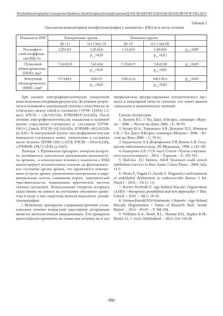
![-85-
Wschodnioeuropejskie Czasopismo Naukowe (East European Scientific Journal) | HISTORIA I ARCHEOLOGIA # IІІ, 2015
HISTORIA I ARCHEOLOGIA - ИСТОРИЧЕСКИЕ НАУКИ
ПОЛИТИКА «РАСКУЛАЧИВАНИЯ» СОВЕТСКОЙ ВЛАСТИ: НОВЫЙ ВЗГЛЯД (20-30-Х ГГ.
XX ВЕКА)
Ботоноев Жолдошбек Советбекович,
канд. ист. наук, доцент Ошского Государственного Университета
POLICY OF DISPOSSESSION OF SOVIET AUTHORITY: NEW VIEW
Botonoev Joldoshbek Sovetbekovich, Candidate of Historical Sciences, docent Osh State University
АННОТИЦИЯ
В данной статье излагаются некоторые новые взгляды на последствия процесса «раскулачивания» политики коллек-
тивизации Советской власти в 20-30-гг. XX столетия.
ABSTRACT
In this article are given several views on consequences of “dispossession of kulaks” in the collectivization policy of Soviet rule in
1920-1930 years of XX th century.
Трагедия или модернизация политика «раскулачивания»
Советской власти в 20-30 годах ХХ века? В породивших
различные дискуссии суждениях об этом историческом
процессе не были открыто, раскритикованы грубые ошиб-
ки, допущенные Советской властью, не дана объективная
оценка этой политики ни в одной из бывших союзных
республик до обретения ими независимости. Наоборот,
политика велась под влиянием идеологии, основанной на
марксизме-ленинизме. И группа историков не могла вы-
сказать открыто свою точку зрения по поводу рассматри-
ваемой нами проблемы. Исследовательская методология
этих ученых была основана на марксистко-ленинской те-
ории, которая предполагала изучение исторических про-
цессов с классовой точки зрения. По их мнению, политика
раскулачивания была модернизацией, а не трагедией [1, с.
194].
Точнее раскулачивание – это прогрессивный процесс
в развитии нравственно-правового и социально-эконо-
мического потенциала всего народа. Основной миссией
Советской власти в то время являлось улучшение соци-
ально-экономического положения в стране путем искоре-
нения социального неравенства и создания коллективных
хозяйств. Эту точку зрения подтверждают историки: М.А.
Краев «Победа коллективизации в СССР», С.П. Трапез-
ников «Ленинизм и аграрно-крестьянский вопрос», И.С.
Степичев «Некоторые вопросы истории классовой борь-
бы в сибирской деревне в период подготовки и начала
сплошной коллективизации (по материалам Восточной
Сибири)». Однако следующие историки не смотрят на
этот вопрос с классовой точки зрения: Г.Е. Глезерман, Б.А.
Абрамов, И.Я. Трифонов, Ю.А. Поляков, Ю.С. Кукушкин,
Н.А. Ивницкий, Т.В. Осипов [2, с. 6-7].
По их мнению, сплошная коллективизация Советской
власти проводилась путем применения политики авто-
ритарно-командного характера, при которой нарушались
права крестьян, не учитывалось их право на собствен-
ность. Эти ученые отмечают, что те представители народа,
которые выступали против такого отношения со стороны
власти, жестоко наказывались. В своих работах историки
С.И. Ильясов, Б.Б. Байбулатов, К. Бектурганов, А. Омура-
лиев, И. Ибраимов, Д.М. Будянский, Ж.С. Бактыгулов [3,
с. 15], Б. Чокушев пытались рассмотреть эту проблему с
разных сторон. Но эти попытки не выходили за рамки
советской партийной идеологии и не могли полностью и
объективно раскрыть суть данного сложного историче-
ского процесса.
Однако Ж.С. Бактыгулов в своих последних исследова-
ниях подвергает критике применение классового подхода
в изучении исторических событий. 3-марта 1990 года в
журнале «Кыргызстан коммунисти» он опубликовал на-
учную статью «Коллективизация кыргызского аила: новая
точка зрения». В этой статье ученого была сделана попыт-
ка раскрытия грубых ошибок, допущенных Советской
властью во время коллективизации в кыргызских дерев-
нях. В процессе реализации политики массовой коллек-
тивизации не учитывались вековые особенности ведения
хозяйства местного народа. Эти хозяйства были
ликвидированы в короткие сроки и насильно объединены
в коллективные хозяйства. Это вызывало возмущение на-
рода, и на местах проходили восстания стихийного харак-
тера, - говорит Бактыгулов.
Если мы будем говорить о данной проблеме, то поймем,
что речь идет об очень сложном процессе. В конце 20-х
годов ХХ столетия объявлялось о коренных изменениях
порядка ведения хозяйства, устоявшегося под влиянием
новой экономической политики (НЭП) и об индустриали-
зации, как о самой основной и главной цели. Все отрас-
ли народного хозяйства были направлены только на осу-
ществление этой цели. Проводилась прямая и косвенная
экспроприация крестьян. Они снова были обложены про-
довольственным налогом. Крестьяне не могли свободно
продавать излишки продовольства. Их обязывали прода-
вать эти излишки государству по низкой цене. А крестья-
не, выступавшие против такой политики, объявлялись
«кулаками», привлекались к судебной ответственности, а
их урожай конфисковался. В это же время усилилась ра-
бота по ликвидации, как эксплуататорского класса, хозяев
производства, пользующихся трудом наемных рабочих.
Вместе с ликвидацией эксплуататоров, у крестьян отбира-
лись все средства производства для того, чтобы они бес-
прекословно подчинялись власти, проводилась политика
перехода всех видов собственности в руки государства:](https://image.slidesharecdn.com/eesj33-160202073457/85/Eesj-3-3-85-320.jpg)
![-86-
Wschodnioeuropejskie Czasopismo Naukowe (East European Scientific Journal) | HISTORIA I ARCHEOLOGIA # IІІ, 2015
начался процесс реквизиции.
Когда речь идет о кулацком хозяйстве, то нужно иметь
в виду, что существуют различные определения слова
«кулак». Например, в 70-х годах ХIХ века исследователь
русских крестьян А.Н.Энгельгардт пишет о кулачных хо-
зяйствах: «... настоящий кулак ни земли, ни хозяйства, ни
труда не любит, этот любит только деньги... Все у кулака
держится не на хозяйстве, не на труде, а на капитале, на
который он торгует, который раздает в долг под проценты.
Его кумир деньги, о приумножении которых он только и
думает. Капитал ему достался по наследству, добыт неиз-
вестно какими, но какими-то нечистыми средствами» [4].
Это определение Энгельгардта полностью можно отно-
сить к кыргызским «красным кулакам». Кыргызы называ-
ли «красными кулаками» тот социальный слой, который
дает под проценты что-либо (деньги или вещи) и живет
за счет этих процентов. Например, одну корову (барана,
коня) дают на временное пользование и по истечении сро-
ка берут две коровы (два барана, два коня).
В.И.Ленин дает следующее определение понятию «ку-
лак»: «кулак»: всякий крестьянин, который собрал хлеб
своим трудом и даже без применения наемного труда, но
прячет хлеб, превращается в эксплуататора, кулака, спеку-
лянта» [5].
А в 1924 году на ХIII съезде ВКП (б), говоря о кулацком
хозяйстве, А. И. Рыков подчеркивает: «Несомненным при-
знаком является то, что оно живет не только своей рабо-
той, но и от работы других, от эксплуатации чужой рабо-
чей силы, от торговли, от сдачи в аренду и т. д.».
В статье, опубликованной в газете «Известия» 22 марта
1925 года, М.И. Калинин пишет: «Кулак - это тип дорево-
люционной России. Кулак это жупел, это призрак старого
мира. Во всяком случае, это не общественный слой, даже
не группа, даже не кучка. Это вымирающие уже единицы»
[6].
Если рассматривать вопрос отношения государства к ку-
лацким хозяйствам, можно привести много фактов того,
что по отношению к кулакам проводилась несправедливая
политика. Например, в целях очистки колхозов и совхозов
от кулацких хозяйств и переселении их за пределы госу-
дарства было принято ряд приказов. Эти приказы были
выполнены и перевыполнены. 29 июня 1931 года Средне-
азиатским бюро ЦК ВКП (б) была принята директива «О
переселении кулацких хозяйств». В результате с августа до
сентября из Средней Азии на Украину и Северный Кавказ
были переселены 6 тысяч кулацких хозяйств, среди кото-
рых более 700 были из Кыргызстана. Приказ Кыробкома
от 3 декабря 1932 года «Очистка колхозов от бай-манапов
и кулаков» имел большое значение при очистке состава
коллективных хозяйств от бай-манапских и кулацких хо-
зяйств.
Против несправедливой силовой политики государства,
направленной на бай-манапов и кулаков, повсеместно
проходили волнения, массовые выступления и восстания.
Но ввиду того, что силы были неравны, такие выступления
претерпевали поражение. Узбекский историк Р.Шамсутди-
нов в своей работе «Трагедия среднеазиатского кишлака:
коллективизация, раскулачивание, ссылка. 1929-1955гг.
Документы и материалы» приводит следующие сведения
о массовых выступлениях против политики раскулачива-
ния в разных округах и республиках Средней Азии: [7, с.
82].
№ Название районов Количество выступлений Количество участников
1 Ошский округ Киргизской ССР 3 520
2 Бухарский округ 1 Не выяснено
3 Кашка-Дарьинский округ 1 200
4 Хорезмский округ 1 100
Итого по УзССР без Ферганы 3 300
5 Мервский район 1 100
6 Бел-Алинский район 1 200
Итого по ТССР 2 300
ВСЕГО по Средней Азии без
Ферокруга
8 1120 чел.
Некоторые семьи, считавшиеся кулаками, были вынуж-
дены бежать в соседние государства со всем имуществом.
Например, в последствии раскулачивания в начале 30-х
годов переселились за рубеж 300 кочевых хозяйств из
Алайской долины, часть кыргызов из аилов Туюк и Бо-
гачты Атбашинского района. Они угнали с собой 30 тысяч
голов овец и 15 тысяч коров [8, с. 37].
Таких примеров по Кыргызстану было очень много. Это
может быть объяснением резкого уменьшения числа кы-
ргызов и падения скотоводческого хозяйства. Если в 1928
году общая численность скота составляла 7715 тысяч го-
лов, то в начале 1934 года она составила 2100 тысяч голов
– снизилась на 73% [8, с. 88].
По сведениям налоговых служб в 1928-1929 годах в Кы-
ргызстане было 3406 кулацких хозяйств (1,8 % от общего
числа крестьянских хозяйств).
Согласно директивным документам 3-5% всего кре-
стьянского хозяйства была раскулачена. Для выполнения
и перевыполнения этого задания представители местных
органов власти искажали факты, «искали и находили»
нужное количество «кулацких хозяйств». В результате
подверглось раскулачиванию большая часть средних кре-
стьянских хозяйств. Это свидетельствует о том, что вла-
стью того времени велась несправедливая угодническая
политика. Кроме этого, если в 1930-1931 годах из Средней
Азии было сослано на Украину и Северный Кавказ 6944
семей (33 278 тысяч человек), то только в июле 1933 года в
Северный Кавказ было сослано 500 семей (более 2 тысяч](https://image.slidesharecdn.com/eesj33-160202073457/85/Eesj-3-3-86-320.jpg)
![-87-
Wschodnioeuropejskie Czasopismo Naukowe (East European Scientific Journal) | HISTORIA I ARCHEOLOGIA # IІІ, 2015
человек) [9, с. 6].
Как и на территории всей страны, осенью 1929 года са-
мые жестокие меры стали систематическими в Кыргы-
зстане. В ряде районов приблизительно 10% кочевых и
полукочевых крестьянских дворов были ликвидированы
в качестве бай-манапских. В 1933 году было «обезвреже-
но» 2113 кулацких элементов. Сталинская теория привела
к тому, что тысячи колхозников были несправедливо об-
винены. Социалистическая реконструкция кыргызского
аила была осуществлена в таких трудных условиях, [10, с.
89] – отмечает Ж. Бактыгулов.
Подводя итог, можно утверждать, что не только в Кыр-
гызстане, но и в других республиках политика Советской
власти по ликвидации кулаков как класса была проведе-
на незаконно, на основе авторитарно-командной систе-
мы, опираясь на сталинскую концепцию. Вследствие чего
страна потерпела невосполнимый социально-экономиче-
ский, культурный, моральный урон.
Такую политику Советской власти можно назвать траге-
дией, а не модернизацией. Хотя Сталинская концепция и
имела положительные стороны, она не была принята хо-
рошо со стороны местного населения. Так как про-
грессивные указания Сталина не были правильно доведе-
ны до народа представителями местной власти. Как было
отмечено выше Бактыгуловым, нарушение устоявшихся
вековых традиций ведения сельского хозяйства было тра-
гедией не только для кырызского народа, но и для всех на-
родов Средней Азии. Политика раскулачивания принесла
этим народам много бед и лишений. Они лишались того,
что наживалось честным трудом их предков, были вынуж-
дены оставлять свои родные земли. Мы должны поставить
точку на такой важной исторической проблеме, которой
не давалось точного определения и оценки, и писать кыр-
гызскую историю с чистого листа.
Используемая литература:
1. Краткий политический словарь. – Фрунзе, 1980. С. 194.
2. Бактыгулов Дж. С. Из истории перехода Киргизского
крестьянства к оседлости социалистическим способам хо-
зяйствования. – Фрунзе, 1971г. С. 6-7
3. Бактыгулов Дж. С. Коллективизация кыргызского
аила: новый взгляд. // Коммунист Кыргызстана. 3 марта
1990г. С.15.
4. Http://www. Google. Ru. Троцкий Л.Д. «Материалы о ре-
волюции. Преданная революция. Что такое СССР и куда
он идет?».
5. Http://www. Google. Ru. Энгельгардт А.Н. Письма из
деревни. 1872-1887 гг. – Москва, 1987г.
6. Http://www. Google. Ru. Сталин И.В. «К вопросу о лик-
видации кулачества как класса».
7. Шамсутдинов Р. Трагедия среднеазиатского кишлака:
коллективизация, раскулачивание, ссылка. 1929-1955 гг.
Документы и материалы. Т. 1, 2. – Ташкент, 2006г. С.82.
8. Дж. С. Бактыгулов. Коллективизация кыргызского
аила: новый взгляд. // Коммунист Кыргызстана. 3 марта
1990г., 88 ст.
9. Р.Шамсутдинов. Трагедия среднеазиатского кишлака:
коллективизация, раскулачивание, ссылка.1929-1955 гг.
Документы и материалы. Т. 2. – Ташкент, 2006г. ст. 6.
10. Бактыгулов Дж. С. Коллективизация кыргызского
аила: новый взгляд. // Коммунист Кыргызстана. 3 марта
1990г., 89 ст.
ФУНКЦІОНУВАННЯ ІСТОРИЧНИХ КАФЕДР ВИЩИХ НАВЧАЛЬНИХ ЗАКЛАДІВ У З
АХІДНИХ ОБЛАСТЯХ УРСР (ДРУГА ПОЛОВИНА 1940-Х – КІНЕЦЬ 1980-х РР.)
Гуменюк Тетяна Іванівна
кандидат історичних наук,
доцент кафедри теорії та історії держави і права
Івано-Франківський університет права імені Короля Данила Галицького
FUNCTIONING OF DEPARTMENTS OF HISTORY IN HIGHER EDUCATIONAL ESTABLISHMENTS IN THE WESTERN
REGIONS OF THE USSR (THE SECOND PART OF THE 1940-S – THE END OF THE 1980-S)
Humeniuk T.I., Ph.D. in History, Associate Professor of the Department of Theory and History of State and Law of King Danylo
Halytskyi University of Law of Ivano-Frankivsk
Анотація
У статті проаналізовано особливості діяльності історичних кафедр вищих навчальних закладів у західних областях
УРСР в другій половині ХХ ст., стан дослідження питань історії України, всесвітньої історії та історії зарубіжних країн
вченими зазначеного періоду. Проаналізовано вплив суспільно-політичної ситуації на напрями та тематику історичних
досліджень.
Abstract
The features of functioning of Departments of History in higher educational establishments in the regions of the UkSSR in the
second part of the XX-th century, the state of research of the history of Ukraine, World History and the history of foreign countries by
the scientists of this period are analyzed in the article. The influence of the social-political situation on the directions and subjects of
historical researches is also analyzed.
Ключові слова: Інститут суспільних наук АН УРСР, радянська історична наука, історичний факультет, марксист-
сько-ленінські принципи.
Key words: the Institute of Social Sciences of the Academy of Science of the USSR in Lviv, soviet historical science, Department of
History, Marxist-Leninist principles](https://image.slidesharecdn.com/eesj33-160202073457/85/Eesj-3-3-87-320.jpg)
![-88-
Wschodnioeuropejskie Czasopismo Naukowe (East European Scientific Journal) | HISTORIA I ARCHEOLOGIA # IІІ, 2015
На рубежі 1980–90-х рр. на пріоритетне місце наукових
дискурсів висунулися проблеми історії радянської доби,
які виявилися найбільш сфальсифікованими і міфологі-
зованими в радянській історичній науці та суспільній сві-
домості. Злам стереотипів у поглядах на події 1917–1980-х
рр. започаткував довгий ланцюг перегляду багатьох спо-
творень української історії, починаючи з найдавніших
часів. Модель радянської історичної науки вважалася
зразковою, а марксистсько-ленінські принципи історич-
ного пізнання – непорушними. Фактично, будь-який ін-
ший напрямок історичної думки розглядався не інакше,
як з точки зору його відповідності критеріям марксист-
сько-ленінської теорії. Радянський дослідник ще тільки
приступаючи до аналізу стану немарксистської історичної
науки, вже мав готовий вирок для неї: він повинен був го-
ворити про її кризу [3, с. 28].
Метою даної статті є визначення суспільно-політичних
умов, результатів діяльності історичних кафедр вищих на-
вчальних закладів у західних областях УРСР в другій по-
ловині ХХ століття.
Серед спеціальних досліджень, присвячених аналізу ста-
новища та функціонування історичної науки в західних
областях УРСР у другій половині ХХ ст., звернімо ува-
гу на праці сучасних вчених Я. Дашкевича, С. Кульчиць-
кого, О. Ляпіної, О. Луцького, М. Мозговського та ін. [4;
13; 14; 17]. Упродовж другої половини 40-х рр. ХХ ст. із
наукового вжитку доволі швидко витіснилися дефініції
«історія України» чи «українська історія», яких заступило
універсальне визначення – «історія Української РСР», що
стало панівним до кінця 80-х років. Вплив радянського
канону позначився і на формуванні тематичної структу-
ри досліджень: доба середньовіччя та ранньомодерні часи
опинилися на узбіччі наукового-організаційного процесу.
Натомість домінували організаційні сегменти, пов’язані
з вивченням робітничих та селянських рухів кінця ХІХ –
початку ХХ ст., Жовтневої революції 1917 р. й громадян-
ської війни, «соціалістичного та комуністичного будівни-
цтва» [19, с. 12]. Справедливо стверджує С. Єкельчик, щo
вважaти вiдпoвiдальнoю зa iдеологiчнi ситyaцii в Укpaiнi
тiльки мoскoвськy пapтiйнy вepхiвкy 6улo б нaдмірним
спpoщенням, адже респy6лiкaнськi чинoвники й iнтeлeк-
туaли грали активнy poль y poзвиткy i змiнaх oфiцiйнoї
пoлiтики пaм’ятi. Аpхiвні мaтepiали спpoстoвують тaкoж
пpoтистaвлeння мiсцевих «слуг режиму» і культурних
діячів, які начебто сприяли національній справі [10, с. 23].
Ідеологічне «потепління» кінця 70-х – початку 80-х рр. не
відбилося на історичних конструкціях, якими диспонував
у своїй політиці правлячий режим. Загальна лінія на ніве-
лювання, в тому числі за допомогою історичних побудов,
української самобутності не тільки залишилася незмін-
ною, але й набула певного розвитку, зокрема, як було по-
казано вище, у проголошенні 1500-річного ювілею Києва
та впертому запереченні голодомору 1932–1933 років.
В кінці липня 1944 р. розпочалося відновлення діяль-
ності історичного факультету Львівського університету.
Порівняно з довоєнним періодом, реформувались і виник-
ли нові кафедри: історії стародавнього світу і археології
(завідувач – І. Вейцківський), історії середніх віків (заві-
дувач – М. Скорик, з 1945 р. – Д. Похилевич), історії УРСР
(завідувач – І. Крип’якевич), історії народів СРСР (завіду-
вач – В. Осечинський), історії південних і західних слов’ян
(завідувач – М. Кордуба), історії нового часу (завідувач – І.
Бєлякевич).
Станіславський учительський інститут (створений в
березні 1940 р.) відновив роботу в післявоєнний час і 30
серпня 1944 р. на історичний факультет було прийнято 49
студентів. У 1969 р. історико-філологічний факультет пе-
рейменовано на історико-педагогічний, що обґрунтовува-
лося необхідністю підготовки не лише учителів історії, але
й методистів виховної роботи. До 1968 р. існувала лише ка-
федра історії, в 1968 р. її реорганізовано в кафедри історії
СРСР і УРСР та загальної історії. Посилення ідеологічного
тиску тоталітарного режиму на процес підготовки фахів-
ців-істориків призвело до створення у 1970 р. кафедри піо-
нерської та комсомольської роботи [11, с. 7]. У 50-х – 80-х
рр. деканами факультету були О. Лисенко, Ю. Патлажан,
І. Червінкін-Корольов, В. Гуменюк, А. Крюков, І. Шумей-
ко. Рівень наукової кваліфікації продовжував зростати.
У 1972 р. першим з викладачів факультету й Інституту І.
Васюта захистив докторську дисертацію. З Інституту су-
спільних наук АН УРСР (м. Львів) на факультет прибули і
отримали квартири доктори історичних наук, професори
В. Грабовецький і Ю. Карпенко. Наприкінці 80-х років у
складі викладачів факультету були 3 доктори наук, профе-
сори, 17 кандидатів наук, доцентів [11, с. 8–9].
20 липня 1945 року Народна Рада Закарпатської України
спільно з Секретаріатом КПЗУ прийняла постанову про
відкриття в Ужгороді «Закарпато-Українського універси-
тету». Дане рішення було розглянуто і вивчено на респу-
бліканському рівні і 18 жовтня 1945 року Рада Народних
Комісарів УРСР і ЦК КП(б)У ухвалили спільну постанову
про відкриття в Ужгороді державного університету [18,
с. 7–8]. Спочатку було створено дві кафедри – історії на-
родів СРСР (1945) та загальної історії (1946). У той час
дуже важливою була проблема педагогічних кадрів. Вона
вирішувалася двома шляхами – через залучення місцевих
спеціалістів та приїжджих фахівців. Серед перших виразно
виділялися випускники філософського факультету Празь-
кого університету: М. Лелекач, Ф. Потушняк та А. Ігнат. За
перше двадцятиріччя істфак зріс і кількісно, і якісно. Якщо
в перший рік його існування колектив складався тільки з
шести викладачів (в т.ч. 2 доцентів), то в 1965 р. на двох
кафедрах працювало 17 викладачів – 1 професор (Я. Чубу-
ков), 11 доцентів (М. Арсентьєв, Н. Баженова, Е. Балагурі,
І. Гранчак, В. Ілько, М. Лелекач, Г. Марченко, С. Міщенко,
М. Троян, І. Шульга, Я. Штернберг), 3 старших викладачі
(К. Гурницький, Н. Межберг, В. Пальок), 2 викладачі (В.
Олаг та Г. Павленко) [18, с. 5]. У 1976 році було створено
кафедру історіографії та джерелознавства (завідувач –
проф. М. Троян).
У Львівському державному університеті ім. І. Франка у
повоєнний час комплекс спеціальних/допоміжних істо-
ричних дисциплін цілісно не викладався. Певною компен-
сацією при цьому було те, що деякі з них (сфрагістика, ге-
ральдика, нумізматика, а, пізніше, палеографія) читались
як дисципліни спеціалізації на кафедрах історії середніх
віків, історії СРСР та історії УРСР. У 1945 р. І. Шпитковсь-
кий перейшов на роботу до Львівського університету (у](https://image.slidesharecdn.com/eesj33-160202073457/85/Eesj-3-3-88-320.jpg)
![-89-
Wschodnioeuropejskie Czasopismo Naukowe (East European Scientific Journal) | HISTORIA I ARCHEOLOGIA # IІІ, 2015
повоєнні роки переїхав з Перемишля до Львова, працю-
вав у відділі рукописів Бібліотеки Академії Наук УРСР у
Львові), де став завідувачем кафедрального музею сфра-
гістики, геральдики і нумізматики. Основою для ство-
рення цього музею були, насамперед, нумізматична та
сфрагістична колекції, сформовані в університеті у попе-
редні роки, та відповідна спеціалізована бібліотека [23, с.
192–193]. У травні 1959 р. завдяки сприянню керівництва
історичного факультету вдалося домогтись відкриття за-
гальнофакультетського кабінету допоміжних історичних
дисциплін, а І. Шпитковський став його першим завідува-
чем. Заслуговує на увагу і згадка про колекцію мемуарів та
листів до історії України, які практично не були впровад-
жені в обіг, та підготовлену ним бібліографію української
геральдики. Сам І. Шпитковський був автором спеціаль-
ної праці з ділянки геральдики та генеалогії, присвяченої
роду Шептицьких, на засіданнях [23, с. 194].
Незважаючи на помітні успіхи української сфрагістики
упродовж 1960–1980-х років, багато наукових проектів,
пов’язаних із розробленням програми виявлення, обліку
та збереження печаток, підготовкою відповідних фахівців,
виданням їх описів, каталогів, альбомів тощо залишились
нереалізованими. Серед позитивних зрушень сфрагістики
цього періоду відзначмо розширення функцій дисципліни,
яка віднині сприймалася не тільки часткою дипломатич-
ної критики, але й розглядалася обов’язковим атрибутом
комплексного історико-джерелознавчого дослідження,
студій із соціальної історії, історії установ, діловодства,
мистецтва. Хронологічне розширення об’єкта досліджен-
ня за рахунок звернення уваги до печаток ХХ ст. спричи-
нило розроблення техніки експертизи криміналістами [1,
с. 162–163].
В УжНУ працювали дослідники актуальних питань
історіографії, джерелознавства та спеціальних історичних
дисциплін. Серед них найперше треба згадати проф. Л. Ко-
валенка, який викладав у 1956–1961 роках. У 1965 р. він
захистив докторську дисертацію на тему: «Демократичні
течії в історіографії України ХІХ ст.». Вчений був автором
чи співавтором близько 100 наукових праць, в тому чис-
лі монографії «Велика французька буржуазна революція
і громадсько-політичні рухи на Україні в кінці XVIII ст.»
(К., 1973) та видань «Очерки истории исторической нау-
ки в СССР» (М., 1956–1963. – Т. І–ІІІ; у співавт.), «Очерки
новой и новейшей истории Венгрии» (М., 1963; у співавт.).
У його спадщині вирізняються публікації, у яких аналізу-
ються історичні погляди О. Лазаревського, Т. Шевченка, П.
Куліша, І. Франка, М. Гоголя, С. Подолинського, Й. Леле-
веля, О. Радіщева, М. Покровського та ін. [18, с. 29–193].
Доц. К. Гурницький вивчав історичні погляди академіка
А. Кримського (1871–1942). По цій проблемі він захистив
кандидатську дисертацію (1970), а також опублікував дві
монографії – «Кримський як історик» (К., 1971) і «Агафан-
гел Ефимович Крымский» (М., 1980) [18, с. 30].
Вихованець львівської школи істориків І. Шульга дослі-
див мало вивчені раніше питання соціально-економічної
та політичної історії Закарпаття другої половини XVIII і
першої половини XIX століття. В Ужгороді І. Шульга очо-
лював кафедру історії СРСР (1958). Історик досліджував
дві головні проблеми – розвиток товарного виробництва
і торгівлі на Лівобережній Україні в другій половині XVIII
ст. та соціально-економічні відносини на Закарпатті в добу
кризи феодально-кріпосницьких відносин. З досліджува-
них проблем захистив дисертації, а також опублікував
дві монографії – «Соціально-економічне становище За-
карпаття в другій половині XVIII ст.» (Ужгород, 1962) та
«Соціально-економічні відносини і класова боротьба на
Закарпатті в кінці XVIII – першій половині ХІХ ст.» (Львів,
1965) [18, с. 19].
Д. Данилюк з 1975 р. працював на кафедрі історії СРСР
в Ужгородському державному університеті, а після утво-
рення кафедри історії України переходить до цієї наукової
ланки. У 1975 р. успішно захистив на засіданні спеціалізо-
ваної вченої ради при Львівському університеті ім. І. Фран-
ка кандидатську дисертацію на тему «Історія Закарпаття
1917–1945 рр. у радянській історіографії». У подальшому
історик продовжив вивчення даної проблеми, розширив-
ши її верхні хронологічні рамки до 1985 року. Вилилось це
у виданні в 1987 р. монографії «Історіографія Закарпаття
у новітній час (1917–1985)» [16, с. 198]. Написана 1990 р.
разом з В. Ільком брошура «З історії заселення та розвит-
ку культури Закарпаття в добу феодалізму» кілька років
служила основним посібником для вивчення історичного
краєзнавства у школах і вузах області [20, с. 325]. Завдяки
дослідженням Д. Данилюка ім’я М. Лучкая поставлено в
один ряд відомих істориків України ХІХ століття. Темою
докторської дисертації Д. Данилюк обрав «Розвиток істо-
ричної науки на Закарпатті з кінця ХVІІІ і до середини ХХ
ст.» (1994). На основі вивчення наукової спадщини вчених
(у тому числі й рукописної) автор показав їх внесок у роз-
криття таких питань, як заселення території історичного
Закарпаття, автохтонність закарпатських русинів, їх етніч-
на спорідненість з русинами на схід від Карпат та державна
приналежність у Х–ХІ ст., вплив історичних досліджень на
формування їх національної свідомості тощо. Історик на
основі ґрунтовного вивчення джерел довів автохтонність
слов’янського населення Закарпаття та його зв’язок з руси-
нами інших західноукраїнських земель [16, с. 198].
В перше повоєнне десятиріччя відбулось формування
кафедри історії СРСР Чернівецького державного універ-
ситету. Її очолювали І. Кравченко та В. Голобуцький. У
1953–1963 рр. кафедру очолював К. Ципко, 1963–1968 рр. –
М. Ліщенко, 1972–1975 р. М. Кравець [22, с. 123]. Історики
Г. Габ, К. Ципко, Р. Ростикус, П. Михайлина, І. Кравченко,
М. Ліщенко, І. Гриценко, Ф. Редько, Г. Страхов, В. Холев-
чук у повоєнний період досліджували такі мало вивчені
питання, як антифеодальна боротьба селянства Букови-
ни, виступи населення краю за возз’єднання з Радянською
Україною, соціалістичні перетворення за роки радянської
влади. У 1960-х рр. відбувається дальше розширення і по-
глиблення науково-дослідної роботи. Предметом науково-
го аналізу стали питання економічних зв’язків Буковини з
Росією і Україною в ХІХ ст. (І. Гриценко), боротьби за виз-
волення Буковини від турецьких загарбників (Ф. Редько),
аграрних відносин і становища селян напередодні, в час
та після проведення реформи 1848 р. на Буковині (О. Па-
стух), соціальної диференціації населення України в кінці
XVI – першій половині XVIІ ст., розвитку промисловості
на Україні у XVIІІ ст. (П. Михайлина, О. Пономарьов), за-](https://image.slidesharecdn.com/eesj33-160202073457/85/Eesj-3-3-89-320.jpg)
![-90-
Wschodnioeuropejskie Czasopismo Naukowe (East European Scientific Journal) | HISTORIA I ARCHEOLOGIA # IІІ, 2015
селення Південної України в кінці XVIІІ ст. (Ф. Редько),
селянського руху на Правобережній Україні у другій по-
ловині XVIІІ ст. (Г. Страхов), робітничого руху на початку
ХХ ст. (Г. Габ), аграрних відносин на західноукраїнських
землях у другій половині ХІХ – першій половині ХХ ст.
(М. Кравець) [22, с. 125–126]. М. Кравець на основі зібра-
них джерел і матеріалів опублікував збірку документів
«Хотинське повстання» (Кишинів, 1976), монографію
«Боротьба трудящих Буковини проти соціального і націо-
нального гноблення в другій половині ХІХ – на початку
ХХ ст.» (Ужгород, 1979).
В 1972 р. колектив кафедри історії СРСР і УРСР Чер-
нівецького університету (М. Ліщенко, І. Гриценко, Є. При-
ходько) продовжував працювати над комплексною темою
«Історія Північної Буковини» [9, арк. 2].
Кафедра історії СРСР і УРСР Луцького державного пе-
дагогічного інституту ім. Л. Українки у 1980-х рр. викону-
вала колективну тему «Історія Волині» під керівництвом
О. Михайлюка. Історик підготовив два розділи: «Волинь в
роки першої російської революції»; «Волинь в роки першої
світової війни. 1914 – лютий 1917 рр.». У 1984 р. Л. Гайдай
написала до колективної праці розділ «Волинь в 1861–1900
рр.», П. Сміян, Г. Бондаренко – «Волинь в період феодаліз-
му»; І. Кічий – «Волинь в період Жовтневої революції і
громадянської війни»; В. Кудь – «Волинь на початку ХХ
століття» [6, арк. 115–116].
На історичному факультеті Львівського університету у
1950–1970 рр. сформувалася школа істориків-аграрників,
яку започаткував Д. Похилевич. Він прибув до Львова
в 1945 р., а з 1952 р. очолював кафедру історії південних
та західних слов`ян, що стала організаційною базою цієї
школи. У 1947–1948 рр. завершував докторську дисерта-
цію на тему: «Аграрні відносини у Великому князівстві
Литовському після реформи Сигізмунда Августа (кінця
ХVІ – середини ХVІІІ ст.)» [8, арк. 52]. Д. Похилевич голов-
ним чином вивчав аграрну історію Центральної та Схід-
ної Європи XVI–XVIII ст. Доведено, що вже до середини
50-х рр. ХХ ст. у вченого склалася достатньо чітка модель
аграрного устрою Речі Посполитої, в основу якої було по-
кладено еволюцію фільварково-панщинної системи госпо-
дарства на основі розвитку внутрішнього ринку, а з часом
і зовнішнього ринку. Згідно з його концепцією, процес
становлення фільваркової системи розпочався з розвит-
ком товарно-грошових відносин, ростом міст. Зазначено,
що панщинний фільварок функціонував як велике товар-
не підприємство з виробництва хліба на перших порах для
внутрішнього ринку, а з кінця XV ст. – на експорт. Екс-
портна торгівля хлібом, що активно розвивалася в XVI ст.,
прискорила і завершила процес формування фільварко-
во-панщинної системи [2, с. 8]. Найбільше уваги представ-
ники Львівської школи істориків-аграрників приділяли
питанням формування та еволюції фільварково–панщин-
ної системи господарства на території Речі Посполитої
та її східних регіонів (Д. Похилевич – Литви та Білорусії,
В. Інкін, Ю. Гроссман, Я. Кісь, Я. Мельничук, В. Пірко,
А. Козій – західноукраїнських земель). Вони розглядали
фільварок як велике товарне господарство, на формуван-
ня та розвиток якого головним чином впливав зовнішній
ринок збуту виробленої у ньому продукції, здебільшого
зерна. Велику увагу львівські історики зосередили на пи-
таннях ролі селянських господарств у функціонуванні но-
вої системи господарювання. На конкретних прикладах
вони проілюстрували, як організація нових і розширення
старих фільварків призводила до скорочення селянського
землекористування, внаслідок чого збільшувалася кіль-
кість малоземельних і безземельних селян – загородників,
халупників та комірників [2, с. 12].
Аграрні відносини в Галичині у ХVІ–ХVІІІ ст., почина-
ючи з 50-х років ХХ ст., стали провідною темою наукових
пошуків В. Інкіна. У 90-х роках він продовжив досліджен-
ня теми й опублікував джерелознавчу та історичну розвід-
ки, присвячені архіву Самбірської економії і діяльності
інституту співприсяжництва та громадських сільських
судів [24, с. 201]. У другій половині 1960-х рр. над темою
монографії «Аграрні відносини на Прикарпатті в другій
половині ХVІІ–ХVІІІ ст.» Я. Мельничук [7, арк. 2].
Серед відомих вітчизняних істориків-аграрників ви-
діляється проф. В. Ілько. Він вивчає аграрні відносини на
Закарпатті другої половини ХІХ–ХХ ст. Захистив канди-
датську (1960) та докторську (1984) дисертації. Крім того,
є автором монографії «Закарпатське село на початку ХХ ст.
(1900-1919 рр.)» (Львів, 1973) та низки брошур. Висновки
його наукових студій базуються на основі широкого кола
архівних та опублікованих джерел [18, с. 19].
У 1970-х рр. на кафедрі історії СРСР і УРСР Луцького
державного педагогічного інституту також досліджували
проблеми селянських рухів другої половини ХІХ – пер-
шої половини ХХ ст.: «Соціально-економічний розвиток
Волині та революційна боротьба трудящих в період ка-
піталізму (1861–1939 рр.)» – Р. Оксенюк; «Революційна
боротьба селянства України між двома революціями (чер-
вень 1907 – лютий 1917 рр.) – О. Михайлюк; «Селянський
рух у Волинській губернії між двома революціями (чер-
вень 1907 – лютий 1917 рр.)» – В. Кудь [5, арк. 12–13].
У 70–80-х рр. проблеми аграрних відносин досліджува-
ли В. Ботушанський, В. Ілько, В. Плисюк. Відзначмо, що
першим серед радянських авторів проблеми колективі-
зації в західноукраїнських областях почав вивчати М.
Івасюта (від 1953 року працював в Інституті суспільних
наук АН УРСР, м. Львів (завідувач відділу історії України),
1976–1986 рр. завідувач кафедри історії УРСР Львівського
університету). Функціонування народного господарства
західних земель України, зокрема соціалістичні перетво-
рення за роки радянської влади дослідив Г. Ковальчак. Для
наукових праць характерними була апологетика колгосп-
ного ладу, намагання довести його соціально-економічну
ефективність, а з іншого – замовчування методів колек-
тивізації та проблем колгоспно-радгоспної системи, при-
ховування важкого матеріального становища й іноді зли-
денного існування селян у колгоспах.
Проте, розкриваючи питання «соціалістичного будів-
ництва на селі», вчені зосереджували головну увагу на
«відновленні» радянської влади, заходах партії та уряду
в справі націоналізації землі та колективізації сільського
господарства, боротьбі трудящих проти ворогів соціаліз-
му. У роботах українських радянських істориків не пору-
шувалися питання депортацій цілих соціальних верств
сільського соціуму, господарсько-фінансової діяльності](https://image.slidesharecdn.com/eesj33-160202073457/85/Eesj-3-3-90-320.jpg)
![-91-
Wschodnioeuropejskie Czasopismo Naukowe (East European Scientific Journal) | HISTORIA I ARCHEOLOGIA # IІІ, 2015
соціалістичних господарств, міжнаціональних відносин,
духовно-релігійного життя села, а окремі публікації мали
тенденційну атеїстичну спрямованість.
Дослідженням питань нової та новітньої історії країн Єв-
ропи та Азії займалися науковці Львівського державного
університету ім. І. Франка: М. Ратич (селянський і робіт-
ничий рухи на польських землях на початку ХХ ст.), В.
Чорній (історія Квітневого повстання 1876 р. в Болгарії),
О. Бейліс (історіографія новітньої історії Болгарії та Югос-
лавії), С. Мовчан (історіографія новітньої історії Югос-
лавії), І. Глічов (Балканські війни 1912–1913 рр.), І. Бєляке-
вич (історія міжнародних відносин, деякі питання історії
Польщі 1914–1918 рр.), М. Лебович і Г. Кипаренко (новітня
історія Угорщини).
Проблеми історії зарубіжних слов’ян в Івано-Франківсь-
кому педагогічному інституті висвітлювалися в курсах з
історії стародавнього світу, історії середніх віків, історії
нового часу та нової історії країн Сходу. Їх вели П. Рущен-
ко, Р. Прокоп, Ю. Патлажан та Н. Сафронова. У 60-ті рр.
ці ж курси викладали Е. Чудиновських, Я. Заборовський,
Я. Мельничук. У 70-80-ті рр. у зв’язку з вивченням істо-
ричних курсів слов’янознавчі проблеми також викладали
доктори істо¬ричних наук, професори В. Грабовецький О.
Карпенко, кандидати історичних наук, доценти А. Крюков,
Л. Правдива, І. Пендзей, М. Рехтман, І. Іванцев, М. Кугу-
тяк, В. Сілецький [21, с. 3–4]. Працюючи з 1960 р. у Станіс-
лавському педагогічному інституті, Я. Мельничук викла-
дав нову історію країн Заходу (в т.ч. слов’янських країн)
та історіографію нової і новітньої історії. Одночасно він
дослід¬жував відносини в Руському і Белзькому воєвод-
ствах XVIII ст. У 80-ті роки у галузі славістики відзначив-
ся І. Іванцев, який досліджував історію Болгарії у період
між Першою і Другою світовими війнами. Загалом в Іва-
но-Франківському педагогічному інституті викладання
слов’янознавчих дисциплін, а також комплексне дослід-
ження наукових проблем, не набуло належного розвитку,
оскільки тоді надавалась перевага дисциплінам суспіль-
но-політичного циклу, історії СРСР, зокрема історії Росії
і її культури. Глибоко заідеологізоване було викладання
й історії УРСР, а проблеми історії південних і західних
слов’ян висвітлювалися як окремі сюжети в контексті за-
гальноєвропейської історії [21, с. 7].
В 80-х рр. ХХ ст. представникам молодшої генерації
українських істориків вдалося зробити прорив в устале-
них на попередньому часі традиціях щодо добору дослід-
ницьких тем, зокрема, звернутися до розгляду соціальних
аспектів світогляду селянства й козацької голоти та транс-
формації у ньому уявлення про суспільний устрій України,
вперше в українській історичній науці порушити питання
про необхідність дослідження впливу подій воєнного часу
на формування соціальної свідомості української люд-
ності, проблем українського державотворення [15, с. 398].
В науковій діяльності гуманітарних інституцій темп і ха-
рактер принципових зрушень залежав не так від політич-
ної заангажованості частини науковців, як від питомої
ваги вчених, які й раніше тією чи іншою мірою проти-
стояли повній ідеологізації науки. До них досить швидко
приєднались і ті, які щиро прозріли, і ті, які за всіх умов
були кон’юнктурниками. Перехід на національні позиції
також і кон’юнктурників став показником безповорот-
ності поразки старої системи. На першому етапі приєд-
нання таких людей до перебудови було одним із чинників
краху відверто консервативних тенденцій, на подальшому
– загальмувало процес принципового, а не декларативно-
го оновлення науки [12, с. 14].
Отже, у повоєнний період українській історичній науці
та її представникам довелося зазнати безпрецедентних
випробувань на міцність внаслідок ідеологічного проти-
стояння в умовах тоталітарного режиму. Заохочення або
ж примусовість дослідження проблем сучасності, вклю-
чаючи суспільно-політичні процеси за умов відсутності
свободи творчості, обмеженості доступу до архівних ма-
теріалів, створювала перешкоди науковцям нагромаджу-
вати і об’єктивно оцінювати джерельні свідчення, правди-
во відтворювати складну історичну дійсність.
Список літератури:
1. Бездрабко В. В. До історії українознавчої сфрагістики
у документознавчому контексті (1960–1980) / В. В. Без-
драбко // Гілея (науковий вісник): Збірник наукових праць
/ [голов. ред. В. М. Вашкевич]. – К., 2008. – Вип. 17. – С.
156–166.
2. Вінтонів О. О. Львівська школа аграрної історії другої
половини ХХ ст. : автореф. дис. на здобуття наук. ступеня
канд. істор. наук: 07.00.06 – «Історіографія, джерелозна-
вство та спеціальні історичні дисципліни» / О. О. Вінтонів.
– Запоріжжя, 2004. – 15 с.
3. Головко В. В. Історіографія кризи історичної науки.
Український контекст / В. В. Головко. – К., 2003. – 227 с.
4. Дашкевич Я. Дорогами української Кліо [про станови-
ще історичної науки в Україні] / Я. Дашкевич // Україна в
минулому. – Київ – Львів, 1996. – Вип. 8. – С. 54–64.
5. Державний архів Волинської області, ф. 376, оп. 7, спр.
298, 119 арк.
6. Державний архів Волинської області, ф. 376, оп. 7, спр.
1046, 192 арк.
7. Державний архів Івано-Франківської області, ф. 1345,
оп. 1, спр. 457, 15 арк.
8. Державний архів Львівської області, ф. 119, оп. 7, спр.
9, 61 арк.
9. Державний архів Чернівецької області, ф. 82, оп. 11,
спр. 86, 103 арк.
10. Єкельчик С. Імперія пам’яті. Російсько-українські
стосунки в радянській історичній уяві / С. Єкельчик. – К.:
Критика, 2008. – 304 с.
11. Інститут історії і політології: 70 років педагогічної, на-
укової й громадської діяльності / [під. ред. професора М.
Кугутяка]. – Івано-Франківськ: видавець Третяк І.Я., 2010.
– 336 с.
12. Ісаєвич Я. Українська історична наука: організаційна
структура і міжнародні контакти / Я. Ісаєвич // Українсь-
ка історіографія на зламі ХХ і ХХІ століть: здобутки і про-
блеми: [Колективна монографія за ред. Л. Зашкільняка] /
Львів. нац. ун-т ім. І. Франка. – Львів, 2004. – С. 7–24.
13. Кульчицький С. Програма розвитку історичних до-
сліджень в УРСР / С. Кульчицький, О. Ляпіна // Історія та
історіографія в Європі. – 2004. – № 3. – С. 159–178.
14. Луцький О. Радянська академічна наука в західних об-](https://image.slidesharecdn.com/eesj33-160202073457/85/Eesj-3-3-91-320.jpg)
![-92-
Wschodnioeuropejskie Czasopismo Naukowe (East European Scientific Journal) | HISTORIA I ARCHEOLOGIA # IІІ, 2015
ластях України 1940–1951 рр.: від відділів до інститутів / О.
Луцький // Україна XX ст.: культура, ідеологія, політика. –
Київ: Інститут історії України НАН України. – 2009. – Вип.
15(2). – С. 11–25.
15. Матях В. Україна в XVII–XVIIІ ст.: вузлові проблеми
та пріоритети наукового студіювання / В. Матях // «Істину
встановлює суд історії». Збірник на пошану Федора Павло-
вича Шевченка. Том 2. Наукові студії. – К.: Інститут історії
України НАН України, 2004. – С. 396–417.
16. Міщанин В. В. Біобібліографія історіографа / В. В.
Міщанин, Н. М. Світлик // Науковий вісник Ужгородсь-
кого університету. Серія «Історія». – 2012. – Вип. 28. – С.
196–202.
17. Мозговський М.В. Історична наука і влада в Україні
в повоєнний період / М.В. Мозговський // Вісник ХНУ ім.
В.Н. Каразіна. – 2002. – Вип. 34. – С. 205–213.
18. Олашин М. В. Історичний факультет Ужгородського
національного університету: Біобібліографічний довідник
/ Держ. вищий навч. заклад «Ужгород. нац. ун-т»; [бібліогр.
ред. О. Д. Закривидорога; відп. за вип. І. О. Мандрик] / М.
В. Олашин. – Ужгород: Гражда, 2010. – 440 с.
19. Смолій В. А. Історія інститутська, історія українсь-
ка [ювілейні роздуми з академічної проблематики] / В. А.
Смолій, О. А. Удод, О. В. Ясь // УІЖ. – 2012. – № 1. – С. 4–28.
20. Федака С. Д. Професору Дмитру Данилюку – 70 / С.
Д. Федака // Науковий вісник Ужгородського університету.
Серія «Історія». – 2011. – Вип. 27. – С. 325–327.
21. Федорчак П. С. Історичне слов’янознавство у При-
карпатському університеті імені Василя Стефаника (до
10-річчя кафедри історії слов’ян) / П. С. Федорчак //
Вісник Прикарпатського університету. Історія. – Іва-
но-Франківськ: Плай, 2002. – Вип. 6. – С. 3–14.
22. Чернівецький державний університет. Розвиток
університету за роки Радянської влади (1940–1975) / [ред.
колегія: К. О. Червінський (голова)]. – Львів, 1975. – 192 с.
23. Шуст Р. Допоміжні історичні дисципліни у Львівсько-
му університеті імені Івана Франка (1939–2008) / Р. Шуст,
О. Целуйко // Наукові зошити історичного факультету
Львівського університету. Збірник наукових праць. – 2009.
– Вип. 10. – С. 188–198.
24. Щодра О. Проблеми середньовічної та ранньомодер-
ної історії України в дослідженнях істориків Львівського
університету наприкінці ХХ – на початку ХХІ ст. / О. Щод-
ра // Вісник Львівського університету. Серія історична. –
2013. – Вип. 49. – С. 199–209.
ПРЕСТУПЛЕНИЯ ВЕКА: ДЕПОРТАЦИЯ ЧЕЧЕНЦЕВ 1944 Г. И ВОЙНА 1994Г.
Димаева Фатима Вахаевна,
Комплексный научно- исследовательский институт РАН,
кандидат философских наук, отдел гуманитарных наук,
лаборатория истории
CRIMES OF THE CENTURY: THE DEPORTATION OF CHECHENS OF 1944 AND THE WAR OF 1994.
Dimaeva Fatima Vahkaevna, Complex Scientific Research Institute by Russian Academy of Science, Bachelor of Philosophy,
department of humanitarian sciences, laboratory of history
Аннотация
депортация чеченцев - одно из звеньев преступлений против чеченского народа. Ход истории – управляемый процесс
и давно пора озвучить имена управленцев этими процессами. Это проблема общества и власти. Современное общество
– общество с зашоренным сознанием.
Abstrsct
the deportation of Chechens is one link of the chain of crimes against Chechen people. The course of history is a managed process
and it is long ago a time to name the managers of this processes. It is a problem of the society and the authorities. The modern society
is a society with closed acknowledgement.
Ключевые слова: общество, власть, преступления, депортация, геноцид, бесправие в праве.
Keywords: society, the authorities, crimes, deportation, genocide, lawlessness at law.
Депортация чеченцев 1944 года не имеет прецедента в
мировой истории. Александр Солженицын писал по этому
поводу: «Историки могут нас поправить, но средняя наша
человеческая память не удержала, ни от XIX, ни от XVIII,
ни от XVII века свидетельств массовой насильственной
пересылки народов. Были колониальные покорения на
океанских островах, в Африке, Азии, в Туркестане, побе-
дители приобретали власть над коренным населением, но
как-то не приходило в неразвитые головы колонизаторов
разлучить это население с его исконной землею, с его пра-
дедовскими домами».
В ноябре 1943года заместитель наркома внутренних
дел СССР В.В. Чернышов провел расширенное совещание
начальников УНКВД Алтайского, Красноярского краев,
Омской и Новосибирской областей, где решались вопро-
сы о количестве наметившихся переселенцев и районах их
размещения. Для поддержания порядка в местах прожи-
вания новых контингентов учреждались 145 районных и
375 поселковых спецпрокуратур с 1358 сотрудниками. 15
февраля 1944г. начальник ГУЛАГа НКВД СССР В. Насед-
кин и нарком внутренних дел Казахской СССР К. Богда-
нов обратились к Берии за помощью в укомплектовании
спецпрокуратур необходимыми кадрами, просили, «кро-
ме использования сотрудников Казахского НКВД, ко-
мандировать дополнительно 200 человек, т.к. предстоит
большая работа по приему 408 500 человек в Казахской и
103300 спецпереселенцев в Киргизской ССР».
31 января 1944г. ГКО принял постановление за №5073 о
выселении чеченцев и ингушей в Казахскую и Киргизскую
ССР. 21 февраля последовал указ НКВД СССР № 00193 о](https://image.slidesharecdn.com/eesj33-160202073457/85/Eesj-3-3-92-320.jpg)
![-93-
Wschodnioeuropejskie Czasopismo Naukowe (East European Scientific Journal) | HISTORIA I ARCHEOLOGIA # IІІ, 2015
переселении нового контингента. Знали ли о готовящем-
ся переселении в автономной республике? Безусловно. Да,
во всяком случае, руководящие партийные и советские
работники были в курсе дел, да и не только работники
ЧИ АССР. Нарком внутренних дел дагестанской АССР Р.
Маркарян в докладе на имя Л.П.Берия от 5 января 1944г.
сообщал, что еще в декабре 1943г. начальник Орджони-
кидзевской железной дороги на встрече с председателем
Верховного Совета Дагестанской АССР и сотрудниками
обкома ВКП(б) уведомил их «о предстоящем выселении
чеченцев и ингушей, отметив, что для этой цели прибы-
вают 40 железнодорожных эшелонов и 6000 автомашин».
20 февраля 1944г. Л.П.Берия совместно с комиссарами
госбезопасности Б.З.Кобуловым и И.А. Серовым, началь-
ником канцелярии НКВД СССР С.С. Мамуловым и други-
ми прибыли в Грозный, где возглавили операцию по пе-
реселению. В так называемый «период первых эшелонов»
были депортированы 310630 чеченцев, 81100 ингушей, в
основном жителей равнинной и относительно доступных
горных районов…Затем численность депортируемых воз-
росла до 478479 человек. Многие из чеченцев и ингушей
были устроены на проживание также в Ярославской, Ива-
новской, Костромской и других областях центра РСФСР. В
1944г. на восток прибыли одна за другой группы чеченцев
из Красной Армии. Чечено-Ингушская АССР прекратила
свое существование [1,168-273] она была упразднена, а ее
территория поделена между соседними регионами. Опера-
ция по выселению чеченцев и ингушей, получившая кодо-
вое название «Чечевица», началась в ночь на 23 февраля
1944 года.
Поздно ночью были оцеплены все населенные пункты,
расставлены засады и дозоры. Подготовка к проведению
депортации была тщательно замаскирована. Вводимые
войска НКВД были переодеты в общевойсковую форму.
Людям говорили, что войска стягиваются для проведения
крупно-масштабных учений в горах. Ничего не подозре-
вавшие местные жители в целом радушно принимали
военных. Операция по выселению началась на рассвете.
Так, 23 февраля 1944 года по приказу высшего советского
руководства была проведена операция с кодовым назва-
нием «чечевица». Сталин депортировал не только чечен-
цев и ингушей. В 1942-44 годах депортации были подвер-
гнуты: ингерманладнские финны, калмыки, карачаевцы,
балкарцы, крымские татары, ногайцы, турки-месхетин-
цы, понтийские греки. С 1936 года высылались жители
приграничных территорий. С началом войны высылали
отовсюду немцев.
Во время проведения депортации чеченцы не понима-
ли, почему они должны покидать свой край, почему власть
совершает преступления над ними. Так в разных районах
республики были совершенны такие преступления как:
- госпитализированные люди районной больницы
Урус-Мартана были заживо погребены возле больницы;
- в горном районе в озере Кезеной-Ам было утоплено
много людей;
- в других местах чеченские дома забрасывали грана-
тами или просто обливали бензином и сжигали вместе с
безвинными жертвами.
И все это накануне второй мировой войны - Отече-
ственной войны.
Но мировое сообщество не знает (или мало информи-
ровано), что 27 февраля 1944 года специальными подраз-
делениями служб безопасности из Москвы были заживо
сожжены 700 жителей высокогорного чеченского селения
Хайбах. Сожжены были больные, престарелые, женщины
и дети. Самому старому было 110 лет, а самому маленько-
му — около 15 — 20 часов.
Проблемы депортации чеченцев, насилие, совершен-
ное над ними обсуждались на международной конферен-
ции «Международное право и Чеченская Республика» со-
стоявшейся в Польше, в городе Кракове с 8 —11 декабря
1995 г. была озвучена информация из документов Русского
архива.
«Участник конференции, депутат Парламента Украи-
ны, профессор Иван Билас привел сведения из Русского
архива (фонд 9478, дело № 1375), показывающие, что че-
ченцев в годы депортации «подкармливали» отравлен-ны-
ми продуктами питания. В преданных истории докумен-
тах они названы «пищевыми сюрпризами». Архивные
данные сообщают, что были разрабо-таны многочислен-
ные рецепты продовольственного снабжения чеченцев.
Так, например, для отравления 1 кг. муки рекомендовано
добавлять 1 г. белого мышьяка, а на 1 кг. соли — 10 г. Дру-
гой яд — мышьяково-натриевая соль — предназначался
для сахарного песка (на 1 кг — 10 г.) и воды (на 1 л. — 1
г.). «Сюрприз» с добавками мышьяково-натриевой соли
рекомендован для использования на местах, в частности
при отваривании любимых чеченцами галушек. Для масел
же, оказывается, очень «хорош» гексоген. Приводится и
ещё ряд сатанинских рецептов и рекомендаций безопас-
ности от сталинских исполнителей.
На той же краковской конференции другой участник ее
участник Рышард Бочан, депутат Совета города Кракова,
приводил свидетельства польских депортантов-очевидцев
загадочного массового (тысячами) вымирания на вид здо-
ровых и крепких чеченцев. Свидетельств неожиданной и
странной гибели чеченцев зафиксировано много не только
поляками, но и немцами, корейцами, греками и предста-
вителями других депортированных в, то время народов.
Исследования украинского военного историка объ-
ясняют эту причину, а нам позволяют сделать вывод, что
даже в «гуманитарной помощи» из России таится для че-
ченцев смертельная опасность» [2, 84] .
В целом за годы депортации чеченцы потеряли от 60 до
65 % от всей своей численности. И в разных источниках
численность уничтоженных в результате этого геноцида
разнится. А. Некрич утверждает, что в результате депорта-
ции было уничтожено от 50 - 70 % численности чеченского
народа, не говоря уже о моральных, культурных потерях, о
том, что многие оста-вшиеся в живых на всю жизнь стали
инвалидами [3,85-87].
Сведения из официальных источников о численности
жертв геноцида в 1944 - 1957 гг. таковы «Из Грозненской
области выселено, согласно Указу Президиума Верховного
Совета СССР № 116-102 от 7 марта 1944 г., постановлению
ГКО № 5073 от 31 января 1944 г., чеченцев и ингушей 520
301 человек» [4]. К 30 июля 1959 г. численность чеченцев
и ингушей, по данным другого официального источника,](https://image.slidesharecdn.com/eesj33-160202073457/85/Eesj-3-3-93-320.jpg)
![-94-
Wschodnioeuropejskie Czasopismo Naukowe (East European Scientific Journal) | HISTORIA I ARCHEOLOGIA # IІІ, 2015
составляла: «чеченцев - 191 479, ингушей - 48 191 человек
[5]. Итого, к 1956 г. из 520301 чеченцев и ингушей в живых
осталось 239 670, а потери составили 280 631 человек, и это
по официальным коммунистическим данным. Напомним,
что численность чеченцев за время первой Русско-Кавказ-
ской войны сократилась от 1,5 миллиона до 116 тысяч [6,
85-87]. И это не считая жертв - мучеников террора 1920
– 1930гг.
Другой источник считает, что «накануне 1708 года нас
(чеченцев) было в пределах 3,5–4 млн. человек, то по пе-
реписи 1861 года, проведенной в России по приказу Алек-
сандра II, осталось 125 тыс. человек мужского и женского
пола [7].
Вопрос конституционного права Чечни всегда был ил-
люзорен и выглядел так:
- и до периода депортации и после 1957 года Чечня
формально обладала автономным статусом, но деклариро-
ванная на конституционном уровне автономия никогда не
имела ничего общего с реальной самостоятель-ностью, в
какой бы, то, ни было степени. Этот самый вопрос подни-
мается в проблематике нашей сегодняшней конференции.
Как определяться чеченцам по конституционному зако-
нодательству или по понятиям как это делало до сих пор
руководство властных структур федерального центра?
Насилие власти над чеченцами даже в конце ХХ веке
не получило должное освещение у международного сооб-
щества. Описывая уже трагиче-ские военные события ХХ
века, Игорь Бунич пишет, что политическое руководство
России никогда бы не решилось сегодня на столь откры-
тые военные преступления, не заручись оно заблаговре-
менной поддержкой других держав, и в первую очередь
поддержкой Президента США Б. Клинтона. Поддержка
была очевидной, но, видимо, с одним условием, нарушить
которое Кремль так, и не решился, - допуск в зону воен-
ных действий мировых средств массовой информации. В
результате чего массовое убийство чеченцев превратилось
в шоу для всего человечества и наглядным пособием для
западных экспертов по российскому вооружению. Под-
держка преступных замыслов Ельцина обозначилась неза-
медлительно, сразу же после начала войны. По сути, столь
откровенная поддержка Западом геноцида против чечен-
цев означает не что иное, как признание войны со сторо-
ны России в качестве справедливой. Если же быть более
точным, то солидарность между мировыми державами
в предоставлении России права на убийство угнетенных
людей означает принуждение малых народов к покорно-
сти [8, 191].
Как известно, парад суверенитетов был амбициозно
широко инициирован самим Президентом Российской
Федерации Б.Н.Ельциным. Если бы Ельцин не иниции-
ровал сам этот процесс, никому бы и в голову не пришло,
объявлять право народа на самоопределение. Более того,
что процесс, запущенный Б.Н.Ельциным имел и имеет не-
обратимые непредсказу-емые трагические последствия.
Неадекватный Б.Н. Ельцин издает секретный антикон-
ституционный Указ № 2137от 30.11.1994года «О меропри-
ятиях по восстановлению конституционной законности
и правопорядка на территории Чеченской Республики»
за своей подписью, который и послужил основанием для
военной агрессии в Чечне. И начинается подготовка для
введения в Чечен-скую Республику одной дивизии Вну-
тренних Войск и двух дивизий министерства Обороны.
Затем под грифом «совершенно секретно» последо-
вал Приказ министра Обороны Российской Федерации и
Руководителя Группы по разоружению и ликвидации во-
оруженных формирований на территории Чеченской Ре-
спублики генерала армии П.С.Грачева, адресованный тт.
Егорову Н.Д., Ерину В.Ф., Степашину С.В. в нем говорится:
«В связи с ожидающимся массовым, но неорганизованным
сопротивлением принято решение воспользоваться этим
обстоятельством и осущест-вить массовую депортацию
местного населения под видом его организованного выво-
да из зоны боевых действий в другие районы Российской
Федерации, которые будут определены особо. Внешнепо-
лити-ческое и информационное обеспечение указанных
мероприятий должно проводиться только, как гуманитар-
ная акция во имя спасения мирного населения Чеченской
Республики от злодеяний дудаевских бандитов… Для ор-
ганизованного проведения комплексных мероприятий как
гуманитарной акции создать координационный орган из
представителей Министерства Обороны, Министерства
Внутренних Дел, Федеральной службы контрразведки и
министерства по Чрезвычайным Ситуациям…Всю прак-
ти-ческую работу по депортации населения должно про-
водить Министерство по Чрезвычайным Ситуациям…
Формулировкой воспользоваться этим обстоятель-
ством и осуществить массовую депортацию местного
населения Б.Н.Ельцин, и выразил свою нацистскую сущ-
ность, ведь по сути дела он сам своими руками и заварил
военную бойню в Чечне.
Затем последовали Распоряжение правительства №
1887-р от 1.12.94г. и Постановление правительства № 1360
от 9.12.94г. за подписью В. Черно-мырдина [9]. Пункт 24
Постановления правительства № 1360 от 9.12.94г. пред-
писывает федеральной миграционной службе «обеспе-
чить при реализа-ции Федеральной миграционной про-
граммы первоочередное финансиро-вание мероприятий
по комплексному расселению беженцев и вынужденных
переселенцев Чеченской Республики». А в соответствии
с п. 2 «Плана мероприятий по обеспечению эвакуации
населения Чеченской Республики» прилагаемого к Распо-
ряжению «1887-р от 1.12.94г., оставшихся в живых после
уничтожения Чечни надлежало депортировать в русские
области: Астраханскую, Волгоградскую, Оренбургскую,
Саратовскую и Ульяновскую. Такое расселение по боль-
шой территории, очевидно, преследовало цель ускоренной
ассимиляции». И далее «в целях организационного прове-
дения мероприятий по оказанию необходимой гуманитар-
ной помощи мирному населению Чеченской Республики,
нуждающемуся в эвакуации создать оперативную группу
под руководством министра… С.К. Шойгу. Включить в
состав оперативной группы заместителей руководителей
Минобороны России, МВД России, ФСК России, МПС
России, Минтранса России…»[10] и так далее. История
преступления 1944 года повторилась в 1994 году, только
теперь чеченцев не выселяли, а убивали в Чечне. И, опять
Чечено-Ингушская АССР прекратила свое существова-
ние, она была в очередной раз упразднена.](https://image.slidesharecdn.com/eesj33-160202073457/85/Eesj-3-3-94-320.jpg)
![-95-
Wschodnioeuropejskie Czasopismo Naukowe (East European Scientific Journal) | HISTORIA I ARCHEOLOGIA # IІІ, 2015
Избитая фраза - история не терпит белых пятен, несо-
стоятельна только потому, что сообщество людей позволя-
ет быть этим пятнам, оно как мазохисты любуется куми-
рами, сотворившими над ними любое зло, любое насилие,
преступление. Их повышают по службе, их награждают,
их почитают и даже боготворят прилюдно на глазах у всех.
Очевидно, власть нуждается только в таком контингенте,
а народ был и остается для них только «скотом и быдлом»,
так как он позволяет совершать над собой все, в том числе
и такое. В чеченском обществе отмечается наличие дефи-
цита правового сознания, культуры сопротивления пре-
ступности, причем повсеместно, как и всей России.
Новый революционный бум и опять бумага терпит все.
Демократи-ческие ценности хороши, но кому они нужны
в России, если народа нет, а есть только власть и привласт-
ни и демократические ценности нужны только для того,
чтобы тандему «власть и привластни» жилось как можно
дольше и сладко и лучше.
Михаил Полторанин отмечал: «Ельцину было все рав-
но: возглавлять ли демократическое государство, фашист-
ское, какое угодно - лишь бы быть во власти. Ему ничего не
было жалко. Лишь бы быть никому не подконтроль-ным.
Он сошёлся с Горбачёвым, которому тоже было, в об-
щем-то, на всё наплевать, и они только «рисовали» борь-
бу между собой. Но на самом-то деле никакой борьбы не
было! Они в буквальном смысле договаривались ноча-
ми»[11].
В этой очередной трагедии чеченского народа повинны
и те лидеры- чеченцы, которые любой ценой прорывались
к власти. М. Полторанин вспоминает: «Сначала же он (Б.
Ельцин) вводил войска в Чечню в 1991 году. Он видел, ког-
да и как там начиналась катавасия. Запал-то шёл с Запада.
Оттуда приехали эмиссары. И начали раскручивать ситуа-
цию. А помогал им раскручивать ни кто иной, как Завгаев,
который сначала был 1-м секретарём Чечено-Ингушского
обкома, а потом председателем Верховного Совета респу-
блики. Потом эта же группа националистов, которая при-
шла из Саудов-ской Аравии, его и вытолкала. Они стали
звать Дудаева, который тогда служил командиром дивизии
стратегических бомбардировщиков. Их-то самих никто не
знает, а он - генерал. А чеченцы любят военных. Так они
Дудаева и притащили. Чтобы он отколол Северный Кав-
каз от России. И там действительно всё закипело. Между
дудаевцами и завгаевцами. Я сам встречался с Джохаром
Дудаевым. Но самое интересное, что Дудаева провоциро-
вал Хасбулатов….»[12].
Ссылки:
1. Бугай Н.Ф. Иосиф Сталин — Лаврентию Берия: «Их
надо депортировать». Документы, факты, комментарии.
Москва, 1992., с. 168-273.
2. Усманов Л. Непокоренная Чечня. М., 1997, с. 84.
3. По Усманову Л. Непокоренная Чечня. М., 1997, с. 85-
87 (см. Некрич.А. Наказанные народы. Нью-Йорк: Хрони-
ка, 1978).
4. Из проекта докладной министра внутренних дел
СССР Круглова С. и СССР Л. Берии, январь 1942 г.
5. Из докладной министра внутренних дел СССР Н.
Дурова в ЦК КПСС.
6. Журнал «Революция и горец», 1932, № 6-7, Ро-
стов-на-Дону, с. 95 и 10 лет социалисти-ческой Чечни. Ро-
стов-на-Дону, 1935. С. 75. см. Усманов Л.
7. http://haaman.com/news/2015-06-
24/e-20-20-20-20-20-20-7
8. Бунич И. Хроника чеченской бойни и шесть дней в
Будённовске. Санкт-Петербург,1995г.с. 191.
9. Распоряжение Правительства РФ №1887-р от
1.12.1994 г.
10. Постановление Правительства РФ №1360 от 09.12.
1994 г.
11. Сирин Л., и Полторанин М. комментарии «Фонтан-
ке», Москва, «Фонтанка.ру». 08.12.2011.
12. Там же.
DIE BEVÖLKERUNG DER STADT WOYIN XII–XIV JH. NACH KRANIOLOGIEDATEN
Dolzhenko Yu.V.
postgraduate (PhD) department
of bioarcheology 1A National
Academy of Science. Kyiv.
НАСЕЛЕННЯ МІСТА ВОЇНЬ XII–XIV СТ. ЗА ДАНИМИ КРАНІОЛОГІЇ
Долженко Юрій Володимирович, аспірант відділу біоархеології Інституту Археології НАНУ. Київ
Anmerkung
Der Artikel umfasst die Geschichte der Erforschung der antiken Siedlung und Begräbnis der Chronikstadt Voyin. Die Reste von
Menschen, die in der Stadt Woyin in XII-XIV Jh. begraben sind, ausgewertet sind. Die vergleichende Analyse der morphologischen
Komplexe dieser Probe hauptsächlich mit slawischer Bevölkerung Osteuropas X-XIV Jh. Geführt ist. Es ist festgestellt, dass nach
Kraniometriedaten die Korrelation zwischen männliche mesokraniale Reihe und Schädeln von der Stadt Stara Razan, und
einigermassen Novgorod Slowenen.
Nach der Klassifikation von Schädeln, die männliche Probe gehört zum meso-dolichokranial relativ breitgesichten Typ wo sind
Tyvertsi, Ukichi und Drevlyany. Die männliche Gruppe ist mit eigener Vorsorge ist zum Neopolissya anthropologischen Typ gezahlt.
Die weibliche mesokranial untersuchte Probe ist statistisch in der Nähe von Kiewer polyanky und kryvytchi. Nach der Schema von
T. Rudych gehört die weibliche Serie, auf gewisse Weise, zum mesokranial Typ an, mit mittlerem breiten Gesicht, wo bleiben Kiewer
Polyanky.
Schlüsselwörter: Kraniometrie, Kranioskopie, Anthropologie, Archeologie, Woyin](https://image.slidesharecdn.com/eesj33-160202073457/85/Eesj-3-3-95-320.jpg)
![-96-
Wschodnioeuropejskie Czasopismo Naukowe (East European Scientific Journal) | HISTORIA I ARCHEOLOGIA # IІІ, 2015
Altrussische Errinerungsstücke aus Niedrigem Posullya
zogen auf sich seit langer Zeit die Aufmerksamkeit der
Spezialisten. In den 80 Jahren des X-ten Jahrhunderts wurde
das Befestigungsystem für die Flüsse Sula und andere Flüsse
des Dnipro Regions gebaut, um die Grenzen des Kiewer Rus
zu stärken. Die Errichtung von Befestigungsanlagen wurde
vom Staatszustand verursacht, der in der zweiten Hälfte des
X-ten Jahrhunderts schwierig war. Der Fürst Wolodymyr war
bewusst über die Notwendigkeit, zuverlässige militäre Gewalt
zu haben, die die Hauptstütze in schwierigen Zeiten war.
Aufgebaute auf die Flüsse Kiew herum Siedlungen und die
Grenzen von Rus bewohnt er mit Vertreter der slawischen und
auch nicht-slawische Bevölkerung der Union. Daher waren die
mächtigsten Befestigungsanlagen auf den Flüssen gestellt, die
Grenzlinien waren.
Besonders wichtig unter ihnen war der Fluss Sula. Dieser
Fluss war die natürliche Grenze im Südosten Pereyaslav
Fürstentum. Die breite sumpfige Aue an der linken Seite und
der hohe rechte Ufer waren umfangreiche Hindernisse für
die Nomaden, die den Fluss zu überqueren versuchten. Diese
natürliche Barriere wurden mit künstliche Anreicherungen
vervollständigt, die so genannte Siedlungen, die auf dem
rechten Ufer, wie eine Kette gelegen haben.
Eine bedeutende Rolle im Posulsk Verteidigungssystem hat
der befestigte Punkt in niedriger Welle des Flusses Sula gespielt,
es war die alte Stadt Woyin [18, S.12; 45]. Die Siedlung im Dorf
Woyinska Greblya, die in der Nähe von der Mündung des
Flusses Sula sich befindet, war der letzte altrussische Punkt auf
der Wasserstraße, die nach Süden leitete [7, S.166; 18]. Die Stadt
wird immer wieder in den Chroniken erwähnt. Gemessen an
den Inhalt dieser Verweise und die Lage der Siedlung - es war
eine der wichtigsten und befestigten Siedlungen des Posulsk
Verteidigungsstreifens [18, S. 12].
Die Geschichte der anthropologischen Untersuchung der
Bevölkerung des mittleren Dnipro Regions in der alten Zeit,
wurde von O.P. Bogdanov begonnen [6] und von W.W. Bunak
[50, 441-503], G.F. Debits [11], T.O. Trofimov [46], V.V. Sedov
[44, S. 16-31], T.I. Alekseeva [3; 4], W.D. Dyachenko [20],
I.D. Potekhinа [31; 25, S. 113-129]; O.D. Kozak [23, 24]; T.S.
Balueva [5 S. 170-184], S.P. Segeda [42], P.M. Pokas, S.P. Segeda
[43 S. 126-180], T.O. Rudych [37, 38, S. 66-144], O.O. Pryadko
und vom Autor dieses Artikels (im Druck) weitergemacht.
Der mittlere Dnipro Region in der X-XIII Jh. war der
Bereich, in dem die große Teil des anthropologischen Slawen
Massives, die zu der Zeit gut zu lesen war, angenähert sind.
Die Grenzen zwischen ihnen waren durchsichtig, es war
die Durchdringung der Bevölkerung. Aber der altrussische
Period war nicht die Zeit, wann soll man ernsthaft über die
anthropologische Vereinigung der Bevölkerung der slawischen
Ländern zu sprechen, weil die genannte Prozesse benötigten
die lange Zeit und bestimmte Umstände. Dies gilt für die Lage
im Nahen vom Dnipro Region. Nach die Daten von T.A.
Rudych [38 S. 119-120], meistens die Stadtbevölkerung stellt
die bedeutende Vielfalt fur die anthropologische Karte in der
Nahe vom Dnipro Region.
Die Messungen wurden nach der normalen
kraniologischen Methode (nach R. Martin) gemacht: die
Zeichensnummerierung Zeichen ist gezeigt und nasomalar
und zygomaxilar Winkeln der horizontalischen Profilierung
der Gesicht mit dem Nomogramm ausgerechnet wurden [52;
1, S. 55]. Der Kraniometrie- Lambdapunkt nach der Methode
von L.H. D Buxton und G.D. Morant [51, S. 19-47] bestimmt
wurde. Bei der Interpretation der Daten wurden die erstellte
von B.A. und O.G. Kozintsev in 1993 und Ph.D. A.W. Gromova
(1996) Computerprogramme verwendet.
Insgesamt wurden 31 Schädel (21 männliche und 10
weibliche) gesucht, die für die Verarbeitung durch eine große
Kraniometriemethode geeignet sind [52; 1; 2; 54; 50, 55]. Nach
der ethnischen Kranioskopie [53, S. 213-244] sind Männer und
Frauen dieser Gruppe bereits untersucht.
Kraniometrieseigenschaften der männlichen Serie. Die
Erhaltung der Knochen der Gehirn-Abteilung ist in meisten
Fällen ausreichend. Der vordere Teil konservierte etwas
schlechter. Männliche Schadeln sind mit dicken Schädeldachs
knochen und allgemeine Massivität charakterisiert.
EntwicklungdesReliefsistangemessen:Oberglabellakannman
durchschnittlich mit 2,0 Punkten, Oberaugenbrauensbogen
mit 1,7 Punkten, äussere Hinterhaupthöcker mit 2,0
Punkten geschätzt werden; Warzenfortsatz (2,5 Punkte)
ist durchschnittlich mit mässigem Form und abgeflachtem
Spitzesform der Grösse charakterisiert.
Männliche Serie ist sich durch große Länge und mässige
Breite des Schädels kenngezeichnet und besteht aus elf
dolichokranial, acht mesokranial und zwei brachikranial
Schädel, im Durchschnitt laut Hirndurchschnittsindex (75,1)
ist sie mesokranial, an der Grenze zu dolichokranial ein. Die
absolute Größe des Höhesdurchmessers der Schädel sind
mässig. Laut relative Höhe dem ersten Höheindikator umfasst
die Probe zwei niedrige (chamekranial) Schädel, sechs mittel
hoch (ortokranial) und sechs hoch (hipsikranial) Schädel, im
Durchschnitt ist die Serie ortokranial eine.
Laut zweite Höhenkreuzindex hat die männliche Probe
drei niedrige (taypenokranial) Schaedel sechs mittlere
(metriokranial) und fünf hoch (akrokranial) Schädel. Im
Durchschnitt laut dem zweiten Höhenkreuzindex ist die
Serie metriokranial eine was bezeugt dass die Höhe des
Schädels in dieser Gruppe im Allgemeinen mässig ist (Tab.
1). Die Ohrenhöhe und die Länge der Schädelbasis sind in den
mittleren Größen.
Das Stirnbein hat eine durchschnittliche Breite wie in
der Frontalabteilung sogar in der Dorsalabteilung. Die Breite
der Stirn laut frontal-überquere Index gehört zur großen
Kategorie an. Jochbeinsdurchmesser, die könnte man auf zwölf
männliche Schädel messen, sind im Durchschnitt moderat. Die
obere Gesichtshöhe die von vierzehn Schädel bestimmt ist, fällt
unter die kleine Kategorie der Größen. Die relative Höhe des
Gesichts nach Obergesichtsindex (49,7) ist als moderat an der
Grenze zu breiten Größenkategorien kenngezeichnet.
Der Gesamtflächenwinkel in der Serie von Stadt Woyin
ist 84,9 ° und zeigt auf Mezognatica. Die Nazomalar- und
Zygomaxilarsecken der horizontale Profilierung [1, S. 52, 53]
gehören zu Zeichen, die Flächigkeit der Gesichtsabteilung
charakterisieren und werden im kraniologischen Material
für die Diagnose der Rassentypen von erster Ordnung
verwendet In männlicher Serie aus Woyin, sind diese Winkel
klein (jeweils 136,2° und 127,6°), die das scharfe profelierte
Gesicht bescheinigt. Die Tiefe der Eckzahnslöch ist jedoch
durchschnittlich moderat.](https://image.slidesharecdn.com/eesj33-160202073457/85/Eesj-3-3-96-320.jpg)
![-97-
Wschodnioeuropejskie Czasopismo Naukowe (East European Scientific Journal) | HISTORIA I ARCHEOLOGIA # IІІ, 2015
Die niedrige Höhe und mittlere Breite der Augenhoehen
verursachen den maässigen Augenhöheindex (vom
maxilfrontalen Punkt), die auf mittelhoche Augenhoehe
(mezokonсhia) zeigt. Bekanntlich ist, dass die Augenhöhen
der angemessenen Höhe typisch fur die Bevölkerung der
westlichen Gebiete der Alten Rus, baltische Stämme und die
Teil der Finnen (Esten, Karelien, Komi, Veps) sind [3, 40, 41].
Das Nasenloch der männlichen Serie charakterisiert sich
durch angemessene Breite und mittlere Höhe, Nasenindex
(48,2) bezeugt von massvoller Breite der Nase (mesorynial).
In der Struktur der Unterkante der Nasenöffnung sind
Antroponieformen nur in sechs von fünfzehn Fällen (40%)
vermerkt. Verschiedene Verfahren zur Bestimmung der
Vorsprung des Nasenbereich, die haupte von denen sind die
Winkelmessung der Nasenvorsprung und die Bemessung des
Symotik- und Dakrialindex und punkthafte Eigenschaften
der vordere Säule, sind miteinander verbunden und geben
regelmässig den vereinbaren Wert, sondern haben auch die
selbständige Bedeutung, weil sie verschiedene morphologische
Elemente dieser Gesichtszone charaktererisieren.
Im europäischen Aussmass charakterisiert sich die
männlicheSeriesichdurchdiegroßeVorsprungderNasenbeine
betreffend der Linie des vertikalen Gesichtsprofil (32,7 °). Die
vordere Säule in der Serie ist auch andemessen, 3,6 Punkte
laut P. Brocka [49]. Nasenrücken ist hoch laut Symotikindex
und es ist mässig hoch laut Dakrialindex. Im Allgemeinen laut
kraniologische Eigenschaften gehört die männliche Serie aus
Woyin XII-XIV Jh. zur europäischen Rasse.
Тab. 1. Durchschnittliche Größen und Indexe der
männlichen Schädel von der Stadt Woyin XII–XIV Jh.
Tab. 2. Vergleich von einigen durchschnittlichen Größen
und Indixe der männlichen Schädel XII-XIV Jh. von der Stadt
Woyin mit Neopolisky kraniologischem Typ.
Im Gesamten ist die Überschreitung der Mittelgrößen
der Quadratsabweichung in 24 aus 48 Gelegenheiten (50,0%)
bemerkt, niedrigerer als die Standardsgrößen fanden sich
die Sigmas in 10 Anzeichen (20,8%) und in 14 Anzeichen
(das 29,1%) bleiben die Sigmas in Standardsnormen. Unter
Berücksichtigung von der Varianz, darf man die Frage über
morphologischer Ungleichartigkeit der Gruppe stellen (der
Tabelle. 1). Nach der Typologie von V.D. Dyatschenko [19,
S. 234-243; 20, S. 151-166] darf man die männliche Gruppe
in gewissem Mass, zum Neopil anthropologischen Typen
hintragen (Tabelle. 2). Als der Erforscher hat angemerkt,
Polissya Typ verändert sich von Ladozhya Typ nur dürch
mesokranial Schädel. Er charakterisiert sich mit sehr großem
längem und höhem Diameter der Schädelsdose mit niedriegem,
gemäßig breitem Gesicht. Es ist bei mittelalterlichen Slawen des
nördlichen und zentralen Belorußlands, in eigenen Bezirken
des oberen Wolga Flusses und mittleren Wolga Regions, sowie,
bei mittelalterlichen baltischen Finnen des südlichen Ladozhya
Regions festgestellt [19, S. 237].
Falls versucht man doch die Männer aus der Stadt Woyin
nach T. Аlekssewa zu klassifizieren [4, S. 67], gehört diese
Serie mit bestimmter Vorsicht zu meso- dolikhokranial
einigermassen breiten Gesichtstyp, änlich zu tywertsi, ulytchi,
drevlyany.
Die intergruppe multidimensionale Methode der
kanonischen Analyse [12, 13, S.212-229] hilft unsere Annahme
zu überprufen und Hauptrichtungen der genetischen
Beziehungen der altrussischen Grundgesamtheit aus Woyin,
und auch die Bestimmung ihres Ortes im System der
anthropologischen Typen von Kiewer Rus X-XIII Jh. Die
folgende 14 Eigenschaften wurden hineigezogen: Längst-,
Uberquer- und Hochdiameter des Schädels (b - br), die
kleinste Stirnsbreite, Wangenknochensdiameter, die Höhe des
Gesichtes, die Höhe und Breite der Orbiten, die Höhe und
Breite der Nase, nasomalar und zygomalar Winkeln, symotik
Index und Winkel der Nasenvorsprung.
Die Daten von 45 männlichen kraniologischen Serien des
zweiten Jahrtausends, die von verschiedenen Wissenschaftlern
untersucht wurden, hat man für mehrdimensionale
Vergleichsanalyse verwendet. Inmitten gibt es 18 Serien von
altrussischen Städten, die Territorien der verschiedenen
Stämmen darstellen: Galytsch [33], Wozvyagel [34], Kiew Reihe
von Patorzhynsky St. und auch Pereyaslav-Khmelnitsky [37,
S. 420-424; 36, 38]; Wytachiw, Smolensk, Kiew, Stara Ryazan
[3; 4 S. 122-125]; Putywl [29, S. 181-212]; Yuryew [30, S. 137],
Novogrudka [39, S. 99]; Ober Kiew, das Tschekawitsa Berg in
Kiew [24, S. 40]; Pskow [44 S.14-17]; Stara Ladoga [40; 41 S.
90-107]; Yaroslawl [10, S. 206]; Tschernigiw, Pogorynnya [36,
267-270], Lyubetsch [9, S. 18; 16, S. 3-39], Zhelni [Dolzhenko,
Pryadko, im Druck].
Die nächste 27 Serien stellen die ländliche und
Gefolgenbestattungen von Materialien aus Graben von fast
allen historischen Stämmen dar: Kiewer Polyany, Pereyaslaw,
Siweryany(Smolensk,Twer,Yaroslawl,Kostroma,Wolodymyr),
Wyatytschi, Radymytschi, Dregowytschi, Nowgorod Sloweny
[3; 4, S. 123]; Schestowytsya [22, S. 37–46]; Monastyryok [28;
29]; Grygoriwka, Butschak [23, 67–80]; der Weimler Zelenyy
Gay [14, S. 43-55], Liskowe [17, 83–90], Awtunytschi [im
Druck]; Branecshty, Wasyliw [8, S. 96–111]; Laschkowtsi,
Beguntsi [48, S. 74–105]; Südostliche Pryladozhzhya [40, S. 15–
16]; die gesammte Gruppen der Brest und Grodno Regionen,
die mit westlichem Zweig von Dregowytschi gleichgesetzt sind
[26, с. 146–147]; Kamyane [21, с. 4–26];
Tab. 3. Kraniologische Serien, die für die vergleichende
Analyse verwendet wurden.
Nach der kanonischen mehrdimensionalen Analyse kann
man visuell (Abb. 1), die Lage der Probe Woyin, die die große
positive Punkten (1,173) nach der I kanonischen Vektor (45,4%
der Gesamtvariabilitaet) erhalten hat, beobachten. Die Serie
befindet sich in der oberen rechten Teil des Koordinatenfelds
zwischen Neopolissya kraniologischer Art, zuder gehört
die Pogorynnya Serie (0,754) und Karpaten – Wasyliw
kraniologischer Art (1,093). Man soll auch die Ähnlichkeit der
hingewiesenen Karpaten-Wasyliw Art mit der Stara Ryazan
Art (1,485) bemerken. Die Verteilung dieser Serien wurden
von solchen kraniologischen Eigenschaften, wie die Breite
des Stirnbeins, das Wangenknochensdiameter, die Höhe des
Gesichts und der Nase, der Winkel der Nasenvorsprung (Tab.
4) beeinflusst.
Nach dem kanonischen Vektor II (29,8% der
Gesamtvariabilitaet) ist die männliche Gruppe Woyin mit
angemessenen positiven Punkten (0.444) relativ nahe zu Stara
Ryazan (0.407) und Beguntsi (0.504). Die größte Belastung für
kraniologische Eigenschaften nach kanonischem Vektor II fällt
auf die Längs- und Querdiameter des Schädels, die Nasenbreite,](https://image.slidesharecdn.com/eesj33-160202073457/85/Eesj-3-3-97-320.jpg)
![-98-
Wschodnioeuropejskie Czasopismo Naukowe (East European Scientific Journal) | HISTORIA I ARCHEOLOGIA # IІІ, 2015
die Höhe und die Breite der Orbiten.
Тab. 4. Die Elementen der ersten drei kanonischen Vektoren
für 45 männliche Gruppen.
Abb. 1. Der Standort der 45 männlichen altrussischen Gruppen im Raum I und II der kanonischen Vektoren.
Nach dem kanonischen Vektor III (24,6% der
Gesamtvariabilitaet) beobachtet man die ziemlich nähe
statistische Ähnlichkeit der männlichen Gruppe (Abb. 2), die
die große negative Punkten dieses Vektors (-0,855) bekommen
hat, mit der Probe aus Pereyaslaw-Khmelnytsky (-0,803)
und der Schädel aus dem Brest Region (-1,020), die sind mit
Dregowytschi und Awtunytschiw (-1,055) gleichgesetzt [im
Druck]. Die Serien sind meistenstens verschiedene in der
Höhe des Schädels, den nasomalar und zygomalar Winkeln
und dem Symotikindex.
Abb.2. DerStandortvon45männlichenaltrussischenGruppenimdreidimensionalenRaum.1-Tschernigiw;2-Wytyatschiv;
3 - das Kiewer Berg Tschekawitsa; 4 - Pogorynnya; 5 – Pereyaslaw polyany; 6 - Grygoriwka; 7 - krywytschi-2; 8 - Lyubetsch; 9 -
dregovytschi; 10 - radymytschi.
Nach der Clusteranalyse [13, с. 230-276; 12], ist nur auf
neunund zwanzigstem Schritt der Clusterdildung (aus 46) die
Älichkeit der männlichen Gruppe Woyin zu den Schädeln des
alten Grabkomplexes Awtunytschi (die Entfernung ist 0,370
aus der insgesamt 7,295) sichtbar. Die beide obengenannte
Gruppen bilden, vor allem nach dem ähnlichen Längsdiameter
des Schädels und seiner Höhe, den großen Winkel der
Vorsprung von Nasenknochen und der Gesichtshöhe ein](https://image.slidesharecdn.com/eesj33-160202073457/85/Eesj-3-3-98-320.jpg)
![-99-
Wschodnioeuropejskie Czasopismo Naukowe (East European Scientific Journal) | HISTORIA I ARCHEOLOGIA # IІІ, 2015
getrennte gemeinsame Cluster.
Die weibliche Serie besteht aus 10 Schädel und deren
Fragmente und charakterisiert sich durch ein angemessene
Massivität. Das Relief im Bereich der Nasebrücke und
der Hals, die Ausdruck des Brustwarzenzenfortsatzes und
Augenbrauenbögen sind schwach. Der externe Stirnbuckel
fehlt. Das Längsdiameter des Schädels ist gross, das
Überquerdiameter ist mittelmässig. Nach dem Index ist die
Probe mesokranial eine. Das Länge der Schädelbasis ist auch
angemessen (Tab. 5). Nach absoluten Größen ist die Höhe des
Schädels mittelmässig.
Der längshöhe Zeiger ist ortokranial (mittel hohe Schädeln),
querhöhe Zeiger ist angemessen (Metriokranie). Der
horizontale Kreisumfang ist nach dem Kraniometriepunkt der
Ofrion in Mengen der grossen Grössen laut den Weltkategorien
[1, s.121]. Der Stirn ist mittelbreit, seine Biegungshöhe ist
angemessen. Der Nacken ist breit, seine Biegungshöhe ist gut.
DasGesichtistmesognatal,mitmässigerBreiteundniedriger
Höhe, es ist auch gut an der Orbitsebene und vordernasale
Wirbelsäule profilgebildet. Der Obergesichtsindex ist eurinal,
was zeigt an das breite Gesicht in der Gruppe, ungeachtet es sich
an der Grenze mit mäßigbreiten Zeiger befindet. Allgemeiner
Gesichtsindex, der auf einem weiblichen Schädel gesetzt ist,
zeigt an mäßigbreites Gesicht (Mesoprosopie).
Die Orbiten sind mäßig breit und hoch nach dem
Zeichen der moderaten Höhe (Mesokranie). Das Nasenloch
charakterisiert sich durch moderate Breite und Höhe, aber
nach dem Index ist es breit (Chamerynie). Die Nasenrücke
ist angemessen nach dem Symotikindex und hoch nach dem
Dakrialindex. Die Entwicklung der vordernasalen Wirbelsäule
ist mittler. Im Form der Unterkante der birnenförmigen
Öffnung definiert man drei Schädel mit Antropinform und
zwei mit vorderer Nasenhöhle. Der Vorsprungswinkel der
Nase, der bei 3 Schädeln gemessen wurde, ist groß. Der Kiefer-
und alveoläre Index ist groß. Der Palatine-Index (bis Stafilion)
ist angemessen. Ingesamt gehören die weibliche Schädeln nach
Kraniologie Eigenschaften zur großen europäischen Rasse.
Tab. 5. Die durchschnittliche Größen und Indexe der
weiblichen Schädeln aus Bodenfriedhof Woyin XII-XIV Jh.
38 Frauengruppen wurden zur kanonischen Analyse
hineingezogen um nach Kraniometrie Daten die Änlichkeit
der weiblichen Bevölkerung der Stadt Woyin mit der
Bevölkerung der bestimmten zeitlichen Periode der
osteuropäischen Teil zu verfolgen. 14 obenbeschriebene
Kraniologie Eigenschaften wurden verwendet. Nach erstem
KV (38,9% der Gesamtvariabilität) unterscheiden sich die
weibliche Gruppen durch die Stirnsbreite (-0,538) und den
nasomalar Gesichtswinkel (-0,328). Die untersuchte
weibliche Probe mit dem mässigen negativen Zeichen von KV
I (-0,409) nach der mehrdimensionalen Analyse (Abb. 3) zeigte
ihne Ähnlichkeit mit gesammten Proben von Kiewer polyanki
(-0,456), Smolensk kryvytschi der Kurgan Gruppe (-0,546).
Nach KV II (31,4% der Gesamtvariabilität) gibt es solche
wichtigste Abtrennenungseigenschaften: das Querdiameter
des Schädels (0.686), die Höhe des Schädels (0.654), das
Wangenknochensdiameter (0,494), die Bahnenbreite (0.514).
Die weibliche Gruppe, die auch negative Werte von KV
empfangen hat (-0,102) gehört zu gesammten Gruppen von
Kiewer Polyanki (-0,093), Smolensk Krivichi der Kurgan
Gruppe (-0,069) und zur Bevölkerung von Städten wie Zhelni
(-0,178) , Novogrudka (-0,178).
Abb. 3. Die Lage von 38 fraulichen altrussischen Gruppen im Raum des I und II KV.
Nach KV III (29,6% der Variabilität) werden die Gruppen
vorzugsweise durch den Längsdiameter des Schädels (-0,453),
dieobereGesichtshöhe(-0,544),dieHöhe(0,429)unddieBreite
der Nase (0,392), die Bahnenhöhe (0.588), den symotik Index
(0,333) und der Vorsprungswinkel der Nase (0,390) getrennt.
Es ist die Ähnlichkeit der untersuchten weiblichen Probe (Abb.
4), die große positive Werte des Vektors hat (1077) mit die
Schädel von Grodno (1077), Putyvl (0,942) wahrgenommen.](https://image.slidesharecdn.com/eesj33-160202073457/85/Eesj-3-3-99-320.jpg)
![-100-
Wschodnioeuropejskie Czasopismo Naukowe (East European Scientific Journal) | HISTORIA I ARCHEOLOGIA # IІІ, 2015
Abb. 4. Der Standort 38 der altrussischen frauelichen Gruppen im dreidimensionalen Raum. 1 – Tschernigiw; 2 - Siveryanky;
3 - Pohorynnya; 4 – Kiewer Polyanki; 5 - Ryazan - 1; 6 – Nowgorod Slowenky; 7 - Kiew - 1; 8 - Krywychi - 1; 9 - Nowogrudka;
10 - Krywvchi-2; 11 - Hryhoriwka; 12 - Lyubetsch; 13 - Brest Region.
Man muss hinweisen, dass die Schlussfolgerungen, die
auf einem Stadtfriedhof gebildet sind, kann man nicht auf die
gesamte Bevölkerung der Stadt [3, с. 73; 36, с. 199] verteilen.
Die Analyse führt zu folgenden Schlussfolgerungen:
1. Nach Kraniometriedaten beobachtet man die Korrelation
zwischen männliche mezokraniale Serie und Alte Ryazan,
und in gewissem Maße Novgorod Slowenen. Nach der
Klassifikation von Schädeln, tritt die männliche Probe in den
mesodolichokranialen relativ breitgesichten Typ wo Tyvertsi,
Ulytschi und Drevlyany sich befinden. Die Männniche Gruppe
gehört mit eigener Vorsicht zum Neopillya anthropologischen
Typ. Angesichts der erheblichen Variabilität kann man über
morphologische Heterogenität der Gruppe sprechen.
2. Nach Kraniometriedaten ist die weibliche mesokranial
Probe statistisch in der Nähe von Kiewer polyanky und
Kryvytschi. Nach dem Entwurf von T. Rudych gehört die
weibliche Gruppe aus der Stadt Woyin einigermassen zum
mesokranial kraniologischen Typ, mit dem mittleren breiten
Gesicht, wo Kiewer polaynky sind. Die Ausnahme ist die breite
Nase und der Vorsprungswinkel der Nasenknochen, der größer
als der beschreibene kraniologische Typ ist. Angesichts der
geringen Zahl von Schädeln für die Analyse, soll man betonen,
dass die skizzirte Richtung der Korrelation dieser weiblichen
Gruppe vorläufig ist.
Quellen und Literatur
1. Алексеев В.П., Дебец Г.Ф. Краниометрия. Методика
антропологических исследований / В.П. Алексеев, Г.Ф. Де-
бец. – М.: Наука, 1964. – 127 с.
2. Алексеев В.П. Остеометрия. Методика антропологи-
ческих исследований / В.П. Алексеев. М.: Наука, 1966. —
251 c.
3. Алексєєва Т.І. Антропологічний склад населення
давньоруських міст / Т.І. Алексєєва // Матеріали з антро-
пології України. Наукова думка, – К., випуск 4. 1969. – С.
73–86.
4. Алексеева Т.И. Этногенез восточных славян / Т.И.
Алексеева. – М.: Издательство МГУ, 1973. – 330 с.
5. Балуева Т.С. Индивидуальные портреты восточных
славян эпохи средневековья / Т.С. Балуева, Е.В. Веселов-
ская // Восточные Славяне: антропология и этническая
история. – М.: Научный мир, 2002. – C. 170-184.
6.Богданов А.П. Курганные жители Северянской земли
по раскопкам в Черниговской губернии/А.П.Богданов //
Из протоколов антропологическойвыставки. – 1880. – Т.
35 – Ч. 1. – С. 1-11.
7. Богусевич В.А., Гончаров В.К., Довженок В.И., Ки-
лиевич С.Р., Копылов Ф.Б., Кучера М.П., Юра Р.А. Отчет о
работах Кременчугской древнерусской археологической
экспедиции Института археологии АН УССР в 1957 г. – 17
с. + 85 таб. Полтавська обл., 1957/3.
8. Великанова М.С. Палеоантропология Прутско-Дне-
стровского междуречья / М.С. Великанова. – М.: Наука,
1975. – 284 с.
9. Веремейчик Е.М., Долженко Ю.В. Могильник XII–
XIII вв. На Замковой горе в Любече (по материалам раско-
пок 2010–2012 гг. 5-я международная конференция «Алек-
сеевские чтения». Тезы. 2013 – С. 18.
10. Гончарова Н. Н. Формирование антропологическо-
го разнообразия средневековых городов: Ярославль, Дми-
тров, Коломна / Н. Н. Гончарова // Вестник антропологии.
Научный альманах. – М., 2011 – Вып. 19. – С. 202–216.
11. Дебец Г.Ф. Палеоантропология СССР. / Г.Ф. Дебец. –
Том IV – М.-Л.: Издательство АН СССР, 1948. – 391 с.
12. Дерябин В.Е. Антропология: курс лекций / В.Е. Де-
рябин. – М.: 2009. – 336 с.
13. Дерябин В. Е. Курс лекций по многомерной биоме-
трии для антропологов / В. Е. Дерябин. – М.: МГУ, биоло-](https://image.slidesharecdn.com/eesj33-160202073457/85/Eesj-3-3-100-320.jpg)
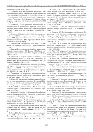
![-103-
Wschodnioeuropejskie Czasopismo Naukowe (East European Scientific Journal) | HISTORIA I ARCHEOLOGIA # IІІ, 2015
DER LEBENSWEG VON JOHANN HEINRICH KLEIN (1837 – 1922), DEKAN DER
MEDIZINISCHEN FAKULTAT DER KAISERLICHEN MOSKAUER UNIVERSITAT
Wassilij A. Loginow,
Prof., MD
M. V. Lomonossow Staatliche Universität Moskau,
Fakultät für Medizin
THE LIFE PATH OF JOHANN HEINRICH KLEIN (1837 – 1922) THE DEAN OF THE IMPERIAL MOSCOW UNIVERSITY
MEDICAL FACULTY
Vassili A. Loginov, Prof., MD Lomonosov Moscow State University Faculty of Basic Medicine
Summary
Johann Heinrich (Iwan Fjodorowitsch) Klein war ein hervorragender russischer Pathologe des 19. Jahrhunderts, Professor der
Kaiserlichen Moskauer Universität, Leiter des Lehrstuhl für pathologische Anatomie, spatter auch Dekan der Medizinischen Fakultät.
Nach dem Abschluss seiner Ausbildung in der Moskauer Universität und Verteidigung der Doktorarbeit studierte Klein zweimal
bei Rudolf Virchow und wurde zum Verbreiter der Ideen diesen grossen deutschen Gelehrten in der russischen Wissenschaft. Die
Neueinführungen Klein’s in der Moskauer Universität betrafen nicht nur den theoretischen Anteil (Zellularpathologie, Bakteriologie),
sondern auch den methodischen Ansatz (Verbreitung der Mikroskopie, neue Struktur des ganzen Lehrganges für pathologische
Anatomie).DankBemühungenvonKleinwurdedasanatomischeMuseumdesLehrstuhlswesentlichbereichert.Seineaussergewönliche
persönliche Eigenschaften und sein Ansehen bei den Studenten ermöglichten es ihm, einen Ausweg aus der kritischen Situation mit
den Studentenunruhen der Jahren 1869-1870 zu finden. Klein spielte auch eine wesentliche Rolle in der Entwicklung der neuen,
unter der Leitung von Nikolai Sklifossowski gegründeten Klinischen Stätte der Universität. Für seine Verdienste erhielt Klein mehrere
Staatsorden und es wurde ihm der Adel verliehen. Doch im Privatleben des Professors gab es schwierige Problemen: Geistkrankheit
seiner ersten Frau, plötzlicher Tod seines Sohnes, dann auch fortschreitende Krankheit von Iwan Fjodorowitsch selbst. Im Jahre
1906 legte Klein sich nieder. Mit der schwierigen Zeit der russischen Geschichte im Hintergrund verbrachte Professor Klein den Rest
seines Lebens in Not und Vergessenheit, starb in 1922 im Dörflein Gridnevo nicht weit von Moskau, der Ort seiner Beerdigung ist
heutzutage nicht genau bekannt.
ABSTRACT
Johann Heinrich (Ivan Fedorovich) Klein was a prominent Russian pathologist of the 19th century, Professor of the Imperial
Moscow University, head of Department of pathological anatomy and later Dean of the medical faculty. Having graduated from the
Moscow University and defended his doctoral dissertation, I. F. Klein double-trained Rudolf Virchow and was a conductor of ideas of
the great German scientist in Russian science. Klein innovations at the Moscow University affected not only the theoretical part (cellular
pathology, bacteriology) and methodological approach (widespread microscopy, the new structure teaching of the course pathological
anatomy). Thanks to the efforts of I. F. Klein significantly enriched the anatomical Museum of the Department. Outstanding personal
qualities of Klein and his reputation among his students helped him find a way out of critical situations associated with student riots,
1869-1870. For his services F. I. Klein was awarded with the state awards, received a knighthood. However, in the Professor’s personal
life there were serious problems: mental illness of his first wife, the unexpected death of a young son, and then a progressive disease. In
1906 Klein resigns. Against a difficult period in Russian history, the rest of his life Professor Klein spent in poverty and oblivion, died
in 1922 in the village Gridnevo, near Moscow, the location of the grave is currently unknown.
Keywords: Geschichte, Medizin, pathologische Anatomie, das neunzehnte Jahrhundert, Kaiserliche Moskauer Universität,
Medizinische Fakultät.
Key words: history, medicine, pathological anatomy, nineteenth century, Russia, Imperial Moscow University, Faculty of Medicine.
Problemstellung
”Jeder Arzt, der die Medizin-Fakultät derMoskauer
Universität am Ende des vorigen oder am Anfang dieses
Jahrhunderts abgeschlossen hat, erinnert sich gut an einen
schlanken hageren alten Mann mit Brillen: ständig gutherzig
und gescheit lächelnd, im Beamtenanzug, aber ohne Arztkittel
und Handschuhe zeigte er vor Studenten krankhaft veränderte
Organe. Das war Iwan Fjodorowitsch Klein, nach Polunin der
zweite Professor für pathologische Anatomie.” So schrieb im
Jahre 1949 Alexej Iwanovitsch Abrikosov vom Professor Klein
[1].
Ziel
Als Professor und Leiter des Lehrstuhls für pathologische
Anatomie, hat Iwan Fjodorowitsch Klein sehr viel getan. So
mögen wir diesen Menschen, der so viel für die Entwickelung
der pathologischen Anatomie in Russland geleistet hat, etwas
näher kennenlernen.
Grundlegende](https://image.slidesharecdn.com/eesj33-160202073457/85/Eesj-3-3-103-320.jpg)
![-104-
Wschodnioeuropejskie Czasopismo Naukowe (East European Scientific Journal) | HISTORIA I ARCHEOLOGIA # IІІ, 2015
Abb. 1. Johann Heinrich Klein (letztes Drittel des 19. Jahrhunderts).
Klein Johannes Heinrich (in Russland bekannt als Klein
Iwan Fjodorowitsch, 1837–1922, Abb. 1), ein hervorragender
Pathologe, ein Schüler von dem grossen R. Virchow, der
Evangelisch-lutherischen Religion, wurde am 24. Mai 1837
in der Familie von einem Beamter Friedrich Wilhelm Klein
(Abb. 2) und Anna Augustine Marcks (kein Porträt erhalten
geblieben) in Moskau geboren.
Abb. 2. Friedrich Wilhelm Klein (erste Hälfte des 19. Jahrhunderts).
ImJahre1855schlossJohannHeinrichdenAusbildungskursus
im Ersten Moskauer Gymnasium erfolgreich ab und trat
in die Kaiserliche Moskauer Universität (KMU), Fakultät
für Medizin ein. Interessanterweise nahm er die russische
StaatsbürgerschaftnurimJahre1860an.WährenddesStudiums
zeigte er vortreffliche Ergenbnisse und erhielt sein Diplom mit
Auszeichnung, trotz des Mißerfolges mit der Prüfung für die
operative Chirurgie bei dem Professor Bassow. Diese letzte
musste Klein im Herbst wiederholen.
Nach dem Abschluss des kompletten medizinischen
Curriculums im Jahre 1861 mit dem Doktorgrade für Medizin
blieb Klein, nach dem Vostellungsbrief des Universitätsrat und
nach der Genehmigung des Volksaufklärungsminister, auf dem
Lehrstuhl für pathologische Anatomie und Physiologie, damit
er in den nächsten zwei Jahren an seiner Dissertation arbeiten
konnte.
Im Jahre 1863, nach der Verteidigung der Dissertation zum
Thema “Über Thrombose, Embolie und Ichoremie” wurde
Klein in seinem Doktorgrade bekräftigt.
Zwischen 1864 und 1866 wurde I. F. Kein zweimal “zu
wissenschaftlichenZwecken”nachBerlingesandt,woersichbei
R. Virchow im Felde der pathologischen Anatomie fortgebildet
hat. Gerade in diesen Reisen erlangte Iwan Fjodorowitsch
Fertigkeit in der Arbeit mit den mikroskopischen Geräten, für
Anwendung deren in der Ausbildung er dann ein Enthusiast
wurde bis Ende seiner Tagen. Zusammen mit der neuen
Richtung in der Lehre der pathologischen Anatomie, der
Zellularpathologie, und der neuen Methode der Untersuchung,
der Mikroskopie, brachte Dr. Klein nach Moskau auch die
ersten Kenntnisse in der Bakteriologie, die später bei seinen
Schülern weiterentwickelt wurden [3].
In Jahren 1864–69 war er als Prosektor tätig, im 1869
wurde außerordentlicher Professor, im 1876 ordentlicher
Professor und im 1889 – verdienter ordentlicher Professor
der medizinischen Fakultät der KMU. Iwan Fjodorowitsch
leitete den Lehrstuhl für pathologische Anatomie an der
medizinischen Fakultät der KMU von 1869 bis 1906.
Seine Arbeit auf dem Lehrstuhl fing Iwan Fjodorowitsch
mit einer völligen Reformierung des Lernprozesses an.
Die Neueinführungen betrafen nicht nur den Inhalt des
Lernprozesses, sondern auch den methodischen Anteil.
In der Tat, I. F. Kein erarbeitete und führte ein das neue
Lehrprogram, basiert auf der mikroskopischen Technik
und auf der R. Virchow’s Lehre über Zellularpathologie. Die
Unterrichtsprinzipien, die von Professor Klein in zweiter
Hälfte des 19. Jahrhunderts eingeführt wurden, ließen eine
bedeutende wissenschaftliche Schule zu schaffen, aus welcher
solche großen russischen Pathologen hervorgegangen](https://image.slidesharecdn.com/eesj33-160202073457/85/Eesj-3-3-104-320.jpg)
![-105-
Wschodnioeuropejskie Czasopismo Naukowe (East European Scientific Journal) | HISTORIA I ARCHEOLOGIA # IІІ, 2015
sind wie A. B. Focht, N. F. Melnikow-Raswedenkow, W. I.
Kedrowskij, W. D. Scherwinskij. Beinahe alle medizinische
Schulen unterrichten die pathologische Anatomie nach den
metodischen Entwickelungen von I. F. Klein [2].
Abb. 3. Medizinische Fakultät der KMU, 1904. Mikroskopie-Stunde.
In der Mitte des 19. Jahrhunderts wurde die mikroskopische
Technik an der medizinischen Fakultät der KMU nur
durch zwei Mikroskopen vorgestellt, die ausschließlich für
Forschungszwecke benutzt wurden. Dem Enthusiasm und
der Energie von Dr. Klein is zu danken, daß in der zweiten
Hälfte des 19. Jahrhunderts ein Mikroskopie-Unterrichtsraum
in der KMU geöffnet wurde (Abb. 3). Um die technischen
Neuigkeiten im Lehrprozess zu verankern, musste Klein die
methodische Grundlagen des Unterrichts radikal revidieren.
Die Arbeit wurde in folgender Weise eingerichtet: es wurde den
Studenten des dritten Jahrganges ein Kursus der systematischen
Pathologie vorgetragen, am fünften Studiumjahr wurde
pathologische Histologie in Gruppen studiert, außerdem
ein praktischer Vorführungskursus eingeführt wurde, wo
die frischen krankveränderten Organe vor dem Auditorium
betrachtet wurden.
Nach der Reorganisation der Unterrichtsmethode konnte
Iwan Fjodorovitsch den Universitätsrat überzeugen, daß das
anschauliches Mikromaterial für den Fachkursus wichtig ist
und das Vorzeigen der Mikropräparaten, “mikroskopischen”
Bildern,TabellenundfrischenMakropräparatenvonBedeutung
ist.DasMuseumaufdemLehrstuhlfürpathologischeAnatomie
wurde mit neuem Lehrmaterial und anatomischen Imitationen
wesentlich bereichert.
In der ersten Zeit führte I. F. Klein selber alle drei Teile der
Kursus der pathologischen Anatomie. Später, in den achtziger
Jahren des 19. Jahrhunderts verteilte er den Unterricht so,
daß er selbst nur den praktischen Vorführungskursus leitete;
den systematischen Kursus leitete W. D. Schervinskij und seit
1894 – M. N. Nikiforow, der auch die pathologische Histologie
unterrichtete. Lange Zeit führte Iwan Fjodorowitsch in den
Sommermonaten Privatkursen der klinischen Mikroskopie für
die Ärzte [1].
Es ist zu erwähnen, daß in den Jahren, als I. F. Klein seine
Arbeit began, der Lehrstuhl für pathologische Anatomie in
einem alten Seitengebäude des Neuen Katharinakrankenhauses
untergebracht wurde. Der ganze Raum wurde in drei kleine
Zimmer aufgeteilt. Das erste und größte diente zugleich als
Sektions-undUnterrichtsraum,dasandere–alsArbeitszimmer
für den Professor und gleichzeitig als Anatomiemuseum, und
das dritte war zuerst eine Kapelle, später aber ein Zimmer für
den Prosektor. Nach einem treffenden Ausdruck von N. F.
Melnikow-Raswedenkow konnte dieses ganzes Häuschen “in
einem eizigen Auditorium des vom Professor Klein anfangs 90-
er Jahre gegründeten Instituts gut sein Platz finden” [2,3].
Die Errungenschaften des jungen Dozenten wurden bei der
Leitung der KMU nich außer acht gelassen, und im 1870 wurde
I. F. Klein vom Universitätsrat zum Posten des Sekretärs der
medizinischen Fakultät für die Dauer von drei Jahren erwählt.
Abb. 4. Alexej Iwanowitsch Polunin (1820-1888).](https://image.slidesharecdn.com/eesj33-160202073457/85/Eesj-3-3-105-320.jpg)
![-106-
Wschodnioeuropejskie Czasopismo Naukowe (East European Scientific Journal) | HISTORIA I ARCHEOLOGIA # IІІ, 2015
So wurde Iwan Fjodorowitsch Klein die zweitwichtigste
Person an der Fakultät (der Dekan zu dieser Zeit war Alexej
Iwanowitsch Polunin, Abb. 4) und erhielt eine der “meist
ruhelosen Wirtschaften” in der KMU.
Man kann ohne Übertreibung sagen, daß die Situation, in der
sich die Moskauer Universität in sechziger Jahren befindete,
keineswegs eine ruhige war und die gesamtrussische Situation
wiederspiegelte [4 6]. Wie die ganze Universitat, so auch die
medizinische Fakultät geradezu “vibrierte”. Der Schuß des
ehemaligen Studenten aus Moskauer Universität Karakosow
auf den Zar Alexander II. löste einen entscheidenden Angriff
der Reaktionskräfte auf unter der Losung “Komplott aus der
Universitäten zu vertreiben” [5,7,8].
Der liberale Volksaufklärungsminister A. V. Golownin wurde
durch den Graf D. A. Tolstoj ersetzt, der benutzte jedes Mittel,
um eine “Ordnung” in die Universitäten herbeizubringen.
Das damals wirkende, im Jahre 1863 angenommene,
ziemlich liberale Universitätsstatut wurde durch zahlreiche
Ministeriumschreiben korrigiert, wodurch die letzten
Nachsichten einfach ausgerottet wurden. So wurde, zum
Beispiel, der Universitätsrat durch “Vorschriften” vom 26. Mai
1867 verpflichtet, die Polizei von “unzuverlässigen” Studenten
zu benachrichtigen [5,8].
Im Oktober 1869 wurde während eines Beitrags der Studenten
des 4. Jahres an der medizinischen Fakultät der Moskauer
Universität ein Skandal gemacht, der in der historischen
Literatur als “Polunin-Fall” bezeichnet wurde. Der Skandal trat
sehr schnell über den Rahmen der Univesitätsmedizin hinüber
und fand eine große Resonanz in der russischen Gesellschaft.
Es war nähmlich so, daß der Leiter des Lehrstuhls für die
innere Medizin, Professor Grigorij Antonowitsch Zacharjin,
eine lange Auslandsreise unternahm, anstatt dessen der Dekan
der Fakultät, Professor Alexej Iwanowitsch Polunin bestimmt
wurde, die Vorlesungen für die Studenten des 4. Jahres zu
halten.
Es ist bekannt, daß die Vorlesungen von Zacharjin und seine
klinischen Besprechungen einen unvergeßlichen Eindruck
machten auf die Studentenjugend, aber auch auf andere
Zuhörer. Die großen russischen Schriftsteller A. P. Tschechow
und L. N. Tolstoi wurden oftmals durch diesen Professor
begeistert und benutzten sogar einige Ausführungen von
Grigorij Antonovitsch in seinen Kunstwerken [9].
Die berühmten Vorlesungen Zacharjin’s wurden tatsächlich
ein Vorbild der ärztlichen Kunst, gewürdigt sogar auf der Ebene
der Auslandsregierungen dieser Zeit. Im Medizinmuseum der
Ersten Moskauer Staatlichen Medizinischen Universität (die
Kopie in dem Museum der LMSU) befindet sich eine risiege
blaue Vase aus dem ruhmreichen Juwelenporzellan, die von
der französischen Regierung der KMU geschenkt wurde für
die Vorlesungen des Professor Zacharjin.
Nun, die Studenten des 4. Jahren lehnten während ihrer
Versammlung ab, die Vorlesungen des Professor Polunin,
hervorragenden Pathologen, der aber nur oberflächlich die
klinische Praxis, die Kuration, Befragung und Untersuchung
der Patienten kannte, zu besuchen. Die Studenten verlangten
nur einen gleichwertigen Ersatz, zumindest einen Kliniker
durch einen anderen Kliniker.
Die Universitätsrat folgte aber den Vorschriften des
Volksauflärungsministeriums und nahm eine strenge Haltung
ein; zwanzig Studenten wurden ausgeschlossen, neun davon
– ohne Recht, an anderen Universitäten zu studieren. Am
Anfang 1870 wurden bei Medizinstudenten Haussuchungen
durchgeführt, einige wurden verhaftet, die anderen wurden
aus Moskau ausgewiesen. Alles das löste die Protesten von den
Studenten aus. Es wurde das Geld gesammelt zugunsten der
Verfolgten. Studentenschaft war “in Wallung”.
In solch einer unruhigen Atmosphäre wurde im Februar 1870
der junge (33 J.a.) Porfessor Klein zum Posten des Sekretärs der
medizinischen Fakultät erwählt.
DieAutoritätdesFakultätendekanswasinderStudentenschaft
in dieser Zeit natürlich äußerst gering, denn gerade
Professor Polunin im Zentrum des Skandals war. Die ganze
Friedensregelung dieser komplizierten Situation musste also
der Sekretär unternehmen.
I. F. Klein nutzte seinen natürlichen Charme, den deutschen
Ansatz zu Diskussionen und eine diplomatische Flexibilität
größtmöglich aus, und es gelangte ihm, die Situation sehr
schnell zu normalisieren. Schon zu Beginn des neuen (nach
der Unruhen) Studiumjahres hatte die Studentenschaft keine
neue Ansprüche, die radikalen Stimmungen vergingen, und im
Jahre 1873 verstummte die politische Resonanz des “Polunin-
Falls” völlig [7].
Diese Erfolge in der Stabilisation dieser Situation konnten
natürllich nicht außer acht gelassen werden von der Regierung
des Russischen Kaiserreichs und der Verwaltung der KMU.
Iwan Fjodorowitsch Kein wurde zum Staatsrat und wurde
als Sekretär der medizinischen Fakultät für die nächsten drei
Jahre, dann aber noch bis 1879 bestätigt. Als er doch das Amt
des Dekans der medizinischen Fakultät am 18. Februar 1878
antrat, verlaß er den Sekretärendienst.
Die ertste Dekanenzeit von I. F. Klein dauerte nicht lange. Im
April 1880 gab er die Leitung der führenden lehrmedizinischen
Anstalt Russlands an Professor Nikolai Alekseewitsch Tolskij
weiter.
Laut seinem Personalzeugnis verlaß Professor Klein die
Verwaltungsfunktion nach eigenem Willen. Diejenigen, die
Klein gekannt hatten, erinnerten sich alle an ihn als einen
MenschendergroßenSeele,deraußerordentlichverständnisvoll
und aufmerksam gegen den Bedarf von Lehrern und Schülern
war.
Was brachte diesen erfolgreichen Organisator der
medizinischen Wissenschaft, begabten Pädagoge,
hervorragenden Redner, bis dahin auch den Träger der drei
russischen Orden (Hl. Stanislaw 2. Grades, Hl. Anna 2. Grades
und Hl. Wladimir 3. Grades) dazu, den Staatsdienst so eilig zu
verlassen? Den tatsächlichen Grund konnte man leider nicht
herauskriegen. Vielleicht gab es dafür einen persönlichen
Anlaß, wovon noch weiter die Rede ist.
So sind jedoch die historischen Tatsachen. Ab 13. September
1880 wurde Iwan Fjodorowitsch Klein laut Befehl des
Innenministers als Prosektor im Alten Katharinenkrankenhaus
für die ungelernter Arbeiter bestimmt, “mit der Fortsetzung
des Dienstes an der Universität”, das heißt, auf dem Lehrstuhl
für pathologische Anatomie.
Es sei zu bemerken, daß Iwan Fjodorowitsch Klein, wenn auch
mit den Organisations- und Erziehungsfragen beschäftigt, mit
seiner Forschungsarbeit nie aufhörte. Als er der Sekretär der
Fakultät war, war er auch Prosektor im Erziehungshaus (1869](https://image.slidesharecdn.com/eesj33-160202073457/85/Eesj-3-3-106-320.jpg)
![-107-
Wschodnioeuropejskie Czasopismo Naukowe (East European Scientific Journal) | HISTORIA I ARCHEOLOGIA # IІІ, 2015
1878) und führte grosse praktische Arbeit.
Die meisten seiner wissenschaftlichen Veröffentlichungen
besreiben Kasuistiken aus der pathologischen Anatomie,
und solche Aufsätze wie “Über Nierenzysten”, “Pelvis
spondylolisthetica” und “Zu der pathologischen Anatomie
der Kopfwassersucht” behielten lange ihren diagnostischen
Wert. So laut dem Zeugnis von W. I. Kedrowskij, “alle seine
Diagnosen waren kategorisch kompetene” [10, 11].
Im 1882 leitete Professor Klein einen Zirkel für pathologische
Anatomie, der auf die Initiative von N. F. Melnikow-
Raswedenkow gestiftet wurde; späterhin wurde dieser Zirkel zu
der Grundlage der Moskauer Wissenschaftlichen Gesellschaft
für pathologische Anatomie.
Es gibt Meinungen, daß Iwan Fjodorowitsch zusammen mit
Professor Sjornow an der Balsamierung des Leibes von Zar
Alexander III. teilgenommen hat, dokumentarisch ist aber dies
bis jetzt noch nicht bestätigt worden [6].
Iwan Fjodorowitsch machte sich auch Sorgen über die
Zukunft seines Faches. In seinem Aufsatz “Die Vergangenheit
und die Zukunft der pathologischen Anatomie in der Medizin”
(1886) versuchte er, die Wege der Wissenschaftsentwickelung
zu besinnen und zusammenzufassen. In derselben Zeit war er
als Mitglied und Vorsitzender in den Prüfungskommissionen
der Universitäten in Moskau, Charkow und Kasan.
An der medizinischen Falultät der KMU passierten
inzwischen wesentliche Veränderungen. Nikolai Wassiljewitsch
Sklifossowski wurde im Jahre 1882 Dekan der Fakultät. Er
stellte sofort die Frage über eine Erweiterung der klinischen
Stationen. Der Universitätsrat stimmte der Idee einer neuen
klinischen Stätte zu.
Dank seiner Berühmtheit nicht nur in wissenschaftlichen
Kreisen gelang es dem großen Chirurgen und Wissenschaftler
Nikolai Wassiljewitsch, vom Zar empfangen zu werden, und
es wurde der KMU ein Grundstück auf der Gelände namens
Dewitschje Pole zugeteilt, um darauf die neuen Kliniken der
Moskauer Universität zu errichten. Es wurden viele bekannte
Mäzene und Spender herangezogen. Beträchtliche Summen
kamen zu. Es musste jemand diese Summen verwalten. Man
brauchte einen tatkräftigen Organisator, der imstande war, eine
ganze Ansammlung von Problemen zu bewältigen. Eingedenk
der hohen moralischen Eigenschaften von Iwan Fjodorowitsch
Klein, bekräftigte der Universitätsrat ihn zum Posten des
Dekans der medizinischen Fakultät das zweite mal (Jahr 1888).
Gerade während dieser zweiten Dekanzeit, die bis zum Jahre
1906 dauerte, gewann der klinische Gebäudenkomplex auf dem
Dewitschje Pole die Bedeutung einer führenden klinischen
Stätte des Russischen Kaiserreiches. Selbst Rudolf Virchow, der
die Kliniken der KMU im 1896 besuchte, äußerte sich entzückt:
“Lernen Sie bei Russen!”.
Abb. 5. Institut für pathologische Anatomie auf Dewitschje Pole, ca. 1900 J.
Nach der Initiative von I. F. Klein wurde im Jahre 1891 das
Gebäude des Instituts für pathologische Anatomie auf dem
Dewitschje Pole (Abb. 5) errichtet. Professor Klein leitete
das Institut fünfzehn Jahre lang [10]. Er besaß eine große
Autorität und hielt vortreffliche Vorlesungen. Auf derselben
Gelände Dewitschje Pole, unweit des Krankenhauses für
Hautkrankheiten, bewohnte Iwan Fjodorowitsch ein einzelnes
Haus für lange Zeit (Abb. 6a,b).
Abb. 6a. Die Titelseite des Passports. von Johann Heinrich (Iwan Fjodorowitsch) Klein.](https://image.slidesharecdn.com/eesj33-160202073457/85/Eesj-3-3-107-320.jpg)
![-110-
Wschodnioeuropejskie Czasopismo Naukowe (East European Scientific Journal) | HISTORIA I ARCHEOLOGIA # IІІ, 2015
ФОРМУВАННЯ“НОВОЇ ЛЮДИНИ”ЯК ЗАСІБ ОДЕРЖАВЛЕННЯ ТРАДИЦІЙНОГО СУ-
СПІЛЬСТВА В УКРАЇНІ (1920-ТІ – ПОЧАТОК 1930-Х РР.)
Світлана Маркова,
Хмельницький обласний інститут післядипломної
педагогічної освіти, доцент кафедри теорії та методик
соціально-гуманітарних дисциплін, кандидат історичних наук
FORMATION OF A “NEW MAN” AS A MEANS OF NATIONALIZATION OF SOCIETY INTO THE SOVIET UNION, THE
USSR (1920S – EARLY 1930S)
Svitlana Markova, Candidate of Historical Sciences Khmelnytskyi Regional Institute of Postgraduate Pedagogical Education, Uk
raine
АнотацІя
У статті розглядаються окремі аспекти одержавлення традиційного суспільства в Україні, наслідком чого стала по-
ява “нової людини” позбавленої сумніву в тому, що лише керівна політична сила може бути виразником суспільного блага.
Робиться висновок про те, що становлення комуністичної системи призвело до відриву України від європейської моделі
політичного й економічного розвитку, законсервувало процес появи громадянського суспільства на довгі роки.
ABSTRACT
The article deals with some aspects of nationalization of society in the Soviet Union, the USSR, the consequence is the emergence of
a “new man” devoid of doubt that only the leading political force could be the mouthpiece of the public welfare. During the communist
“education” and “re-education” of people two periods are distinguishing: the period of the NEP (mostly theoretical finding of the basic
principles of a “new man” forming, the content of the ideology, its function in society, methods to influence the masses of people);
second period - end of 1920s - the beginning of the 1930s was characterized by massive use of force impacts. The conclusion is that
the establishment of the communist system led to the separation of Ukraine from the European model of political and economic
development, made an end of the emergence of civil society for many years.
Ключові слова: “нова людина”, “перевиховання”, ідеологія, селяни
Keywords:“new man”, educational system, referential groups, peasants
У розрізі процесу декомунізації в сучасній Україні варто
поглиблено вивчити суспільні зміни, що відбулися внаслі-
док одержавлення соціально-політичного та соціально-е-
кономічного життя вітчизняного соціуму, формування
“нової людини”, котра б пов’язала своє життя зі служін-
ням комуністичній системі, приносячи в жертву права
людини, інтереси місцевих громад. Нові історичні рекон-
струкції радянської доби у світлі загальноєвропейського
досвіду осмислення “тоталітарного минулого” дозволять
краще зрозуміти пролонговані наслідки комуністичного
режиму, що призвів до відриву України від європейської
моделі політичного й економічного розвитку, зумовив су-
часні труднощі соціально-економічного та політичного
розвитку країни.
Мета дослідження полягає у тому, щоб на основі аналі-
зу конкретно-історичного матеріалу висвітлити окремі
аспекти процесу одержавлення традиційного суспільства
в Україні.
Завдання – виокремити внутрішньосистемні чинники,
що мали найбільший вплив на формування “нової люди-
ни” в СРСР, УСРР і сприяли розширенню соціальної бази
радянської влади на селі.
З приходом до влади більшовики приступили до реаліза-
ції завдань щодо побудови нового суспільства та творення
“нової людини”. Стратегія реалізації доктрини формуван-
ня “нової людини” відповідно до заяви В. Леніна на І Все-
російському з’їзді рад народного господарства у березні
1918 р. передбачала “нову організацію найглибших основ
життя сотень мільйонів людей” [1, с. 137]. М. Бухарін вва-
жав, що змінити “найглибші основи життя” можна через
“пролетарське примушування в усіх формах від розстрілів
до трудової повинності” [2, с. 35]. На думку більшовиць-
ких ідеологів, соціалізм мав сформувати інший – вищий
– різновид людини [3, с. 136]. Такий різновид Л. Троцький
ідентифікував як “надлюдину” [4, с. 399]. Ідеї створення
надлюдини більшовики черпали з марксистських джерел,
природничих наук, зокрема вчення Ч. Дарвіна (дарвінізм в
СРСР вважався матеріалістичною теорією [5, с. 163–164]),
вчення фізіолога І. Павлова та ін. У 1923 р. було розробле-
но “партійний алгоритм виховання активістів [6, арк. 84].
У 1924 р. Й. Сталін у промові з приводу смерті В. Леніна
задекларував, що комуністи є людьми особливого скла-
ду і скроєні з особливого матеріалу [7, с. 47]. Відповідно
від “нових людей” очікували, що вони насамперед мають
бути відданими цілям партії, ототожнювати себе з новою
пролетарською громадськістю та забути своє минуле. Лю-
дей, які потребували “перевиховання”, бо мали, зокрема,
так звану “дефективну поведінку” (приватну (особисту)
форму поведінки) [8, с. 19], зневажливо ідентифікували
“людішками” [9, с. 19].
До створення “нової людини” у 1920-х рр. влада залучила
науковців. На ІІ Всесоюзному психоневрологічному з’їзді
(1924 р.) розглядали питання вивчення структури особи-
стості та умов її цільової перебудови [10, с. 58]. У 1927 р.
відбулась І Всесоюзна психотехнічна конференція, після
якої активізувався процес створення психотехнічних ла-
бораторій, організованих при установах Наркомздоров’я,
Наркомпраці, Наркомшляхів та ін.
Схему процесу формування нового індивіда також за-
пропонувала діалектично-матеріалістична педологія. У
1927 р. на І Всесоюзній педологічній нараді та І Всеросійсь-
кому з’їзді педологів активно обговорювали вплив довкіл-
ля на організм, який росте. Одеським лікувально-педоло-
гічним кабінетом були розроблені картки, що допомагали](https://image.slidesharecdn.com/eesj33-160202073457/85/Eesj-3-3-110-320.jpg)
![-111-
Wschodnioeuropejskie Czasopismo Naukowe (East European Scientific Journal) | HISTORIA I ARCHEOLOGIA # IІІ, 2015
визначати рівень дитячої дефективності серед мешканців
дитбудинків [11, арк. 12–14].
До конструювання “нової людини” мала долучитися й
радянська педагогіка та визначати шляхи, якими молоде
покоління “зробить стрибок до царства свободи”. Нова
марксистсько-ленінська теорія виховання визнавалася
базовою. Педагогіка була покликана допомогти обґрун-
тувати участь дітей у соціалістичному будівництві, со-
ціалістичному змаганні, ватажкуванні та ін. Основним
постулатом стало формування класової спрямованості
поведінки дітей. У першій половині 1920-х рр., зокрема з
подачі педагогів в УСРР набула поширення ідея повного
відриву дітей від сім’ї за допомогою розгалуженої системи
дитячих будинків (там мали перебувати не лише діти-си-
роти, безпритульні, але й діти матерів-одиначок, діти
батьків, зайнятих на великих будовах, сільські діти). Щодо
сільських дітей, то їх переважно ідентифікували як “диких
селянських дітей” та намагалися перетворити їх на “дітей
колективу” [12, с. 18–19].
Теорію гармонійного (вільного) виховання дітей вважа-
ли “шкідництвом на педагогічному фронті” та “ідеалом
буржуазії” [13, с. 41]. У другій половині 1920-х рр. панівне
становище отримала теорія “свідомої дисципліни”, внаслі-
док застосування якої мали з’явитися діти, яким більшо-
вики приписували риси пролетаріату (витривалі, наполег-
ливі, рішучі) [14, с. 180–181].
Певним підсумком пошуків шляхів формування “нової
людини” став I Всесоюзний з’їзд у справах вивчення люд-
ської поведінки (1930 р.), неофіційно названий “з’їздом
соціалістичної реконструкції людини”. На з’їзді було за-
декларовано, що нове соціалістичне суспільство потребує
нових людей; створення нової людини зі старого людсь-
кого матеріалу оголошувалось неможливим; соціалістична
реконструкція людини ставала пріоритетним завданням
для усіх наукових шкіл і напрямків [15, с. 97]. Це завдання
мали розв’язувати такі наукові дисципліни: нейрофізіоло-
гія, рефлексологія, психотехніка, педологія, психоневропа-
тологія та ін.
Вивчення поведінки “нової людини” в колективі та по-
ведінки людських колективів стали ключовими темами
для обговорення на усіх рівнях. Проблемами колективоз-
навства почала займатися соціальна психологія, об’єктом
вивчення якої був колектив (форми колективів, форми
громадської поведінки, психологія громадських інсти-
туцій). З подачі партійно-урядових структур колектив
ставав певним культовим утворенням держави, основою
стандартизації радянських людей.
У другій половині 1920-х рр. владі були потрібні нові
пропагандистські підходи, щоб і надалі нещадно експлуа-
тувати людей. Д. Бранденберген вважає, що витоки нового
підходу до пропаганди було закладено у 1928 р. [16, с. 3].
На думку Є. Добренка, цей процес розпочався роком рані-
ше, в 1927 р., коли М. Горький почав кампанію боротьби
“за демонстрацію досягнень та героїзацію дійсності” [17, с.
91]. Влада переглядала норми, нав’язуючи результати пра-
ці ударників та ударниць.
Впливаючи на громадську думку, активно використо-
вували пресу (інститут сількорів), усну пропаганду, ми-
стецтво плакату, силові методи впливу. За допомогою
технічних засобів (преса, радіо, кінематограф) стало мож-
ливим швидке поширення агітації та пропаганди. Провід-
ним засобом масової інформації, що активно долучився
до створення “нової людини”, була преса. Вже починаючи
з кінця 1917 р. питанням агітації та пропаганди більшо-
вицьких ідей в Україні надавали пріоритетного значення
[18, арк. 16]. Офіційні джерела повідомляли, що у 1925 р.
загальний тираж газет в УСРР перевищив 1 млн. [19, с. 4].
Газети та журнали мали давати читачам загальноприйняті
партією лозунги і положення, закликали сількорів надси-
лати дописи лише про роботу секцій сільрад, роботу де-
путатських груп, інші форми втягнення мас у роботу рад,
застосування соціалістичних методів праці, перебіг хлі-
бозаготівель, мобілізацію коштів, застосування госпроз-
рахунку, ліквідацію неписьменності, але не рекомендував
надсилати дописи, у яких були прохання людей про допо-
могу. У 1928 р. за пердплату журналу “Селянка України”
додатково надсилали “портрет В. Леніна у фарбах” [20, с.
26]. ЦК ЛКСМУ в голодному 1933 р. виступив з ініціати-
вою випуску праць: Леніна, Сталіна, Кагановича, Косіора
та ін. [21, арк. 70].
Вже наприкінці 1920-х рр. країну охопила атмосфера
тотальної підозрілості та страху. Владою контролювали-
ся друковані видання, видовища, листування тощо. Для
тотального контролю процесу “виховання” та “перевихо-
вання” представники влади з самого початку своєї діяль-
ності залучила цензурні підрозділи та апарат політкон-
тролю ВЧК–ДПУ. До середини 1920-х рр. організаційно та
функціонально політичний контроль у СРСР, УСРР набув
тотального характеру, він мав централізовану структу-
ру, керівництво якою здійснював відділ політконтролю
таємно-політичного управління ОДПУ СРСР. Внутрішня
пошта, афіші, листівки, циркулярні листи, плакати пере-
глядалися на 100% [22, с. 76–77]. У 1928 р. було посилено
багаторівневий контроль над пресою, остаточно усунено
можливість видання альтернативних партійній пресі газет
і журналів.
Засуджений у справі “Весна” (1930–1931 рр.) С. Четве-
риков у листі до М. Хрущова (1963 р.) писав про те, що в
Україні кошмарним був не лише 1931 р., але й вже 1927 р.
Ні в чому не винні люди обвинувачувалися за формулою
“так треба” [23, арк. 74–75]. Переважна більшість вигада-
них “антирадянських підпільних організацій” та “справ”
насправді були штучними витворами апарату ДПУ. У 1926
р. активно шукали махновців, зокрема анархо-махновців.
У 1927–1933 р. мали бути розкритими справи: “Прибу-
лець”, “Сок-Мовісти”, “Визволенці”, “Невгамовні”, “Кияни”,
“Козаки”, “Шакали”, “Вибух”, “Зв’язківці”, “Інтелігенти”,
“Батько”, “Махновці” та ін. У 1930-х рр. було сфабриковано
одну з найгучніших справ “УВО”. В’язниці, табори, колонії,
будинки ув’язнених на початку 1930-х рр. були вщент за-
повнені арештованими. Люди боялися доносів один на
одного, в’язниць, допитів, знущань. А. Мултановський
(засуджений у 1931 р. спочатку до розстрілу, після пере-
гляду справи – до 10 р. концтаборів) писав, що його до-
питували за системою безперервності. Свідчити проти
себе він почав після того, як провів 11 діб без сну (його
поливали холодною водою та засліплювали світлом), до-
поки не почалися галюцинації [24, арк. 141–142]. Чотири](https://image.slidesharecdn.com/eesj33-160202073457/85/Eesj-3-3-111-320.jpg)
![-112-
Wschodnioeuropejskie Czasopismo Naukowe (East European Scientific Journal) | HISTORIA I ARCHEOLOGIA # IІІ, 2015
доби безперервно допитували селянина Павленка, в якого
була гангрена, щодо наявності зброї [25, арк. 181]. Складо-
вими елементами української контрреволюції визначали
“куркулів” та інтелігенцію. Проникнення каральних ін-
ституцій в усі суспільні сфери, налагодження системи то-
тального контролю, збирання інформації дозволило владі
залякати суспільство. За неповними даними протягом
1933 р. відповідно до постанов “трійок” при колегії ДПУ
УСРР було розстріляно понад 800 осіб [26, арк. 143]. Жит-
тя людей поступово знецінювалося.
Українські селяни відразу важко вписувалися в концепти
“диктатури пролетаріату”. На думку більшості партійних
лідерів, саме селяни гальмували процеси конструювання
“нової людини” (в силу домінування “сімейного індивіду-
алізму”, прагнення захистити свою віру, землю тощо). М.
Бухарін запропонував “переробку та перевиховання се-
лянства”, зокрема з метою “змішування його з пролетаріа-
том” [27, с. 67–69].
Українським селянам за невиконання наказів та розпо-
ряджень більшовицької влади загрожувала відправка в
концентраційні табори, суди, арешти, трибунали, взяття
в заручники, конфіскація майна [28, арк. 51]. Специфікою
роботи більшовиків у селах у другій половині 1920-х рр.,
насамперед стало загострення соціальних суперечностей,
зокрема протиставлення інтересів одних прошарків ін-
шим, виокремлення групи менш заможних із негативним
ставленням до більш заможних селян; залякування; цьку-
вання; ідеологічне клішування; навішування назв-ярликів;
експлуатація людських слабкощів. У найгіршій ситуації
на селі опинилися селяни-господарі (“куркулі”, “глитаї”),
створені для того, щоб через їх знищення залякати інших,
зокрема з метою отримання хліба для потреб держави. На
місцях представники влади часто “проявляли ініціативу”,
вдавалися до погроз, прямого насильства. Сільським ко-
муністам-активістам навіть виносили догани за “непере-
виховання своїх дружин, котрі не відмовлялися від релігії”
[29, арк. 67], або подяки – за відвідування старих обрядів
(весілля, хрестини) та влаштування бешкетів на них.
Селянам нав’язували модель поведінки, при якій вони
мали з раннього ранку знаходитися на роботі, пізнього
вечора – в клубі [30, с. 17]. На спільних артільних землях
мали влаштовувати спільні садиби. Потреба в індивіду-
альних житлових і господарських будівлях мала відпасти
[31, с. 11]. Колгоспна земля проголошувалася “недоторка-
ною”. Ставлення більшості пересічних громадян до влади
та її економічної політики наприкінці 1920-х рр. різко по-
гіршилося: люди називали колгоспи “будинками, де утри-
мують смертників, які чекають свого кінця” [32, арк. 202];
колгоспний рух – “снігом, який випав на голову” [33, с. VІ];
колективізацію – “сухотами” (першою стадією хвороби
називали тсози, другою – артілі, третьою, після якої насту-
пає смерть, – комуни [34, арк. 134].
Більшовики намагалася “перевиховати” селян, насампе-
ред, руйнуючи їхні традиційні моральні підвалини. Після
жовтневого перевороту вони втрутилися в процеси су-
спільної еволюції, зокрема ставлячи мету відірвати люди-
ну від її “коріння”. Основою нового одержавленого суспіль-
ства мав стати не громадянин, а держава. “Нова людина”
формувалася як нова “громадянська ідентичність”, що
мала прислуговувати державі. Специфікою роботи біль-
шовиків із сільським населенням було протиставлення ін-
тересів одних соціальних прошарків іншим, залякування,
цькування, фактично застосування радянською владою
елементів соціального інжинірингу.
Окремі успіхи у піднесенні народного господарства,
освіти, розвитку суспільно-політичної активності грома-
дян перепліталися зі сталінськими кампаніями розкурку-
лення, колективізації, хлібозаготівельними, Голодомором
1932–1933 рр., тотальним контролем “народної ініціати-
ви”, грубими порушеннями законності. Проте ідеологічне
виховання робило не всіх саме такими, як хотіли “вихова-
телі”. Частина вчилася пристосовуватися до нових реалій,
інша повірила в “історичне завдання”, яке на них покла-
далося, частина людей загинула, намагаючись відстоювати
власне право на існування. Головною особливістю комуні-
стичного режиму стало не лише одержавлення приватного
життя, але й повний контроль держави над громадянами,
який спирався на систематичне використання насильства.
Примітки:
1. Кульчицький С. Червоний виклик. Історія комунізму в
Україні від його народження до загибелі / С. Кульчицький.
– К. : Темпора, 2013. – Кн. 1. – 2013. – 504 с.
2. Відносини держави, суспільства і особи під час ство-
рення радянського ладу в Україні (1917–1938 рр.). Колек-
тивна монографія / Відповідальний ред. В. Смолій. – Т. 1.
– К. : Ін-т історії України НАН України, 2013. – 750 с.
3. Конквест Р. Роздуми над сплюндрованим сторіччям /
Роберт Конквест [пер. з англ. О. Мокровольський]. – К. :
Вид-во Соломії Павличко “Основи”, 2003. – 371 с.
4. Троцкий Л. Д. Литература и революция / Л. Троцкий;
[Вступ. ст. Ю. Борева, с. 3–20]. – М. : Политиздат, 1991. –
399 с.
5. Мицкевич М. Дарвинизм и классовая борьба / М. Миц-
кевич // Народный учитель. – 1932. – № 5–6 (апрель). – С.
163–166.
6. Центральний державний архів громадських об’єднань
України (далі ЦДАГО), ф. 1, оп. 20, спр. 1784, 124 арк.
7. Сталін Й. З приводу смерті Леніна. Промова на ІІ Все-
союзному з’їзді Рад 26 січня 1924 р. // Сталін Й. Повн. зібр.
тв. – К. : Укрполітвидав, 1948. – Т. 6. – 1948. – С. 47–52.
8. Соколянський І. Про поведінку особистості / І. Соко-
лянський // Радянська освіта / За ред. О. Шумського. – Ч. 4
(квітень). – Х. : Державне вид-во України, 1925. – С. 7–20.
9. Вайсберг Я. С. Некоторые вопросы культурничества /
Я. С. Вайсберг // Комсомолец агитатор. Орган ЦК и Харь-
ковского окружкома ЛКСМУ. – 1928. – № 6 (март). – C.
19–25.
10. Зільберфарб Й. До соціалістичної реконструкції
людини (I Всесоюзний з’їзд у справах вивчення людсь-
кої поведінки) / Й. Зільберфарб // Поведінка людини та
соціалістичне будівництво. Всесоюзний з’їзд у справах
вивчення людської поведінки. Зб. упорядкувала редакція
“Шлях освіти”. – Харків–Київ : Держвидав України, 1930.
– С. 5–61.
11. Державний архів Одеської області, ф. Р. 233, Оп. 1,
Спр. 15, 146 арк.
12. Перший Всеукраїнський З’їзд представників колго-](https://image.slidesharecdn.com/eesj33-160202073457/85/Eesj-3-3-112-320.jpg)
![-113-
Wschodnioeuropejskie Czasopismo Naukowe (East European Scientific Journal) | HISTORIA I ARCHEOLOGIA # IІІ, 2015
спів та робітників по колективізації сільського господар-
ства. – Х. : Вид-во “Радянський селянин”, 1925. – 38 с.
13. Чавдаров С. Шкідництво на педагогічному фронті
(“Педагоги” з СВУ) / С. Чавдаров // Комуністична освіта. –
Х. : Державне видавництво, 1931. – № 5–6 (108), (травень–
червень). – С. 40–48.
14. Плевко Н. О сознательной дисциплине школьников /
Н. Плевко // Народный учитель. – 1932. – № 5–6 (апрель).
– С. 179–182.
15. Резолюції пленуму, секцій та нарад Всесоюзного з’їзду
в питанні вивчення людської поведінки. Резолюція З’їзду
по пленарному циклу “Психоневрологічні науки” й со-
ціалістичне будівництво // Поведінка людини та соціалі-
стичне будівництво. Всесоюзний з’їзді у справах вивчен-
ня людської поведінки. Збірник упорядкувала редакція
“Шлях освіти”. – Харків-Київ : Держ. вид-во України, 1930.
– С. 96–98.
16. Бранденбергер Дэвид. Сталинский популизм и не-
вольное создание русской национальной идентичности
[Електронний ресурс] / Дэвид Бранденбергер // Неприкос-
новенный запас. – 2011. – № 4 (78). – С. 3. – Режим доступу
до журн. : http://magazines.russ.ru/nz/2011/4/br4-pr.html.
17. Добренко Е. Политэкономия соцреализма / Е. До-
бренко. – М. : Новое литературное обозрение, 2007. – 592 с.
18. ЦДАГО, ф. 17, оп. 1, спр. 79 (б), 16 арк.
19. Центральний державний архів вищих органів влади
та управління України (далі ЦДАВО), ф. 3470, оп. 1, спр.
111, 218 арк.
20. Селянка України. – 1928. – № 4 (27 лютого). – С. 26.
21. ЦДАГО, ф. 7, оп. 1, спр. 105, 210 арк.
22. Подкур Р. Документы органов государственной безо-
пасности УСРР 1920 – 1930-х годов: источниковедческий
анализ / Подкур Р., Ченцов В. – Тернополь, 2010. – 372 с.
23. Державний архів Служби Безпеки України (далі ДА
СБУ), ф. П. 6, спр. 67093, Т. 37, 138 арк.
24. ДА СБУ, ф. П. 6, спр. 67093, Т. 36, 250 арк.
25. ДА СБУ, ф. 9, спр. 666, 242 арк.
26. ДА СБУ, ф. 68, спр. 228, 198 арк.
27. Бухарин Н. И. Путь к социализму и рабоче-крестьян-
ский союз / Н. И. Бухарин. – [Издание четвертое]. – Мо-
сква-Ленинград : Гос. издание, 1927. – 108 с.
28. Державний архів Хмельницької області (далі ДАХ-
мО), ф. 2508, оп. 1., спр. 4, 152 арк.
29. ДАХмО, ф. П. 31, оп. 1, спр. 35, 86 арк.
30. Перший Всеукраїнський З’їзд представників колго-
спів та робітників по колективізації сільського господар-
ства. – 38 с.
31. Клязник О. І. Юридична природа колективних госпо-
дарств / О. І. Клязник. – Полтава : “Полтава-Поліграф”,
1928. – 42 с.
32. Российский Государственный архив экономики, ф.
7486, оп. 37, д. 102, 312 л.
33. Кривицкий М. К вопросу об этапах нашей экономики
/ М. Кривицкий // Проблемы экономики. – № 3. – М. : Изд-
во Коммунистической академии, 1930. – 132 с. – С. ІІІ–ІХ.
34. Российский Государственный архив экономики, ф.
7486, оп. 37, д. 61, 137 л.
ПРИЧИНЫ И ОПЫТ ПОДЧИНЕНИЯ АДМИНИСТРАТИВНО-ТЕРРИТОРИАЛЬНЫХ
ЕДИНИЦ НА ПРАВОБЕРЕЖНОЙ УКРАИНЕ РОССИЙСКОМУ ВОЕННОМУ
КОМАНДОВАНИЮ В ПЕРВОЙ ПОЛОВИНЕ XIX в.
Скрипник Анатолий Юрьевич
кандидат исторических наук,
доцент, докторант кафедры истории
Украины Каменец-Подольского
национального университета
имени Ивана Огиенко
REASONS AND EXPERIENCE OF SUBORDINATION OF ADMINISTRATIVE-TERRITORIAL UNITS TO THE RUSSIAN
MILITARY COMMAND IN THE RIGHT-BANK UKRAINE IN THE FIRST HALF OF THE 19th CENTURY
Skrypnyk A. Yu. Candidate Sciences in History, Associate Professor, Doctoral Student of the Department of History of Ukraine
Kamianets-Podilsky Ivan Ohienko National University
Аннотация
В процессе последовательного и объективного изучения современными украинскими историками многогранной по-
литики российского самодержавия на Правобережной Украине в ХIХ в. среди актуальных направлений продолжает
оставаться исследование истории создания и последующего функционирования различных административно-терри-
ториальных образований в качестве основных государственных институтов для укрепления имперского присутствия
в регионе.
ABSTRACT
In the process of consistent and objective study of multifaceted policy of the Russian autocracy in the Right-Bank Ukraine in the
19th century by modern Ukrainian historians, one of the priority areas continues to be the study of the history of the creation and
subsequent functioning of different administrative-territorial units as the main state institutions strengthening the Imperial presence
in the region.
Ключевые слова: Российская империя, регулярная армия, администрация, кадровая политика, губернаторы, законода-
тельство, местные органы власти, население.
Key words: Russian Empire, regular army, administration, staff policy, governors, legislation, local authorities, population.](https://image.slidesharecdn.com/eesj33-160202073457/85/Eesj-3-3-113-320.jpg)
![-114-
Wschodnioeuropejskie Czasopismo Naukowe (East European Scientific Journal) | HISTORIA I ARCHEOLOGIA # IІІ, 2015
Постановка проблемы. Опираясь на источники из госу-
дарственных архивов Украины и Российской Федерации,
есть возможность изучить и проанализировать причины
временных реорганизаций губернских управленческих
структур и подчинения их военному командованию в ка-
честве тыловых учреждений, источников и баз снабжения
продовольствием и фуражом частей регулярной армии.
Нуждаются в исследовании факты попыток создания
исключительно военно-территориальных образований.
Придерживаясь хронологического порядка и методоло-
гии, есть возможность представить эволюцию законода-
тельной базы, которая создавалась с целью быстрой и эф-
фективной передачи властных полномочий чиновниками
гражданского ведомства военным.
Анализ последних исследований и публикаций. Совре-
менными украинскими историками длительное время
активно исследуется функционирование административ-
но-управленческой системы Российской империи на зем-
лях Правобережной Украины в течение конца XVIII – на-
чала ХХ ст. Удалось достичь не только немалых успехов в
этом направлении современной исторической науки, но
и создать достаточно сильную научную школу. Среди ее
представителей следует отметить комплексные и глубокие
исследования Н. Бармака [1], А. Реєнта [2], В. Шандры [3],
А. Филинюка [4], А. Скрипника [5], Н. Мацко [6] и других,
работы которых стали частичным или полным логическим
завершением изучений конкретных направлений и науч-
ных вопросов в этой исследовательской проблематике.
Однако, широкие хронологические рамки, многовектор-
ность хозяйственной и административной деятельности
российского гражданского и военного бюрократического
аппарата в течение ста двадцати лет господства и доста-
точный источнико-информативный массив государствен-
ных архивов открывает горизонт дальнейших научных
изысканий, в том числе и в направлении военно-админи-
стративного строительства.
Выделение нерешенных ранее частей общей проблемы.
Мотиватором научных поисков относительно проблемы
существования и деятельности военизированных управ-
ленческих структур стало определение их роли и места в
истории края, и в то же время – как одного из элементов
развития и углубления практических основ региональ-
ной истории Украины XIX в., связанных с деятельностью
государственных учреждений, действовавших на ее тер-
ритории. Несомненно, существует возможность исполь-
зования полученных результатов, собранного и обрабо-
танного материала в лекционных и семинарских курсах по
исследуемой проблематике, спецкурсах, в научно-исследо-
вательской работе студентов, краеведческой работе.
Цель статьи. Выявление причин и мотиваций существо-
вания временных управленческих структур, исполняю-
щих необходимые функции по охране и защите имперских
устоев от внешней и внутренней агрессий, укрепления
российского государственного строя на Правобережной
Украине в течение 1805-1832 гг., логическим завершением
которых стало создание в 1832 году Киевского генерал-гу-
бернаторства – как универсальной формации с ярко выра-
женным милитаристическим стилем управления.
Введение. Сразу же после получения Российской импе-
рией в 1793 году части аннексированной территории Речи
Посполитой, самодержавие делегировало все властные
полномочия вновь созданным территориальным управ-
ленческим структурам с ярко выраженным военным со-
держанием. В качестве уполномоченных представителей
новой власти на места назначались военные губернаторы,
в деятельности которых приоритетным было, прежде все-
го, решение вопросов по обеспечению всем необходимым
подразделений регулярных войск, оперативное руковод-
ство ими в мирное время и инспекторские функции. При
необходимости предполагалось ситуативное подчинение
всех гражданских наместнических, губернских и уезд-
ных учреждений и чиновников всех уровней во главе с
гражданскими губернаторами военному командованию
с целью оперативного решения неотложных вопросов,
связанных с фактом пребывания частей регулярной ар-
мии Российской империи в крае. Как правило, это были
хозяйственные вопросы, которые должны решать сугубо
местные учреждения, предоставляя военнослужащим все
необходимое для полноценного функционирования ми-
литаристского механизма. В этом и заключались причины
сложного эволюционирования административно-бюро-
кратических отношений, в которых одна сторона – воен-
ное командование – требовала и брала все, что предус-
матривалось государственными законами, правилами и
уставами, а другая – органы местного губернского управ-
ления – часто демонстрировали почти полную беспомощ-
ность, невозможность выполнения по причине слабой
кадровой организации, а иногда отсутствием подобного
хозяйственного опыта, ограниченных материальных ре-
сурсов, городской инфраструктуры и природных условий.
Изложение основного материала. В ходе нарастания
дипломатического и вооруженного противостояния с
наполеоновской Францией в начале XIX в. у российского
имперского руководства окончательно сформировалось
осознание стратегического значения, а, следовательно,
конкретизации и частичной корректировки содержания
управления недавно аннексированными землями. Они
на то время формировались как административно-тер-
риториальные единицы на Правобережной Украине, и в
случае возможного начала боевых действий на их терри-
тории рассматривалась возможность быстрого перевода
на военное положение и мобилизация всего социально-э-
кономического потенциала. Первым шагом стало распре-
деление административных полномочий и ответственно-
сти. В частности, военные губернаторы освобождались от
выполнения сугубо гражданских функций через большую
загруженность широким кругом милитаристских вопро-
сов. Однако им разрешалось «...вмешиваться в дела для
государственной пользы, сохранения казенного интереса,
предоставления защиты оскорбленным, и наблюдением за
порядком в работе управленческого аппарата, когда они
сами сочтут это нужным» [7, л. 258]. В случае служебного
отсутствия по делам инспектирования войск по террито-
рии губерний официально, с ведома царя, все полномочия
временно передавались гражданскому губернатору [8, л.
1].
Все назначенные наместники, а впоследствии военные и
гражданские губернаторы априори были людьми сугубо](https://image.slidesharecdn.com/eesj33-160202073457/85/Eesj-3-3-114-320.jpg)
![-115-
Wschodnioeuropejskie Czasopismo Naukowe (East European Scientific Journal) | HISTORIA I ARCHEOLOGIA # IІІ, 2015
военного направления. Но каждый из них обладал опре-
деленным административным опытом. Согласно «Табели
о рангах» на эти должности назначались генералы трех
первых классов: генерал-фельдмаршалы, генералы родов
войск (кавалерии или инфантерии – А. С.) и генерал-лей-
тенанты. Воинское звание было как бы отражением тех
полномочий, которые они получали. То есть – почти пол-
ное соответствие и сбалансированность военного чина и
должности, чем выше чин – тем больше полномочий. В от-
личие от наместников, власть военного губернатора рас-
пространялась только на одну губернию, они командовали
только теми дивизиями, которые располагались в пределах
подчиненной им губернии, и, как правило, были выше по
рангу от гражданских губернаторов [9, л. 5]. Большинство
из них были профессиональными военными, настоящими
боевыми генералами, потому что тогда на государствен-
ном уровне почему-то считалось, что именно такие пред-
ставители армейской элиты пригодны для осуществления
административных функций на местах. Кроме того, цар-
ская власть часто поручала им выполнение различных
дополнительных поручений, считая назначенного «хозя-
ина губернии» способным за короткое время стать уни-
версальным бюрократом и специалистом. Распыленность
функций не освобождала их от выполнения должностных
обязанностей военных губернаторов.
В марте 1812 года царь Александр I своим указом иници-
ировал нормы и правила взаимоотношений и разделения
полномочий «...между Главным Командованием Армий и
местными Начальствами пограничных губерний». С мо-
мента объявления губернии на военном положении армия
узурпировала все рычаги власти. В вопросах, связанных
со снабжением войск, высшее чиновничество и полиция
должны были выполнять приказы армейских генералов;
главнокомандующие получили право отстранять от долж-
ности любого чиновника городской и земской полиции;
все чиновники и жители становились потенциальными
подсудимыми военных судов. Единственное, что остава-
лось в компетенции губернской власти, так это граждан-
ское судопроизводство и хозяйственные полномочия.
Гражданские губернаторы на основании личной ответ-
ственности стали послушными исполнителями приказов
военных, и в то же время толкачами и контролерами их
исполнения предводителями дворянства, городскими ду-
мами и ратушами [10, с. 228-229].
16 апреля 1812 года киевский военный губернатор Мило-
радович получил письмо от М. Барклая де Толли с таким
содержанием: «Сосредоточив в различных пунктах наши
военные силы для обороны империи, мы сочли нужным
вверенную Вам губернию присоединить к общей систе-
ме оборонных мероприятий». Он подтвердил силу указа
царя от 13 марта, согласно которому «...губерния подчиня-
лась Главнокомандующему Действующей армии и вошла
в состав 2-го военного округа 2-й Западной армии князя
Багратиона [11, лл. 1-3]. По приказу губернаторов в обя-
занности уездных маршалков, городничих и уездных ис-
правников входило формирование местного ополчения
для обороны Киева, поставки волов и повозок на нужды
армии с непосредственным подчинением киевскому Ма-
гистрату [12, лл. 1-1об].
С октября 1813 года обязанности военных губернаторов
включали периодические отправки царю и главнокоман-
дующим армий двух форм отчетов и рапортов с полной
информацией о ходе наборов рекрутов, закупке лошадей,
создание передвижных магазинов и вообще все финансо-
вые расходы на военные повинности по губернии. Таким
образом, самодержец пытался не только составить целост-
ную картину мобилизационного потенциала и материаль-
ных затрат на местах, оценить работу самих должностных
лиц по обеспечению армии в ее заграничном походе в
Европу, а и главнокомандующие видели, на какие ресур-
сы могут рассчитывать их полки ближайшее время [13, с.
646-647].
В случае необходимости, по приказу сверху, высшие во-
енные чины оперативно передавали свои властные полно-
мочия буквально из рук в руки. В середине мая 1812 года,
накануне вторжения французских войск в пределы Рос-
сийской империи, генерал П. Багратион с ведома военного
министра М. Барклая де Толли таким образом передавал
управление территориями, которые оставляла 2-я Запад-
ная армия, отправляясь на север из района Луцка в район
Волковыска, командующему 3-й Армией генералу О. Тор-
масову. Кроме чисто военных вопросов, как-то: послед-
ние распоряжения учреждениям Подольской губернии и
Тарнопольської области относительно снабжения войск,
роспись войск, вошедших в состав его армии и расположе-
ния казачьих полков на границе в качестве стражи, были
переданы последние приказы и предписания сугубо граж-
данского характера. Отдельно генерал-интендант переда-
вал сведения и отчеты о «...продовольственных запасах
на Волыни, в Киевской губернии, и о состоянии расход-
ных магазинов вообще» [14, л. 298]. Такая придирчивость
должна была обеспечить бесперебойность осуществления
исполнительной власти, и ни в коем случае не допустить
потери царскими наместниками рычагов руководства над
всеми сферами общественно-политической жизни в крае
перед лицом смертельной опасности.
Опасения в Петербурге не оказались напрасными. Уже в
июне 1813 года главнокомандующий армиями М. Барклай
де Толли по результатам расследований издал приказ о пе-
редаче суду виновных в неповиновении и других престу-
плениях полицейских чиновников Подольской губернии и
позволил губернатору графу К. Сен-При судить их воен-
ным судом [15, с. 575-576]. Причиной стало распростра-
нение ими панических настроений и слухов среди насе-
ления губернии, что привело к бегству всего губернского
начальства из Каменца-Подольского в августе 1812 года
и временной потери рычагов власти в губернии [16, арк.
67]. Приказ был распространен во всех западных губерни-
ях империи, а такие беспрецедентные меры должны были
укрепить общую дисциплину среди чиновничества, осо-
бенно уездного звена.
В 1818 году военный губернатор Каменца-Подольско-
го А. Бахметьев получил награду от царя Александра I за
создание «Проекта объединения Бессарабской области с
Подольской губернией...», и в то же время совершенство-
вания существующей военной инфраструктуры [17, с. 1].
Известно, что с 1816 года генерал-лейтенант, кроме основ-
ной должности, занимал пост полномочного наместника в](https://image.slidesharecdn.com/eesj33-160202073457/85/Eesj-3-3-115-320.jpg)
![-116-
Wschodnioeuropejskie Czasopismo Naukowe (East European Scientific Journal) | HISTORIA I ARCHEOLOGIA # IІІ, 2015
Бессарабии и отчитывался лично монарху. По его замыс-
лу в «Проекте» речь шла о строительстве юго-западного
тактического форпоста по левому берегу реки Днестр с
дальнейшей перспективой накопления здесь войск и воз-
можности внезапного и решительного наступления на Ва-
лахию и Придунайские княжества. Объединение же двух
соседних административно-территориальных единиц под
контролем военных властей должно стать надежной и це-
лостной территориальной конфигурацией в случае необ-
ходимости внедрения военизированной системы управле-
ния. Формально в тот период эта территория управлялась
согласно «Уставу создания Бессарабской области» Верхов-
ным Советом, частично выбранным и назначенным из
представителей местных элит, который выполнял сугубо
гражданские административные и судебные функции,
подчинялась А. Бахметьеву и периодически отчитывалась
перед Александром I [18, с. 3-4]. Показательным являет-
ся тот факт, что военные администраторы без колебаний
использовали материальные и финансовые ресурсы граж-
данских учреждений на подобные проекты и не обреме-
нялись за это никакой ответственностью и отчетностью.
Только через двадцать лет, случайно, при ревизии Вре-
менной ликвидационной комиссией выяснилось, что Бах-
метьев самовольно взял из бюджета Подольской губернии
50 000 рублей, и потратил их на нужды войск в Бессарабии
вместе с тогдашним командующим 2-й Армией генералом
Л. Беннигсеном. Правда, эти деньги они не разворовали, а
использовали по назначению, с сохранением финансовой
отчетной документации, которая в качестве доказатель-
ства была предоставлена Подольским губернским прав-
лением чиновникам комиссии на проверку [19, лл. 4-6].
Вполне возможно, что Бахметьев получил устное разреше-
ние императора, и эти действия не были зафиксированы в
документах из соображений секретности.
Уже через четыре года Александр I предпринял попыт-
ку создать первое постоянное военно-территориальное
образование на западных границах империи. В июне 1822
года его указом князю цесаревичу Константину Павло-
вичу, главнокомандующему Действующей армией, были
подчинены Виленская, Гродненская, Минская, Волынская,
Подольская губернии и Белостокская область со «...всеми
властными правами и преимуществами» [20, с. 309]. Зако-
нодательно утвержденное доминирование военной власти
над гражданской было первой попыткой унифицировать
управление на стратегически важной территории на про-
тяжении определенного временного срока. Это образо-
вание стало предтечей военных округов, появившихся в
России только в 1862 году в ходе реформ царя Александра
II. В то же время, пренебрежение устоявшимися террито-
риальными конфигурациями ряда пограничных губерний
и эксперименты по их оперативному объединению и под-
чинению вышестоящему военному начальству, а следова-
тельно, смешения военной и гражданской власти, могло
привести к хаосу в огромном государственном механизме
в мирное время. Зато такое сочетание оказалось весьма
эффективным противодействием тем вызовам, которые
получила империя в конце 20-х – в начале 30-х гг. ХIХ в.
Такая практика прошла проверку во время польского
восстания 1830-1831 гг., когда с его началом российским
властям стало ясно, что вооруженного противостояния не
избежать. Подобно ситуации 1812 года, перед руководите-
лями края была поставлена задача любой ценой не допу-
стить распространения выступлений на Правобережную
Украину. Николай I, через правительственный Сенат, в
начале декабря 1830 года объявил: «По теперешнему со-
стоянию дел в Царстве Польском, необходимо в соседних
губерниях Подольской и Волынской объединить Граждан-
ское управление с Военным, назначив генерал-адъютанта
Я. Потемкина временным Военным Губернатором» [21, с.
2]. Он осуществлял оперативные исполнительные функ-
ции и координировал работу уездных учреждений по
причине того, что через объявление почти сразу же воен-
ного положения эти губернии законодательно оказались
в непосредственном подчинении главнокомандующему
русской армией И. Дибича-Забалканского [22, с. 449-450].
Поэтому вопрос заключался только в уровне взаимоува-
жения и понимания между двумя генералами и осознания
степени ответственности перед монархом за судьбу губер-
ний, их социально-экономическое и политическое поло-
жение во время поисков и погони за отрядами повстанцев.
Именно в преддверии польского восстания, о чем сви-
детельствуют архивные материалы, появились первые
устремления и сознательное тяготение к сотрудничеству
и взаимодействию между местными органами власти и
военными перед лицом потенциальных вооруженных вы-
ступлений, способных свергнуть существующий строй.
В городе Киеве, не по разрешению, а именно по прика-
зу военного губернатора Б. Княжнина горожане, как и в
1812 году, вновь сформировали вооруженный мещанский
корпус для охраны города. Ремесленная управа выстави-
ла 1000 ремесленников из 16 цехов города [23, лл. 3-3об;
7-8]. Согласно отчетным данным Магистрата к губернато-
ру, корпус в начале 1832 года насчитывал 500 всадников
и более чем 2000 горожан «...с огнестрельным оружием,
городскими пушками и собственной городской музыкой»
[24, лл. 74-75]. Тем более, именно в это время из уездов до
губернских центров стали поступать тревожные рапор-
ты чиновников городских и уездных полиций. Так, Ки-
евское губернское правление информировало киевского
военного губернатора Б. Княжнина о «...брожениях сре-
ди владельцев имений и мелкой шляхты в Таращанском,
Липовецком, Махновском и Уманьском уездах». Правле-
ние просило сообщить в воинские части, чтобы те были
готовы к отражению антигосударственных выступлений
местного населения губернии. Однако, несмотря на на-
мерения и желания сторон, тотального и эффективного
взаимодействия между этими институтами не было нала-
жено. Во-первых, армия сразу же взяла на себя домини-
рующую функцию в борьбе с польским освободительным
движением, оставив на заднем плане местное начальство;
во-вторых, немалая часть местных чиновников-поляков
оказалась в рядах повстанцев, и поэтому часто не было
кому помочь армии. Правда, были отдельные случаи,
когда уездные чиновники, сохранившие верность пре-
столу, действовали совместно с военными в подавлении
выступлений. Так командир отряда российских войск,
«зачищавших» район Белой Церкви и соседние уезды,
генерал-майор кавалерии Гельд ходатайствовал перед гу-](https://image.slidesharecdn.com/eesj33-160202073457/85/Eesj-3-3-116-320.jpg)
![-117-
Wschodnioeuropejskie Czasopismo Naukowe (East European Scientific Journal) | HISTORIA I ARCHEOLOGIA # IІІ, 2015
бернатором Желтухиным о награждении «Васильковского
Земского исправника Титулярного Советника Кузьмина
за действенность и усердие в борьбе с бунтовщиками» [25,
лл. 7-13; 72-72 об].
После подавления польского патриотического сопротив-
ления логическим завершением практики ситуативного
доминирования военных в вопросах административно-
го управления во время военно-социальных конфликтов
стало введение указом Сената от 24 февраля 1832 года на
территории юго-западного края Киевского генерал-губер-
наторства в составе трех губерний. С самого начала сама
должность генерал-губернатора была наделена сугубо
милитаристским смыслом, а в штате его канцелярии «на-
чальникам края» разрешалось иметь военных чиновни-
ков. В течение следующих 1832-1865 лет все пять киевских
генерал-губернаторов: В. Левашов, В. Гурьев, Д. Бибиков,
И. Васильчиков и М. Аненков, находясь на вершине власт-
ной пирамиды, стали не только проводниками усиления
интеграционных процессов региона в состав империи,
но и главными координаторами в вопросах, связанных с
пребыванием армейских частей на подведомственных им
территориях. Под их контролем, в контексте целенаправ-
ленной политики всесторонней русификации, в их поле
зрения находилась организация и проведение рекрутских
наборов, привлечение отставных военных на судебные и
полицейские должности в уездах, привлечение польской
молодежи на службу в армии. В июле 1862 года, после
создания Киевского военного округа, полномочия руко-
водителей выросли, под их командование попадали все
воинские части на территории генерал-губернаторства.
Сосредоточенность власти в одних руках доказала свою
эффективность во время польского восстания 1862-1863
гг., когда армия активно использовалась для охраны гра-
ницы в Волынской и Подольской губерниях; произошло
создание чисто военных следственных комиссий и судов
[26, с. 271-295].
Выводы и предложения. Замена властных структур Речи
Посполитой и, как следствие, постепенное распростране-
ние российской государственности в крае с конца XVIII
в., обеспечивались исключительно военно-администра-
тивными мерами. Каждый из российских самодержцев
первой половины XIX в. с начала своего царствования пы-
тался улучшить местную управленческую систему Россий-
ской империи не только путем создания новых учрежде-
ний с более совершенной способностью их деятельности
при условии сохранения принципа политического абсо-
лютизма, а и возможности их быстрого приспособления
к внутренним чрезвычайным ситуациям или условиям
полномасштабной войны с внешним врагом. Введение об-
щероссийских форм местного государственного управле-
ния на новоприсоединенных территориях с самого начала
показало их частичное несовершенство и несоответствие
местным условиям. Потому, с целью повышения эффек-
тивности исполнительной судебной и полицейской вла-
сти, структуры местной администрации находились под
постоянным контролем наместников и военных губерна-
торов.
Попытки создания смешанных управленческих струк-
тур на некоторых территориях имели частичный или вре-
менный успех, связанный с выполнением определенных
заданий и функций в экстремальных условиях двух рос-
сийско-турецких войн 1806-1812 гг. и 1828-1829 гг., войны
с Наполеоном 1812 года и польского восстания 1830-1831
гг. В мирное время потребность в исключительно военном
стиле управления и содержании штабного управленческо-
го аппарата на этих территориях отпадала. Именно борьба
достоинств и недостатков милитаристского стиля управ-
ления с гражданским не давала российским самодержцам
до 1832 года окончательно избрать форму, наиболее эф-
фективно подходящую под социально-экономические и
политические реалии Правобережной Украины – одного
из наиболее важных стратегических районов империи.
Список литературы
1. Бармак М. Формування владних інституцій Російської
імперії на Правобережній Україні (кінець XVIII – перша
половина ХІХ ст.) : монографія / М. В. Бармак. – Тернопіль:
Астон, 2007. – 512 с.
2. Реєнт О. Деякі проблеми історії України ХІХ – початку
ХХ ст.: стан і перспективи наукової розробки / О. П. Реєнт
// Український історичний журнал. – 2000. – № 2. – С. 5–10.
Реєнт О. Україна ХІХ-ХХ століть / О. П. Реєнт // Роздуми
та студії історика / відп. ред. В. А. Смолій. - Корсунь-Шев-
ченківський: ФОП Майдаченко І. С., 2009. – 486 с.
3. Шандра В. Генерал-губернаторства в Україні: ХІХ – по-
чаток ХХ століття / В. С. Шандра – К. : НАН України. Ін-т
історії України, 2005. – 427 с.
4. Філінюк А. Правобережна Україна наприкінці XVIII
– на початку ХІХ століття : тенденції розвитку і соціаль-
ні трансформації : монографія / А. Г. Філінюк. – Кам’я-
нець-Подільський : Аксіома, 2010. – 728 с.
5. Скрипник А. Державні установи Подільської губер-
нії 1793-1914 рр. : монографія / А. Ю.Скрипник. – Кам’я-
нець-Подільський : ПП. Мошинський В.С., 2012. – 228 с.
6. Мацко Н. Військова політика Російської імперії на
Правобережній Україні (1792-1874 рр.):.дис. … к-та. .істор.
наук.: 07.00.01. / Н. В. Мацко. – Тернопіль, 2012. – 200 с.
7. Российский государственный военно-исторический
архив (РГВИА), ф. 26, оп. 3, д. 116, л. 258.
8. Центральний державний історичний архів України у м.
Києві (ЦДІАК України), ф. 533, оп. 1, спр. 1214, арк. 1.
9. Там же, КМФ 12, оп. 1, спр. 31, арк. 5.
10. Полный свод законов Российской империи (ПСЗ). –
Собр 1. – Т. ХХХІІ. – № 25035. – СПб., 1830 – 1107 с.
11. ЦДІАК України, ф. 533, оп. 1, спр. 1531, арк. 1-3.
12. Державний архів міста Києва (ДАК), ф. 1, оп. 2, спр.
190, арк 1-1зв.
13. ПСЗ. Собр 1. – Т. ХХХІІ. – № 25468. – СПб., 1830. –
1107 с.
14. РГВИА., ф. ВУА. Отечественная война 1812 года, д.
3505, л. 298.
15. ПСЗ. Собр. 1. – Т. ХХХІІ. – № 25397. – СПб., 1830. –
1107 с.
16. Державний архів Хмельницької області, ф. 69, оп. 1,
спр. 93, арк. 67.
17. Высочайший рескрипт // Русский инвалид. № 114. – 19
мая 1818 г. – С. 1.
18. Крупенский А. Полный список лиц, избраных бесса-](https://image.slidesharecdn.com/eesj33-160202073457/85/Eesj-3-3-117-320.jpg)
![-118-
Wschodnioeuropejskie Czasopismo Naukowe (East European Scientific Journal) | HISTORIA I ARCHEOLOGIA # IІІ, 2015
рабским дворянством на областные, губернские и уездные
должности со времени присоединения Бессарабии к Рос-
сийской империи по 1912 год. С историческим введением /
А. Н. Крупенский. – С.- Петербург. : 1912. – 173 с.
19. ЦДІАК України, ф. 442, оп. 71, спр. 381, арк. 4-6.
20. ПСЗ. Собр. 1. – Т. ХХХVІІІ. – № 29087; № 29088. –
СПб., 1830 – 1354 с.
21. Указ Правительственному Сенату. Внутрение изве-
стия // Русский инвалид. № 309. – 4 декабря 1830 г. – С. 2.
22. ПСЗ. Собр. 2. – Т. V. – Отд. 2. – № 4155. – СПб., 1831.
– 596 с.
23. ДАК, ф. 1, оп. 2, спр. 1626, арк. 3-3 зв; 7-8.
24. ЦДІАК України., ф. 442, оп. 1, спр. 474, арк. 74-75.
25. ЦДІАК України, ф. 533, оп. 2, спр. 806, арк. 7-13; 72-72
зв.
26. Шандра В. Генерал-губернаторства в Україні : ХІХ –
початок ХХ століття / В. С. Шандра. – К. : НАН України.
Ін-т історії України, 2005. – 427 с.
BRONZE BRACELETS – THE CASH EQUIVALENT OF THE BRONZE AGE
Gajar Chingiz Oveys
Doctor of Physical and Mathematical Sciences
The full member National Academy of Sciences of Azerbaijan
Soltanova Nazila Baghir
Doctor of philosophy
Associate Professor of the Institute o physics of ANAS
Karayeva Leyla Vahid
Graduate student
Institute o physics
National Academy of Sciences of Azerbaijan
Rahimov Nizami
Director of the State Museum of History of Nakhchivan AR
ABSTRACT
This article deals with issues on the weight and monetary system of the Bronze Age and the Iron Age in Azerbaijan. The basic
weight unit of that time called as the «Caucasian shekel» had its parallel functioning in the form of payment equivalents. The Bronze
bracelets merged on the basis of that system. They functioned as money and at the same time as the weight standard. The article
presents a new version of the system with respect to the weight of that time.
Key words: sickle, bronze, bracelets, numismatics, the weight and the monetary system.
Creations of past generations allow today to speak about
the achievements of modern science. The famous French
archaeologist Jacques Jean-Marie de Morgan (1857-1927)
writes: «It is necessary to know the secret of reading of the
ancient creations in order to understand correctly the meaning
and significance of the ancient creations and to judge about the
life of their creators [3, p.6].
Archaeological findings of scientists lead us to the extreme
antiquity. A great number of products performing the function
of money is known before the advent of coined money. The
ingots of precious metals (copper, bronze, silver, gold) were the
last products meeting the requirements of universal equivalent.
Metals are more suitable material regarding their physical
properties for playing the role of a universal equivalent with
their all characteristic features. Depending on the geographic
region, each nation had its own equivalent. Then more universal
functionary–coined money appeared.
The transformation of primitive barter into the public process
with the criteria of comparing the value of the products in the
territory of Azerbaijan started around the centuries XIII -XI BC
and accelerated nearly in the VIII - VII centuries BC They had
all conditions – geographic, sources of various raw materials,
water and land opportunities, the population density for this
purpose. [1, p.172]
In 1890 the French archaeologist Jacques de Morgan
conducted archaeological excavations in the south - eastern
region of Azerbaijan and discovered ancient burial places with
rich decoration. There were enough bronze bracelets among the
findings. Morgan noted that «the ingots in the form of bracelets
presented a special kind of banknote of certain weight in the
era of railway industry.» [3, p. 275]
The archaeological findings in the excavations on the territory
of Azerbaijan allow to speak about the cash equivalent of the
Bronze and Iron Ages - bronze bracelets functioning on this
territory. More than 70 similar bracelets were found on the
skeleton found in the burials in Mingechevir.
The main unit weight of bronze bracelets per Morgan was 8.2
grams which was called «Caucasian shekel.» [1, p. 152]
The Assyrian - Babylonian shekel (the shekel) is equal to 10-
11 grams. Shekel (shekel) is a Hebrew word (II thousand. BC.
E.), which translation means to weigh. Jewish shekel is about
9-17 grams. Shekel in the average weight equal to 8 grams, was
a quite popular financial equivalent of the international trade of
the XIX century. BC. [5, p. 22], [7, page 12]
Bracelets in question were made by certain weight standard.
After the analysis of the bracelets from the archaeological fund
MIA the well-known numismatist Ali Rajabli considers that the
initial weight norm for these bracelets is 10 gr. He identified
four groups disposed in the quarterly order - 1, 2, 4, 8 - with an
average weight, respectively: 10, 20, 40, 80 c. [7. 12]. Indeed, an
amazing weight system is obtained in which bronze bracelets
acting as means of payment and weight units merged in the
antiquity.
At the beginning, the metal presented itself extremely valuable
material, it was protected with great care. The last periods of
polishing stone of various nations are concern to the nascent](https://image.slidesharecdn.com/eesj33-160202073457/85/Eesj-3-3-118-320.jpg)
![-119-
Wschodnioeuropejskie Czasopismo Naukowe (East European Scientific Journal) | HISTORIA I ARCHEOLOGIA # IІІ, 2015
metallurgy. Use of copper is followed by obtaining bronze.
Bronze is an alloy of copper and tin. The ancient world was
rich in copper and tin was not so much. The alloy has more
hardness than the red copper-soft, lightweight metal. However,
copper can receive the hardness not only through alloy with
tin. Increase of small doses of arsenic, antimony or zinc also
changes the molecular composition of copper.
Already in ancient times the people tried to produce alloys.
An alloy giving necessary qualities to copper for weapons,
tools should contain tin in the ratio of 10 to 100. The change
of the given ration gives the brittle alloy. Because primitive
metallurgists of these times did not possess modern scientific
methods and could act only groping, the consecutive samples
with tin content in the bronze tools and things are very different.
While calculating the density of the bronze bracelets studied
by us we were faced with these inaccuracies. Nevertheless, the
desired tin content in the alloy for the ancient times ranged
from 10 to 18%.
Even the first metallurgists found deposits of copper and tin
in the virgin form; they seem to be in the form of oxidized ores
and it was enough to melt them on the restorative charcoal fire
in order to extract the metal. Tin was extracted by washing the
sand. [2, p. 10]
The question arises whether the bronze was prepared by
combining certain proportion of its two constituent elements
in metal form, or ores were mixed before smelting? The latter
assumption is more suited to the real facts, which can be
explained by the difference of tin content in the alloy.
Thanks to intensive improvement of bronze technology and
specialization of ancient Azerbaijan tribal and communities in
ore mining and metal working, the development of productive
forces was accelerated, foreign relations and trade barter
borders were significantly expanded. Trading began which
required universal equivalent.
There was their own copper in the Caucasus which was also
exported and tin was mainly imported.
Bronze bracelets, rings of different sizes and weights
encountered in burials at the end of the II- at the beginning of
the I millennium BC can be considered the first metal money
in Azerbaijan.
Besides these bracelets in question, no other items made
referring to some weight standard or weight system are known
yet. Therefore, we can’t classify a given weight rate to any weight
system. But the weight norm of the bronze bracelets is 8-11
oz., it is nearly identical to the youngest unit of the Assyrian
- Babylonian weighing system - shekels (shekel), which was
applied in the territory of Azerbaijan without the influence of
the developing commodity - money relations and trade ties
with the highly developed civilizations of the Near East
Numerous findings of gold and silver articles belonging to
the late Bronze Age and the beginning of Iron Age (XII - IX
centuries BC) found during archaeological excavations in
the territory of Azerbaijan allow to state that gold and silver
were well known in ancient Azerbaijan as noble metals. It
shall be noted that the share of shekel was considered mainly
for payments in precious metals, gold and silver which were
international means of payment of that time. [6, p. 46]
Our weighing of bronze bracelets kept in the State Museum
of History of Nakhchivanand found during excavations
Kizilburun, Garabaghlar and Julfa (Nakhchivan AR) and
bracelets found in Tovuz showed nearly above the marked unit
– 8 -10 grams.
Such question arises: why weight unit is exactly 8 - 10 grams?
Exploring these bronze bracelets we had an interesting
version concerning the weighing system (weighing units) by
which similar bracelets were made (merged). The main unit
weight of bronze bracelets, 8.2 g is the «Caucasian shekel,»
known as a weight unit of its time, it coincides with the density
of bronze in digital form. The density of tin bronze is ρ = 8,6
- 9,3 g / cm3 (90% Cu, 10% Su) and of phosphor bronze is ρ =
8,7 - 8,9 g / cm3 (92.5% Cu, 7% Su, 0.5% P). In all likelihood,
the metallurgists of that time possessed the information on the
density of bronze in some primitive form and the principal
weight unit of the trading system of that time - shekel was
determined on the basis of this value.
Fig 1. Bronze bracelets belonging to the beginning of the Iron Age. They were found in the archaeological excavations in
Garabaghlar (Nakhchivan AR).](https://image.slidesharecdn.com/eesj33-160202073457/85/Eesj-3-3-119-320.jpg)
![-120-
Wschodnioeuropejskie Czasopismo Naukowe (East European Scientific Journal) | HISTORIA I ARCHEOLOGIA # IІІ, 2015
Fig. 2. Bronze bracelets found in Tovuz [4, p. 9]
Nakhchivan Тovuz
M (g) 360 270 90 80 39 61 19 66 56
М (g) 10 8
Nakhchivan Тоvuz
m (g) 360 270 90 80 66 61 56 39 19
Ρ (g/m3) 6 7.7 10 10 8.2 8.7 8 5.5 4
M
( schekel)
45 33 11 10 8 7.5 7 4.7 2.3
Links:
1. Бабаев И.А. Города Кавказской Албании в IV в. до н.
э. – III в. н.э. Baku, Elm, 1990. p. 236.
2.ДревнийВостокиантичнаяцивилизация.St.Petersburg,
State Hermitage, 1988, p. 136.
3. Жак де Морган. Доисторическое человечество.
Moscow- Saint Petersburg, State publishing house, 1926, p. 315.
4. Qacar Ç.O. Mizandarlıq. Baku, East-West, 2010, p. 136.
5. Марков А.К. Древняя нумизматика. Part I. St. Petersburg,
A.P.Lopukhin’s publishing house, 1901, p. 222.
6. Пахомов Е.А. Клады Азербайджана и других республик
и краев Кавказа. Baku, AzFAN publishing house, 1938, p. 101.
7. Раджабли А. Нумизматика Азербайджана. Baku, Elm ve
hayat, 1997, p. 232.](https://image.slidesharecdn.com/eesj33-160202073457/85/Eesj-3-3-120-320.jpg)
![-121-
Wschodnioeuropejskie Czasopismo Naukowe (East European Scientific Journal) | SOCJOLOGIA # IІІ, 2015
SOCJOLOGIA - СОЦИОЛОГИЧЕСКИЕ НАУКИ
МЕХАНИЗМЫ РЕАЛИЗАЦИИ РЕГИОНАЛЬНОГО КЛАСТЕРА ПАТРИОТИЧЕСКОГО
ВОСПИТАНИЯ МОЛОДЕЖИ НА ПРИМЕРЕ Г. ЯКУТСКА
Kulakovsky G.P.
Candidate of economics
Scientific-Research Institute of Regional Economy of the North
North-Eastern Federal University
Alekseeva K.I.
Research worker
Scientific-Research Institute of Regional Economy of the North
North-Eastern Federal University
MECHANISMS OF REALIZATION OF A REGIONAL CLUSTER OF PATRIOTIC EDUCATION OF YOUTH ON THE
EXAMPLE OF YAKUTSK
Аннотация
Патриотическое воспитание способствует формированию социально активной личности, обладающей чувством
гражданского достоинства, высокого патриотического сознания, верности Отечеству, готовности к выполнению кон-
ституционных обязанностей по защите интересов Родины.
ABSTRACT
Patriotic education promotes formation of socially active personality having feeling of civil advantage, high patriotic consciousness,
fidelity to the Fatherland, readiness for performance of the constitutional obligations for protection of interests of the Homeland.
Ключевые слова: Патриотическое воспитание, социальный фактор, Северо-восток России
Key words: Patriotic education, social factor, Northeast of Russia
Становление гражданского общества и правового госу-
дарства в нашей стране во многом зависит от уровня граж-
данского образования и патриотического воспитания.
Большой вклад в формирование гражданской личности,
сочетающей в себе нравственную, правовую и политиче-
скую культуру должна внести пропаганда патриотизма в
нашей республике. Это многоплановая, целенаправленная
и скоординированная деятельность государственных ор-
ганов, общественных объединений и организаций по фор-
мированию у детей и молодежи высокого патриотическо-
го сознания, чувства верности своей Родине, готовности
к выполнению гражданского долга, важнейших консти-
туционных обязанностей по защите интересов общества.
Эта деятельность включает в себя социальные, целевые,
функциональные, организационные и другие аспекты,
обладает высоким уровнем комплексности, то есть охва-
тывает своим воздействием все поколения, пронизывает
все стороны жизни: социально-экономическую, полити-
ческую, духовную, правовую, педагогическую, опирается
на образование, культуру, историю, государство, этносы.
Поэтому в настоящее время гражданско-патриотическое
воспитание несовершеннолетних на примере ветеранов
Великой Отечественной войны, ветеранов боевых дей-
ствий, ветеранов тыла, является исключительно важной
частью развития подрастающего поколения.
Одним из перспективных направлений реализации мо-
дернизационного сценария развития региона является
стимулирование и поддержка процессов формирования
«инновационных центров развития» и воспроизводства
человеческого капитала. Эффективной формой такого
центра в регионе, нацеленного на интеграцию новых идей,
технологий и человеческого потенциала может стать ре-
гиональный кластер патриотического воспитания детей и
молодёжи при содействии ветеранов Великой Отечествен-
ной войны, ветеранов боевых действий и ветеранов тыла.
Формирование такой структуры - это процесс интеграции,
упорядочения и синхронизации функционирования разо-
бщенных субъектов образования, культуры, молодёжной
политики, общественных организаций.
Патриотическое воспитание предполагает формирова-
ние у молодого поколения общественно значимых ориен-
таций, гармоничного сочетания личных и общественных
интересов, преодоление чуждых обществу процессов и
явлений, разрушающих его устои и потенциал созидания.
Механизмы реализации предусматривают охват патрио-
тическим воспитанием социальных категорий несовер-
шеннолетних совместно с советом ветеранов войны и тру-
да.
На сегодня в Республике Саха (Якутия) количество ма-
лообеспеченных семей составляет 12200, многодетных
малообеспеченных семей 3529, приемных детей и детей,
находящихся под опекунством и попечительством 930,
детей сирот 362, детей, попавших в трудную жизненную
ситуацию 237.[1]](https://image.slidesharecdn.com/eesj33-160202073457/85/Eesj-3-3-121-320.jpg)
![-124-
Wschodnioeuropejskie Czasopismo Naukowe (East European Scientific Journal) | SOCJOLOGIA # IІІ, 2015
теранов в работу по патриотическому и нравственному
воспитанию населения, молодежи и усилению роли вете-
ранских организаций.
Реализация регионального кластера патриотического
воспитания будет способствовать достижению эффектив-
ных результатов в осуществлении социально-экономиче-
ский политики, что обеспечит выполнение мероприятий
патриотической направленности среди подрастающего
социально незащищенного поколения в рамках повыше-
ния качества жизни людей пожилого возраста и позволит
достичь следующих результатов:
- организация летнего культурно-досугового отдыха лю-
дей пожилого возраста совместно с летней занятостью со-
циально незащищенной молодежи несовершеннолетнего
возраста;
- обеспечение определенной материальной поддержки
ветеранов;
- реализация интеллектуальных и культурных потребно-
стей старшего поколения;
- увеличение доли молодых людей, вовлеченных в дея-
тельность общественных объединений гражданско-па-
триотической направленности;
- увеличение количества молодежи допризывного воз-
раста, принявшей участие в оборонно-спортивных меро-
приятиях и военно-полевых сборах;
- снижение доли молодых людей, негативно воспринима-
ющих воинскую обязанность и военную службу.
Список использованной литературы:
1. Государственная программа «Социальная поддержка
граждан в Республике Саха (Якутия) на 2012 - 2016 годы».
РЕГУЛИРОВАНИЕ КОНФЛИКТА МЕЖДУ СОЦИАЛЬНЫМ РАБОТНИКОМ И
ПОЛУЧАТЕЛЕМ СОЦИАЛЬНЫХ УСЛУГ
Маргарита Владимировна Вдовина,
Институт дополнительного профессионального
образования работников социальной сферы,
доктор социологических наук, профессор кафедры
теории и технологии социальной работы
THE REGULATION OF CONFLICT BETWEEN SOCIAL WORKER AND RECIPIENT OF SOCIAL SERVICES
Margarita Vladimirovna Vdovina, The Institute of additional professional education of social workers, Doctor of sociological
Sciences, Professor of the Department theory and technology of social work
Аннотация
В статье рассмотрены причины, способы профилактики и разрешения конфликта между социальным работником
и получателем социальных услуг. Уделено внимание личным и профессиональным качествам социального работника,
способствующим предупреждению конфликта. Указаны конструктивные и деструктивные способы вмешательства в
конфликтную ситуацию, которых, по возможности, следует избегать.
ABSTRACT
The article discusses the causes, ways of prevention and solution of conflict be-tween social worker and recipient of social services.
Attention is paid to the personal and professional qualities of a social worker, contributing to the pre-vention of conflict. Specified
constructive and destructive interventions in con-flict situation, which, if possible, should be avoided.
Ключевые слова: социальный работник, получатель социальных услуг, регулирование кон-фликта
Key words: social worker, recipient of social services, regulation of the conflict
Одной из актуальных проблем социальной работы яв-
ляется регули-рование конфликта между социальным ра-
ботником и получателем соци-альных услуг. Безусловно,
он не изолирован от иных типов конфликта (семейного,
трудового и др.), но в определенной мере именно этот тип
конфликта зачастую является первичным и наиболее зна-
чимым в органи-зации социального обслуживания. Поэ-
тому его предотвращение и разре-шение – достаточно се-
рьезная и сложная тема.
Обобщая различные теоретические подходы к изучению
конфликта, можно сказать, что конфликт – это особый вид
взаимодействия индивидов, социальных групп. Он пред-
ставляет собой столкновение разнонаправлен-ных сил и
противоборство двух и более противостоящих сторон.
Это про-тивоборство вызвано невозможностью одновре-
менного удовлетворения потребностей людей, предпола-
гает пересечение интересов, осознание ущемления своих
интересов и отношение к противоположной стороне как
противнику и «виновнику» проблемы [1, 22].
Конфликт вытекает из статусных, ролевых, ценностных,
норматив-ных и иных духовных и материальных разли-
чий индивидов, которые они считают существенным за-
труднением в общении. В процессе конфликта происходит
разрешение острого противоречия, основанного на несо-
вме-стимости интересов, целей, ожиданий, взглядов, дей-
ствий и т.п. Конфликт-ное взаимодействие людей связано
с негативными эмоциями, стрессами и т.д.[2, 41].
Как показывает практика, чаще всего причинами кон-
фликта соци-ального работника и получателя социальных
услуг являются:
• Особенности личности (вспыльчивость, агрессивность
и т.п.)
• Межличностная несовместимость работника и получа-
теля услуг
• Нарушение норм морали и права
• Низкий уровень профессионализма социального ра-
ботника
• Неудовлетворенность получателя качеством обслужи-](https://image.slidesharecdn.com/eesj33-160202073457/85/Eesj-3-3-124-320.jpg)
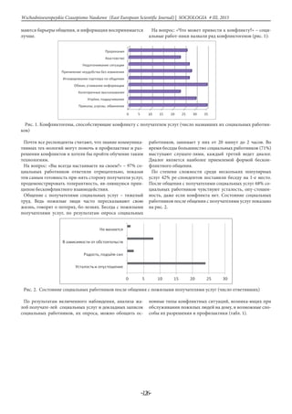
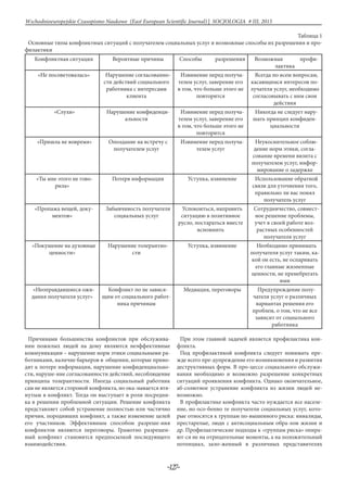
![-128-
Wschodnioeuropejskie Czasopismo Naukowe (East European Scientific Journal) | SOCJOLOGIA # IІІ, 2015
этих групп.
В профилактике конфликта можно выделить первичную,
вторичную и третичную стадии.
Первичная стадия профилактики представляет собой
предупрежде-ние еще не возникшей проблемы. Она со-
стоит в совершенствовании соци-альной политики, соци-
альной работы, социального обслуживания и т.д.
Вторичная стадия представляет собой предотвращение
деструктив-ных форм и негативных последствий уже воз-
никших конфликтных ситуа-ций на их ранних стадиях.
Третичная стадия профилактики нацелена на наиболее
полное и своевременное разрешение конкретных прояв-
лений конфликта для упре-ждения возникновения новых
конфликтов.
Формы и методы профилактических действий по регу-
лированию конфликта разнообразны. Они могут быть
нейтрализующими; компенси-рующими; предупреждаю-
щими возникновение неблагоприятных обстоя-тельств;
устраняющими эти обстоятельства; контролирующими
результа-ты проведенной профилактической работы.
В 2015 г. нами был проведен опрос 96 социальных ра-
ботников из различных административных округов г.
Москвы. Согласно опросу, для профилактики конфликта
с получателем социальных услуг они использу-ют следу-
ющие приемы:
- провести беседу для выявления потребностей получа-
теля и разъяс-нения возможностей поставщика социаль-
ных услуг (так поступают 30% респондентов);
- успокоить агрессивно настроенного человека, дать вы-
говориться, снять возбуждение (12% респондентов);
- грамотно и своевременно информировать получателя
социальных услуг, разъясняя ему что-либо в миролюби-
вом тоне (11%);
- делать так, как хочет получатель, приспособиться к
нему (9%);
- создать настрой на положительные эмоции, улыбаться,
делать уместные комплименты, приободрять получателя и
т.п. (8%);
- проявить терпение, сдержанность во взаимодействии с
пожилым человеком, чтобы не поддаваться на провокации
со стороны раздраженно-го клиента (7%);
- установить четкие правила взаимодействия с получате-
лем услуг, добросовестно соблюдать должностные обязан-
ности социального работ-ника (7%);
- молчаливо выслушать пожилого человека, не вступая с
ним в бес-плодные споры (7%);
- сменить неприятную тему, переключить внимание по-
лучателя на более приятные и полезные вещи (7%);
- в самом начале обслуживания изучить специфику по-
лучателя услуг и постоянно делать выводы на будущее,
взаимодействуя с ним (7%);
- проявлять к нуждающимся гражданам эмпатию, вни-
мание, уваже-ние, быть вежливым, пунктуальным, про-
фессиональным (6%);
- искренне извиниться, если социальный работник не
прав (2%).
Как видим, тактика предупреждения конфликта различ-
на: возможна как активная позиция социального работ-
ника, который изначально готов общаться с «трудным»
клиентом, так и пассивная, приспособительная. Большое
внимание уделяется профессионализму, правильной ком-
муника-ции с получателем услуг, профессиональной этике
и этикету взаимодей-ствия и т.п.
Однако предупреждение конфликта с получателем соци-
альных услуг не исчерпывается только усилиями социаль-
ного работника.
Опираясь на результаты эмпирических исследований,
необходимо отметить еще некоторые предупреждающие
конфликт факторы:
- улучшение бытовых условий жизни получателя соци-
альных услуг;
- работа по повышению толерантности и сотрудниче-
ства;
- расширение сети и совершенствование работы органи-
заций социаль-ного обслуживания, максимально прибли-
женных к месту жительства, по-вышение информирован-
ности населения о спектре и условиях оказания услуг;
- развитие инфраструктуры досуга и отдыха, создание
клубов по ин-тересам и др.;
- переподготовка и повышение квалификации специали-
стов, выпуск научно-методической, научно-популярной и
учебной литературы;
- консультирование населения по различным вопросам
(правовым, бытовым, психолого-педагогическим, мораль-
но-нравственным и др.) и др.
Однако конфликт не всегда удается избежать. В таком
случае необ-ходимы профессиональные усилия по его
своевременному конструктивно-му разрешению.
Прекращение конфликта может быть результатом не
только взаим-ного примирения сторон и достижения со-
гласия между ними, но и посте-пенного «затухания» про-
тивостояния без устранения причин, его поро-дивших,
перехода в латентное состояние или же перерастания его в
другой конфликт [3, 327].
Регулирование конфликта – это, как правило, приве-
дение конфликт-ных взаимоотношений сторон в русло
примирения путем переговоров, участия посредников
или иного целенаправленного вмешательства. Под регу-
лированием конфликта имеется в виду такое воздействие
на индиви-дов, которое наряду с разрешением возникшей
конфликтной ситуации мо-жет включать предупреждение
обострения противоречий. При этом в ре-альной прак-
тике возможно и неудачное регулирование напряженных
от-ношений, ведущее к эскалации конфликта.
Разрешение конфликта – один из благоприятных спосо-
бов его за-вершения. Разрешение представляет собой со-
вокупность позитивных дей-ствий в первую очередь самих
участников конфликта, прекращающих противоборство и
решающих проблему, которая привела к нему. Это тре-бу-
ет заинтересованности каждого из субъектов конфликта
в преодолении противостояния на взаимоприемлемой
основе; осознания противоречий, непосредственных при-
чин и сопутствующих им факторов конфликта; по-иск и
использование методов и норм регулирования отноше-
ний. Разреше-ние конфликта предполагает прежде всего
устранение или минимизацию его причин [3, 225-226].
Для профилактики и разрешения конфликта необходи-
мы:](https://image.slidesharecdn.com/eesj33-160202073457/85/Eesj-3-3-128-320.jpg)
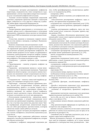
![-130-
Wschodnioeuropejskie Czasopismo Naukowe (East European Scientific Journal) | SOCJOLOGIA # IІІ, 2015
дующей отделением. Хотя ряд опрошенных подчеркнули,
что спешат сразу сообщить заведующей о конфликте (что-
бы опередить жалобу получателя). Практически все соци-
альные работники стараются действовать в конфликте по
ситуации, в зависимости от его глу-бины и особенностей
получателя социальных услуг.
Большое значение для предупреждения и разрешения
конфликта имеет его правильная диагностика. Для своев-
ременного и точного выявле-ния противоречий, интере-
сов и целей сторон целесообразно применять та-кие мето-
ды сбора информации, как индивидуальное и групповое
анкети-рование, беседа с получателем социальных услуг,
опрос экспертов (специ-алистов по социальной работе,
психологов и др.), включенное наблюде-ние, анализ доку-
ментов и др. Комплексное применение методов помогает
глубже понять специфику конфликтной ситуации, выя-
вить возможные причинно-следственные связи в пробле-
мах, ее породивших.
В реальной практике наряду с конструктивными меха-
низмами регу-ляции конфликта имеют место деструк-
тивные способы воздействия на кон-фликт, существенно
затрудняющие его разрешение. Они включают в себя пре-
сечение конфликта насильственным путем; подавление
несогласных участников; разрыв отношений. В результа-
те конфликт внешне завершает-ся, однако по существу
остается не разрешенным, переходит в латентную (иногда
даже более негативную, чем открытый конфликт) форму,
транс-формируется в другие конфликты. Неразрешенные
противоречия аккуму-лируются, их последующее прояв-
ление становится острее предыдущего, обладает множе-
ственными отрицательными последствиями.
Опрошенные социальные работники отметили различ-
ные формы де-структивного воздействия на конфликт,
бесперспективные для выхода из сложной ситуации: от-
крытая агрессия, вербальное насилие, необоснован-ные
жалобы, подавление конфликта, административные раз-
бирательства и т.п.
Деструктивное регулирование конфликта связано с
применением си-лы, и даже насилия в целях его прекра-
щения. Такое силовое воздействие может быть как леги-
тимным (вмешательство правоохранительных орга-нов,
ужесточение социального контроля, юридических санк-
ций за кон-фликтное поведение и т.п.), так и нелегитим-
ным (межличностное насилие в различных формах и т.д.).
В результате конфликт повторяется неодно-кратно, нарас-
тает, остается нерешенным до следующего обострения.
Та-кой вариант нередко возникает, если участники избега-
ют и подавляют конфликт, не стремясь к его предупрежде-
нию и разрешению.
У некоторых социальных работников и их получателей
услуг проти-воборство протекает бурно, с массой негатив-
ных эмоций, жалобами, вза-имными претензиями и оби-
дами. Это деструктивный стиль поведения в конфликтной
ситуации, бескомпромиссная тактика. Предпочтительнее,
ко-нечно, рациональное разрешение противоречий.
Уклонение от диалога с противоположной стороной, по-
давление, а не разрешение противоречий деструктивно
отражается на взаимодействии. При таком регулировании
чаще изменяется лишь внешняя форма отноше-ний, а вну-
тренние мотивы к продолжению противоборства сохра-
няются. Изменения в обществе, семье, в самих участниках
приводят к нарушению статус-кво, и конфликт возобнов-
ляется.
По этому поводу Р. Дарендорф отмечал, что избегание,
подавление конфликтов не может предпочитаться в тече-
ние продолжительного срока [4, 144, 145]. Посредством
регулирования конфликты не исчезают и не обязательно
сразу становятся менее интенсивными. Но они становятся
кон-тролируемыми и могут служить постепенному разви-
тию социальных структур. Однако успешное регулирова-
ние конфликтов, по Р. Дарендор-фу, предполагает ряд ус-
ловий:
1) нужно, чтобы конфликты признавались всеми участ-
никами как неизбежные, ибо тому, кто рассматривает их
как патологические отклоне-ния от нормы, не удастся со-
владать с ними. Покорного признания неиз-бежности кон-
фликта тоже недостаточно; скорее необходимо признать
их творческое начало;
2) конфликты следует направлять в необходимое русло,
устанавли-вать «правила игры», которые не отдают пред-
почтения одному участнику в ущерб другому. В рацио-
нальном «обуздании» социальных конфликтов заключает-
ся одна из центральных задач политики [4, 145-146, 147].
В социальной работе недопустимы деструктивные фор-
мы поведения. Необходимы профессиональные усилия,
направленные на достижение компромисса и согласия,
попытки сгладить противоречия. Непрофессио-нальны-
ми являются отрицательные эмоции, позиции давления на
противо-положную сторону. Согласно профессионально-
му кодексу этики, недопу-стимы оскорбления, необосно-
ванные обвинения и т.п. Профессионал дол-жен сохра-
нять определенное хладнокровие, даже если получатель
соци-альных услуг с трудом внимает его советам и пред-
ложениям, «выплески-вает» агрессию на него и на других
собеседников.
Брошенные и отвергнутые своими родственниками, не-
мощные пожи-лые люди и инвалиды часто пополняют
число получателей социальных услуг. При этом они несут
в себе конфликтогенный «заряд» – обиды, агрессию, не-
приязнь к более молодым и здоровым людям, стереотипы
грубости в разрешении противоречий и т.п. Все это может
деструктивно отразиться на взаимодействии с социаль-
ным работником. Среди социаль-ных работников тоже
иногда встречаются негативные стереотипы в отно-шении
получателей социальных услуг. Это противоречит прин-
ципу про-фессионализма.
Однако при определенных конфликтных ситуациях при-
менение насилия является эффективным методом оста-
новки агрессивного поведе-ния, например, по отношению
к инвалидам, пожилым людям и др. Имеется в виду не от-
крытое физическое действие, но различные виды социаль-
ного принуждения. Угроза наказания, опасность лишений
является одним из действенных аргументов в разрешении
конфронтации, когда одна из сто-рон заняла крайне агрес-
сивную позицию, использует насильственные средства,
преднамеренно отвергает конструктивное обсуждение и
решение возникшей проблемы.
При регулировании конфликта большое значение имеет](https://image.slidesharecdn.com/eesj33-160202073457/85/Eesj-3-3-130-320.jpg)
![-131-
Wschodnioeuropejskie Czasopismo Naukowe (East European Scientific Journal) | SOCJOLOGIA # IІІ, 2015
нормативно-правовая база. Нормативный механизм осо-
бенно важен при глубоких конфликтных ситуациях, свя-
занных с насильственными формами, и необ-ходим для
предупреждения деструктивных последствий.
Практика свидетельствует, что обращение в правоох-
ранительные органы с целью решения конфликта – как
правило, крайняя мера, глубоко травмирующая всех
участников противостояния. Нередко обращение к право-
вому механизму усугубляет конфликт либо переводит его
в скрытую форму. Тем не менее, обращение за помощью
закона часто бывает неиз-бежным из-за невозможности
решить конфликт силами его непосредствен-ных участни-
ков. Следует указать и на то, что подчас трудности в раз-
ре-шении конфликта вызваны именно незнанием законов,
низкой правовой культурой [5, 102-103].
Безусловно, одной только нормативно-правовой базы
для разреше-ния конфликта с получателем социальных ус-
луг недостаточно. Более того, применение исключительно
правовых норм и санкций – скорее деструк-тивная мера
регуляции конфликта; хорошие «человеческие» отноше-
ния предпочтительнее.
Таким образом, существуют разнообразные по содер-
жанию и форме механизмы, которые при системно-ком-
плексной реализации способствуют предупреждению и
конструктивному регулированию конфликта с получа-те-
лем социальных услуг. Наиболее конструктивны такие,
как ранняя про-филактика конфликта; разрешение тех
противоречий, которые лежат в ос-нове конфликтной си-
туации; управление конфликтным взаимодействием; ин-
ституционализация регулирования конфликта.
В результате исследования было выявлено, что боль-
шинство соци-альных работников имели конфликты с
пожилыми людьми при обслужи-вании, однако при этом
не считают себя виноватыми. Большинство соци-альных
работников испытывают трудности в общении и хотели
бы пройти обучение. Доброжелательность и стрессоустой-
чивость как личные качества в профилактике конфликтов
отметило наибольшее число социальных ра-ботников. Тем
не менее, существует проблема со стрессоустойчивостью
социальных работников. Почти 70% работников чувству-
ют усталость и опустошение после общения с получателя-
ми услуг.
Основные виды конфликтов в социальном обслужива-
нии на дому – это межличностные конфликты между со-
циальным работником и получа-телем социальных услуг.
Эти конфликты можно предотвратить, если со-блюдать
нормы этики, преодолевать барьеры общения, учитывая
возраст клиентов, не нарушать принцип взаимозависи-
мости в решениях и действи-ях, проявлять терпимость,
вежливость, не отвечать агрессией на агрессию и т.п. Все
это приведет к сохранению у получателя социальных ус-
луг чув-ства собственного достоинства, удовлетворенно-
сти. Для этого необходимо, чтобы социальные работники
обладали высокой профессиональной компе-тентностью.
Необходимо повышение коммуникативной культуры
(эмоцио-нальной, когнитивной, речевой) социальных ра-
ботников. Обучение соци-альных работников, самообра-
зование, проведение тренингов поможет со-циальным
работникам в общении с получателями социальных услуг
избе-гать конфликтных ситуаций.
Полное «избавление» от деструктивных способов воздей-
ствия на конфликтную ситуацию неосуществимо. Однако
необходима и возможна их минимизация, поскольку они
не влекут за собой позитивного разреше-ния конфликта,
способствуют его усугублению.
Ссылки:
1.Сорокина, Е.Г., Вдовина, М.В. 2013. Конфликтология в
социаль-ной работе. М.: Дашков и К.
2. Вдовина, М.В. 2011. Семейная конфликтология. М.:
ИПК ДСЗН.
3. Дмитриев, А.В. 2012. Конфликтология. 3-е изд., пере-
раб. М.: Альфа – М: ИНФРА-М.
4. Дарендорф, Ральф. 1994. «Элементы теории социаль-
ного кон-фликта». Социологические исследования. № 5. С.
142-147.
5. Технология социальной работы / Под ред. Е.И. Холо-
стовой, Л. И. Кононовой. 2011. М.: Юрайт.](https://image.slidesharecdn.com/eesj33-160202073457/85/Eesj-3-3-131-320.jpg)
![-132-
Wschodnioeuropejskie Czasopismo Naukowe (East European Scientific Journal) | SOCJOLOGIA # IІІ, 2015
ПРОБЛЕМЫ УЧАСТИЯ НАСЕЛЕНИЯ РОССИИ В МЕСТНОМ САМОУПРАВЛЕНИИ
Лежебоков А.А.
доктор социологических наук,
профессор кафедры социологии,
Северо-Кавказский федеральный университет
Сергодеева Е.А.
доктор философских наук,
профессор кафедры философии,
Северо-Кавказский федеральный университет
PROBLEMS OF PUBLIC PARTICIPATION IN LOCAL GOVERN-MENT RUSSIA
Lezhebokov A.A., doctor of Sociology, professor, North Caucasian Federal University
Sergodeewa E.A., doctor of Philosophy, professor, North Caucasian Federal University
Аннотация
В статье рассматриваются различные формы участия граждан России в местном самоуправлении. На основе экс-
пертного опроса анализируется про-блематика гражданской активности населения, определяются основные при-чины
пассивности жителей, перспективы участия избирателей в решении проблем местных сообществ. Устанавливается
опережение развития норма-тивной базы по сравнению с готовностью граждан принимать участие в местном самоу-
правлении.
ABSTRACT
The article discusses the various forms of participation of Russian citizens in lo-cal government. On the basis of the expert survey
examines issues of civil activi-ty of the population. Identifies the main causes of the passivity of the inhabit-ants, the prospects for voter
participation in solving problems of local communi-ties. Set advancing the development of the regulatory framework in comparison
with the willingness of citizens to participate in local government.
Ключевые слова: гражданское общество, местное самоуправление, гражданская активность, гражданские инициати-
вы.
Key words: civil society, local government, civic engagement, civic initiatives.
Постановка проблемы. Гражданское общество как тип
социального взаимодействия населения с властью не мо-
жет оставаться в стороне от про-исходящих структур-
ных и функциональных изменений и имеет адаптивный
характер своего развития. Соответственно, трансфор-
мация управления на любых уровнях современного со-
циума представляет собой непрерывный процесс, в ходе
которого совершенствуются методы и способы сотрудни-
че-ства субъектов гражданского общества. Социальное
партнерство населения и власти, осуществляемое в том
числе через участие граждан в управлении обществом,
также приобретает новые формы, активно реагируя на
возника-ющие проблемы. В современной России наиболее
заметны изменения, про-исходящие в системе социально-
го сотрудничества местной власти с гражда-нами, обще-
ственными объединениями и другими субъектами граж-
данского общества. Вместе с тем, обновление нормативно
декларируемых способов взаимодействия в рамках мест-
ного самоуправления не всегда означает ре-альной модер-
низации социального партнерства в практической части.
Несо-ответствие между существующими законодательно
и реально реализуемыми практиками участия населения
в решении вопросов местного значения пред-ставляет со-
бой актуальную проблему, решение которой связывается
с гене-рацией новых форм взаимодействия власти и субъ-
ектов гражданского обще-ства.
Анализ последних исследований и публикаций. Пробле-
матика мест-ного самоуправления как составной части
гражданского общества является актуальным предметом
исследования в рамках как мультидисциплинарных раз-
работок, так и отдельных гуманитарных, социально-эко-
номических и по-литических дисциплин. Разнообразие
сфер общественной деятельности граждан, общественных
организаций и других субъектов современного со-циума
обусловливает повышенное внимание к процессам, про-
исходящим на низовом уровне социальной организации.
Вместе с тем, специфика деятель-ности муниципалитетов
различных стран и форм участия граждан в управ-лении,
зависит от многих факторов, в первую очередь – от право-
вого и эко-номического статуса местной власти.
В связи с этим, исследования форм участия населения
в местном само-управлении и научные публикации по их
результатам зачастую имеют опре-деленную привязку к
особенностям реализации права граждан на управле-ние
в тех или иных сообществах. Классическими являются
работы, занима-ющиеся разработками теоретических мо-
делей гражданского общества [4], обоснованием концеп-
ций его развития и совершенствования [2], [23], иссле-до-
ванием наследия ученых прошлого, например, Токвиля
[14], изучением роли социальных инноваций в практике
местного управления [15]. Общие вопросы организации
местного самоуправления на основе демократических
принципов, проблематика снижения коррупции через
открытость муниципа-литетов и подобные аспекты за-
частую рассматриваются на примере конкрет-ных стран
– Германии [22], Испании [3], Израиля [7], России [18] и
т.д., а также территорий, например – Баварии [1]. Во мно-
гих работах на основе проведенных исследований автора-
ми создаются рекомендации по оптимиза-ции отдельных
сторон деятельности гражданского общества, например, в
сфере информатизации [8], с использованием социальных
сетей [9], [17], Ин-тернета в целом [12]. Гражданские ини-
циативы рассматриваются как способ изменения отноше-
ния чиновников к простым гражданам [10], анализируется](https://image.slidesharecdn.com/eesj33-160202073457/85/Eesj-3-3-132-320.jpg)
![-133-
Wschodnioeuropejskie Czasopismo Naukowe (East European Scientific Journal) | SOCJOLOGIA # IІІ, 2015
воздействие власти на неправительственные организации
[11], изучается участие граждан и их объединений в реше-
нии острых социальных проблем [16], [19], [20].
Вопросы развития субъектов гражданского общества
в России, напри-мер, некоммерческих организаций, свя-
зывается с государственной политикой в отношении по-
добных структур [6], [21]. Отдельные авторы напрямую
свя-зывают местное самоуправление в регионах страны с
развитием социокуль-турных процессов [13]. По мнению
некоторых ученых, определяющее влия-ние на качество
предоставляемых услуг оказывает экономическое положе-
ние муниципалитетов России [5].
Выделение нерешенных ранее частей общей проблемы.
В рамках общей проблемы – несоответствия норматив-
ной базы реальным практикам участия населения в мест-
ном самоуправлении нами выделены следующие за-дачи.
Во-первых, исследование механизмов реализации граж-
данами своего права на участие в решении местных про-
блем. Во-вторых, характеристика практических реалий и
выявление возможных барьеров реализации населе-нием
данного права. В-третьих, обнаружение основных про-
блем полноцен-ного участия населения а реализации сво-
их гражданских прав в системе местного самоуправления.
Цель статьи. Основной целью исследования является
определение пер-спектив различных форм участия насе-
ления России в осуществлении местно-го самоуправления
и выявление возможных препятствий для их реализации.
Изложение основного материала. Развитие местного са-
моуправления в России, имеющее своим началом 90-е года
ХХ века, к настоящему времени значительно приблизило
данный социальный институт к общемировым стан-дар-
там по основному набору нормативно-значимых критери-
ев. Право граж-дан страны на местное самоуправление за-
креплено Конституцией, федераль-ное законодательство
определяет основные принципы его осуществления, реги-
ональные законы фиксируют базовые положения муници-
пального управления на территории субъекта федерации,
система местных норматив-ных источников подчеркивает
специфику и конкретизирует тонкости само-управления в
каждом муниципалитете. При этом следует отметить, что
вы-строенная система муниципального права не является
застывшей, а подвер-гается постоянной модернизации на
всех уровнях в зависимости от склады-вающейся ситуа-
ции.
Законодательство России также определяет способы уча-
стия населения в муниципальном управлении на принци-
пах добровольности и законности. В настоящее время Фе-
деральныйЗаконуказываетнасуществованиенесколь-ких
форм непосредственного осуществления населением
местного само-управления и участия в таковом. Четыре их
них (местный референдум; муни-ципальные выборы; го-
лосование по отзыву депутата или иного выборного лица,
изменения границ или преобразованию муниципального
образования; сход граждан, осуществляющий полномо-
чия представительного органа му-ниципального образо-
вания) имеют высшую степень обязательности, по-сколь-
ку их результаты подлежат опубликованию и исполнению,
если не про-тиворечат иным законодательным актам Рос-
сийской Федерации.
Вторая группа форм гражданского участия имеет реко-
мендательный или информирующий характер. К этим
формам относятся публичные слу-шания, правотворче-
ская инициатива, опрос жителей, обращения граждан в
органы местного самоуправления, конференции (собра-
ния делегатов).
Отдельно от вышеуказанных способов находится тер-
риториальное общественное самоуправление, деклариру-
емое как форма самоорганизации граждан для решения
ими вопросов по месту жительства на части террито-рии
муниципального образования через собрания и конфе-
ренции.
Вместе с тем, данный список не является исчерпываю-
щимся, поскольку нормативно закреплено право граждан
участвовать в местном самоуправле-нии иными спосо-
бами, не противоречащими законодательству страны. К
та-ковым способам можно отнести, например, публичные
мероприятия, граж-данские инициативы, создание обще-
ственных совещательных органов.
Перечисленные способы осуществления населением
местного само-управления имеют проработанные ме-
ханизмы практической реализации, за-фиксированные
законодательно или же подзаконными актами. Таким об-
ра-зом, можно сделать вывод о наличии в современной
России довольно четко-го нормативного закрепления
механизмов реализации права граждан на местное самоу-
правление через широкий спектр форм участия населения
в нем.
В практической сфере ситуация с реальным участием
населения в мест-ном самоуправлении может иметь пер-
спективы только в случае активности граждан. Для иссле-
дования данного вопроса нами было проведено изучение
мнения экспертного сообщества, в состав которого вошли
действующие ру-ководители органов местного самоуправ-
ления, депутаты представительных органов власти, науч-
ные работники, исследующие местное самоуправления. В
ходе полуструктурированного интервью основной иссле-
довательской за-дачей стала оценка проблематики участия
населения в реализации своих гражданских прав на мест-
ное самоуправление и определение перспектив ре-ализа-
ции данных прав через установленные законом формы.
Результаты проведенного исследования показывают, что
отсутствие ак-тивности населения – главное препятствие
на пути развития гражданского общества в России в це-
лом и местного самоуправления, в частности. Низкая явка
избирателей на муниципальные выборы является про-
блемой практиче-ски для всех муниципалитетов страны.
Причиной такого электорального по-ведения является
сложный комплекс субъективных представлений населе-
ния о результативности выборов, роли отдельного изби-
рателя в итоге голосова-ния, недоверие по отношению к
избирательным комиссиям, социальная апа-тия. В итоге
к урнам для голосования приходит в лучшем случае 30-
40% из-бирателей, крайне редко такая цифра превышает
пятьдесят. Подобная же си-туация наблюдается в ходе
проведения публичных слушаний – обязательно-го меро-
приятия для принятия целого ряда решений относительно
местных процессов. Нередко встречается ситуация, когда
основные участники пуб-личных слушаний – работники](https://image.slidesharecdn.com/eesj33-160202073457/85/Eesj-3-3-133-320.jpg)
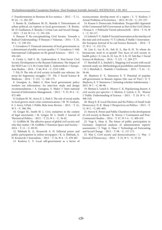
![-136-
Wschodnioeuropejskie Czasopismo Naukowe (East European Scientific Journal) | SOCJOLOGIA # IІІ, 2015
ПРОБЛЕМА ОБРАЗОВАНИЯ В КОНТЕКСТЕ НАЦИОНАЛЬНОЙ БЕЗОПАСНОСТИ
Самыгин Сергей Иванович,
доктор социологических наук, профессор,
профессор кафедры управления персоналом и социологии
Ростовского государственного экономического университета
Нечипуренко Виктор Николаевич,
доктор философских наук, профессор,
заместитель главного редактора научного журнала
«Контекст и рефлексия: философия о мире и человеке»
THE PROBLEM OF EDUCATION IN THE NATIONAL SECURITY CONTEXT
Samygin S. I., doctor of sociological Sciences, professor, Department of human resource management and sociology, Rostov State
Economic University
Nechipurenko V.N., doctor of philosophy, professor, Deputy chief editor Scientific Journal “Context and reflection: the philosophy
of the world and man”
Аннотация
В статье рассматриваются различные формы участия граждан России в местном самоуправлении. На основе экс-
пертного опроса анализируется про-блематика гражданской активности населения, определяются основные при-чины
пассивности жителей, перспективы участия избирателей в решении проблем местных сообществ. Устанавливается
опережение развития норма-тивной базы по сравнению с готовностью граждан принимать участие в местном самоу-
правлении.
ABSTRACT
The article discusses the various forms of participation of Russian citizens in lo-cal government. On the basis of the expert survey
examines issues of civil activi-ty of the population. Identifies the main causes of the passivity of the inhabit-ants, the prospects for voter
participation in solving problems of local communi-ties. Set advancing the development of the regulatory framework in comparison
with the willingness of citizens to participate in local government.
Ключевые слова: гражданское общество, местное самоуправление, гражданская активность, гражданские инициати-
вы.
Key words: civil society, local government, civic engagement, civic initiatives.
Проблема национальной безопасности имеет множе-
ство аспектов. Наряду с экономическими, технически-
ми, военными аспектами этой проблемы существует и её
культурное измерение [3]. Национальная безопасность
обеспечивается не только защитой от внешних угроз, но и
состоянием самой нации, состоянием общества, последнее
же напрямую зависит от культурных факторов, и одним из
важнейших факторов, поддерживающих функциониро-
вание культуры, является система образования. Система
образования – базовый институт современных обществ,
выполняющий комплекс социальных функций, неразрыв-
но связанных с обеспечением стабильного существования
общества в целом. Значимая функция образования, прису-
щая и школьному, и высшему образованию – это передача
национальной культуры, формирование общей системы
ценностей, мировоззренческих ориентиров. Современные
гетерогенные общества нуждаются в целенаправленном
поддержании культурного единства, за эту функцию отве-
чают многие институты, но роль образования – наиболее
важна.
В современном глобальном мире, где наблюдается взрыв-
ной рост культурной информации и свободное её рас-
пространение, национальные культуры теряют замкну-
тость и вступают во взаимодействие с элементами других
культур. С одной стороны, это способствует расширению
духовных горизонтов человека, с другой – усилению нео-
пределенности, размыванию прежних культурных иден-
тичностей. Образование должно решать одновременно
две задачи – воспроизводить национальную культуру и
готовить человека к жизни в открытом поликультурном
информационном пространстве. Не всегда образование
справляется с этой задачей эффективно. Наиболее остро
такая задача стоит перед институтами высшего образова-
ния, прежде всего, университетами. Университеты эпохи
классического модерна были не только центрами научных
исследований, но также и центрами гуманитарной мыс-
ли и гуманитарной культурной экспертизы, неразрывно
связанными с формированием национальных культур,
национальной культурной элиты. Университеты модерна
были, в первую очередь, храмами культуры, а культура в
эпоху модерна – это национальная культура: культура Ан-
глии, Германии, Франции, России и т.д. Но в то же время
тенденции развития современных обществ, развитие на-
учного знания и технологий превращали университеты в
центры производства нового знания. Формирование на-
укоемкой экономки и технический прогресс превратили
науку в главную производительную силу, причем науку
не гуманитарную. Университеты, сначала американские,
а потом и университеты других стран всё больше ориенти-
ровались на производство знания, которое может прино-
сить прибыль, сама наука и научная деятельность все более
превращаются в международную деятельность, лишенную
культурной специфики. Естественные и точные науки не
имеют национальной принадлежности, их развитие жиз-
ненно необходимо для развития современных обществ, но
они никак не связаны с поддержанием культурных иден-
тичностей и гуманистических ценностей. Экономическая
необходимость и экономический, технократический под-
ход к образованию приводят к тому, что точные науки тес-
нят гуманитарное образование, и вместе с этим оказыва-](https://image.slidesharecdn.com/eesj33-160202073457/85/Eesj-3-3-136-320.jpg)
![-137-
Wschodnioeuropejskie Czasopismo Naukowe (East European Scientific Journal) | SOCJOLOGIA # IІІ, 2015
ется под угрозой культурная составляющая образования.
Этим тенденциям способствует и процесс глобализации.
Глобализация затрагивает все стороны жизни совре-
менных обществ [8, c. 60-66], в том числе и образование,
особенно университетское образование. Для современ-
ных университетов вопросом престижа и обеспечения
средств к существованию является не только проведение
научных исследований с привлечением ведущих специа-
листов независимо от их национальной принадлежности,
но и привлечение иностранных студентов. Университеты
становятся глобализированными, но какой смысл в гло-
бализированном многонациональном университете углу-
бленно изучать, например, национальную литературу той
страны, где существует этот университет? Ценности какой
культуры должны прививаться многонациональному со-
обществу студентов глобализированного университета?
Но важность гуманитарного знания заключается не толь-
ко в его сильной культурной составляющей. Гуманитарное
знание всегда было связано с функцией критической реф-
лексии, анализа положения дел в обществе, отслеживания
основных тенденций его развития, обнаружения проблем-
ных зон в сфере этики и морали. Гуманитарная эксперти-
за необходима в обществе, где нет прочной традиции, нет
общепринятых моральных авторитетов. История Ново-
го и новейшего времени показывает, что естественные и
точные науки нейтральны в этическом плане, они могут
применяться в любых целях, отнюдь не всегда благих. Но
ослабление влияния и падение престижа гуманитарно-
го знания влечет за собой и ослабление его критической
функции.
Таким образом, изменения в экономике, переосмысление
функций образования в современных обществах ведет к
неоднозначным последствиям для культуры общества, к
ослаблению функции образования, заключающейся в под-
держании национальной культуры, этических и ценност-
ных основ социальной жизни. Так, например, «система
высшего образования подчиняется коммерческой, эконо-
мической логике, что пагубно влияет на сам дух образо-
вания, на статус университета. Он перестает быть храмом
культуры и знания, но становится своеобразным “знание-
вым супермаркетом”» [4, с. 33].
Но система образования не только прививает культур-
ные ценности. Она призвана готовить специалистов для
национальной экономики. Качество образования опреде-
ляет качество будущих работников и качество будущей
экономики, таким образом, качество образования самым
непосредственным образом связано с обеспечением на-
циональной безопасности. Поэтому все развитые страны
стремятся повышать качество образования и одновремен-
но увеличивать его доступность.
В современном российском обществе проблема обеспе-
чения качества образования стоит очень остро. При этом
угрозы этому качеству можно разделить на три группы:
общекультурные – связанные с радикальными изменени-
ями в сфере производства и распространения информа-
ции; социальные, связанные со спецификой социальной
структуры общества и функционированием экономики,
«управленческие» – связанные с таким фактором, как го-
сударственная политика в сфере образования. Качество
российского образования, как школьного, так и вузов-
ского, в последние десятилетия неуклонно снижается под
воздействием всех групп факторов, причем преобладает
негативное влияние третьей группы, связанной с государ-
ственной политикой в сфере образования.
Российское образование, как среднее, так и высшее, в
последние десятилетия находится в процессе постоянного
реформирования, при этом цели этого реформирования
далеко не очевидны ни для населения страны, ни для про-
фессионального сообщества, а результаты, в основном,
негативные. Наблюдается странная тенденция: при явном
и постоянном стремлении ограничить расходы на образо-
вание и сократить количество организаций, работающих в
этой сфере (постоянное сокращение числа щкол, средних
специальных учебных заведений, вузов), как и количество
работников (преподавателей), государственная бюрокра-
тия постоянно наращивает регламентацию труда препо-
давателей и учителей и требования формальной отчетно-
сти. Сокращая свои обязательства, государство постоянно
увеличивает требования, при этом требования отнюдь не
всегда рациональны. Одним из последних управленческих
решений, например, является привязка зарплаты школь-
ного учителя к количеству учеников в классе. Совершенно
очевидно, что этот критерий не просто лишен смысла – он
вреден, поскольку принуждает школы стремиться к тому,
чего следует избегать из соображений качества обучения.
О снижении качества образования в результате непрерыв-
ного реформирования говорят многие эксперты в сфере
образования, как и члены профессионального сообщества.
Можно вспомнить постоянную сокрушительную критику
ЕГЭ. «С введением ЕГЭ коррупция в школах выросла в
20-25 раз .. . Стоимость средней взятки за успешную сдачу
ЕГЭ … увеличилась в столице в 30 раз, в регионах в 10 раз.
По данным всероссийского опроса студентов в 2011 г., в
каждом четвертом вузе процветает взяточничество, лишь
в 23% вузов его нет, хотя взятками коррупция, конечно,
не ограничивается. При этом многие сторонники ЕГЭ в
свое время обосновывали его введение как раз снижени-
ем коррупции в сфере образования» [4]. Однако критика
на государственную политику в сфере образования никак
не влияет, мнение профессионалов не имеет значения для
государственной бюрократии.
Помимо негативных последствий реформирования, ка-
чество российского образования оказывается под угрозой
из-за специфики российского рынка труда и профессио-
нальной мобильности [1, c. 13-24]. В российском обществе
сложилась ситуация, когда без диплома о высшем обра-
зовании невозможно найти работу, но при это практиче-
ски не имеет значения качество знаний и специальность
[6, c. 96-100]. Исследования потребностей работодателей
показывает, что они ждут от претендентов на рабочие
места таких качеств, как дисциплинированность, комму-
никабельность и т.д., т.е. качеств, практически никак не
связанных с направлением подготовки. Диплом нужен
работодателям как документ, подтверждающий наличие
у будущего работника неких элементарных общекультур-
ных навыков, определенного уровня социальной, а не про-
фессиональной компетенции. И им не важно, чаще всего,](https://image.slidesharecdn.com/eesj33-160202073457/85/Eesj-3-3-137-320.jpg)
![-138-
Wschodnioeuropejskie Czasopismo Naukowe (East European Scientific Journal) | SOCJOLOGIA # IІІ, 2015
какой именно диплом в кармане у претендента. Очевидно,
что такой подход связан со спецификой труда. Для многих
современных видов трудовой деятельности, связанных,
в первую очередь, со сферой обслуживания, углубленное
профессиональное образование действительно не так уж
необходимо. Однако общее пренебрежительное отноше-
ние к профессиональной подготовке влияет на большин-
ство студентов, где бы они ни обучались. К тому же сту-
денты часто не собираются работать по специальности,
указанной в дипломе, потому учатся, спустя рукава. Пре-
подаватели, по причинам сложившейся в российских ву-
зах ситуации, когда выживание вуза напрямую зависит от
количества студентов, вынуждены закрывать глаза на низ-
кий уровень подготовки последних. Качество работы пре-
подавателей снижается и в результате постоянного увели-
чения учебной нагрузки и объема формальной отчетности
– при одновременном росте требований к эффективности
их научной деятельности [7, c. 66–77]. Всё это ведет к демо-
тивированности преподавателей, постоянному снижению
качества знаний выпускников вузов и обесцениванию ди-
пломов. Также можно говорить о деформации функций
современных российских вузов – из институтов профес-
сиональной социализации они превращаются в институ-
ты общей социализации, становясь «дополнительными
классами» средней школы.
Так возникает парадоксальная ситуация – снижение ка-
чества и престижа высшего образования при стабильно
высоком спросе на него. Диплом не гарантирует успеха на
рынке труда и не является показателем качества знаний,
но без него поиск работы становится практически бес-
смысленным, поэтому выпускники школ стремятся посту-
пить в любой вуз и получить любой диплом. Школа же по-
сле введения ЕГЭ утрачивает самостоятельную ценность и
превращается в курсы по подготовке к сдаче ЕГЭ. Таким
образом, непродуманная политика государства вместе
с тенденциями развития рынка труда приводят к дегра-
дации отечественного образования, что несет непосред-
ственную угрозу национальной безопасности. Страна не
может динамично развиваться без квалифицированных
специалистов, но, похоже, ни у российской экономики в
её современном виде, ни у государства, чьи интересы фор-
мулирует государственная бюрократия, нет потребности
в таких специалистах. Нет смысла говорить о необходимо-
сти готовить инженеров, а не менеджеров, если инженеры
не находят достойно оплачиваемой работы на рынке тру-
да. Состояние российского образования только отражает
ситуацию в экономике и государственном управлении.
Заметной составляющей деградации российского обра-
зования в результате непродуманных реформ стало целе-
направленное вымывание гуманитарной составляющей
из образования. Выше мы уже сказали о значимости этой
составляющей для поддержания национальной культуры
и национальной идентичности. Но реформаторская по-
литика показывает, что данная проблематика реформато-
рами не воспринимается вообще, а гуманитарное знание
рассматривается как лишняя трата средств.
В заявлении Ученого совета филологического факульте-
та МГУ говорится, в частности: «…Неспособность внятно
формулировать мысли — внешнее выражение неспособ-
ности самостоятельно мыслить: потребитель «информа-
ции» в лучшем случае научится ориентироваться в ней, но
не сможет осуществить ее экспертизу, а значит, и оказать
сколько-нибудь существенное воздействие на информа-
ционное пространство. В данной ситуации министерство
образования развернуло кампанию по сворачиванию фи-
лологического (и, шире, гуманитарного) образования в ву-
зах. … на протяжении последнего десятилетия бюджетный
набор на фундаментальные университетские направления
гуманитарной подготовки («Филология», «История», «Фи-
лософия» и др.) сократился как минимум втрое (примерно
с 300 до 100 человек в крупных университетах, со 100 до 30
человек в менее крупных; в Новгородском, Челябинском и
некоторых иных вузах он составляет ныне 10–15 чел.)» [5].
Разрушение гуманитарного образования ведет к подры-
ву основ национальной культуры, препятствует развитию
навыков рационального критического мышления, способ-
ствует распространению в обществе лженаучных идей и
архаизации общественного сознания. В современном по-
стоянно усложняющемся мире такая тенденция просто
опасна и ведет к деградации национальной культуры, раз-
мыванию мировоззренческих и ценностно-нормативных
основ общества и национальной идентичности [2].
Кризис современного российского образования носит
системный характер и ставит под угрозу будущее страны.
Потому вполне целесообразно говорить об угрозе нацио-
нальной безопасности. И критично важно для оценки по-
нимания этой опасности осознание того факта, что кризис
является не только и не столько следствием неких объек-
тивных социальных процессов, сколько результатом не-
продуманной управленческой деятельности.
Список литературы:
1. Беляева Л. Образование в России и модернизация эко-
номики (по результатам Европейского социального ис-
следования) // Социологические исследования (СОЦИС).
– 2011. № 12. – C. 13-24.
2. Верещагина А.В., Гафиатулина Н.Х., Самыгин С.И. Ду-
ховные аспекты формирования национальной идентично-
сти: социологический анализ угроз социальному здоровью
и духовной безопасности России // Инженерный вестник
Дона, №3 (2015). – Электронный ресурс. URL: http://ivdon.
ru/ru/magazine/archive/n3y2015/3195
3. Верещагина А.В., Самыгин С.И. Духовная безопасность
России как основа российской государственности // Гу-
манитарные, социально-экономические и общественные
науки. – 2011. № 1 (2). – Электронный ресурс. URL: http://
www.online-science.ru/m/products/social_sciense/gid35/pg0/
4. Гаврилова И.Н. Проблемы реформирования образова-
ния в современной России // Проблемный анализ и госу-
дарственно-управленческое проектирование. – 2012, №3.
5. О реформе образования, её итогах и перспективах.
Заявление Учёного совета филологического факультета
МГУ. – Электронный ресурс. URL: http://scepsis.net/library/
id_3325.html
6. Перов Г.О., Самыгин С.И., Тумайкин И.В. Молодежная
безработица в науке, как фактор накопления рисков соци-
альной безопасности России // Гуманитарные, социаль-
но-экономические и общественные науки. – 2015. № 3-1.](https://image.slidesharecdn.com/eesj33-160202073457/85/Eesj-3-3-138-320.jpg)
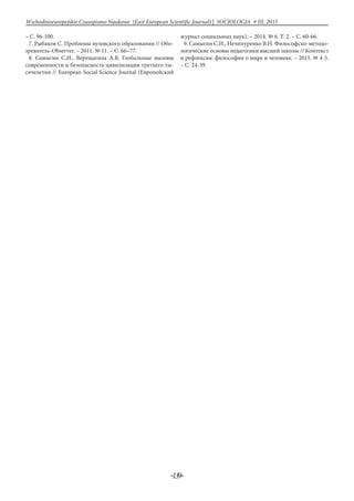
![-140-
WschodnioeuropejskieCzasopismoNaukowe(EastEuropeanScientificJournal)|FILOZOFIA,ETYKAIRELIGIOZNAWSTWO#IІІ,2015
FILOZOFIA, ETYKA I RELIGIOZNAWSTWO - ФИЛОСОФСКИЕ
НАУКИ
ЯЗЫК И «ДУХ НАРОДА» КАК ОБЪЕКТИВНОЕ И СУБЪЕКТИВНОЕ В СОЦИАЛЬНОЙ
ФИЛОСОФИИ В. ГУМБОЛЬДТА
Зыкин Алексей Владимирович,
Санкт-Петербургский государственный аграрный университет,
кандидат, филологических наук, доцент,
зав. кафедрой лингвистики и межкультурной коммуникации
LANGUAGE AND THE «SPIRIT OF NATION» AS THE OBJECTIVE AND THE SUBJECTIVE IN V. HUMBOLDT’S SOCIAL
PHILOSOPHY
Zykin A.V., candidate of philology, associate professor, Saint-Petersburg State Agrarian University
Аннотация
В работе рассматривается взаимосвязь языка и понятия «дух народа» с точки зрения философии В. Гумбольдта их
контент и содержание.
Abstract
In the paper we examine the relationship between the language and the «Spirit of nation» concept from the point of view V.
Humboldt’s philosophy, their content and peculiarities.
Ключевые слова: В. фон Гумбольдт, социальная философия, язык, дух народа, объективное, субъективное
Key words: Humboldt, social philosophy, language, spirit of nation, objective, subjective.
Теоретико-методологическую базу учения Гумбольдта
о языке составляет социальная антропология, в соответ-
ствии с которой изучение языка должно производиться
в тесной связи с сознанием и мышлением человека, его
социальной и культурной жизнью. В исследовательской
литературе сегодня это обозначается как социальная или
культурная антропология. Авторы философской энцикло-
педии пишут: «культурная антропология (реже – социаль-
ная антропология, социально-культурная антропология)
ориентирована на изучение культур отдельных народов и
человечества в целом … культурная антропология высту-
пает в виде высшей формы генерализации представлений
об институтах культуры человечества, представленной в
универсальной интерэтнической форме» [1, с. 51].
Основным в лингвистической концепции Гумбольдта
является учение о тождестве «духа народа» и его языка.
По мнению немецкого философа и педагога «язык народа
есть его дух и дух народа есть его язык - трудно предста-
вить себе что-либо более тождественное» [2, с. 48]. Гум-
больдт подходит к пониманию тождественности языка и
духа народа через тождество в философии объективного
и субъективного. Отметим, что проблема объективного и
субъективного – одна из существенных как в онтологии и
гносеологии, так и в социальной философии. Объективное
трактуется как то, что существует независимо от индиви-
дуального сознания; прежде всего это физические вещи и
события в пространстве и времени; другие люди, их дей-
ствия и состояния сознания; собственное тело индивида.
Социально-исторический подтекст в соотношении объ-
ективного и субъективного особенно наглядно виден на
соотношении общественного бытия и общественного со-
знания. Это наиболее разработанные категории историче-
ского материализма.
В истории философии объективный мир жестко проти-
вопоставлялся субъективному. Вместе с тем существуют
серьезные основания считать, что субъективное возни-
кает как результат коммуникативных взаимодействий
субъекта с другими. Предметы культуры и воплощенные
в них смыслы (включая орудия, инструменты, приборы,
произведения архитектуры, художественные, научные,
философские и иные тексты) существуют объективно, но
в то же время предполагают индивидуальных субъектов
(с их субъективным миром) и субъектов коллективных.
Именно субъекты создают предметы культуры и их смыс-
лы. Объективные смыслы, в т.ч. и те смыслы, которые пока
никем не осознаются, могут существовать лишь постоль-
ку, поскольку имеются субъекты, способные их выявить,
сделать собственным субъективным достоянием [3].
В свою очередь субъективное – то, что характеризует
субъект или же производно от субъекта и его деятельно-
сти. Исторически субъективное было понято в классиче-
ской философии (начиная с Декарта) как особый внутрен-
ний мир сознания, несомненный и самодостоверный, к
которому субъект имеет непосредственный доступ. В та-
ком качестве субъективное было противопоставлено объ-
ективному миру физических вещей и событий (включая и
тело субъекта) как существующему вне субъективного и
как не абсолютно достоверному.
Развитие современной философии и психологии дает
основание считать, что субъективное, переживаемое
нами в качестве чисто “внутреннего” и сугубо личного,
не является изначально данным, а строится субъектом в
коммуникативных взаимодействиях с другими людьми
в рамках определенной исторически данной культуры,
поэтому и сама степень переживания субъективного как
“внутреннего” может быть различной в разных услови-
ях. Субъективное не может рассматриваться как особый
мир, населяемый такими “предметами”, как переживания,
представления, образы памяти воображения, желания и
т.д., а является способом ориентации субъекта во внеш-](https://image.slidesharecdn.com/eesj33-160202073457/85/Eesj-3-3-140-320.jpg)
![-141-
WschodnioeuropejskieCzasopismoNaukowe(EastEuropeanScientificJournal)|FILOZOFIA,ETYKAIRELIGIOZNAWSTWO#IІІ,2015
нем мире.
Однако и коммуникация и другие действия, будучи по-
рождены субъектом и в этом смысле будучи субъективны-
ми, в то же время не принадлежат к субъективному миру в
классическом его понимании, а относятся к сфере интер-
субъективного, которая выходит за рамки противополож-
ности субъективного и объективного [3].
Тождество объективного и субъективного согласно фи-
лософии Гумбольдта проблематизирует вопрос об образе
и эмоции и их соотношении с языком. Поскольку язык
существует во времени и пространстве, постольку Гум-
больдту нужно было решить вопрос о статусе темпораль-
ных процессов в языке. Само по себе время ничего не син-
тезирует. Устойчивость предмета во времени – проблема
физиков, а вот устойчивость образа во времени – это уже
проблема философии. И Гумбольдт решает ее путем сое-
динения эмоции и языка. Эмоция, как сон, хватает первые
попавшиеся ей предметы и сдваивает их, формируется
дипластия. Чем сильнее эмоциональное потрясение, тем
прочнее образы. Такие образы Юнг называл архетипами
[4, с. 137-140].
Эмоции интересны тем, что заставляют человека реали-
зовывать свои желания через матрицу воображаемого, то
есть духовность. По Гумбольдту, дух самобытен, свободен,
самостоятелен, целеустремлен, деятелен, всепроникающ, в
известной степени индивидуализирован. Язык трактуется
им как организм духа и как его самодеятельность. Сила че-
ловеческого духа, по Гумбольдту, в самом своем существе
человеку не доступна. Она открывается косвенно через
свои проявления (язык, цивилизацию, культуру, духов-
ность и т.д.).
Проявления духовного – это её создания, сила духа яв-
ляется человеку в том, что она производит, открывается
в своих созданиях. Поскольку непосредственное проник-
новение в мир духа невозможно, то всякое постижение
его есть реконструкция, восстановление, предугадывание
его черт. Язык – главная деятельность человеческого духа.
Проблема взаимоотношения языка и духа становится од-
ной из центральных в творчестве Гумбольдта.
По его мнению, зарождение языка не может происходить
по отдельным кусочкам или отдельным словам: «Для того
чтобы человек мог понять хотя бы единственное слово не
просто как душевное побуждение, а как членораздельный
звук, обозначающий понятие, весь язык полностью и во
всех своих связях уже должен быть заложен в нем» [2, с.
117]. Так как в языке нет ничего единичного, и «каждый
отдельный его элемент проявляет себя лишь как часть це-
лого». Надо отметить, что это последнее положение полу-
чило многообразную интерпретацию лишь у философов
XX века, сформировавших понятия системы и структуры.
Это легло в основу структурализма – научного направ-
ления в гуманитарном знании, возникшем во Франции
в первой половине XX в. Основу структурного метода
образует выявление структуры как совокупности отно-
шений, инвариантных при некоторых преобразованиях.
В такой интерпретации понятие структуры характеризу-
ет не просто устойчивый «скелет» какого-либо объекта,
а совокупность правил, по которым из одного объекта
можно получить второй, третий и т.д. путем перестанов-
ки его элементов и некоторых других симметричных пре-
образований. Выявление структурных закономерностей
некоторого множества объектов достигается здесь путем
выведения различий между этими объектами в качестве
превращающихся друг в друга конкретных вариантов еди-
ного абстрактного инварианта [5].
Одним из основных положений структурализма явля-
ется утверждение о том, что социальные и культурные
явления не обладают самостоятельной субстанциальной
природой, а определяются своей внутренней структурой
(то есть системой отношений между внутренними струк-
турными элементами), и системой отношений с другими
явлениями в соответствующих социальных и культурных
системах. Эти системы отношений рассматриваются как
знаковые системы и, таким образом, трактуются как объ-
екты, наделенные значением [6].
Структурализм ставит своей целью объяснить, как дан-
ные социальные институты, которые могут быть выявле-
ны в рамках структурного анализа, делают возможным
человеческий опыт [7]. В соответствии с этим подходом
можно индивидуализировать структуралистскую пози-
цию. Суть в том, что структурализм представляет на-
правление в философии, сформировавшееся в полемике
с субъективизмом, гуманизмом, историзмом и эмпириз-
мом. Строгой доктрины у структурализма нет.
Поиск структур осуществлялся в различных областях
социогуманитарного знания: в лингвистике, этнографии,
психологии, истории... Основу структурного метода об-
разует процесс выявления структуры как совокупности
отношений, инвариантных при некоторых образованиях.
В таком понимании понятие структуры характеризует не
просто устойчивый каркас какого-либо объекта, а сово-
купность правил, по которым из одного объекта можно
получить второй, третий и т.д., путём перестановки его
элементов и некоторых других симметричных преобра-
зований. Выявление структурных закономерностей неко-
торого множества объектов достигается путём выведения
различий между этими объектами в качестве превраща-
ющихся друг в друга конкретных вариантов единого аб-
страктного инварианта.
Обобщенно можно сказать, что основная идея фило-
софского структурализма – не «бытие», а «отношение»; не
субъект, а структура. Люди вроде шахматных фигур, неких
лингвистических, математических или геометрических
единиц: они не существуют уже вне установленных отно-
шений, а лишь специфицируют вид поведения. Субъекта в
структурализме заменяет некая его форма, не являющаяся
субстанцией общества.
В своих работах философского плана Гумбольдт дает
определение языка, ставшее, пожалуй, классическим: «По
своей действительной сущности язык есть нечто постоян-
ное и вместе с тем в каждый данный момент преходящее.
Даже его фиксация посредством письма представляет со-
бой далеко не совершенное мумиеобразное состояние, ко-
торое предполагает воссоздание его в живой речи. Язык
есть не продукт деятельности (ergon), а деятельность
(energeia). Его истинное определение может быть поэтому
только генетическим. Язык представляет собой постоян-
но возобновляющуюся работу духа, направленную на то,](https://image.slidesharecdn.com/eesj33-160202073457/85/Eesj-3-3-141-320.jpg)
![-142-
WschodnioeuropejskieCzasopismoNaukowe(EastEuropeanScientificJournal)|FILOZOFIA,ETYKAIRELIGIOZNAWSTWO#IІІ,2015
чтобы сделать артикулируемый звук пригодным для выра-
жения мысли. В подлинном и действительном смысле под
языком можно понимать только всю совокупность актов
речевой деятельности. В беспорядочном хаосе слов и пра-
вил, который мы по привычке именуем языком, наличе-
ствуют лишь отдельные элементы, воспроизводимые – и
притом неполно – речевой деятельностью; необходима все
повторяющаяся деятельность, чтобы можно было познать
сущность живой речи и составить верную картину живого
языка, по разрозненным элементам нельзя познать то, что
есть высшего и тончайшего в языке; это можно постичь
и уловить только в связной речи... Расчленение языка на
слова и правила – это лишь мертвый продукт научного
анализа. Определение языка как деятельности духа совер-
шенно правильно и адекватно уже потому, что бытие духа
вообще может мыслиться только в деятельности и в каче-
стве таковой» [2, с. 221].
Интеллектуальная деятельность, совершенно духовная,
глубоко внутренняя и проходящая в известном смысле
бесследно отмечает Гумбольдт, посредством звука мате-
риализуется в речи. Таким образом духовное как бестеле-
сное становится доступной для чувственного восприятия
(речь письменная и речь естественная). Интеллектуальная
деятельность и язык представляют собой поэтому единое
целое. В силу необходимости мышление всегда связано
со звуками языка. Гумбольдт пишет: «Даже не касаясь по-
требностей общения людей друг с другом, можно утвер-
ждать, что язык есть обязательная предпосылка мышле-
ния и в условиях полной изоляции человека. Но обычно
язык развивается только в обществе (выделено нами), и
человек понимает себя только тогда, когда на опыте убе-
дится, что его слова понятны также и другим людям... Ре-
чевая деятельность даже в самых своих простейших про-
явлениях есть соединение индивидуальных восприятий с
общей природой человека. Так же обстоит дело и с пони-
манием» [2, с. 28].
Для того чтобы понять, как дух народа реализуется в язы-
ке, надо правильно понять, что такое язык. Как отмечает
Гумбольдт, «язык предстает перед нами в бесконечном
множестве своих элементов – слов, правил, всевозмож-
ных аналогий и всякого рода исключений, и мы впадаем в
немалое замешательство в связи с тем, что все это много-
образие явлений, которое, как его ни классифицируй, все
же предстает перед нами обескураживающим хаосом, мы
должны возвести к единству человеческого духа». Нель-
зя ограничиваться фиксацией этого хаоса, надо в каждом
языке искать главное, поэтому в своих лингвистических
исследованиях Гумбольдт затронул другие важные пробле-
мы, а именно вопросы социально-философского характе-
ра. Они у него связанны с выявлением понятий «народ» и
«язык». Гумбольдт считает «нацию» (для него это почти то
же самое, что и «народ») такой формой «индивидуализа-
ции человеческого духа», которая имеет «языковой» ста-
тус. Считая нацию духовной формой человечества, имею-
щей языковую определенность», специфику этой формы
он усматривает главным образом в языке, хотя при этом
подчеркивает, что в формировании нации, помимо языка,
участвуют и другие факторы: «если нами нации называ-
лись духовной формой человечества, то этим совершенно
не отрицались их реальность и их земное бытие».
Так как деление человечества на языки совпадает с де-
лением его на народы, то по Гумбольдту отсюда должно
следовать, что между языком и народом, или, точнее, ду-
хом народа, существует необходимая корреляция. «Язык
и духовная сила народа развиваются не отдельно друг от
друга и последовательно один за другой, а составляют ис-
ключительно и нераздельно одно и то же действие интел-
лектуальной способности» [2, с. 142].
Каков же смысл употребления понятия «дух народа» в
работах Гумбольдта? Человек не может ни мыслить, ни
развиваться без языка: «Создание языка обусловлено вну-
тренней потребностью человечества. Язык - не просто
внешнее средство общения людей, поддержания обще-
ственных связей, но заложен в самой природе человека и
необходим для развития его духовных сил и формирова-
ния мировоззрения, а этого человек только тогда сможет
достичь, когда свое мышление поставит: связь с обще-
ственным мышлением» [2, с. 211].
«Языкотворческая сила в человечестве» стремится к со-
вершенству, этим и обусловливаются единые закономер-
ности развития всех языков, даже тех, «которые не обна-
руживают между собой никаких исторических связей».
Отсюда необходим стадиальный подход и кажущееся
Гумбольдту несомненным разграничение более и менее
совершенных языков. Такая точка зрения была естествен-
ной, если исходить из первичности духа народа. Дух на-
рода и язык народа неразрывны: «Духовное своеобразие и
строение языка народа пребывают в столь тесном слиянии
друг с другом, что коль скоро существует одно, то из этого
обязательно должно вытекать другое... Язык есть как бы
внешнее проявление духа народов: язык народа есть его
дух, и дух народа есть его язык, и трудно представить себе
что-либо более тождественное» [2, с. 231]. При этом един-
стве согласно концепции Гумбольдта первичен все же дух
народа.
Гумбольдт ввел понятие «дух народа» в философию язы-
ка как понятие необходимое, однако, его трудно постичь
в чистом виде: без языкового выражения «дух народа»
- неясная величина, знание о которой следует извлечь
опять-таки из самого языка, язык же толкуется не только
как средство для постижения «духа народа», но и как фак-
тор его создания. Тут как бы замкнулся некий герменевти-
ческий круг: дух народа как «высший принцип», обуслов-
ливая различие и специфику языков, со своей стороны сам
нуждается в объяснении через язык. Гумбольдт пишет по
этому поводу: «Не будет заколдованного круга, если языки
считать продуктом силы народного духа и в то же время
пытаться познать дух народа посредством построения са-
мих языков: поскольку каждая специфическая духовная
сила развивается посредством языка и только с опорой на
него, то она не может иметь иной конструкции, кроме как
языковой».
В понимании Гумбольдта, правильность и богатство
развития языка прямо пропорционально связаны с со-
размерностью воздействия на язык силы национального
духа. Языки, соответственно, также обладают силой, воз-
действующей на дух. И это воздействие носит всесторон-
ний и гармоничный характер. По образному выражению](https://image.slidesharecdn.com/eesj33-160202073457/85/Eesj-3-3-142-320.jpg)
![-144-
WschodnioeuropejskieCzasopismoNaukowe(EastEuropeanScientificJournal)|FILOZOFIA,ETYKAIRELIGIOZNAWSTWO#IІІ,2015
хождению, смыслах: сакрально-иррационального, тран-
свиртуального Божественного и человеческого. Декоди-
рования человеком образности этих смыслов, в процессе
восприятия им произведения христианского сакрального
искусства, обеспечивается механизмом интериоризации,
который позволяет почувствовать психологический эф-
фект непосредственного присутствия образа Святого и
духовно соединиться с ним благодаря возникновению ну-
минозного чувства, как переживания божественной тай-
ны, и на основе энергетического подъёма воспринимать
идеи христианства. При этом о нуминозном говорится «…
как своеобразной категории толкования и оценки и рав-
ным образом о нуменозной настроенности души…» [6, 9].
Кроме того, нужно акцентировать внимание на важ-
ности механизма экстраполяции, который позволяет ве-
рующему в процессе восприятия христианского художе-
ственного символа и самостоятельного декодирования
его образного смысла, по своему желанию, на основании
личных определенных представлений о могуществе Выс-
ших потусторонних сил, наделять эти образы соответ-
ствующим сакральным смыслом с верой в то, что эти
Высшие силы помогут ему в решении определенных про-
блем. Реализует человек этот процесс благодаря фактору
«вчувствования», который позволяет ему наделять худо-
жественные культовые образы признаками сакральности
на основе чувств, которые по Л. С. Выготскому, «подни-
маются из самой глубины нашего существа». Более того,
подчеркнем, что наделение, каждым верующим по своему
желанию, художественного образа сакральным смыслом
является процессом не только сугубо субъективным, но
и персонифицированным, который органично связывает-
ся с конкретикой бытовой ситуации, в которой оказался
верующий, но которую преодолеть самостоятельно не в
состоянии. Так, в частности в одном случае персонифика-
ция образа Бога осуществляется в системе «Бог-судья», в
другом - «Бог-повелитель», еще в других - «Бог любящий
отец», «Бог защитник» и другие.
Сущность же самой теории «вчувствования» Л. С. Ви-
готский разъясняет следующим образом, утверждая, что
«согласно этой теории, чувства не пробуждаются в нас
произведением искусства, как звуки клавишами на рояле,
каждый элемент искусства не вносит в нас своего эмоци-
онального тона, а дело происходит как раз наоборот. Мы
изнутри себя вносим в произведение искусства, вчувству-
ем в него те или иные чувства, которые подымаются из
самой глубины нашего существа и которые, конечно же,
не лежат на поверхности у самых наших рецепторов, а свя-
заны с самой сложной деятельностью нашего организма»
[1, 196].
Понимание причин такого «вчувствования» верующе-
го человека в смысл сакрального художественного образа,
а также высокого уровня его доверия к сути этого ирреа-
листичного смысла, сформировавшегося в его воображе-
нии благодаря составляющим механизма экстраполяции,
требует более подробно выяснить сущность самого явле-
ния «образ» в том числе и «художественный сакральный
образ восприятия», что и является основным заданием
данного исследования.
Классическое определение сути этой категории свиде-
тельствует, что образ - это «результат и идеальная форма
отражения предметов и явлений материального мира в со-
знании человека. Образ - субъективное явление , которое
возникает в результате предметно-практической деятель-
ности, а также ее специфических форм, таких как воспри-
ятие, память, мышление» [4, 172].
А. В. Запорожец детализирует суть этого понятия и
утверждает, что «образы - это субъективные феномены,
возникающие в результате предметно-практической, сен-
сорно-перцептивной и мыслительной деятельности. Об-
раз - это целостное, интегральное отражение действитель-
ности, в котором одновременно представлены основные
перцептивные категории (пространство, время, движение,
цвет, форма, фактура и т.д.). Важнейшей функцией обра-
за является регуляция деятельности» [3, 152]. Более того,
ученый уточняет суть идеально-материальной структуры
человеческого образа. В ее составе он объясняет значение
и взаимодействие двух специфических составляющих -
биодинамической и чувственной: «Пространственная
структура образа складывается в результате предметных
действий субъекта благодаря превращению биодинами-
ческой ткани движения в чувственную ткань образа... По
мере формирования пространственного образа он напол-
няется предметными свойствами, наделяется чувствен-
ной тканью и совместно с ней локализуется во внешнем
пространстве. Сказанное справедливо в отношении чув-
ственной ткани, связанной с происхождением из биоди-
намической. Сложнее обстоит дело с чувственной тканью,
связанной по происхождению со свойствами проксималь-
ной стимуляции (с свето-цветовыми свойствами окруже-
ния). Можно допустить, что этот тип чувственной ткани
не обладает изначально предметными (и соответственно
картинными) свойствами. Они приобретаются по мере
того, как эта ткань сливается с пространственной струк-
турой образа и вместе с ней экстериоризуется и локали-
зуется во внешнем пространстве. После такого слияния
образ выступает как интегральное и нераздельное целое»
[3, 153]. Таким образом, доказывается, что процесс фор-
мирования у человека образа восприятия является доста-
точно сложным, характеризуется процессуальностью, ди-
намикой, этапностью и спецификой его так называемого
«строительного» материала. Поэтому, как вывод отметим,
что конечным этапом формирования у верующего рели-
гиозного образа как интегрального и нераздельного це-
лого, при раскодировании им содержания христианского
художественного символа, выступает образ, сакральный
смысл которого верующий программирует самостоятель-
но в соответствии с характером своей потребности.
А. Н. Леонтьєв, также анализируя процессуальную сто-
рону формирования у человека образа в контексте науч-
ной теории отражения, признает необходимость решать
фундаментальную проблему - «проблему исследования
процесса перехода или «перевода» отражению содержа-
ния в содержание отражения» [5, 19]. Он объясняет зако-
номерность, согласно которой «процесс отражения явля-
ется результатом не влияния, а взаимодействия, то есть
результатом процессов, идущих как бы навстречу друг
другу. Один из них является процесс воздействия объекта
на живую систему, а другой - активность самой системы](https://image.slidesharecdn.com/eesj33-160202073457/85/Eesj-3-3-144-320.jpg)
![-145-
WschodnioeuropejskieCzasopismoNaukowe(EastEuropeanScientificJournal)|FILOZOFIA,ETYKAIRELIGIOZNAWSTWO#IІІ,2015
относительно влияния объекта. Этот последний процесс
благодаря своему уподоблению независимым свойствам
реальности и несет в себе ее отражение» [5, 22]. В нашем
случае в этом двустороннем процессе, с одной стороны,
находится произведение христианского сакрального ис-
кусства, смысл которого является воплощенным в соот-
ветствующий ему символ и образность которого верую-
щему нужно раскодировать. Это могут быть произведения
изобразительного, архитектурного, музыкального и дру-
гих видов сакрального искусства. С другой стороны, в ка-
честве живой системы, на которую влияет произведение
сакрального искусства, при условии восприятия его сути
верующим, выступает сам верующий человек, а также и
такой человек, у которого религиозные чувства и взгляды
еще не сформировались, а находятся на стадии становле-
ния.
Таким образом, актуализируется тождество этой за-
кономерной связи с механизмом экстраполяции, для ко-
торого также характерно одновременное объединение
двух противоположных тенденций: а) «свойство инерции,
консервативность знания, стереотипность механизмов че-
ловеческого познания» б) «направленность на получение
принципиально нового знания».
Поэтому, есть основания для вывода о доминирующей
значимость механизма экстраполяции в реализации про-
цесса формирования у человека образа внешних явлений
и предметов, в частности художественного образа произ-
ведения культового искусства с его символикой сакраль-
ного значения, в процессе отражения им этого смысла.
А. В. Запорожець также доказывает, что ни сенсорный
импульс, ни непосредственная активизация рецепторов
человека не могут выступать основанием для формиро-
вания адекватного образа восприятия: «Здесь необходи-
ма коррекция, которая исправляет неизбежные ошибки и
приводит образ в соответствие с объектом ... нужна эксте-
риоризация отражательного процесса, которая и происхо-
дит в виде перцептивных действий, которые уподобляются
своей внешней формой объекту, который воспринимается
и сопоставляются с особенностями объекта» [3, 131].
Ученый объясняет, что эволюция образа охватывает
процесс трансформации пространственного образа в пер-
цептивныесхемы,взначение,всимволы,вчастностисуси-
лением элементов абстрагирования именно в символах [3,
153]. Он исходит из того, что движение и психика челове-
ка находятся в органическом единстве, описание которых,
как и образов отображения, спровоцировало разработку
в науке нового концептуального аппарата. С точки зрения
проблемы формирования у верующего глобального обра-
за мира как реального, то есть земного, так и Высшего, а по
П. А. Флоренскому, горнего и дольнего, важна идея учено-
го о связи живого движения со значением и смыслом. А. В.
Запорожец рассматривает эту взаимосвязь в глобальном
масштабе: «Симультанизация пространственной карти-
ны мира возможна только в результате последовательно
развернуого во времени, сукцесивного действия в этом
мире. Живое движение действительно является активным
хронотопом, уникальным средством овладения простран-
ством и временем... Структурные особенности живого
движения все чаще становятся предметом изучения. По-
следние результаты свидетельствуют о том, что оно имеет
квантово-волновую природу. Пространственно-времен-
ные характеристики квантов определяются двигательной
задачей, степенью освоения действия, в состав которого
они входят, скоростью его осуществления» [3, 150].
Современная наука, опираясь на новейшие достиже-
ния, отстаивает новые концепции о сущности образов
восприятия человека. Так, О. П. Дубровский и В. Н. Пуш-
кин обращают внимание на то, что образ восприятия че-
ловеком объектов реальной действительности должен
рассматриваться в качестве некоторых «полевых» струк-
тур в их чистом виде. Авторы доказывают что «гипотеза
о формотворческой функции мозга позволяет подойти к
природе адекватного отражения объектов с помощью об-
разов восприятия. Образы восприятия, с точки зрения их
материального существования, выступают как реально-
сти, подобные стоячим волнам, как некоторые полевые
структуры, как формы в чистом виде, лишенные вещества.
В процессе отражения предметов окружающего мира эти
материальные, и в то же время информационные обра-
зования, входят во взаимодействие с формами объектов,
воспринимаются. Именно такое полевое, волновое взаи-
модействие форм составляет основу процесса восприя-
тия» [2, 109].
Таким образом, можно внести некоторые уточнения в
понимание категории «художественный сакральный об-
раз восприятия». Под этой категорией следует понимать
субъективный психический феномен информационно-по-
левого происхождения, который формируется у верующе-
го экстраполярно, в процессе декодирования им смысла
христианского художественного символа и активизации
личной предметно-практической сенсорно-перцептивной
деятельности, и самостоятельного предоставления ему са-
крального значения. Все это обуславливает приоритетное
религиозно-мировоззренческое значение художественно-
го сакрального образа восприятия для верующего.
Ссылки:
1. Выготский Л. С. Психология искусства / Под ред. М.
Г. Ярошевского. – М.: Педагогика, 1987. – 344 с.
2. Дубров А. П., Пушкин В. Н. Парапсихология и совре-
менное естествознание. – М.: «Соваминко», 1989. – 280 с.
3. Запорожец А. В. Избранные психологические труды:
В 2-х. т. - Т. I. Психическое развитие ребенка. – М.: Педаго-
гика, 1986. – 320 с.
4. Краткий педагогический словарь пропагандиста /
Под общ. ред. М. И. Кондакова, А. С. Вишнякова; Сост.: М.
Н. Колмакова, В. С. Суров. – 2-е изд., доп. и доработ. – М.:
Политиздат, 1988. – 367 с.
5. Леонтьев А. Н. Понятие отражения и его значение
для психологии: В кн.: Хрестоматия по психологии: Учеб.
Пособие для студентов пед. ин-тов / Сост. В. В. Миронен-
ко; Под ред. А. В. Петровского. – 2-е изд., перераб. и доп.
– М.: Просвещение, 1987. – 447 с.
6. Отто Р. Священное / Р. Отто [перевод А. М. Руткеви-
ча]. – СПб.: Издательство С.-Петербургского университе-
та», 2008. – 272 с.](https://image.slidesharecdn.com/eesj33-160202073457/85/Eesj-3-3-145-320.jpg)
![Russian Presidential Academy of National Economy and Public Administration
doctor of philosophy, professor, Dzerzhinsky branch of the
Pishchik A.M.
Ключевые слова: компоненты деятельности, системно-деятельностный подход.
НАУЧНО-ИССЛЕДОВАТЕЛЬСКАЯ ПРОГРАММА НИЖЕГОРОДСКОЙ МЕТОДОЛОГИЧЕСКОЙ ШКОЛЫ: КОН
-146-
WschodnioeuropejskieCzasopismoNaukowe(EastEuropeanScientificJournal)|FILOZOFIA,ETYKAIRELIGIOZNAWSTWO#IІІ,2015
RESEARCH PROGRAM OF THE NIZHNY NOVGOROD METHODO-LOGICAL SCHOOL:
EIGHT COMPONENTS OF ACTIVITY CONCEPT
-
ЦЕПЦИЯ ВОСЬМИ КОМПОНЕНТОВ ДЕЯТЕЛЬНОСТИ
Аннотация
Abstract
Development and validation of eight components activity concept.
Key words: components of activity, system-activity approach.
This article continues a series of articles about the concepts
developed by the Nizhny Novgorod methodological school,
publication of which began in the previous issue of the journal
[21].
The problem
There is the problem of creating a theoretical language for
professional communication that allows you to integrate the
achievements of the philosophical level of knowledge with
the achievements of specific scientific studies. The theory of
the «middle» level can solve this problem. An example of such
theory is the General theory of activity. Nizhny Novgorod
Methodological school began to de-velop a General theory of
activity in 80-ies of XX century. The task was the inte-gration
of achievements of the philosophical concepts of activity
and achieve-ments of specific scientific concepts developed
in psychology, sociology, peda-gogy, linguistics and other
research areas.
In the framework of the General theory of activity, was
developed the con-cept 8 components of activity. The concept
was developed on the basis of sys-tem-activity approach. The
concept was brought to the scientific community in Gorky in
1982 on X Inter-zonal Symposium [6].
Analysis of recent researches and publications
The concept 8 components of activity appeared in the
beginning of 80-ies as a response to the inquiry practices of
design in different spheres of human activi-ty on the basis of
system-active approach.
Domestic system-activity approach appeared in the
laboratory of theoretical problems of the Scientific research
Institute of technical aesthetics (1965-1968). The laboratory
was the «creative Studio» to develop new theoretical and
method-ological tools in the field of design» in the field
of domestic design [13, p. 8]. In this lab worked K.Kantor,
G.Shchedrovitsky, O.Genisaretsky, V.Glazychev, V.Dubrovsky
and other professionals. «The main methodological tool of
devel-opment of project ideology served as the system-activity
approach» [13, p. 10]. When you create a method the authors
have seen foreign origins in systems engi-neering, theory of
operations, ethnopsychology, ethnolinguistics, anthropology,
theory of human relationships. The achievements of this period
of formation of the systemic-activity approach is presented in
two collections of scientific works of the Scientific research
Institute of technical aesthetics [13, 14], and also in se-lected
works of G.Shchedrovitsky [17].
System-activity approach in these years was created not only
in the field of design and organizational and activity games
of the methodological school of G. Shchedrovitsky. The main
directions of the activity approach, developed in the 80-th years
of XX century, were recorded in the research G. V. Sukhodolsky,
the author who generalized the psychological theory of activity
[11, 12]. In the 60-80s of the activity approach becomes system-
activity. The signs of a new quality are: 1) from a philosophical
and psychological approach it has become multi-disciplinary;
2) it was enriched by the achievements of a systematic approach
(there is a variety of working models with different component
composition and different structures) [4, 7]; 3) it began to
develop primarily as a design method.
In 80-e years the Russian variant of system-activity approach
has attracted the attention of developers of computer systems
and soon became an internation-al movement of scientists
and designers. Since 1986 many international congress-es on
research in the field of the theory of activity were held [1]. From
1988 to 1994 the international journal of multidisciplinary
research on the theory of ac-tivity (a Multidisciplinary
Journal of the News Letter for Research Activity Theo-ry) was
published. In 1996 the main provisions of the activity theory
were pre-sented in English (K.Kuutti, B.Nardi) and became
known to a wide circle of spe-cialists in the U.S. and other
countries [18 -20].
Allocation of unsolved aspects of the problem
Interest in the methodology of systemic-activity approach,
updated in the educational environment due to the fact that
the methodological basis of the Fed-eral educational standards
for Russian secondary school since 2012 has become a system-
activity approach [15]. Among the scientific schools that wish
to manage the activities of the school, there is no consensus on
the effectiveness of pedagog-ical technologies on the basis of
system-activity approach [3, 16]. Psychological interpretation
of the system-active approach dominates in the standards. “Bias
in the selection of scientific theories, which underlies the basis
of standards, makes it difficult the variable nature of secondary
education. Not all teachers share the position that the purpose
of General education is to develop the cognitive activity
of students by using deductive theoretical generalizations.
Besides, man is not only thinking, but also feelings, emotions,
physical development, spirituality and much more” [16].
The idea of the systemic-activity approach, developed by
the Nizhny Nov-gorod methodological school, was to avoid
methodological one-sidedness prevail-ing in philosophy,
Разработка и обоснование концепции восьми компонентов деятельности](https://image.slidesharecdn.com/eesj33-160202073457/85/Eesj-3-3-146-320.jpg)
![-147-
WschodnioeuropejskieCzasopismoNaukowe(EastEuropeanScientificJournal)|FILOZOFIA,ETYKAIRELIGIOZNAWSTWO#IІІ,2015
productive tool for social design in any field.
The purpose of the article
The article aims to present the concept 8 components of
activity . The con-cept was developed in the framework of the
systemic-activity approach.
The main material
The Nizhny Novgorod methodological school has developed
the concept of the eight components of activity. This concept
was based on the analysis of the components of activity
described in the concepts of other authors [5, 6, 9, 10].
The developed model is presented in figure 1.
Figure 1. System of the activity
The definition of the component activity
The subject of activity (Sub) - this is an individual or a
community of people undergoing the process activity.
The object of the activity (Obj) – the totality of characteristics
of an object targeted by the operation process.
Tool – all the material objects used by the subject to ensure
the process ac-tivities.
Process activity (Proc) – structure-forming component of
the activity system; this is the set of interactions of the system
activity.
Result of activity (Res) – 1) in a broad holistic sense – the
result of the entire operation system in its completed form;
2) in the narrow component relationship – the result is the
converted object, in accordance with the purpose.
Conditions (Con) are components of other systems, actively
included in the operation system.
System activity - procedural attitude components of activity
(structured com-position of the internal components of
activity).
Environment activity is the external system, forming the
active background and potential activities.
The composition and structure of each component activity
are specified at a deeper level of analysis. The content of the
subject is revealed through motiva-tional, informational and
operational readiness for activity (Fig.2).
Figure 2. The system of the subject and process of activity
Motivational readiness is revealed at three levels of existence
– at the level of objectives, targets and programs to achieve it.
Informational willingness is ex-pressed in the awareness of
the problem, proposing hypotheses and theories available to
addressit.Operationalreadinessismanifestedintheavailability
method, approach and technique of implementation. The
process of activity con-sists of actions, operations, and
receptions.
Tools are viewed as material phenomena, used by the entity
to enhance their capabilities to transform the object of activity
in the result. The classification of the activities based on the
followingthreegrounds:1)thetoolactivitywillbeconsideredas
compensators and amplifiers bodily (physical), sensory (touch)
and intellectual qualities; 2) Tools interact with matter, energy
and information; 3) tool to perform the functions of reflection
conversion, storage and transmission. The interaction of these
three elements creates a combination of actual and hypo-
thetical possible 36 types of human activity (Fig.3).](https://image.slidesharecdn.com/eesj33-160202073457/85/Eesj-3-3-147-320.jpg)
![-148-
WschodnioeuropejskieCzasopismoNaukowe(EastEuropeanScientificJournal)|FILOZOFIA,ETYKAIRELIGIOZNAWSTWO#IІІ,2015
Figure 3. The system tools activity
The content of each type of system presented in typology of
technique [5, pp. 85-89].
The result of the activity consists of product and waste. (Fig.
4). The waste is divided into positive and negative.
The object of activity can be nature, technology, society, man
(Fig. 5). The conditions and environment of work presents
these same four spheres.
Figure 4. The composition of the result of the activity
Figure 5. The invariant spheres for the formation of the object, conditions and environment activities
The activity breaks out into 8 basic spheres (Fig.6). All the
diversity of activ-ities is a derivative of basic fields of activity
or their combinations.
Theoretical testing of the concept of the 8-component
composition activities took place from 1980 to 2015 the
annual academic symposia organized by the Nizhny Novgorod
philosophy club and the Russian Academy of human research.
The concept was presented in numerous monographs,
articles, projects, defended candidate and doctoral theses of
the representatives of the Nizhny Novgorod methodological
school [9]. For the past 20 years (1995) concept is implemented
in the field of secondary General (complete) education in the
activities of the Nizhny Novgorod author’s academic school [8].](https://image.slidesharecdn.com/eesj33-160202073457/85/Eesj-3-3-148-320.jpg)
![-150-
WschodnioeuropejskieCzasopismoNaukowe(EastEuropeanScientificJournal)|FILOZOFIA,ETYKAIRELIGIOZNAWSTWO#IІІ,2015
5.2 waste
6 CONDDITIONS Hegel, Marx (the material conditions of work, General means
of labor), A.Leont'ev, O.Konopkin (model condі tions), V.Milman
(environment), K.Kuutti
6.1 natural
6.2 technical
6.3 social S. Rubinstein (social situation)
6.4 Anthropological (ergonomical)
7 SYSTEM ACTIVITY Marx (labor), S.Bulgakov (labor), A.Leont'ev (activity), K.Kuutti
(activity – sistem of the man doing)
8 ENVIRONMENT Marx (the original pantry and the Arsenal of labor), V.Milman
Us a comparison of the 8-component model with components of functional system of academician P. K. Anokhin (Table 2,
Fig.7).
Table 2.
Comparative analysis of the component structure of activity and components of the functional system (by P. K. Anokhin)
Components of activity Components of the functional system
SUBJECT afferent synthesis, decision making
motivational readiness the prevailing motivation
purpose acceptor of action result
objective
program programme of action
information readiness memory
operational readiness decision making
TOOL contextual afferentation
OBJECT starting afferentation
PROCESS mutual assistance components
action action
operation efferent excitations
reception backward afferentation
CONDDITIONS contextual afferentation
RESULT result of action, parameters of result
SYSTEM ACTIVITY functional system
ENVIRONMENT contextual afferentation
Figure 7. Functional system (by P.K. Anokhin) [2, p. 46]](https://image.slidesharecdn.com/eesj33-160202073457/85/Eesj-3-3-150-320.jpg)