Double-Eyelid Surgery Using Septoaponeurosis Junctional Thickening Results in Dynamic Fold in Asians.pptx
•Download as PPTX, PDF•
0 likes•41 views

Double-Eyelid Surgery Using Septoaponeurosis Junctional Thickening Results in Dynamic Fold in Asians

Recommended
Recommended
More Related Content
Similar to Double-Eyelid Surgery Using Septoaponeurosis Junctional Thickening Results in Dynamic Fold in Asians.pptx
Similar to Double-Eyelid Surgery Using Septoaponeurosis Junctional Thickening Results in Dynamic Fold in Asians.pptx (20)
More from Gierelma J.T.
More from Gierelma J.T. (17)
Recently uploaded
Recently uploaded (20)
Double-Eyelid Surgery Using Septoaponeurosis Junctional Thickening Results in Dynamic Fold in Asians.pptx
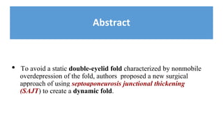
- 1. Abstract • To avoid a static double-eyelid fold characterized by nonmobile overdepression of the fold, authors proposed a new surgical approach of using septoaponeurosis junctional thickening (SAJT) to create a dynamic fold.
- 2. Abstract • 680 patients underwent double-eyelid surgery using the SAJT fixation technique. • Patients were followed for 2–8 years (mean, 3.6 y). • Anatomic study with 28 upper eyelids from 28 Korean adult cadavers was performed to confirm the histological structure of the SAJT.
- 3. Abstract • This technique created a dynamic fold: eyes were open- moderate depth eyes were closed - fold not depressed. • The surgery had a 95% patient satisfaction rate • Postoperative complications: - partial / complete loss of the double-eyelid line 14 / 4 cases - hypertrophic scar formation in 7 cases - asymmetric fold in 8 cases.
- 4. Natural double eyelid Congenital or “natural” double eyelid: 1) a smooth upper eyelid with a shallow fold line when eyes are closed 2) a fold line that is not fixated onto the tarsus and thus capable of changing with eye movement 3) equally distributed tension at the upper and lower flaps of the fold line; 4) appropriate depth of the fold when the eyes are fully open.
- 5. Static fold • The tarsal fixation technique (dermis of upper lid to tarsus) is the oldest and more commonly performed method, but the technique over-resects pretarsal tissue, resulting in a deep, immovable, depressed fold.
- 6. Static fold Fig. 1. Photographs of a 27-y-old woman exhibiting a static fold after she underwent double-eyelid surgery using the tarsal fixation technique. A static fold is characterized by deep fold formation (A) a visibly depressed line along the incision with eyes closed, and an immobile line (B).
- 7. Static fold Video Graphic 1. See video, Supplemental Digital Content 1, which shows the front and side views of a 29-year-old woman exhibiting a static fold after she underwent double eyelid surgery using the tarsal fixation technique as she opens and close her eyes
- 8. SAJT - Dynamic fold • Authors devised a technique that uses the septo-aponeurosis junctional thickening (SAJT). • It creates a fold line that changes with the movement of the eyelids. • Eyes are closed - the SAJT extends toward the skin and prevents formation of a depressed fold. Eyes are open - the levator excursion pulls the SAJT, and the fold occurs.
- 9. Static fold • The tarsal fixation technique (dermis of upper lid to tarsus) is the oldest and more commonly performed method, but the technique over-resects pretarsal tissue, resulting in a deep, immovable, depressed fold.
- 10. Patients and Methods - Anatomic Study 28 upper eyelids from 28 Korean adult cadavers were used to confirm the histological structure of the SAJT. ❏ Mean age, 65.8 y (SD, 10.8 y) ❏ 13 males, 15 females
- 11. Patients and Methods - Anatomic Study The septum inner layer joins the levator aponeurosis at the conjoined junction above the tarsal plate. The septum outer layer extends inferiorly to the tarsus where it connects with the orbicularis oculi muscle fascia and interdigitates with the distal portion of the levator aponeurosis to the ciliary margin. SAJT
- 12. Patients and Methods - Anatomic Study As both the inner and outer layers of the septum fuse at the distal levator aponeurosis (LA) interosuperior to the tarsal plate, thick septal tissue exists in this region. This portion of the septum was named the SAJT.
- 13. Operative method Between Jan 2004 - Feb 2010 - 680 patients (without blepharoptosis) • 586 women • 94 men • Mean age, 31.5 y 488 female / 52 male patients epicanthoplasty.
- 14. Design 1. Upright position + facing forward 1. The optimal central height (Fig. 4A) was marked at a point of midpupillary line as the surgeon manually elevated the eyebrow. This point was determined by observing fold height that was aesthetically appropriate for the patient. The extent of skin excision was determined by releasing the elevated eyebrow from the first marked point (Fig. 4B).
- 15. Design 3. The medial height was determined by gently tapering from the central height to the medial canthus (Fig. 4C). 4. From the central marked point A to point D at the same height above the lateral canthal angle, the lower line was drawn parallel to the curvilinear shape of the eyes. At this point, the lateral line extended parallel to the lateral canthus slant to the lateral raphe (Fig. 4E).
- 16. Design 5. The upper incision line was drawn by gently connecting the epicanthus (Fig. 4C) to point B and tapering from point B to the lateral endpoint E.
- 17. (Exposure of SAJT) video OF THE PROCEDURE
- 19. RESULTS ● The skin sutures were removed 5 days after the operation. ● The evaluation criteria were based on fold size, scar depression, and patient satisfaction. ● The first measurement was taken 5–7 days after the surgery when the skin sutures were taken out.
- 20. RESULTS ● Consecutive measurements were taken at 3, 6, and 9 weeks and at 1, 2, 4, and 8 years postoperatively
- 21. RESULTS
- 22. RESULTS
- 25. COMPLICATIONS
- 26. CONCLUSIONS The double-eyelid surgery using SAJT fixation requires meticulous dissection and has a longer operative time compared with the classic technique using tarsus fixation, septal fixation, or levator fixation. Using the SAJT fixation technique can create a dynamic and long- lasting double eyelid in Asian patients with puffy eyelids or eyelids with excess skin.
- 27. THANK YOU FOR YOUR ATTENTIONs ANY QUESTIONS?