This article discusses a study of 124 patients with differentiated thyroid cancer who underwent dosimetric evaluation to determine the optimal radioactive iodine dose for treatment. Dosimetry calculations were performed using MIRD methodology to estimate radiation doses to bone marrow, lungs, and tumor metastases. The goal was to determine the maximum safe dose that would deliver less than 3 Gy to bone marrow or 30 Gy to lungs, while aiming to deliver over 100 Gy to metastases for a curative intent. No patients experienced permanent bone marrow suppression when doses were under 3 Gy. The largest administered dose was 38.5 GBq (1,040 mCi) based on bone marrow dose limitations. Dosimetry-guided treatment allows administration of the maximum possible
![Dosimetry-Guided Radioactive Iodine Treatment
in Patients with Metastatic Differentiated Thyroid
Cancer: Largest Safe Dose Using a Risk-Adapted
Approach
Robert Dorn, MD1; Juergen Kopp2; Harry Vogt, MD1; Peter Heidenreich, MD1; Robert G. Carroll, MD3;
and Seza A. Gulec, MD4
1Department of Nuclear Medicine, Augsburg Clinic, Augsburg, Germany; 2Department of Medical Physics, Augsburg Clinic,
Augsburg, Germany; 3Nuclear Medicine Service, University Community Hospital, Tampa, Florida; and 4John Wayne Cancer
Institute, Santa Monica, California
This study is a retrospective analysis of 124 differentiated thy-
roid cancer patients who underwent dosimetric evaluation using
MIRD methodology over a period of 15 y. The objectives of the
study were to demonstrate the clinical use of dosimetry-guided
radioactive iodine ([RAI] 131I) treatment and the safe and effec-
tive application of a 3-Gy bone marrow (BM) dose in patients
with differentiated thyroid cancer. Methods: Tumor and BM
dose estimates were obtained. The administered activity that
would deliver a maximum safe dose to the organ at risk (red BM
or lungs) was determined as well as the resulting doses to the
metastases. The clinical benefit of an individual RAI treatment
was predicted on the basis of the dose estimates and the
expected therapeutic response. Each patient’s response to
treatment was assessed clinically and by monitoring the hema-
tologic profile. Results: One hundred twenty-four patients un-
derwent 187 dosimetric evaluations. One hundred four RAI
treatments were performed. A complete response at metastatic
deposits was attained with absorbed doses of Ͼ100 Gy. No
permanent BM suppression was observed in patients who re-
ceived absorbed doses of Ͻ3 Gy to BM. The maximum admin-
istered dose was 38.5 GBq (1,040 mCi) with the BM dose
limitation. Conclusion: Dosimetry-guided RAI treatment allows
administration of the maximum possible RAI dose to achieve
the maximum therapeutic benefit. Estimation of tumor dose
rates helps to determine the curative versus the palliative intent
of the therapy.
Key Words: thyroid cancer; 131I therapy; largest safe dose;
dosimetry
J Nucl Med 2003; 44:451–456
The optimal radioactive iodine (RAI) dose (administered
activity) in the treatment of differentiated thyroid cancer
(DTC) has been a subject of controversy since its first use
by Seidlin et al. (1) in 1946. A dosimetric approach and
administration of the maximum safe dose were first intro-
duced in 1962 by Benua et al. (2), who observed that
repeated subtherapeutic doses of RAI might induce dedif-
ferentiation and loss of iodine-concentrating ability of tu-
mors. The dose-limiting toxicity of RAI treatment is mainly
on the bone marrow (BM), and the limit has been set as the
dose (administered activity) that delivers 2 Gy (200 rad) to
the blood as an equivalent of BM and whole-body retention
of Ͻ4.44 GBq (Ͻ120 mCi) at 48 h (3).
There has been a significant improvement in dosimetric
techniques over the past decades. Earlier dosimetric tech-
niques involved blood and urine measurements. Image-
based whole-body dose determinations have remarkably
improved the accuracy and reproducibility of dosimetric
calculations. The development of MIRD methodology has
yielded a new paradigm in dosimetry (4). MIRD dosimetry
has been successfully used in 131I-metaiodobenzylguanidine
therapy and radioimmunotherapy (RIT) (5,6). More sophis-
ticated techniques beyond the macrodosimetry of MIRD
have evolved over the years, and new methods such as
patient-specific Monte Carlo simulation and dose-point ker-
nel convolution dosimetry have been described (7–9).
We report the experience with dosimetry-guided RAI
therapy in the management of DTC patients. This report is
a retrospective analysis of combined BM and tumor dosim-
etry application in clinical practice and addresses the safety
and efficacy of high-dose 131I administration.
MATERIALS AND METHODS
One hundred twenty-four patients with diagnosis of DTC un-
derwent 187 RAI surveys and dosimetry in hypothyroid condition
for evaluation or treatment of their metastatic disease between
Received Mar. 25, 2002; revision accepted Sep. 25, 2002.
For correspondence or reprints contact: Seza A. Gulec, MD, John Wayne
Cancer Institute, 2200 Santa Monica Blvd., Santa Monica, CA 90404.
E-mail: gulecs@jwci.org
DOSIMETRY-GUIDED RADIOIODINE TREATMENT • Dorn et al. 451
only.
by University of Miami School of Medicine on March 21, 2014. For personal usejnm.snmjournals.orgDownloaded from](https://image.slidesharecdn.com/gulec-dosimetry-guidedi-131treatment-160717124820/85/Dosimetry-guided-i-131-treatment-2-320.jpg)
![remaining 18 treatments (44%), the therapeutic endpoint of
achieving a dose to the metastases of Ն100 Gy was reached
delivering Ͻ3-Gy BM or Ͻ30-Gy lung limiting doses.
The administered activity delivering a 3-Gy BM dose
ranged from 7.4 to 37.9 GBq (200–1,040 mCi; mean, 22.1
GBq [597 mCi]). The calculated doses to metastases ranged
from 100 to Ͼ1,000 Gy.
Outcome of Patients Who Underwent Salvage
Treatment
Thirty-two patients underwent at least 1 curative-intent
RAI treatment (Table 4). Post-RAI treatment survival in this
group ranged from 0.6 to 10.9 y (mean, 4.4 y). Six patients
(19%) died of disease, and 4 patients (13%) died of other
causes (bronchial asthma, suicide caused by depression,
incarcerated hernia, sudden cardiac event) during the fol-
low-up. Twelve patients (38%) showed decreased thyro-
globulin (Tg) levels of Ͻ1 ng/mL after RAI treatment. Nine
of these 12 patients maintained their Tg level at or Ͻ1
ng/mL over a mean follow-up of 4.3 y. In 4 patients (13%),
Tg levels remained stable and Ͻ30 ng/mL over a mean
follow-up of 5.6 y. Tg levels before and after therapy are
given in Table 4.
Mild-to-moderate xerostomia was the most common side
effect among the patients who underwent curative-intent
therapy. One patient, first treated by risk-adapted RIT with
curative intent, received 5 treatments over a 10-y period
with a cumulated activity of 94.7 GBq (2.6 Ci). All metas-
tases (bone) were eradicated at the first therapy (Tg, Ͻ1
ng/mL). However, new metastatic sites were identified 2 y
after the initial treatment. The patient underwent a second
therapy with curative intent. Unfortunately, a complete re-
sponse was not obtained with the second treatment. There-
fore, all further therapies were given with palliative intent.
This patient later developed a secondary cancer and died as
a result of a pathologic hip fracture secondary to metastatic
disease.
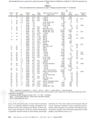
BM Toxicity
All patients (24 curative therapies and 1 palliative ther-
apy) who received 131I doses delivering 3 Gy to the BM
developed transient BM depression. BM depression reached
its nadir at approximately 3–5 wk after the RAI treatment
and manifested with thrombocytopenia followed by leuko-
penia. A spontaneous complete recovery was observed
within the next 3–5 wk (Figs. 1 and 2). Four patients (2 of
whom received a Ͻ3-Gy BM dose but had pretherapeutic
impaired BM function) required admission to the hospital; 2
of them received transfusion of platelets and red blood cells
for pancytopenia. No permanent BM failure was observed
and none of the patients required stem cell treatment for
recovery of their BM.
Lung Function
None of the patients in whom lung was the dose-limiting
organ showed symptoms of impaired lung function. Pulmo-
nary function tests were not performed because they were
not clinically indicated. A patient who had 3 prior RAI
treatments at another institution, presenting with pulmonary
fibrosis, was not selected for further RAI treatment.
DISCUSSION
Despite the generally good prognosis in most cases, ap-
proximately 10% of the patients with DTC, some with an
unexpectedly aggressive clinical course, die of their disease.
The indications and appropriate use of available treatment
options (i.e., surgery, RAI treatment, and thyroid hormone)
have been subjects of controversy for many decades. Be-
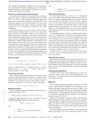
TABLE 3
Reasons for No-Treatment Action in
41 Patients (83 Dosimetries)
Reason for no-treatment action
No. of RAI
surveys or
dosimetry
Negative RAI localization of tumor 2
Low uptake in target 42
High remnant uptake (repeat surgery) 4
Surgery of recurrence or metastases 5
Contraindication for treatment (low platelet count) 1
Death before planned treatment 1
Success of previous therapy 11
No metastases after surgery 17
TABLE 1
Patient and Tumor Characteristics in 104 Patients
Who Underwent Dosimetry
Patient or tumor feature Distribution
Age (y) 4–86* (59)
Sex
Male 42† (34)
Female 82† (66)
Histopathology
Papillary 54† (44)
Follicular 63† (50)
Hu¨ rthle cell 7† (6)
*Value in parentheses is mean age.
†Value in parentheses is percentage.
TABLE 2
Tumor Localization in 95 Patients with Proven Tumor
or Metastases at Time of Dosimetry
Distribution of metastases No. of patients
Lung only 44
Bone only 18
Lung and bone 12
Lung and other 7
Cervical 6
Other 8
DOSIMETRY-GUIDED RADIOIODINE TREATMENT • Dorn et al. 453
only.
by University of Miami School of Medicine on March 21, 2014. For personal usejnm.snmjournals.orgDownloaded from](https://image.slidesharecdn.com/gulec-dosimetry-guidedi-131treatment-160717124820/85/Dosimetry-guided-i-131-treatment-4-320.jpg)
![maximizing the administered dose, one has a better chance
of overcoming the anatomic and physiologic obstacles of
nonhomogeneous distribution.
The potential impact of the stunning effect on the efficacy
of RAI treatment has been a subject of controversy clini-
cally. Furthermore, the stunning effect might also alter the
projected dose estimates. Both issues require more experi-
mental and clinical data for a rational discussion. In our
patients, no stunning effect could be objectively demon-
strated. Additionally, no decrease in thyroid uptake was
seen after dosimetry with a 370-MBq test activity (with
estimated absorbed doses as high as 50–100 Gy to the
thyroid remnants).
Stem cell procurement may offer additional safety for
patients undergoing high-dose (salvage) therapy with RAI.
Although none of the patients in our series required stem
cell treatment after RAI, such supportive backup might be
appropriate for more aggressive salvage therapies.
The risk of leukemia from high-dose RAI treatment has
also been a subject of controversy. The incidence of leuke-
mia appears to be related to the cumulated administered
activity or, more exactly, to the cumulated red BM dose
rather than to a single RAI treatment dose (especially if the
activity is Ͻ18.5 GBq [Ͻ500 mCi]). An incidence of acute
myelocytic leukemia of 1 or 2 per 100,000 per year has been
reported after a mean cumulated activity of 40.7 GBq (1,100
mCi), equaling a 3.2-Gy red BM dose. The mean latency
was 42 mo (22). To date, we have not observed a single case
of leukemia in our patients. One could even speculate that a
longer recovery period for the BM after RAI treatment
might promote cell repair mechanisms and lower the inci-
dence of leukemia. More data and longer follow-up are
needed to answer these questions more conclusively.
In the early post-RAI treatment follow-up (the first 2 mo),
the possibility of severe hematologic complications requires
intense cooperation with the hematologist and the primary
care physician. The patient’s strict compliance to monitor-
ing of the blood count is also crucial.
CONCLUSION
Dosimetry-guided high-dose (BM absorbed dose up to 3
Gy) RAI therapy is a safe approach in the treatment of
patients with DTC. This approach might also reduce the
cumulated administered activity compared with the re-
peated, limited dose schedules (3.7–7.5 GBq [100–200
mCi] every 3 or 6 mo) and, hence, may reduce the delivery
of unnecessary radiation dose to marrow and other tissues.
A risk–benefit assessment before high-dose RAI therapy is
essential. The therapeutic benefits certainly outweigh the
cost and labor associated with radiation protection measures
and potential stem cell procurement applications. Decreased
total hospital stay and pretreatment preparation period (es-
pecially when a thyroid hormone withdrawal protocol is
applied) favorably affect the quality of life in patients
with DTC.
REFERENCES
1. Seidlin SM, Marinelli LD, Oshry E. Radioactive iodine therapy: effect on
functioning metastases of adenocarcinoma of thyroid. JAMA. 1946;132:838–
847.
2. Benua RS, Cicale NR, Sonenberg M, Rawson RW. The relation of radioiodine
dosimetry to results and complications in the treatment of metastatic thyroid
cancer. AJR. 1962;87:171–182.
3. Benua RS, Leeper RD. A method and rationale for treating thyroid carcinoma
with the largest safe dose of I-131. In: Meideros-Neto GA, Gaitan E, eds.
Frontiers of Thyroidology. Vol. II. New York, NY: Plenum; 1986:1317–1321.
4. Loevinger R, Budinger TF, Watson EE, in collaboration with the MIRD Com-
mittee. MIRD Primer for Absorbed Dose Calculations. New York, NY: The
Society of Nuclear Medicine; 1988.
5. Beierwaltes WH. Update on basic research and clinical experience with metaio-
dobenzylguanidine. Med Pediatr Oncol. 1987;15:163–169.
6. Rao DV, Howell RW. Time-dose-fractionation in radioimmunotherapy: implica-
tions for selecting radionuclides. J Nucl Med. 1993;34:1801–1810.
7. Kolbert KS, Sgouros G, Scott AM, et al. Implementation and evaluation of
patient-specific three-dimensional dosimetry. J Nucl Med. 1997;38:301–308.
8. Akabani G, Hawkins WG, Eckblade MB, Leichter PK. Patient-specific dosimetry
using quantitative SPECT imaging and three-dimensional discrete Fourier trans-
form convolution. J Nucl Med. 1997;38:308–314.
9. Furhang EF, Larson SM, Buranapong P, Humm JL. Thyroid cancer dosimetry
using clearance fitting. J Nucl Med. 1999;40:131–136.
10. Mazzaferri EL. Long-term outcome of patients with differentiated thyroid carci-
noma: effect of therapy. Endocr Pract. 2000;6:469–476.
11. Taylor T, Speckler B, Robbins J, et al. Outcome after treatment of high risk
papillary and non-Hurthle-cell follicular thyroid carcinoma. Ann Intern Med.
1998;129:622–627.
12. VanNostrand D, Neutze J, Atkins F. Side effects of “rational” iodine-131 therapy
for metastatic well-differentiated thyroid carcinoma. J Nucl Med. 1986;27:1519–
1527.
13. Thomas SR, Samaratunga RC, Sperling M, Maxon HR. Predictive estimate of
blood dose from external counting data preceding radioiodine therapy for thyroid
cancer. Nucl Med Biol. 1993;20:157–162.
14. Sisson JC, Ackermann R, Zempel S, Spaulding S. Treatment with 131I imports
less absorbed radiation to thyroid cancer than predicted by dosimetry [abstract].
Thyroid. 1994;4:S-61.
15. Stabin MG. MIRDOSE: personal computer software for internal dose assessment
in nuclear medicine. J Nucl Med. 1996;37:538–546.
16. Kopp J, Heidenreich P. Clinical whole-body dosimetry and therapy of metastases
with 131I. Proceedings, Sixth International Radiopharmaceutical Dosimetry Sym-
posium. Gatlinburg, TN: Oak Ridge Associated Universities; 1999:137–139.
17. Beierwaltes WH, Nishiyama RH, Tompson NW, Copp JE, Kubo A. Survival time
and “cure” in papillary and follicular thyroid carcinoma with distant metastases:
statistics following University of Michigan therapy. J Nucl Med. 1982;23:561–
568.
18. Maxon HR, Thomas SR, Samaratunga RC. Dosimetric considerations in the
radioiodine treatment of macrometastases and micrometastases from differenti-
ated thyroid cancer. Thyroid. 1997;7:183–187.
19. Miller RC, Hiraoka T, Kopecky KJ, et al. Sensitivity to radiation of normal,
hyperthyroid, and neoplastic thyroid epithelial cells in primary culture. Radiat
Res. 1987;111:81–91.
20. Maxon HR. Quantitative radioiodine therapy in the treatment of differentiated
thyroid cancer. Q J Nucl Med. 1999;43:313–323.
21. Flower MA, Schlesinger T, Hinton PJ, et al. Radiation dose assessment in
radioiodine therapy. 2. Practical implementation using quantitative scanning and
PET, with initial results on thyroid carcinoma. Radiother Oncol. 1989;15:345–
357.
22. Guenter H-H, Schober O, Schwarzrock R, Hundeshagen H. Hematologic long-
time modifications after radioiodine therapy of the carcinoma of the thyroid
gland. II. Modifications of the bone marrow including leukemia [in German].
Strahlenther Onkol. 1987;163:475–485.
456 THE JOURNAL OF NUCLEAR MEDICINE • Vol. 44 • No. 3 • March 2003
only.
by University of Miami School of Medicine on March 21, 2014. For personal usejnm.snmjournals.orgDownloaded from](https://image.slidesharecdn.com/gulec-dosimetry-guidedi-131treatment-160717124820/85/Dosimetry-guided-i-131-treatment-7-320.jpg)


![11.[42 53]effectiveness of gefitinib as additional radiosensitizer to convent...](https://cdn.slidesharecdn.com/ss_thumbnails/11-42-53effectivenessofgefitinibasadditionalradiosensitizertoconventionalchemoradiationforlocallyadvancednon-metastaticsquamouscellcarcinomaofheadandneck-120512235836-phpapp01-thumbnail.jpg?width=640&height=640&fit=bounds)


![Cells and Organs of immune system [Autosaved].pptx](https://cdn.slidesharecdn.com/ss_thumbnails/cellsandorgansofimmunesystemautosaved-260123152717-ea0cb261-thumbnail.jpg?width=640&height=640&fit=bounds)


