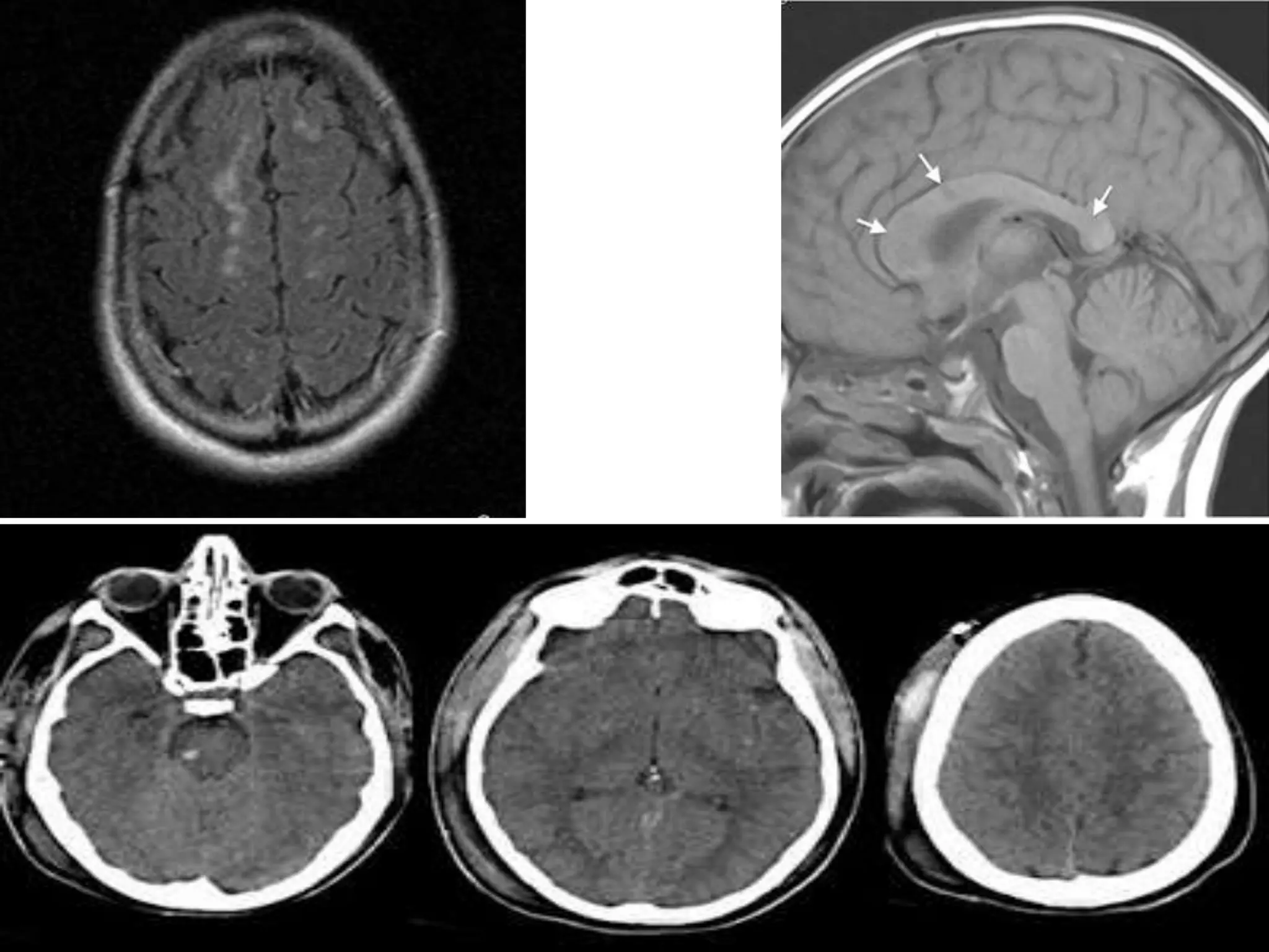
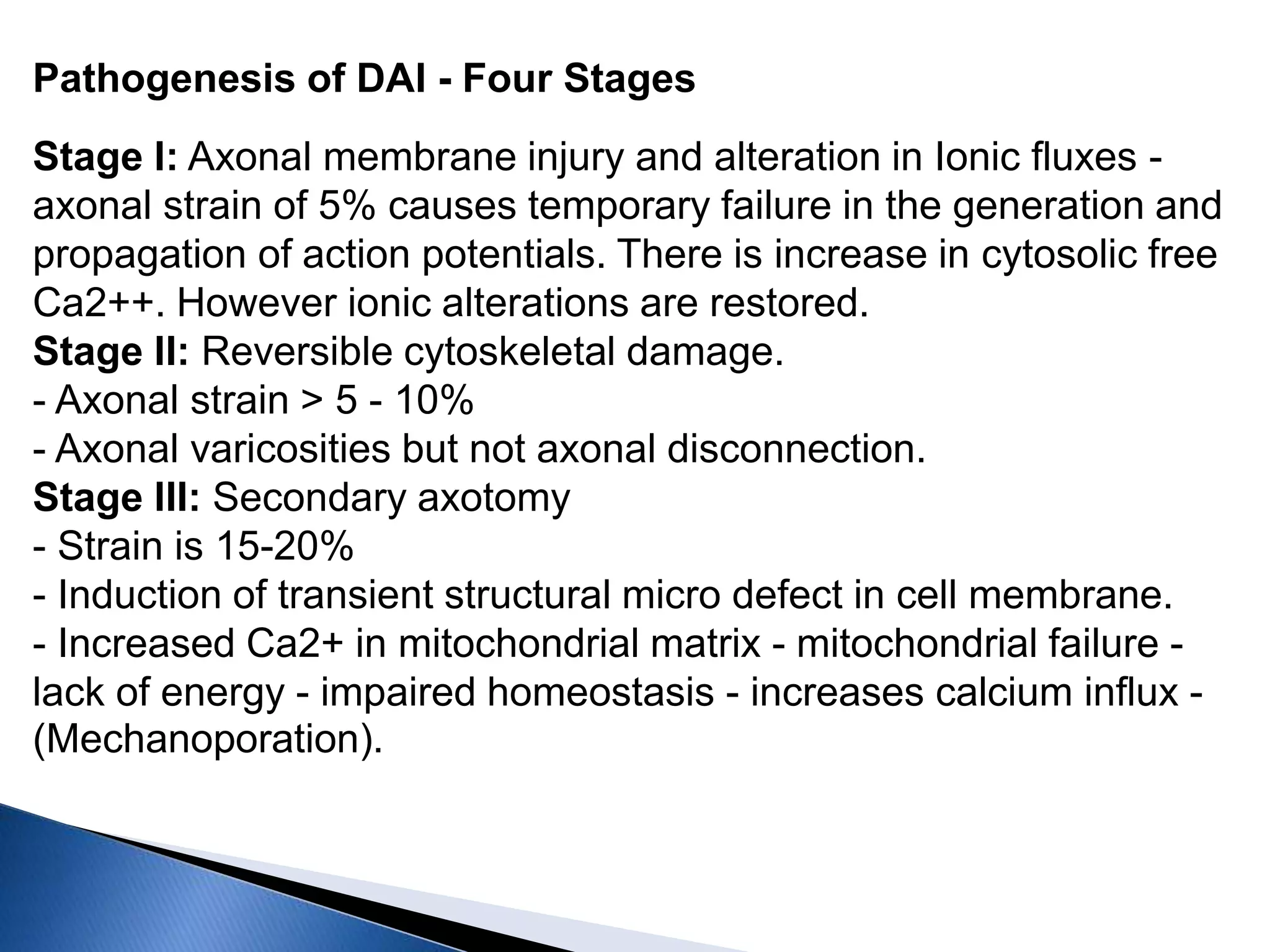
DR.Kalakoti chandra sekhar reddy
 A form of post-
traumatic brain injury which occurs over a
broad swath of myelinated tracts of the CNS,
resulting in significant neurologic effects
ranging from loss of consciousness to
persistent vegetative state.
Diffuse axonal injury
 - Occurs in 50% of severely head injured
patients and accounts for 35% of all death from
Head injury.
 Triad - Lesion in corpus callosum.
 - Dorsolateral quadrant of brain stem.
 - Axonal rolling of white matter.
 The susceptibility of axons to mechanical
injury appears to be due to both their
viscoelastic properties and their high
organization in white matter tracts.
 Two basic mechanisms
-contact injuries
-Inertial injuries
 physical stretch, or mechanoporation at the
time of injury, results in damage to the
axolemma and related axoplasm at the
injured node of Ranvier
 Either following impact or from impulse
loading
 Brain lags behind due to inertia
 Movement away from skull cause low pressure
[ tensile strain] - contrecoup injuries
 Strain within parenchyma cause diffuse injuries
[concussions and DAI], tissue tear
hemorrhages.
Pathogenesis of DAI - Four Stages
Stage I: Axonal membrane injury and alteration in Ionic fluxes -
axonal strain of 5% causes temporary failure in the generation and
propagation of action potentials. There is increase in cytosolic free
Ca2++. However ionic alterations are restored.
Stage II: Reversible cytoskeletal damage.
- Axonal strain > 5 - 10%
- Axonal varicosities but not axonal disconnection.
Stage III: Secondary axotomy
- Strain is 15-20%
- Induction of transient structural micro defect in cell membrane.
- Increased Ca2+ in mitochondrial matrix - mitochondrial failure -
lack of energy - impaired homeostasis - increases calcium influx -
(Mechanoporation).
Stage IV:
- Primary axotomy
- Strain is > 20%
- Most severe form
- Tearing of axons
Axonal swelling in corpus
callosum
Disruption& globular swelling
of axons in fornix.
Dilated axonal spheroids in
thalamus.
Diffuse Axonal injury (DAI)
Focal axonal swelling without
disconnection (Bodian silver)
Dystrophic changes in
cortical neurons proximal
to damage
Normal Axons in white
matter
Neurofilament
immunostain
 grade I: involves grey-white matter interfaces
◦ most commonly: parasagittal regions of frontal lobes,
periventricular temporal lobes
◦ less commonly: parietal and occipital lobes, internal and external
capsules, and cerebellum
◦ often inapparent on conventional imaging
◦ may have changes on MR spectroscopy (MRS)
 grade II: involves corpus callosum in addition to grade I
locations
◦ observed in approximately 20% of patients
◦ most commonly: posterior body and splenium but does advance
anteriorly with increasing severity of injury
◦ most frequently unilateral
 grade III: involves brainstem in addition to grade I and II
locations
◦ most commonly: rostral midbrain, superior cerebellar peduncles,
medial lemnisci and corticospinal tracts
Grading of DAI: [ Gennarelli et al 1982]
 I. Microscopic axonal damage in the white matter, corpus
callosum, brain stem or cerebellum without any macroscopic
evidence.
 II. Macroscopic or Microscopic detected focal lesions in the
CC and diffuse axonal damage.
 III. Macroscopic or microscopic injury, focal injury to CC +
dorsolateral quadrant of rostral brain stem.
 Wakefulness and alertness-RAS
 Swallowing -9,10,12 nerves
 Breathing –Apneustic center
 Arrhythmias-Chemoreceptor trigger zone
 Vision impairement:MLF and PPRF
 The amount of axonal injury in the brainstem
is predictive of long-term vegetative state,
 supratentorial injury can result in focal
neurological or neuropsychiatric deficits
 MILD :coma for 6 to 24 hrs
 MODERATE :coma for more than 24 hrs but
no clinical sign of brainstem dysfunction.
 SEVERE :more than 24 hrs with brainstem
signs
 Injury depends on rate and magnitude of
acceleration
No injury subdural hematomas DAI
Short duration longer duration high magnitude
Well damped falls, assaults motor vehicle accidents
 Transient reversible dysfunction due to trauma
Loss of consciousness < 6 hours ,confusion, easy
distractibility, amnesia
 Angular or horizontal acceleration deforming
deeper aspects of the brain
 No permanent structural damage unless repeated
 Precise location of the functional derangement
remains undetermined
 The outcome of patients after DAI has been
linked to the number of lesions identified
through imaging.
 Volume of the lesions and
 Location of lesion
 MRI is the imaging modality of choice as it
identifies the lesions not identified on CT.
 Paroxysmal autonomic instability with dystonia
(PAID) appears to be a unique syndrome
following brain injury.
 elevated temperature (≥ 38.5°C),
 Tachycardia (≥ 130 beats per minute),
 Tachypnoea (≥ 140 breaths per minute),
 Agitation
 Diaphoresis and
 Dystonia (rigidity or decerebrate posturing).
 The duration of an episode is cyclic, with at least
one cycle per day for at least three days.
Managed with
 Antipyretics
 Morphine
 Benzodiazepines
 Betablockers
 ICP measurement
 multimodality monitoring (MMM)
 PbtO2 and cerebral microdialysis are
independent markers of mortality.
 Main goal to prevent secondary brain injury
like cerebral ischemia, intracranial
hypertension, and energy dysfunction.
Includes
 serial neurological examinations,
 imaging studies,
 ICP analysis, and
 measurements from other bedside
physiologic monitors.
 MMM can be used in evaluating adequate
 hyperventilation,
 osmotherapy,
 glycemic control,
 transfusion,
 CPP augmentation,
 therapeutic temperature modulation, and
 oxygen-based therapies
 Pt with GCS <8
 Motor posturing on presentation
 Abnormal ct on admission
 Age >40
 1 episode of BP <90mm of hg
 In decompressive craniectomy following
delayed surgery or failed medical
management
Invasive methods:
 Epidural catheter
 Intraparenchymal
 Intraventricular
Non invasive:
 Mri based indirect icp measurement
 Optic nerve diameter measurement
 elevating the patients head to 30°
 hyperventilation only to a pCO2 level of
25 mmHg
 Mannitol effect seen in 10-20 mins
 Barbiturates only for barbiturate induced
coma last resort
 EVD
 Decompression
 VP shunt
Diffuse axonal injury

Diffuse axonal injury

  • 1.
  • 2.
     A formof post- traumatic brain injury which occurs over a broad swath of myelinated tracts of the CNS, resulting in significant neurologic effects ranging from loss of consciousness to persistent vegetative state.
  • 3.
    Diffuse axonal injury - Occurs in 50% of severely head injured patients and accounts for 35% of all death from Head injury.  Triad - Lesion in corpus callosum.  - Dorsolateral quadrant of brain stem.  - Axonal rolling of white matter.
  • 4.
     The susceptibilityof axons to mechanical injury appears to be due to both their viscoelastic properties and their high organization in white matter tracts.
  • 5.
     Two basicmechanisms -contact injuries -Inertial injuries  physical stretch, or mechanoporation at the time of injury, results in damage to the axolemma and related axoplasm at the injured node of Ranvier
  • 6.
     Either followingimpact or from impulse loading  Brain lags behind due to inertia  Movement away from skull cause low pressure [ tensile strain] - contrecoup injuries  Strain within parenchyma cause diffuse injuries [concussions and DAI], tissue tear hemorrhages.
  • 7.
    Pathogenesis of DAI- Four Stages Stage I: Axonal membrane injury and alteration in Ionic fluxes - axonal strain of 5% causes temporary failure in the generation and propagation of action potentials. There is increase in cytosolic free Ca2++. However ionic alterations are restored. Stage II: Reversible cytoskeletal damage. - Axonal strain > 5 - 10% - Axonal varicosities but not axonal disconnection. Stage III: Secondary axotomy - Strain is 15-20% - Induction of transient structural micro defect in cell membrane. - Increased Ca2+ in mitochondrial matrix - mitochondrial failure - lack of energy - impaired homeostasis - increases calcium influx - (Mechanoporation).
  • 8.
    Stage IV: - Primaryaxotomy - Strain is > 20% - Most severe form - Tearing of axons
  • 9.
    Axonal swelling incorpus callosum Disruption& globular swelling of axons in fornix. Dilated axonal spheroids in thalamus. Diffuse Axonal injury (DAI) Focal axonal swelling without disconnection (Bodian silver) Dystrophic changes in cortical neurons proximal to damage Normal Axons in white matter Neurofilament immunostain
  • 11.
     grade I:involves grey-white matter interfaces ◦ most commonly: parasagittal regions of frontal lobes, periventricular temporal lobes ◦ less commonly: parietal and occipital lobes, internal and external capsules, and cerebellum ◦ often inapparent on conventional imaging ◦ may have changes on MR spectroscopy (MRS)  grade II: involves corpus callosum in addition to grade I locations ◦ observed in approximately 20% of patients ◦ most commonly: posterior body and splenium but does advance anteriorly with increasing severity of injury ◦ most frequently unilateral  grade III: involves brainstem in addition to grade I and II locations ◦ most commonly: rostral midbrain, superior cerebellar peduncles, medial lemnisci and corticospinal tracts
  • 12.
    Grading of DAI:[ Gennarelli et al 1982]  I. Microscopic axonal damage in the white matter, corpus callosum, brain stem or cerebellum without any macroscopic evidence.  II. Macroscopic or Microscopic detected focal lesions in the CC and diffuse axonal damage.  III. Macroscopic or microscopic injury, focal injury to CC + dorsolateral quadrant of rostral brain stem.
  • 13.
     Wakefulness andalertness-RAS  Swallowing -9,10,12 nerves  Breathing –Apneustic center  Arrhythmias-Chemoreceptor trigger zone  Vision impairement:MLF and PPRF
  • 14.
     The amountof axonal injury in the brainstem is predictive of long-term vegetative state,  supratentorial injury can result in focal neurological or neuropsychiatric deficits
  • 16.
     MILD :comafor 6 to 24 hrs  MODERATE :coma for more than 24 hrs but no clinical sign of brainstem dysfunction.  SEVERE :more than 24 hrs with brainstem signs
  • 17.
     Injury dependson rate and magnitude of acceleration No injury subdural hematomas DAI Short duration longer duration high magnitude Well damped falls, assaults motor vehicle accidents
  • 18.
     Transient reversibledysfunction due to trauma Loss of consciousness < 6 hours ,confusion, easy distractibility, amnesia  Angular or horizontal acceleration deforming deeper aspects of the brain  No permanent structural damage unless repeated  Precise location of the functional derangement remains undetermined
  • 19.
     The outcomeof patients after DAI has been linked to the number of lesions identified through imaging.  Volume of the lesions and  Location of lesion  MRI is the imaging modality of choice as it identifies the lesions not identified on CT.
  • 24.
     Paroxysmal autonomicinstability with dystonia (PAID) appears to be a unique syndrome following brain injury.  elevated temperature (≥ 38.5°C),  Tachycardia (≥ 130 beats per minute),  Tachypnoea (≥ 140 breaths per minute),  Agitation  Diaphoresis and  Dystonia (rigidity or decerebrate posturing).  The duration of an episode is cyclic, with at least one cycle per day for at least three days.
  • 25.
    Managed with  Antipyretics Morphine  Benzodiazepines  Betablockers
  • 26.
     ICP measurement multimodality monitoring (MMM)  PbtO2 and cerebral microdialysis are independent markers of mortality.
  • 27.
     Main goalto prevent secondary brain injury like cerebral ischemia, intracranial hypertension, and energy dysfunction.
  • 28.
    Includes  serial neurologicalexaminations,  imaging studies,  ICP analysis, and  measurements from other bedside physiologic monitors.
  • 29.
     MMM canbe used in evaluating adequate  hyperventilation,  osmotherapy,  glycemic control,  transfusion,  CPP augmentation,  therapeutic temperature modulation, and  oxygen-based therapies
  • 30.
     Pt withGCS <8  Motor posturing on presentation  Abnormal ct on admission  Age >40  1 episode of BP <90mm of hg  In decompressive craniectomy following delayed surgery or failed medical management
  • 31.
    Invasive methods:  Epiduralcatheter  Intraparenchymal  Intraventricular Non invasive:  Mri based indirect icp measurement  Optic nerve diameter measurement
  • 33.
     elevating thepatients head to 30°  hyperventilation only to a pCO2 level of 25 mmHg  Mannitol effect seen in 10-20 mins  Barbiturates only for barbiturate induced coma last resort
  • 34.