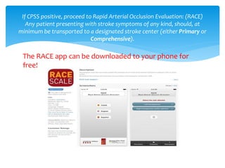
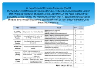
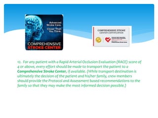
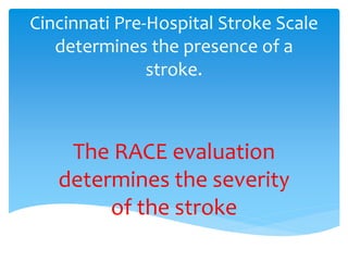
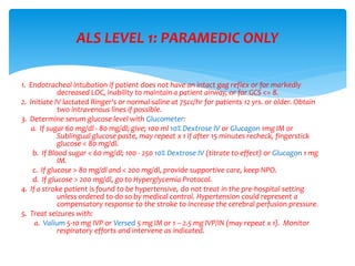
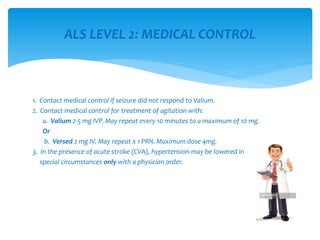
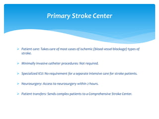
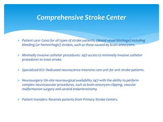
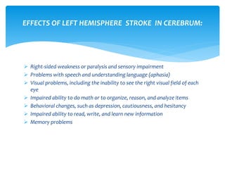
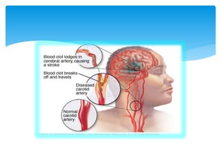
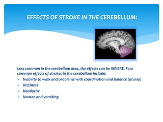
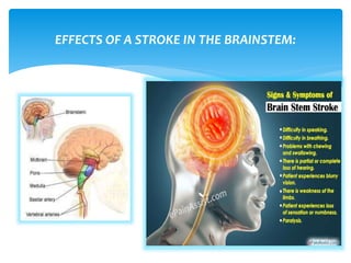
CVA/STROKE
Purpose:
This protocol is used for those patients exhibiting signs
consistent with acute stroke/CVA/”Brain Attack” (altered
mental status, slurred speech, loss of function of any
body part, hemiplegia, loss of vision, weakness of facial
muscles, loss of sensation, drooling, etc.). Other causes
should be ruled out (hypoglycemia, drug overdose,
hypoxia, etc.).
2.5.5 CVA/Stroke Adult Medical Protocol
BASIC LEVEL: EMT and PARAMEDIC
1. Initial Patient Assessment Protocol 2.1.1.
2. Airway Assessment/Management Protocol 2.1.2. Oxygen via nasal cannula
@2 - 4 LPM to maintain pulse ox of > 94% (non-rebreather @15 LPM if SpO2 <
90%).
3. When CVA is suspected, transport to the hospital should not be delayed.
Determine if patient has facial droop, abnormal speech, or arm drift.
4. If possible place in Semi-Fowler’s position with head of bed elevated 30
degrees for transport (if patient unable to tolerate, transport flat).
Procedure
5. Assess for and document Glasgow Coma Scale.
6. Attach cardiac monitor and pulse oximeter.
7. Keep patient NPO.
8. Determine time last seen normal. If onset of symptoms is within 6 hours
notify hospital of a “stroke alert”.
9. Try to determine if patient had a seizure prior to onset of “stroke”
symptoms as he/she may have a condition called Todd’s paralysis, which is
NOT treated with thrombolytics. Relay this information to the hospital.
Procedure cont.
10. Cincinnati Pre-hospital Stroke Scale: (CPSS)
a. Assess for the unilateral presence of at least one of the following:
Item Description
1. Facial droop: Ask the patient to smile. Watch for weakness on one side
of the face.
2. Arm drift: Ask the patient to hold both arms out with palms up and
eyes closed for 10 seconds. Watch for a drift of one side. A positive
result is present if there is weakness in one arm. Weakness in
both arms or normal strength is a negative test result.
3. Slurred speech: Ask the patient to repeat a simple sentence such as
“The sky is blue in Cincinnati.” Inability to repeat the words correctly
and intelligibly is a positive result.
TRANSPORT: DESTINATION DETERMINATION
The RACE app can be downloaded to your phone for
free!
If CPSS positive, proceed to Rapid Arterial Occlusion Evaluation: (RACE)
Any patient presenting with stroke symptoms of any kind, should, at
minimum be transported to a designated stroke center (either Primary or
Comprehensive).
11. Rapid Arterial Occlusion Evaluation (RACE)
The Rapid Arterial Occlusion Evaluation (R.A.C.E.) is based on an abbreviated version
of the National Institutes of Health Stroke Scale (NIHSS), the “gold standard” for
evaluating stroke victims. The maximum score is 9 (not 11) because the evaluation of
the final two components is done based on the left or right side presentation, not
both simultaneously.
12. For any patient with a Rapid Arterial Occlusion Evaluation (RACE) score of
4 or above, every effort should be made to transport the patient to a
Comprehensive Stroke Center, if available. (While transport destination is
ultimately the decision of the patient and his/her family, crew members
should provide the Protocol and Assessment based recommendations to the
family so that they may make the most informed decision possible.)
Cincinnati Pre-Hospital Stroke Scale
determines the presence of a
stroke.
The RACE evaluation
determines the severity
of the stroke
1. Endotracheal intubation if patient does not have an intact gag reflex or for markedly
decreased LOC, inability to maintain a patient airway, or for GCS <= 8.
2. Initiate IV lactated Ringer's or normal saline at 75cc/hr for patients 12 yrs. or older. Obtain
two intravenous lines if possible.
3. Determine serum glucose level with Glucometer:
a. If sugar 60 mg/dl - 80 mg/dl; give; 100 ml 10% Dextrose IV or Glucagon 1mg IM or
Sublingual glucose paste, may repeat x 1 if after 15 minutes recheck, fingerstick
glucose < 80 mg/dl.
b. If Blood sugar < 60 mg/dl; 100 - 250 10% Dextrose IV (titrate to effect) or Glucagon 1 mg
IM.
c. If glucose > 80 mg/dl and < 200 mg/dl, provide supportive care, keep NPO.
d. If glucose > 200 mg/dl, go to Hyperglycemia Protocol.
4. If a stroke patient is found to be hypertensive, do not treat in the pre-hospital setting
unless ordered to do so by medical control. Hypertension could represent a
compensatory response to the stroke to increase the cerebral perfusion pressure.
5. Treat seizures with:
a. Valium 5-10 mg IVP or Versed 5 mg IM or 1 – 2.5 mg IVP/IN (may repeat x 1). Monitor
respiratory efforts and intervene as indicated.
ALS LEVEL 1: PARAMEDIC ONLY
1. Contact medical control if seizure did not respond to Valium.
2. Contact medical control for treatment of agitation with:
a. Valium 2-5 mg IVP. May repeat every 10 minutes to a maximum of 10 mg.
Or
b. Versed 2 mg IV. May repeat x 1 PRN. Maximum dose 4mg.
3. In the presence of acute stroke (CVA), hypertension may be lowered in
special circumstances only with a physician order.
ALS LEVEL 2: MEDICAL CONTROL
 Patient care: Takes care of most cases of ischemic (blood vessel blockage) types of
stroke.
 Minimally invasive catheter procedures: Not required.
 Specialized ICU: No requirement for a separate intensive care for stroke patients.
 Neurosurgery: Access to neurosurgery within 2 hours.
 Patient transfers: Sends complex patients to a Comprehensive Stroke Center.
Primary Stroke Center
 Patient care: Cares for all types of stroke patients, (blood vessel blockage) including
bleeding (or hemorrhagic) strokes, such as those caused by brain aneurysms.
 Minimally invasive catheter procedures: 24/7 access to minimally invasive catheter
procedures to treat stroke.
 Specialized ICU: Dedicated neuroscience intensive care unit for unit stroke patients.
 Neurosurgery: On-site neurosurgical availability 24/7 with the ability to perform
complex neurovascular procedures, such as brain aneurysm clipping, vascular
malformation surgery and carotid endarterectomy.
 Patient transfers: Receives patients from Primary Stroke Centers.
Comprehensive Stroke Center
Ischemic stroke (Clots) occurs
as a result of an obstruction
within a blood vessel supplying
blood to the brain. It accounts
for 87 percent of all stroke
cases
Hemorrhagic stroke occurs
when a weakened blood
vessel ruptures. Two types of
weakened blood vessels
usually cause hemorrhagic
stroke: aneurysms and
arteriovenous malformations
(AVMs). But the most
common cause of
hemorrhagic stroke is
uncontrolled hypertension
(high blood pressure)
TIA (transient ischemic attack)
is caused by a temporary clot.
Often called a “mini stroke”,
these warning strokes should
be taken very seriously.
.
SAVE THE PENUMBRA!

Depending on which region of the brain the stroke occurs, the effects may be
very different. The brain is divided into 3 main areas:
Cerebrum (consisting of the right and left sides or hemispheres)
Cerebellum
Brainstem
Brainstem
Hemorrhagic Stroke
There are two types of hemorrhagic strokes:
Intracerebral hemorrhage is the most
common type of hemorrhagic stroke. It occurs
when an artery in the brain bursts, flooding
the surrounding tissue with blood.
Subarachnoid hemorrhage is a less common
type of hemorrhagic stroke. It refers to
bleeding in the area between the brain and
the thin tissues that cover it.
There are two kinds of thrombosis that can lead to a stroke: large vessel
thrombosis and small vessel disease, also called “lacunar infarction.” Thrombotic
stroke is caused by an artery disease called atherosclerosis, which is followed by
the formation of blood clots.
According to the National Stroke Association (NSA), large vessel thrombosis is the
most common of the thrombotic strokes.
Types of Thrombotic Stroke
Small vessel disease, or “lacunar infarction,” is the other type of stroke-causing
thrombosis. Small vessel disease develops from a blood clot in a small artery.
According to the NSA, researchers are uncertain about the exact cause of lacunar
infarction. But they do know that the condition is related to high blood pressure.
Both types of thrombotic stroke have been linked to coronary artery disease,
the NSA reports.
Lacunar Infarction
Depending on the area and side of the cerebrum affected by the stroke, any,
or all, of these functions may be impaired:
 Movement and sensation
 Speech and language
 Eating and swallowing
 Vision
 Cognitive (thinking, reasoning, judgment, and memory) ability
 Perception and orientation to surroundings
 Self-care ability
 Bowel and bladder control
 Emotional control
 Sexual ability
In addition to these general effects, some specific impairments may occur when a particular
area of the cerebrum is damaged.
EFFECTS OF STROKE IN THE CEREBRUM:
 Left-sided weakness or paralysis and sensory impairment.
 Denial of paralysis or impairment and reduced insight into the problems
created by the stroke (this is called “left neglect”).
 Visual problems, including an inability to see the left visual field of each
eye.
 Spatial problems with depth perception or directions, such as up or down
and front or back.
 Inability to localize or recognize body parts.
 Inability to understand maps and find objects, such as clothing or toiletry
items.
 Memory problems.
 Behavioral changes, such as lack of concern about situations,
inappropriateness, and depression.
EFFECTS OF RIGHT HEMISPHERE STROKE IN CEREBRUM:
 Right-sided weakness or paralysis and sensory impairment
 Problems with speech and understanding language (aphasia)
 Visual problems, including the inability to see the right visual field of each
eye
 Impaired ability to do math or to organize, reason, and analyze items
 Behavioral changes, such as depression, cautiousness, and hesitancy
 Impaired ability to read, write, and learn new information
 Memory problems
EFFECTS OF LEFT HEMISPHERE STROKE IN CEREBRUM:
Less common in the cerebellum area, the effects can be SEVERE. Four
common effects of strokes in the cerebellum include:
 Inability to walk and problems with coordination and balance (ataxia)
 Dizziness
 Headache
 Nausea and vomiting
EFFECTS OF STROKE IN THE CEREBELLUM:
EFFECTS OF A STROKE IN THE BRAINSTEM:

CVA

  • 1.
  • 2.
    Purpose: This protocol isused for those patients exhibiting signs consistent with acute stroke/CVA/”Brain Attack” (altered mental status, slurred speech, loss of function of any body part, hemiplegia, loss of vision, weakness of facial muscles, loss of sensation, drooling, etc.). Other causes should be ruled out (hypoglycemia, drug overdose, hypoxia, etc.). 2.5.5 CVA/Stroke Adult Medical Protocol
  • 3.
    BASIC LEVEL: EMTand PARAMEDIC 1. Initial Patient Assessment Protocol 2.1.1. 2. Airway Assessment/Management Protocol 2.1.2. Oxygen via nasal cannula @2 - 4 LPM to maintain pulse ox of > 94% (non-rebreather @15 LPM if SpO2 < 90%). 3. When CVA is suspected, transport to the hospital should not be delayed. Determine if patient has facial droop, abnormal speech, or arm drift. 4. If possible place in Semi-Fowler’s position with head of bed elevated 30 degrees for transport (if patient unable to tolerate, transport flat). Procedure
  • 4.
    5. Assess forand document Glasgow Coma Scale. 6. Attach cardiac monitor and pulse oximeter. 7. Keep patient NPO. 8. Determine time last seen normal. If onset of symptoms is within 6 hours notify hospital of a “stroke alert”. 9. Try to determine if patient had a seizure prior to onset of “stroke” symptoms as he/she may have a condition called Todd’s paralysis, which is NOT treated with thrombolytics. Relay this information to the hospital. Procedure cont.
  • 5.
    10. Cincinnati Pre-hospitalStroke Scale: (CPSS) a. Assess for the unilateral presence of at least one of the following: Item Description 1. Facial droop: Ask the patient to smile. Watch for weakness on one side of the face. 2. Arm drift: Ask the patient to hold both arms out with palms up and eyes closed for 10 seconds. Watch for a drift of one side. A positive result is present if there is weakness in one arm. Weakness in both arms or normal strength is a negative test result. 3. Slurred speech: Ask the patient to repeat a simple sentence such as “The sky is blue in Cincinnati.” Inability to repeat the words correctly and intelligibly is a positive result. TRANSPORT: DESTINATION DETERMINATION
  • 6.
    The RACE appcan be downloaded to your phone for free! If CPSS positive, proceed to Rapid Arterial Occlusion Evaluation: (RACE) Any patient presenting with stroke symptoms of any kind, should, at minimum be transported to a designated stroke center (either Primary or Comprehensive).
  • 7.
    11. Rapid ArterialOcclusion Evaluation (RACE) The Rapid Arterial Occlusion Evaluation (R.A.C.E.) is based on an abbreviated version of the National Institutes of Health Stroke Scale (NIHSS), the “gold standard” for evaluating stroke victims. The maximum score is 9 (not 11) because the evaluation of the final two components is done based on the left or right side presentation, not both simultaneously.
  • 8.
    12. For anypatient with a Rapid Arterial Occlusion Evaluation (RACE) score of 4 or above, every effort should be made to transport the patient to a Comprehensive Stroke Center, if available. (While transport destination is ultimately the decision of the patient and his/her family, crew members should provide the Protocol and Assessment based recommendations to the family so that they may make the most informed decision possible.)
  • 9.
    Cincinnati Pre-Hospital StrokeScale determines the presence of a stroke. The RACE evaluation determines the severity of the stroke
  • 10.
    1. Endotracheal intubationif patient does not have an intact gag reflex or for markedly decreased LOC, inability to maintain a patient airway, or for GCS <= 8. 2. Initiate IV lactated Ringer's or normal saline at 75cc/hr for patients 12 yrs. or older. Obtain two intravenous lines if possible. 3. Determine serum glucose level with Glucometer: a. If sugar 60 mg/dl - 80 mg/dl; give; 100 ml 10% Dextrose IV or Glucagon 1mg IM or Sublingual glucose paste, may repeat x 1 if after 15 minutes recheck, fingerstick glucose < 80 mg/dl. b. If Blood sugar < 60 mg/dl; 100 - 250 10% Dextrose IV (titrate to effect) or Glucagon 1 mg IM. c. If glucose > 80 mg/dl and < 200 mg/dl, provide supportive care, keep NPO. d. If glucose > 200 mg/dl, go to Hyperglycemia Protocol. 4. If a stroke patient is found to be hypertensive, do not treat in the pre-hospital setting unless ordered to do so by medical control. Hypertension could represent a compensatory response to the stroke to increase the cerebral perfusion pressure. 5. Treat seizures with: a. Valium 5-10 mg IVP or Versed 5 mg IM or 1 – 2.5 mg IVP/IN (may repeat x 1). Monitor respiratory efforts and intervene as indicated. ALS LEVEL 1: PARAMEDIC ONLY
  • 11.
    1. Contact medicalcontrol if seizure did not respond to Valium. 2. Contact medical control for treatment of agitation with: a. Valium 2-5 mg IVP. May repeat every 10 minutes to a maximum of 10 mg. Or b. Versed 2 mg IV. May repeat x 1 PRN. Maximum dose 4mg. 3. In the presence of acute stroke (CVA), hypertension may be lowered in special circumstances only with a physician order. ALS LEVEL 2: MEDICAL CONTROL
  • 12.
     Patient care:Takes care of most cases of ischemic (blood vessel blockage) types of stroke.  Minimally invasive catheter procedures: Not required.  Specialized ICU: No requirement for a separate intensive care for stroke patients.  Neurosurgery: Access to neurosurgery within 2 hours.  Patient transfers: Sends complex patients to a Comprehensive Stroke Center. Primary Stroke Center
  • 13.
     Patient care:Cares for all types of stroke patients, (blood vessel blockage) including bleeding (or hemorrhagic) strokes, such as those caused by brain aneurysms.  Minimally invasive catheter procedures: 24/7 access to minimally invasive catheter procedures to treat stroke.  Specialized ICU: Dedicated neuroscience intensive care unit for unit stroke patients.  Neurosurgery: On-site neurosurgical availability 24/7 with the ability to perform complex neurovascular procedures, such as brain aneurysm clipping, vascular malformation surgery and carotid endarterectomy.  Patient transfers: Receives patients from Primary Stroke Centers. Comprehensive Stroke Center
  • 14.
    Ischemic stroke (Clots)occurs as a result of an obstruction within a blood vessel supplying blood to the brain. It accounts for 87 percent of all stroke cases Hemorrhagic stroke occurs when a weakened blood vessel ruptures. Two types of weakened blood vessels usually cause hemorrhagic stroke: aneurysms and arteriovenous malformations (AVMs). But the most common cause of hemorrhagic stroke is uncontrolled hypertension (high blood pressure) TIA (transient ischemic attack) is caused by a temporary clot. Often called a “mini stroke”, these warning strokes should be taken very seriously. .
  • 15.
  • 16.
     Depending on whichregion of the brain the stroke occurs, the effects may be very different. The brain is divided into 3 main areas: Cerebrum (consisting of the right and left sides or hemispheres) Cerebellum Brainstem Brainstem
  • 17.
    Hemorrhagic Stroke There aretwo types of hemorrhagic strokes: Intracerebral hemorrhage is the most common type of hemorrhagic stroke. It occurs when an artery in the brain bursts, flooding the surrounding tissue with blood. Subarachnoid hemorrhage is a less common type of hemorrhagic stroke. It refers to bleeding in the area between the brain and the thin tissues that cover it.
  • 18.
    There are twokinds of thrombosis that can lead to a stroke: large vessel thrombosis and small vessel disease, also called “lacunar infarction.” Thrombotic stroke is caused by an artery disease called atherosclerosis, which is followed by the formation of blood clots. According to the National Stroke Association (NSA), large vessel thrombosis is the most common of the thrombotic strokes. Types of Thrombotic Stroke
  • 19.
    Small vessel disease,or “lacunar infarction,” is the other type of stroke-causing thrombosis. Small vessel disease develops from a blood clot in a small artery. According to the NSA, researchers are uncertain about the exact cause of lacunar infarction. But they do know that the condition is related to high blood pressure. Both types of thrombotic stroke have been linked to coronary artery disease, the NSA reports. Lacunar Infarction
  • 20.
    Depending on thearea and side of the cerebrum affected by the stroke, any, or all, of these functions may be impaired:  Movement and sensation  Speech and language  Eating and swallowing  Vision  Cognitive (thinking, reasoning, judgment, and memory) ability  Perception and orientation to surroundings  Self-care ability  Bowel and bladder control  Emotional control  Sexual ability In addition to these general effects, some specific impairments may occur when a particular area of the cerebrum is damaged. EFFECTS OF STROKE IN THE CEREBRUM:
  • 21.
     Left-sided weaknessor paralysis and sensory impairment.  Denial of paralysis or impairment and reduced insight into the problems created by the stroke (this is called “left neglect”).  Visual problems, including an inability to see the left visual field of each eye.  Spatial problems with depth perception or directions, such as up or down and front or back.  Inability to localize or recognize body parts.  Inability to understand maps and find objects, such as clothing or toiletry items.  Memory problems.  Behavioral changes, such as lack of concern about situations, inappropriateness, and depression. EFFECTS OF RIGHT HEMISPHERE STROKE IN CEREBRUM:
  • 22.
     Right-sided weaknessor paralysis and sensory impairment  Problems with speech and understanding language (aphasia)  Visual problems, including the inability to see the right visual field of each eye  Impaired ability to do math or to organize, reason, and analyze items  Behavioral changes, such as depression, cautiousness, and hesitancy  Impaired ability to read, write, and learn new information  Memory problems EFFECTS OF LEFT HEMISPHERE STROKE IN CEREBRUM:
  • 25.
    Less common inthe cerebellum area, the effects can be SEVERE. Four common effects of strokes in the cerebellum include:  Inability to walk and problems with coordination and balance (ataxia)  Dizziness  Headache  Nausea and vomiting EFFECTS OF STROKE IN THE CEREBELLUM:
  • 26.
    EFFECTS OF ASTROKE IN THE BRAINSTEM: