Call Girls Varanasi Just Call 9907093804 Top Class Call Girl Service Available
Crow cognition
1. CONSCIOUSNESS
A neural correlate of sensory consciousness
in a corvid bird
Andreas Nieder*, Lysann Wagener, Paul Rinnert
Subjective experiences that can be consciously accessed and reported are associated with the cerebral
cortex. Whether sensory consciousness can also arise from differently organized brains that lack a
layered cerebral cortex, such as the bird brain, remains unknown. We show that single-neuron responses
in the pallial endbrain of crows performing a visual detection task correlate with the birds’ perception
about stimulus presence or absence and argue that this is an empirical marker of avian consciousness.
Neuronal activity follows a temporal two-stage process in which the first activity component mainly
reflects physical stimulus intensity, whereas the later component predicts the crows’ perceptual
reports. These results suggest that the neural foundations that allow sensory consciousness
arose either before the emergence of mammals or independently in at least the avian lineage and do
not necessarily require a cerebral cortex.
S
ensory consciousness, the ability to have
subjective experience that can be ex-
plicitly accessed and thus reported, arises
from brain processes that emerged through
evolutionary history (1, 2). Today, the neu-
ral correlates of consciousness are primarily
associated with the workings of the primate
cerebral cortex (3–6), a part of the telencephalic
pallium that is laminar in organization
(7–9). Birds, by contrast, have evolved a differ-
ent pallium since they diverged from the mam-
malian lineage 320 million years ago (10, 11).
The bird pallium retains organizational prin-
ciples reminiscent of the mammalian brain
(12) but is distinctively nuclear and lacks a
layered cerebral cortex (13–15). Despite this,
birds demonstrate sophisticated perceptual
and cognitive behaviors that suggest conscious
experiences (16, 17).
The associative endbrain area called nidopal-
lium caudolaterale (NCL) is linked to high-level
cognition in birds (18, 19) and is considered a
putative avian analog of the mammalian pre-
frontal cortex (20), which plays a predominant
role in sensory consciousness in primates
(21–23). To signify a “neural correlate of con-
sciousness” in primates, brain activity that
systematically changes with the subject’s report
of whether or not it had perceived identical
stimuli is identified (24, 25). We hypothesized
that conscious experience originates from ac-
tivity of the NCL in corvids and used a corre-
sponding experimental protocol in which only
the crows’ internal state, not the physical stim-
ulus properties, determined their subjective
experience.
We trained two carrion crows (Corvus corone)
to report the presence or absence of visual
stimuli around perceptual threshold in a rule-
based delayed detection task (Fig. 1A and
supplementary materials and methods). At
perceptual threshold, the internal state of
the crows determined whether stimuli of
identical intensity would be seen or not per-
ceived. After a delay, a rule cue informed
the crow about which motor action was re-
quired to report its percept. Thus, the crows
could not prepare motor responses prior to
the rule cues, which enabled the investi-
gation of neuronal activity related to sub-
jective sensory experience and its lasting
accessibility.
The crows’ proportion of “yes” responses in
relation to increasing stimulus intensity gave
rise to classical psychometric functions (Fig. 1,
RESEARCH
Nieder et al., Science 369, 1626–1629 (2020) 25 September 2020 1 of 4
Animal Physiology, Institute of Neurobiology, University of
Tübingen, Auf der Morgenstelle 28, 72076 Tübingen, Germany
*Corresponding author. Email: andreas.nieder@uni-tuebingen.de
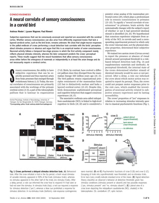
Fig. 1. Crows performed a delayed stimulus detection task. (A) Behavioral
task. After the crow initiated a trial in the Go period, a brief visual stimulus
of variable intensity appeared in 50% of the trials (stimulus trials), whereas
no stimulus appeared in the other half of the trials (no stimulus trials). After
a delay period, a rule cue informed the crow how to respond if it had seen or
had not seen the stimulus. In stimulus trials (top), a red cue required a response
for stimulus detection (“yes”), whereas a blue cue prohibited a response for
stimulus detection. In no-stimulus trials (bottom), rule-response contingencies
were inverted. (B and C) Psychometric functions of crow O (B) and crow G (C).
Grouping of trials into suprathreshold, near-threshold, and no-stimulus trials.
Error bars (very small) indicate standard error of the mean. (D) Signal detection
theory classifies an observer’s behavior at detection threshold, given two
stimulus conditions (stimulus present or absent) and two possible responses
(“yes, stimulus present” and “no, stimulus absent”). (E) Lateral view of a
crow brain depicting the nidopallium caudolaterale (NCL, shaded) in the
telencephalon. Cb, cerebellum; OT, optic tectum.
onOctober8,2020http://science.sciencemag.org/Downloadedfrom
2. B and C). Trials were classified into supra-
threshold (the two highest stimulus intensi-
ties), near-threshold (the two lowest stimulus
intensities at perceptual threshold), and no-
stimulus categories (Fig. 1C). The crows’ re-
sponses were classified according to signal
detection theory into “hit” (correct “yes” re-
sponse to a stimulus), “correct rejection” (cor-
rect “no” response for stimulus absence), “miss”
(erroneous “no” response to stimulus pres-
ence), and “false alarm” (erroneous “yes” re-
sponse for stimulus absence) (Fig. 1D).
While the crows performed the task, we rec-
orded single-cell activity of 480 neurons (n =
306 for crow G; n = 174 for crow O) from the
NCL (Fig. 1E and supplementary materials and
methods). We first identified 262 task-selective
neurons that showed differences in firing rates
for suprathreshold trials versus no-stimulus
trials (Mann-Whitney U test, p < 0.01). The
selective time intervals of these neurons that
together bridged the total trial period were
classified into stimulus-related (n = 155) (Fig. 2A)
and delay-related (n = 165) (Fig. 2B).
Next, we compared the discharges during
the crows’ “yes” versus “no” responses in the
different trial categories (Fig. 1C and supple-
mentary materials and methods). If neurons
signal stimulus intensity, the responses to
near-threshold stimuli should be indistinguish-
able irrespective of the crow’s response. In ad-
dition, the responses during “false alarms” are
expected to be similar to “correct rejections” in
the no-stimulus condition. However, if neu-
rons represent the crows’ percept, they are ex-
pected to change activity as a function of the
crows’ later report. In this case, firing rates
to near-threshold “no” responses should re-
semble those during “correct rejections” in
no-stimulus trials. Likewise, discharges for
near-threshold“yes”responsesand“falsealarms”
should be more similar to those of supra-
threshold “yes” responses.
During stimulus presentation, neurons re-
sponded mainly to stimulus intensity and only
mildly to the crow’s later reported conscious
percept. The example neuron in Fig. 2C dis-
charged exclusively to the presentation of a
salient stimulus, without a correlation with
the crow’s “yes/no” responses. The neuron in
Fig. 2D showed some correlation with the
crow’s later report because firing rates during
near-threshold “yes” responses were similar to
supra-threshold “yes” responses, whereas dis-
charges during near-threshold “no” responses
resembled “correct rejections.”
During the subsequent delay period, however,
many neurons responded according to the crows’
impending report, rather than to stimulus in-
tensity. The neuron in Fig. 2E showed cat-
egorically higher firing rates for all “yes”
responses (suprathreshold and near-threshold
“hits,” as well as “false alarms” in the absence
of stimuli) compared to all “no” responses (“no”
responses to near-threshold stimuli, “correct
rejections” in the absence of stimuli) during
the first half of the delay period. A similar
effect can be witnessed for the neuron in Fig.
2F, which showed discharges that correlated
with the report at the beginning and end of
the delay period.
To find out whether the activity of the 262
task-selective neurons was related to the crows’
report for the same near-threshold stimuli, we
compared the firing rates in the neurons’ re-
spective selectivity intervals for different trial
outcomes. We used receiver operating char-
acteristic (ROC) analysis from signal detection
theory (26) (supplementary materials and
methods). We derived the area under the ROC
curve (AUC), termed choice probability, as the
probability of predicting the subjective “yes/
no” responses for identical stimuli for the stim-
ulus and the delay phases separately (27).
We first compared the mean (rectified) ac-
tivity during “hit” and “miss” trials for near-
threshold stimuli in the stimulus presentation
period. Choice probability was higher than the
chance level of 0.5 (mean: 0.55; p < 0.001; one-
sample Wilcoxon signed-rank test; n = 155
neurons; compared to a mean of 0.69 for supra-
threshold “hits” and no-stimulus “correct rejec-
tions”) (Fig. 3A). In addition, we compared the
choice probability for “correct rejections”
and “false alarms” during no-stimulus trials,
which was comparable to chance (mean: 0.51;
p = 0.08; one-sample Wilcoxon signed-rank
test; n = 155 neurons) (Fig. 3B). Thus, during
Nieder et al., Science 369, 1626–1629 (2020) 25 September 2020 2 of 4
Fig. 2. Single-neuron
responses in NCL. (A and
B) Pattern of task selectivity
for all stimulus-selective
neurons during the stimulus
(A) and delay period (B).
Bottom: Color-coded traces
of significance values (every
line represents a neuron);
neurons sorted according to
selectivity latency. Top:
Cumulative time-resolved
histogram of task-selective
intervals. (C and D) Activity
of two stimulus-period task-
selective example neurons
in relation to the crow’s
behavioral responses. Top
panels depict dot raster
histograms (every line is
a trial, every dot is an action
potential); bottom panels
represent the corresponding
averaged and smoothed
spike density histograms.
The vertical gray shading
indicates the presentation of
the stimulus (onset at 0 ms),
the vertical dotted line signi-
fies the end of the delay.
The color code represents
the five different trial catego-
ries, with red, orange, and pink
colors representing “yes”
response trials, and dark and
light blue colors “no” response
trials. The horizontal bars in
each spike-density histogram
signify the task-selective
interval. (E and F) Activity of
two delay-period task-selective
example neurons in relation
to the crow’s behavioral
responses. Same layout as in
(C) and (D).
RESEARCH | REPORT
onOctober8,2020http://science.sciencemag.org/Downloadedfrom
3. stimulus presentation, the neurons signaled
the crows’ subsequent report only mildly.
However, the primarily stimulus-based ac-
tivity changed to a predominantly report-driven
representation during the delay. Both the choice
probabilities for near-threshold “hit” and “miss”
trials (mean: 0.56; Fig. 3C), as well as the choice
probability for no-stimulus “correct rejections”
and “false alarms” (mean: 0.53; Fig. 3D), were
higher than expected by chance (p < 0.001 for
both values; one-sample Wilcoxon signed-rank
test; n = 165 neurons). On the background of
a mean AUC of 0.64 for suprathreshold “hits”
and no-stimulus “correct rejections,” both choice
probabilities predicted the crows’ perceptual
report rather than the physical stimulus. No-
tably, this effect was found not only for the very
same faint stimuli, but also on “false alarm”
trials, when the crows mistakenly reported
perceiving a stimulus when in fact no stimulus
was present. Thus, shortly after stimulus pres-
entation, the neurons represented the crows’
later report.
To explore the time course of choice predic-
tion from stimulus onset to delay offset irre-
spective of neuronal selectivity, we applied
time-resolved population analyses based on
the activity of all NCL neurons with sufficient
trials per trial type (n = 152). We first trained a
support vector machine (SVM) classifier to dis-
criminate “yes” versus “no” responses on the
basis of the spiking activity (28) (supplemen-
tary materials and methods). Cross-validation
on “hits” in suprathreshold trials and “correct
rejections” in no-stimulus trials indicated reli-
able information differentiating the crows’ al-
ternative responses (fig. S1). To minimize the
influence of stimulus intensity, we next trained
the classifier with discharges exclusively from
near-threshold trials in which crows subjective-
ly made “yes” and “no” responses for identical
stimulus intensities. After training, the classi-
fier was tested with new data from the same
neuronal population, but for suprathreshold
“hits” versus “correct rejections” in the ab-
sence of stimuli. Indeed, the classifier was able
to correctly assign the new trials into “yes”
versus “no” responses, with particularly high
accuracy at stimulus offset and toward the
end of the delay (Fig. 4A). This indicates that
a population of neurons contained information
about the crows’ subjective experience through-
out the trial.
Finally, we quantified how much informa-
tion about the physical stimulus and the later
report was carried by the activity of the same
population of NCL neurons across the trial.
We calculated the percent explained variance
(w2
, PEV) for stimulus intensity and “yes/no”
response (29, 30) (supplementary materials
and methods). We found that stimulus inten-
sity information increased sharply after stim-
ulus presentation, but then rapidly decayed
and vanished during the following delay (Fig.
4B). Instead, the neurons increasingly encoded
the crows’ perceptual report until it reached a
peak level toward the end of the delay (Fig. 4B).
A similar response pattern was found for pre-
dictions on near-threshold trials of a SVM-
classifier trained on population responses of
“yes” responses in suprathreshold trials (“hits”)
and “no” responses in no-stimulus trials (“cor-
rect rejections”) (fig. S2). The neuronal popu-
lation results suggest that NCL neurons switch
from initially mainly representing stimulus
intensity to predominantly encoding the crows’
subjective experience later in the trial and
before a required behavioral report.
A difference between the neuronal activities
of one reported perceptual state versus the
other for equal visual stimuli is considered to
be a “neural correlate of visual consciousness”
(3, 5, 21–23). Our finding thus constitutes an
empirical marker of avian sensory conscious-
ness. As for any animal, the qualitative nature
of this subjective experience—“what it is like”
for a crow to be consciously aware of sensory
data—remains hidden (31). Moreover, whether
pure subjective experience itself (“phenome-
nal consciousness”) can and should be disso-
ciated from its report (“access consciousness”)
remains intensely debated (1, 32).
Our report of a two-stage process in aware-
ness in the corvid NCL is markedly similar to
findings in the primate cerebral cortex, where
the initial sweep of activity is also mainly in-
volved in unconscious vision, whereas activity
correlating with consciousness is delayed rela-
tive to stimulus onset activity (21, 33–36). To
explain these effects, the global neuronal work-
space theory (25, 37) posits that only sensory
activity that is strong enough can access
awareness by causing a state termed “global
ignition” in higher brain centers such as pre-
frontal cortex. “Ignition” causes information
about a brief stimulus to become sustained
and broadcasted back through recurrent inter-
actions between many brain areas, thereby
also characterizing the transition of a sensory
representation into the explicit working mem-
ory state (1, 23). The NCL may very well con-
stitute the avian brain site of an “all-or-none”
ignition process that leads either to a high
degree of activation causing and maintaining
Nieder et al., Science 369, 1626–1629 (2020) 25 September 2020 3 of 4
Fig. 3. Neuronal activity predicts “yes” versus “no” responses. Distribution of neuronal choice probabilities
according to signal detection theory. (A and B) Choice probabilities during the stimulus period (155 neurons).
(C and D) Choice probabilities during the delay period (165 neurons). Gray arrow indicates mean of choice
probabilities for near-threshold hits versus near-threshold misses [(A) and (C)] and for correct rejections versus
false alarms, respectively [(B) and (D)]. Choice probabilities in (A), (C), and (D) were significantly larger than
chance level indicated by dotted vertical line (***p < 0.001; n.s., not significant). Black arrows indicate mean AUC
values for suprathreshold hits versus correct rejections for comparison.
RESEARCH | REPORT
onOctober8,2020http://science.sciencemag.org/Downloadedfrom
4. information about conscious experience across
a temporal gap for a future goal, or to a van-
ishing response. Combining report-based be-
havioral protocols in crows with no-report
protocols may help to disentangle the neural
mechanisms involved in generating, maintain-
ing, and reporting conscious experience (38, 39).
This two-stage process in awareness could
prove to be a general and evolutionarily stable
principle of how sensory consciousness is
achieved in vertebrates in general.
Our finding also provides evidence for the
phylogenetic origins of consciousness (2). It
excludes the proposition that only primates
or other mammals possessing a layered cereb-
ral cortex are endowed with sensory conscious-
ness. To reconcile sensory consciousness in
birds and mammals, one scenario would post-
ulate that birds and mammals inherited the
trait of consciousness from their last-common
ancestor. If true, this would date the evolution
of consciousness back to at least 320 million
years when reptiles and birds on the one hand,
and mammals on the other hand, evolved from
the last common stem-amniotic ancestor (40).
Alternatively,consciousnessemergedindepend-
ently on the basis of convergent evolution on
different branches of the vertebrate “tree of
life.” According to this hypothesis, conscious-
ness was absent in the common stem-amniotic
ancestor, but—comparable to homeothermy—
evolved later and independently during the rise
of, at least, birds and mammals. Yet another
scenario would predict a gradual emergence of
consciousness. Here, different degrees of con-
served pallial connectivity patterns in verte-
brates could give rise to aspects of sensory
consciousness across phylogeny. Combining
measurements of brain signals with controlled
behavioral protocols will help to delineate the
origins of conscious experience in the animal
kingdom.
REFERENCES AND NOTES
1. G. A. Mashour, P. Roelfsema, J. P. Changeux, S. Dehaene,
Neuron 105, 776–798 (2020).
2. D. B. Edelman, A. K. Seth, Trends Neurosci. 32, 476–484
(2009).
3. D. A. Leopold, N. K. Logothetis, Nature 379, 549–553
(1996).
4. G. Kreiman, I. Fried, C. Koch, Proc. Natl. Acad. Sci. U.S.A. 99,
8378–8383 (2002).
5. V. de Lafuente, R. Romo, Nat. Neurosci. 8, 1698–1703 (2005).
6. C. Koch, M. Massimini, M. Boly, G. Tononi, Nat. Rev. Neurosci.
17, 307–321 (2016).
7. L. Puelles et al., J. Comp. Neurol. 424, 409–438 (2000).
8. E. D. Jarvis et al., Nat. Rev. Neurosci. 6, 151–159 (2005).
9. L. Puelles, Int. J. Dev. Biol. 62, 207–224 (2018).
10. S. Kumar, S. B. Hedges, Nature 392, 917–920 (1998).
11. S. B. Hedges, Nat. Rev. Genet. 3, 838–849 (2002).
12. M. Shanahan, V. P. Bingman, T. Shimizu, M. Wild, O. Güntürkün,
Front. Comput. Neurosci. 7, 89 (2013).
13. H. J. Karten, Philos. Trans. R. Soc. Lond. B Biol. Sci. 370,
20150060 (2015).
14. J. Dugas-Ford, C. W. Ragsdale, Annu. Rev. Neurosci. 38,
351–368 (2015).
15. S. Olkowicz et al., Proc. Natl. Acad. Sci. U.S.A. 113, 7255–7260
(2016).
16. N. J. Emery, N. S. Clayton, Science 306, 1903–1907
(2004).
17. A. Nieder, Curr. Opin. Behav. Sci. 16, 8–14 (2017).
18. L. Veit, A. Nieder, Nat. Commun. 4, 2878 (2013).
19. H. M. Ditz, A. Nieder, Nat. Commun. 11, 686 (2020).
20. O. Güntürkün, Curr. Opin. Neurobiol. 15, 686–693 (2005).
21. V. de Lafuente, R. Romo, Proc. Natl. Acad. Sci. U.S.A. 103,
14266–14271 (2006).
22. T. I. Panagiotaropoulos, G. Deco, V. Kapoor, N. K. Logothetis,
Neuron 74, 924–935 (2012).
23. B. van Vugt et al., Science 360, 537–542 (2018).
24. V. A. Lamme, Trends Cogn. Sci. 10, 494–501 (2006).
25. S. Dehaene, J. P. Changeux, Neuron 70, 200–227 (2011).
26. D. M. Green, J. A. Swets, Signal Detection Theory and
Psychophysics (Wiley, 1966).
27. K. H. Britten, W. T. Newsome, M. N. Shadlen, S. Celebrini,
J. A. Movshon, Vis. Neurosci. 13, 87–100 (1996).
28. C.-C. Chang, C.-J. Lin, ACM Trans. Intell. Syst. Technol. 2, 1–27
(2011).
29. M. R. Warden, E. K. Miller, Cereb. Cortex 17 (suppl. 1), i41–i50
(2007).
30. S. N. Jacob, A. Nieder, Neuron 83, 226–237 (2014).
31. T. Nagel, Philos. Rev. 83, 435–456 (1974).
32. N. Block, Trends Cogn. Sci. 9, 46–52 (2005).
33. H. Supèr, H. Spekreijse, V. A. Lamme, Nat. Neurosci. 4,
304–310 (2001).
34. R. Q. Quiroga, R. Mukamel, E. A. Isham, R. Malach, I. Fried,
Proc. Natl. Acad. Sci. U.S.A. 105, 3599–3604 (2008).
35. K. G. Thompson, J. D. Schall, Nat. Neurosci. 2, 283–288
(1999).
36. V. A. Lamme, P. R. Roelfsema, Trends Neurosci. 23, 571–579
(2000).
37. B. J. Baars, Trends Cogn. Sci. 6, 47–52 (2002).
38. N. Tsuchiya, S. Frässle, M. Wilke, V. Lamme, Trends Cogn. Sci.
20, 242–243 (2016).
39. N. Block, Trends Cogn. Sci. 23, 1003–1013 (2019).
40. P. Århem, B. I. B. Lindahl, P. R. Manger, A. B. Butler, in
Consciousness Transitions: Phylogenetic, Ontogenetic, and
Physiological Aspects, H. Liljenstrom, P. Arhem, Eds. (Elsevier,
2008), pp. 77–96.
ACKNOWLEDGMENTS
We thank D. Liao for reading an earlier version of the manuscript.
Funding: This work was supported by a DFG grant NI 618/6-1
to A.N. Author contributions: A.N., L.W., and P.R. designed the
experiment. A.N. and L.W. conducted the experiments. L.W.,
P.R., and A.N. analyzed the data. A.N., L.W., and P.R. wrote the
paper. A.N. supervised the study. Competing interests: The
authors declare no competing financial interests. Data
and materials availability: All data necessary to assess the
conclusions of this study are available in the main text or the
supplementary materials. All behavioral and electrophysiological
data are archived at the Institute of Neurobiology, University of
Tübingen, Germany.
SUPPLEMENTARY MATERIALS
science.sciencemag.org/content/369/6511/1626/suppl/DC1
Materials and Methods
Figs. S1 and S2
View/request a protocol for this paper from Bio-protocol.
3 February 2020; accepted 27 July 2020
10.1126/science.abb1447
Nieder et al., Science 369, 1626–1629 (2020) 25 September 2020 4 of 4
Fig. 4. Time-resolved neuron
population analyses. (A) A
support vector machine (SVM)
classifier trained on near-
threshold trial activity predicts
the crows’ “yes” responses
from suprathreshold “hit” trials
and “no” responses from correct
rejection no-stimulus trials. Chance
level is 50%. (B) Sliding-window
percent explained variance (w2
)
analysis quantifying the information
about the stimulus intensity and
report-associated subjective percept.
RESEARCH | REPORT
onOctober8,2020http://science.sciencemag.org/Downloadedfrom