CONTENT
◦ INTRODUCTION
◦ PERSONALDATA
◦ HISTORY – CHIEF COMPLAIN, MEDICAL HISTORY, PREVIOUS DENTURE HISTORY,
HOUSE CLASSIFICATION
◦ EXTRAORAL EXAMINATION
◦ INTRAORAL EXAMINATION
◦ REMARKS ON EXISTING DENTURE
◦ TREATMENT PLAN
◦ CONCLUSION
3.
INTRODUCTION
◦ Successful completedenture therapy begins with a thorough assessment of the
patient’s physical and psychological condition and determining a treatment that will
deliver a functional complete denture that will satisfy the expectations of the patient.
◦ Evaluation of patients for complete-denture therapy should be thorough and well
documented.
4.
DIAGNOSIS
◦ According toBoucher - Diagnosis consists of planned observations to
determine and evaluate the existing conditions, which lead to decision making
based on the conditions observed.
◦ According to Sheldon Winkler – The examination of the physical state and
evaluation of the mental or psychological make up and understanding the
needs of each patient to ensure a predictable result.
5.
TREATMENT PLAN
◦ Accordingto GPT 9 - The sequence of procedures planed for the treatment of a
patient after diagnosis.
6.
PERSONAL DATA
◦ Name:It is useful for establishment of patient’s identity. Addressing by name
gains patience confidence and psychological security to patient. For record
purpose
◦ OPD number – Required for thorough record keeping
7.
Age
◦ Indicator ofthe patient's ability to wear and to use dentures.
◦ Till fourth decade of life- Tissues are relatively resilient and heal rapidly. Individuals adapt
to new conditions more readily and aesthetics are of major importance.
◦ Beyond the fifth decade- Tissues do not heal as rapidly. The body does not adapt readily to
new situations.
◦ For teeth selection
◦ For determining the prognosis of the treatment
◦ Some age related diseases like - Scleroderma , Rheumatoid arthritis, Hypertension, Diabetes
8.
◦ Women facingthe physiologic and psychological problems of menopause
often present as exacting or hysterical patients who are very concerned with
esthetics.
◦ Men at this age often are preoccupied with their careers. It is not unusual for
them to present as indifferent patients who are concerned only with comfort or
function.
9.
Sex
◦ Generally, appearanceis a higher priority for women than for men.
◦ Though younger men often are concerned with esthetics, males often grow
indifferent to their own appearances as they age and shift focus to comfort and
function.
◦ For teeth selection according to SPA FACTOR men will have sharp and broad
teeth and women will have round and small teeth
10.
Address
◦ Helps infuture communication, knowledge of patient’s social status and
setting up of appointments.
◦ To determine location related diseases like fluorosis which is an endemic
disease. So such people may want characterization of teeth that is pattern
staining for natural appearance.
◦ To know the living style of the individual for teeth selection
11.
Occupation
◦ A patient'sjob and social standing often determine the value he or she places
on oral health, as well as the esthetics and other qualities desired in denture.
◦ Tooth position is very important for a musician who plays a wind instrument.
◦ Public speakers, teachers and singers are more particular about phonetics.
◦ People with high socio economic status will have more expectations and
requirements than people with low socioeconomic status.
12.
HISTORY
◦ Chief Complaint
◦Reason for teeth loss
◦ Duration of complete edentulousness
◦ Weather a previous denture wearer
◦ Patients comments on present dentures
◦ Patient’s expectations with the new dentures
13.
CHIEF COMPLAIN
◦ shouldbe recorded in patients own words
◦ According to DeVan, "The dentist should meet the mind of the patient before he meets
the mouth of the patient.
◦ Reasons for seeking this information:-
1. If this is not done, the chief complaint may be overlooked during therapy.
2. The response allows the practitioner to assess whether the patient's expectations are
"realistic" or "attainable.“
3. The response provides information regarding the patient's psychological classification
14.
◦ Reasons forteeth loss
1. Provide insight into their appreciation of the dentistry and contribute to the
prognosis for prosthodontic success.
2. The patient should be questioned regarding the reasons for tooth loss (e.g.,
periodontal disease, gross caries, trauma, etc.).
3. Patients who lost their teeth in an accident might be more unhappy about
their edentulous state than those who lost teeth as a consequence of decay
resulting from neglect.
15.
DURATION OF EDENTULOUSNESS:
1.Provide information about bone resorption patterns, progression, timing of
tooth loss.
2. Large, rapid changes occurs in the alveolar ridge morphology during the first
year after extraction.
16.
PREVIOUS DENTURE
◦ Questionedregarding the number and types of previous dentures.
◦ Patients should be asked to comment on the reasons for replacement.
◦ Patients displaying consistent patterns of remarks should be educated
regarding the realities of denture service.
◦ A patient with a history of several dentures over a short period of time is a
poor prosthodontics risk.
17.
CURRENT DENTURE
◦ Thepatient should be questioned about the length of time he or she has worn
the current dentures.
◦ Careful observation may provide valuable information about denture
experience, denture care, dental knowledge, parafunctional habits
18.
◦ Denture Success:The patient should be asked about the esthetics and function
of existing maxillary and mandibular dentures.
◦ Responses may indicate the patient's ability to wear or adjust to complete
dentures.
◦ Denture success for each arch should be noted as "favourable" or
"unfavourable."
19.
MEDICAL HISTORY
◦ GeneralHealth: A thorough and accurate medical history must be obtained during
the diagnostic phase of complete- denture therapy and must be updated as necessary.
◦ The medical history provides important insights regarding the patient's dental
prognosis.
◦ Hence, the practitioner must be aware of local and systemic factors and must
consider them during treatment planning.
◦ Knowledge of medications that patient is taking is important to avoid any conflict in
the therapy.
20.
◦ Systemic factorsthat may affect complete-denture therapy include: arthritis,
Bell's palsy, diabetes and diseases, conditions, or therapies leading to
xerostomia
◦ Localized lesions, defects, and abnormalities provide a wide array of clinical
situations that may necessitate alteration of the treatment plan to serve the
patient better.
21.
◦ Debilitating Diseases
1.Complete denture patients, most of whom are geriatric, are bound to be
suffering from debilitating diseases like diabetes, blood dyscrasias and
tuberculosis. These patients require specific instructions on denture/tissue
care.
2. They also require special follow-up appointments to observe the response of
the soft tissues to the denture.
3. Diabetic patients show excessive rate of bone resorption, hence, frequent
relining may be necessary.
22.
◦ Diseases ofthe Joints
1. The most common disease of the joint in old age is osteoarthritis.
2. Complete denture patients with osteoarthritis affecting the finger joints may find it
difficult to insert and clean dentures.
3. Osteoarthritis plays an important role in complete denture construction when it affects
the TMJ.
4. With limited mouth opening and painful movements of the jaw, it becomes necessary
to use special impression trays.
5. It may also become necessary to repeat jaw relations and make postinsertion occlusal
adjustments due to changes in the joint.
23.
◦ Diseases ofthe Skin
1. Skin diseases like Pemphigus have oral manifestations, which vary, from
ulcers to bullae.
2. Such painful conditions, make the denture use impossible without medical
treatment.
3. Constant use of the prosthesis should be discouraged for these patients.
24.
◦ Neurological Disorders
1.Diseases such as Bell’s palsy and Parkinson’s disease can influence denture
retention and jaw relation records.
2. Patients should understand the difficulty in denture fabrication and usage.
25.
PERSONAL HISTORY
◦ Oralhygiene habits
◦ Other habits
◦ Cosmetic index
◦ Mental attitude- Philosophical - Exacting - Hysterical - Indifferent
26.
◦ Tobacco smokingand alcohol consumption
◦ Patient should be informed about their systemic effects, potential local impacts
e.g. detrimental effect on wound healing, soft tissue health, or the durability of
tissue conditioners
27.
COSMETIC INDEX
◦ Classifyfrom class 1 (high cosmetic index) to class 3 (low cosmetic index).
◦ Patients with high cosmetic indices, though often exacting, usually are
appreciative and cooperative.
◦ Patients with low cosmetic indices often are indifferent, uncooperative, and
place little value on the efforts of the prosthodontist.
28.
HOUSE CLASSIFICATION
◦ Philosophic:Those patients are easy going, congenial, mentally well-adjusted,
cooperative, and confident in the dentist. Prognosis is excellent.
◦ Exacting: These patients are precise, above average in intelligence, immaculate in
dress and appearance, often dissatisfied with past treatment, doubt the ability of
the practitioner to satisfy him or her, and often want written guarantees or
remakes at no additional charge. Once satisfied, an exacting patient may become
the practitioner's greatest supporter.
29.
◦ Hysterical: Thesepatients submit to treatment as a last resort, have a negative
attitude, are often in poor health, are poorly adjusted, often appear "exacting"
but with unfounded complaints, have failed at past attempts to wear dentures,
and have unrealistic expectations (hysterical patients often demand esthetics
and function equal to or greater than natural teeth). Prognosis is poor
30.
◦ Indifferent: Thesepatients are not concerned with appearance, often go
without dentures for years (or wear poor or worn-out dentures far beyond
serviceability) do not persevere, and do not adapt well. Such patients have no
desire to wear dentures and do not value the efforts or skills of the dentist.
FACIAL MUSCLE TONE
◦BY HOUSE
◦ Class 1: The patient exhibits normal tension, tone, and placement of the muscles
of mastication and facial expression. No apparent degenerative changes. The
majority of edentulous patients have experienced some degree of degeneration.
Usually, only immediate- denture patients have normal musculature.
◦ Class 2: The patient displays approximately normal function but slightly
impaired muscle tone. Maximum muscle function cannot be used following the
loss of all natural teeth.
38.
◦ Class 3:The patient exhibits greatly impaired muscle tone and function. This
impairment usually is coupled with poor health, inefficient dentures, and loss
of vertical dimension, wrinkles, decreased biting force, and drooping
commissures.
39.
COMPLEXION AND EYES
◦Hair, eye, and skin color provide useful guides in shade selection.
◦ Skin color also can reveal underlying disease and pathology.
◦ Pale, anemic-looking patients may have underlying systemic diseases and may
require further investigation
40.
◦ Heavy wrinklesat the commissures and nasolabial fold usually suggest
decreased Vertical Dimension of Occlusion (VDO) or poor support of facial
musculature by the denture.
41.
LIPS
◦ The contourand appearance of the vermillion border usually are altered by
tooth loss.
◦ Restoration of lip support and vermillion border width must be considered
during placement of anterior teeth.
42.
◦ Lip support:-Adequately supported, Unsupported
◦ Lip mobility:-
1. Normal (class 1)
2. Reduced mobility (class 2)
3. Paralysis (class 3)
◦ Lip length:- Long, Normal or Medium, Short
◦ Patients withminimal lip mobility show very little of the anterior teeth.
◦ Some stroke victims may have paralysis of half the lip, leading to unilateral
mouth droop and facial asymmetry. These patients must be counseled regarding
treatment limitations. If not, they may have unrealistic expectations regarding
functional and esthetic results.
◦ A long lip reveals little of the anterior teeth
◦ A very short lip allows the display of the denture base
◦ Mold selection and denture characterization can be critical factors in these cases
46.
TEMPORO MANDIBULAR JOINT
◦Any crepitus or clicking
◦ Any history of TMJ discomfort or locking
◦ smoothness of mandibular movements
◦ Deviation of the mandible
◦ Severe joint pain can indicate a severe discrepancy in the VDO.
NEUROMUSCULAR EVALUATION
◦ Speech:- Note as "normal" or "affected"
◦ Patients who are capable of articulate speech with existing dentures; (or
natural teeth) usually have no problem producing articulate speech with new
dentures.
◦ Patients with speech impediments or those who cannot articulate optimally
with their existing dentures require special attention when the dentist places
the anterior teeth and forms the palatal portions of the denture base.
49.
◦ Coordination
◦ Noteas- Class 1: Excellent Class 2: Fair Class 3: Poor
◦ Patients with good neuromuscular coordination can be expected to learn to
manipulate dentures quickly and adapt readily to new dentures.
◦ Patients with poor coordination or a neurologic deficit (such as from a stroke)
may never adapt to a denture completely.
EXAMINATION OF RESIDUAL
RIDGES
◦Arch Form: Classified according to House:
◦ Class 1: Square
◦ Class 2: Tapering
◦ Class 3: Ovoid
◦ Many arches are combinations of the aforementioned categories (e.g., square-
tapering)
52.
◦ Arch Size
◦1) Class 1: Large (best for retention and stability)
◦ Class 2: Medium (good retention and stability but not ideal)
◦ Class 3: Small (difficult to achieve good retention and stability)
53.
◦ Ridge Form:Maxillary ridge and vault form should be classified as follows:
◦ Class 1: Square to gently rounded
54.
◦ Class 2:Tapering or "V" shaped Class
◦ Class 3: Flat
55.
◦ Mandibular ridgeform is classified as follows:
◦ Class 1: Inverted "U" shaped parallel walls from medium to tall with broad
crest
◦ Class 2: Inverted "U" shaped Short with flat crest
DEFECTS
◦ Defects: Noteridge defects, such as exostoses or divots, that may pose
problems for complete-denture patients or may warrant preprosthetic surgery.
59.
TORI
◦ Class 1:Tori are absent or minimal in size. Existing tori do not interfere with
denture construction.
◦ Class 2: Clinical examination reveals tori of moderate size. Such tori offer mild
difficulties in denture construction and use. Surgery is not required.
◦ Class 3: Large tori are present. These tori compromise the fabrication and
function of dentures. Such tori usually require surgical recontouring or removal.
60.
◦ Ridge Relationship:BY ANGLE
◦ Class 1: Normal
◦ Class 2: Retrognathic
◦ Class 3: Prognathic
61.
◦ Ridge Parallelism:Classify ridge parallelism as follows:
◦ Class 1: Both ridges are parallel to the occlusal plane.
62.
◦ Class 2:The mandibular ridge is divergent from the occlusal plane anteriorly.
63.
◦ Class 3:The maxillary ridge is divergent from the Occlusal plane anteriorly or
both ridges are divergent anteriorly,
64.
◦ Interach distance:Classify interach space as follows:
◦ Class 1: Ideal interach space to accommodate the artificial teeth.
65.
◦ Class 2-Excessive interarch space to accommodate the artificial teeth
MUCOSA
◦ COLOUR
◦ Rangesfrom healthy pink to angry red
◦ Redness indicative of inflammation: related to ill fitting denture, underlying
infection, systemic disease or chronic smoking.
◦ Pigmented spots or lesions
◦ White patches
◦ keratotic areas caused by denture irritation.
68.
◦ Mucosa conditionaccording to House
◦ Class I: Healthy
◦ Class II: Irritated
◦ Class III: Pathologic
69.
◦ Mucosal Thicknessaccording to House
◦ Class I: Normal uniform density (1 mm) Investing membrane is firm but not
tense and forms an ideal cushion for the basal seat of a denture.
◦ Class II: Thin investing membrane - Soft tissues have thin investing
membranes and are highly susceptible to irritation under pressure
◦ Class III: Thick investing membrane - Soft tissues have excessively thick
investing membranes filled with redundant tissues. At the very least, this
requires tissue treatment. Such conditions may require surgical correction
70.
◦ QUALITY OFMUCOSA COVERING RESIDUAL RIDGE
◦ Firm Mucosa
◦ Hard Mucosa and Keratinized
◦ Soft Mucosa
71.
QUALITY OF MUCOSACOVERING
RESIDUAL RIDGE
◦ Ideally, the residual ridges should be of moderate height with a rounded shape.
On gentle palpation, the mucosa should be firm and not painful
◦ The patient may present with a sharp bony residual ridge or mylohyoid ridge,
which are painful when lightly pressed.
72.
◦ If theresidual ridge is uneven and irregular then movement of the lower
denture during function can be painful. Incorporation of a resilient liner in the
fitting surface of the new lower denture can do much to relieve these
symptoms
73.
CLASSIFICATIONS OF RESIDUALRIDGE RESORPTION
◦ According to BRANEMARK et al in 1985, ridges were classified on the basis of
bone quantity and bone quality by radiographic means
◦ BONE QUANTITY:
◦ CLASS A: Most of the alveolar bone is present
◦ CLASS B: Moderate residual ridge resorption occurs
◦ CLASS C: Advance residual ridge resorption occurs
◦ CLASS D: Moderate resorption of the residual bone is present
◦ CLASS E: Extreme resorption of the basal bone
74.
◦ Bone quality:
◦CLASS 1- Almost entire jaw is composed of homogenous compact bone
◦ CLASS 2- A thick layer of compact bone surrounds a core of dense trabecular
bone
◦ CLASS 3- A thin layer of cortical bone surrounds a core of dense trabecular
bone
◦ CLASS 4- A thin layer of cortical bone surrounds a core of low density
trabecular bone
LATERAL THROAT FORM
◦Neil defined as the contour of the hard lingual surfaces of the mandibular ridge
and the velum like tissue distal to the mylohyoid ridge in the retromylohyoid
fossa. It functions under the influence of tongue
◦ Examination • The lateral throat form depth and width in moderate function is
estimated by placing a mouth mirror in the disto- lingual vestibule. This has
been classified by Ewell Neil
77.
◦ CLASS I:The mouth mirror is not visible when the tongue is in a slightly
protruded position; most favorable for retention and stability
◦ CLASS II: One half of the mouth mirror is visible; less favorable
◦ CLASS III: The entire mouth mirror is visible; least favorable
79.
TONGUE
◦ BY HOUSE
◦Class 1: Normal in size, development, and function. Sufficient teeth are
present to maintain normal form and function.
◦ Class 2: Teeth have been absent long enough to permit a change in the form
and function of the tongue.
80.
◦ Class 3:Excessively large tongue. All teeth have been absent for an extended
period of time, allowing for abnormal development of the size of the tongue.
Inefficient dentures sometimes can lead to the development of a class 3 tongue.
81.
◦ Tongue Position
◦BY WRIGHT
◦ Class 1– Tongue lies in the floor of the mouth with the tip forward and slightly
below the incisal edges of mandibular anterior teeth.
◦ CLASS 2- The tip is in a normal position but the tongue is broadened and
flattened
◦ CLASS 3- The tongue is retracted and depressed into the floor of the mouth
with the tip curled upward, downward or assimilated into body of tongue.
83.
PALATE
◦ Palatal ThroatForm: BY HOUSE
◦ Class 1: Large and normal in form, with a relatively immovable band of
resilient tissue 5 to 12 mm distal to a line drawn across the distal edge of the
tuberosities.
84.
◦ Class 2:Medium size and normal in form, with a relatively immovable
resilient band of tissue 3 to 5 mm distal to a line drawn across the distal edge
of the tuberosities
85.
◦ Class 3:Usually accompanies a small maxilla. The curtain of soft tissue turns
down abruptly 3 to 5 mm anterior to a line drawn across the palate at the distal
edge of the tuberosities
86.
◦ SOFT PALATECLASSIFICATION
◦ CLASS I - It is horizontal and demonstrates little muscular movement. In this
case more tissue coverage is possible for posterior palatal seal
87.
◦ CLASS II:• Soft palate makes a 45 angle to the hard palate. Tissue coverage
ᵒ
for posterior palatal seal is less than that class I
◦ CLASS III: • Soft palate make a 70 angle to the hard palate. Tissue coverage
ᵒ
for posterior palatal seal is minimum
88.
◦ Palatal Sensitivity:BY HOUSE
◦ Some patients may have an exaggerated gag reflex, the cause of which can be due
to a systemic disorder, psychological, extraoral, intraoral or iatrogenic factors.
◦ The management of such patients is through clinical, psychological and
pharmacological means.
◦ If the patient lacks progress he/she should be referred to a specialized consultant
◦ Class 1: Normal
◦ Class 2: Subnormal (hyposensitive)
◦ Class 3: Supernormal (hypersensitive)
89.
◦ HARD PALATE
◦U-shaped palatal vault; most favourable for retention & lateral stability.
◦ V-shaped vault: less favourable for retention.
◦ Flat palatal vault: also unfavourable.
90.
FRENUM ATTACHMENT
◦ BYHOUSE
◦ Class 1: High in the maxilla or low in the mandible with respect to the crest of
the ridge.
◦ Class 2: Medium
◦ Class 3: Freni encroach on the crest of the ridge and may interfere with the
denture seal. Surgical correction may be required.
92.
BORDER ATTACHMENTS
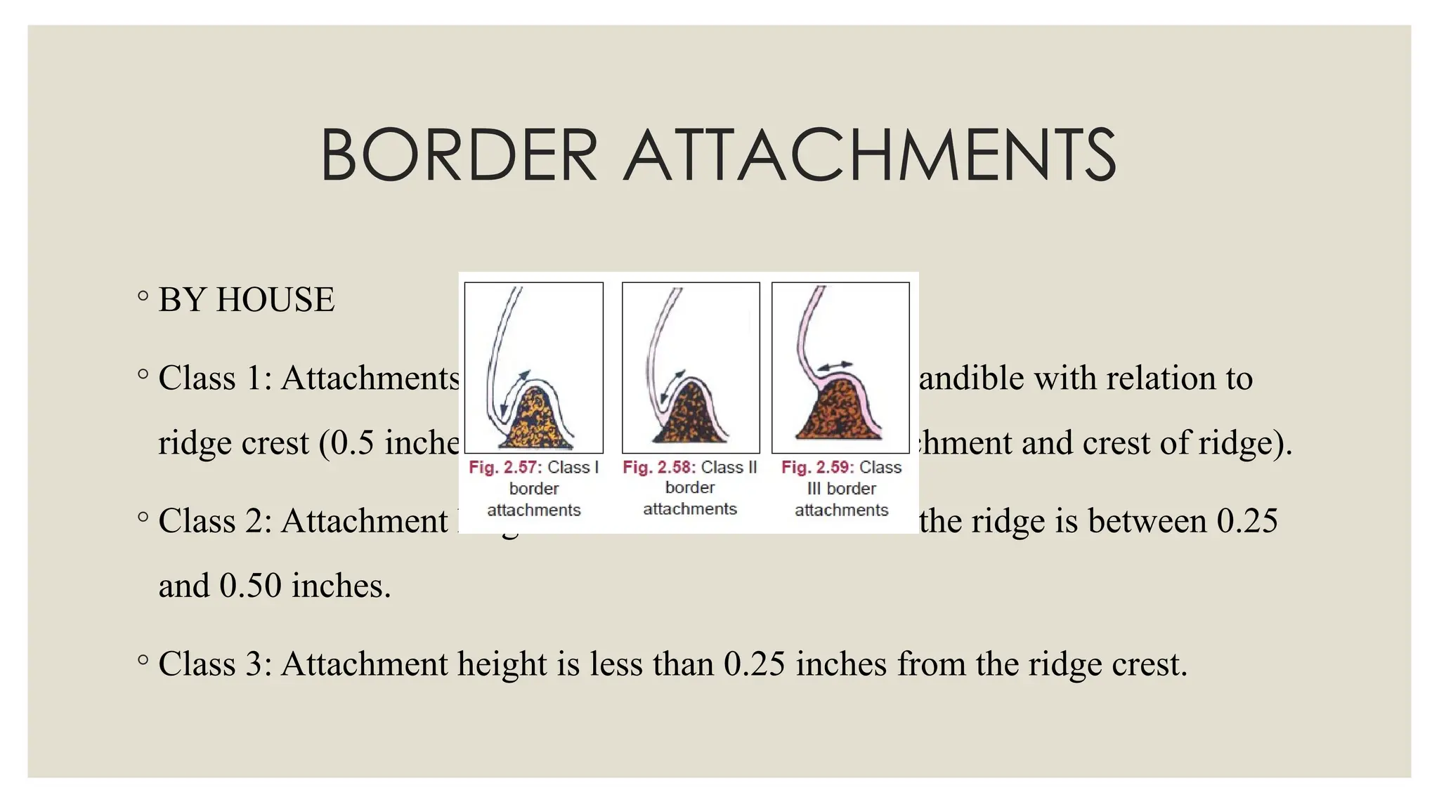
◦ BYHOUSE
◦ Class 1: Attachments are high in maxilla or low in mandible with relation to
ridge crest (0.5 inches or more between level of attachment and crest of ridge).
◦ Class 2: Attachment height in relation to the crest of the ridge is between 0.25
and 0.50 inches.
◦ Class 3: Attachment height is less than 0.25 inches from the ridge crest.
93.
SALIVA
◦ Class 1:Normal quality and quantity of saliva. Cohesive and adhesive
properties of saliva are ideal.
◦ Class 2: Excessive saliva; contains much mucus.
◦ Class 3: Xerostomia; remaining saliva is mucinous.
94.
SALIVA
◦ Thin waterysaliva may affect retention.
◦ Thick ropy saliva complicates impression making and is annoying to the
patient as it clings to the denture.
◦ Abundant saliva is common when the denture is first inserted but usually
improves with time.
95.
◦ QUANTITY OFSALIVA:
◦ A pre- weighed cotton ball is placed in the mouth at the orifices of the major
salivary glands (mostly in the sub lingual area) and is removed for reweighing
at the end
96.
◦ QUALITY: Cottonball or blunt end of the instrument is placed in the
sublingual region
◦ 3 types- • Thin serous saliva- There is no string formation by lifting the
instrument
◦ Mixed type- Formation of strings by lifting the instrument
◦ Thick mucous saliva- Thick saliva pooled and lifted by the instruments
97.
Increase in salivaryrate can be due to:-
◦ Direct cholinergic / muscarinic agonists -Bethanechol, Pilocarpine
◦ Antipsychotics -Haloperidol, Fluphenazine, Clozapine
◦ Medications irritating the esophagus -Tetracycline, Iron preparations
98.
Decrease in thesalivary rate may be due to:-
◦ Anticholinergic/ antimuscarinic- Atropine, Belladona, Benzotropine
◦ Anti hypertensives- Enalapril, Lisinopril
◦ Antihistamines- Chlorpheniramine, Diphenhydramin
◦ Psychoactive agents- Amitriptyline
◦ Opioids and analgesic agents- Codeine, Meperidine, Methadone
◦ Nonsteroidal anti-inflammatory agents- Ibuprofen, Naproxen
99.
REMARKS OF EXISTINGDENTURES
◦ Anterior Tooth Shade, Mold, and Material
◦ Posterior Tooth Shade, Mold, and Material
◦ Physical, esthetic, and anatomic characteristics should be determined. If the
mold cannot be determined, the general shape of the teeth should be recorded
(e.g., square, square-tapering, tapering, ovoid, etc.).
◦ Existing esthetics, phonetics, retention, stability, extensions, and contours
should be evaluated. Rated as good, fair, and poor
100.
◦ Centric Relationand Vertical Dimension of Occlusion:- Rated as "acceptable"
or "unacceptable,"
◦ If unacceptable, it should be noted whether the existing VDO is "inadequate"
or "excessive”
101.
◦ Occlusal PlaneOrientation:- Improper orientation as a result of tooth setting or
changes in bony architecture often creates a "reverse smile line." This
condition is characterized by teeth that slope downward as one progresses
posteriorly. Consequently, the anterior teeth assume a curvature that does not
follow the arc of the lower lip.
102.
◦ Palate: -Note the denture base material and thickness
◦ Anatomic features -The presence or absence of rugae on the cameo surface of
the denture base
◦ Should listen to speech patterns.
103.
◦ Post dam:note the Soft tissues in the vicinity of the "vibrating line”
◦ The seal of the existing maxillary denture
◦ The post dam should be rated "acceptable" or "unacceptable."
104.
◦ Base Adaptation:-The fit of maxillary and mandibular bases should be assessed
using an appropriate disclosing medium, Noted as "acceptable" or "unacceptable."
◦ Midline:- Noted as "acceptable" or "unacceptable.'
◦ Discrepancies in midline placement create noticeable facial disharmonies.
◦ The existing maxillary midline should be evaluated using intraoral (e.g., incisive
papilla) and extraoral landmarks (e.g., nasion, filtrum, middle of the chin).
◦ Deviations of the maxillary midline should be recorded by direction and amount
(e.g., maxillary midline 2 mm to the right of the facial midline).
105.
◦ Buccal Vestibule:-It is an important esthetic and functional component. The
buccal vestibule should be judged "acceptable" or "unacceptable." Corrective
actions should be proposed.
◦ Crossbite:- The presence of a unilateral or bilateral crossbite should be
observed and entered into the diagnostic record using the categories "none “, “
unilateral," or "bilateral."
◦ Characterization:- Characterization or staining of existing denture bases should
be evaluated and recorded. • Noted as "characterized" or ' 'uncharacterized."
106.
◦ Wear:- Wearis an indicator of parafunctional habits or an abrasive diet.
◦ The wear process must be assessed with respect to time.
◦ Wear should be classified as (1) minimal, (2) moderate, or (3) severe.
107.
◦ Attachments andHardware:- Attachments and hardware usually are limited to
overdenture situations. When working under these constraints, it is important
to know the specific system in use and the availability of components
108.
SPECIAL INVESTIGATION
◦ RADIOGRAPHS:-OPG should be advised
◦ Check for
1. Root pieces
2. Foreign bodies
3. Impacted/Embedded teeth
4. Rarefaction of bone
5. TMJ-Findings
109.
◦ DIAGNOSTIC CASTSAid in determining the inter ridge space, ridge
relationships, ridge shape and form that cannot be adequately determined by
clinical examination alone.
110.
PROGNOSIS
◦ A forecastas to the probable result of a disease or a course of therapy- GPT 9
◦ A number of factors affect the prognosis
1. gross appraisal of the patient
2. patient’s needs and expectations
3. medical, psychological and behavioral considerations
4. anatomic factors
5. physiological factors etc.
◦ It can be rated as - most favourable prognosis/ integral / least favourable prognosis
TREATMENT PLANNING
◦ Eliminationof Infection
◦ Sources of infection like infected necrotic ulcers, periodontally weak teeth, and
nonvital teeth should be removed.
◦ Infective conditions like candidiasis, herpetic stomatitis, and denture stomatitis
should be treated and cured before commencement of treatment.
123.
TREATMENT PLANNING
◦ Eliminationof Pathology
◦ Pathologies like cysts and tumours of the jaws should be removed or treated
before complete denture treatment begins.
◦ The patient should be educated about the harmful effects of these conditions
and the need for the removal of these lesions.
124.
TREATMENT PLANNING
◦ PreprostheticSurgery
◦ Labial frenectomy.
◦ Lingual frenectomy.
◦ Excision of denture
granulomas.
◦ Excision of flabby tissue.
◦ Reduction of enlarged
tuberosity.
◦ Excision of hyperplastic
retromolar pad.
◦ Alveoloplasty.
◦ Alveolectomy.
◦ Reduction of genial
tubercle.
◦ Reduction of mylohyoid
ridge.
◦ Excision of tori.
◦ Vestibuloplasty.
◦ Lowering the mental
foramen.
◦ Ridge augmentation
procedures.
◦ Implants
125.
TREATMENT PLANNING
◦ TissueConditioning
◦ The patient should be requested to stop wearing the previous denture for at least 72 hours
before commencing treatment.
◦ He/she should be taught to massage the oral mucosa regularly.
◦ Special procedures should be done in patients who have adverse tissue reactions to the denture.
◦ Denture relining material should be applied on the tissue side of the denture to avoid denture
irritation.
◦ Treatment dentures or acrylic templates can be prepared to carry tissue-conditioning material
during the treatment of abused tissues.
126.
TREATMENT PLANNING
◦ NutritionalCounseling
◦ Nutritional counseling is a very important step in the treatment plan of a
complete denture.
◦ Patients showing deficiency of particular minerals and vitamins should be
advised a proper balanced diet.
◦ Patients with vitamin B2 deficiency will show angular cheilitis.
◦ Prophylactic vitamin A therapy is given for xerostomic patients.
127.
TREATMENT PLANNING
◦ Articulator:• Instrument Number and Manufacturer .
◦ Tooth Selection:- The shade, mold, and material of the maxillary anterior,
mandibular anterior, maxillary posterior, and mandibular Posterior should be
selected
◦ Denture Base Material and Shade
◦ Characterization: Establish the stains to be used; draw a "map" of the proposed
stain placement.
128.
CONCLUSION
◦ Successful completedenture therapy is obtained by thorough assessment of patients physical
and psychological condition and determining a treatment plan that will satisfy patient’s
expectations.
◦ All the facts must be known before they can be correlated in such a way that decision can be
made. Only then can treatment plans be developed to best serve the needs of each individual
patient.
◦ For the patient to be happier the dentist should not only require the skills of complete denture
construction but also the skills to treat a patient’s aspirations & expectations
129.
REFRENCES
◦ Examination, diagnosisand treatment planning- chester perry, university of
detroit, school of dentistry, vol 10, no 6
◦ Boucher’s: Prosthodontic treatment for edentulous patients, 11th edn.
◦ Winkler: Essentials of complete denture prosthdontics, 2nd edn.
◦ J.J. Sharry: Complete denture prosthodontics, 2nd edn.
◦ Bouchers: Prosthodontic Treatment for edentulous patients, 10th edn.
Editor's Notes
#26 A pack year is the equivalent of smoking one standard pack of cigarettes (20 cigarettes) per day for the whole year.
#35 Frontal plane. Intercanthal distance – equal to width of nose
Inter pupillary distance –width of the mouth
Nose and the chin should be centered . Forehead tip of the nose and chin in straight line. Worms eye view, birds eye view
#36 If denture present, check in occlusion, if not check in rest
#42 Adequately supported – teeth arrangement can be done according to the clinician
#61 SHUNTING EFFECT – OCCLUSAL FORCES, THE DENTURE TENDS TO SLIDE OVER EACH OTHER
#111 Type I (most favorable): residual bone height of 21 mm or greater measured at the least vertical height of the mandible (Fig 1); Type II: residual bone height of 16 to 20 mm measured at the least vertical height of the mandible (Fig 2); Type III: residual alveolar bone height of 11 to 15 mm measured at the least vertical height of the mandible (Fig 3); Type N: residual vertical bone height of 10 mm or less measured at the least vertical height of the mandible (Fig 4).
#113 Type A (most favorable) (Fig 5) Anterior labial and posterior buccal vestibular depth that resists vertical and horizontal movement of the denture base. 0 Palatal morpholog resists vertical and horizontal movement of the denture base. Sufficient tuberosity definition to resist vertical and horizontal movement of the denture base. 0 Hamular notch is well defined to establish the posterior extension of the denture base. Absence of tori or exostoses.
#114 Type B (Fig 6) Loss of posterior buccal vestibule. 0 Palatal vault morphology resists vertical and horizontal movement ofthe denture base. Tuberosity and hamular notch are poorly defined, compromising delineation of the posterior extension of the denture base. 0 Maxillary palatal tori and/or lateral exostoses are rounded and do not affect the posterior extension of the denture base.
#115 Loss of anterior labial vestibule. Palatal vault morpholog~7 offers minimal rcsistance to vertical and horizontal movement of the denture base. 0 Maxillary palatal tori and/or lateral exostoses with bony undercuts that do not affect the posterior extension of the denture base. Hyperplastic, mobile anterior ridgc offcrs minimum support and stabilit).-ofthe denture basE
REDUCTIN OF THE POSTMALAR SPACE BY OPENING OF MAXILLA
#116 0 Loss of anterior labial and posterior buccal vestibules. 0 Palatal vault morpholoa does not resist vertical or horizontal movement of the denture base. Maxillary palatal tori and/or lateral exostoses" (rounded or undercut) that intcrferc with the posterior border of the denture. Hyperplastic, redundant anterior ridge. Prominent anterior nasal spine.


![HISTORY TAKING IN COMPLETELY EDENTULOUS PATIENTS [Autosaved] [Autosaved].pptx](https://cdn.slidesharecdn.com/ss_thumbnails/historytakingincompletelyedentulouspatientsautosavedautosaved-250408092503-84de7c34-thumbnail.jpg?width=640&height=640&fit=bounds)


