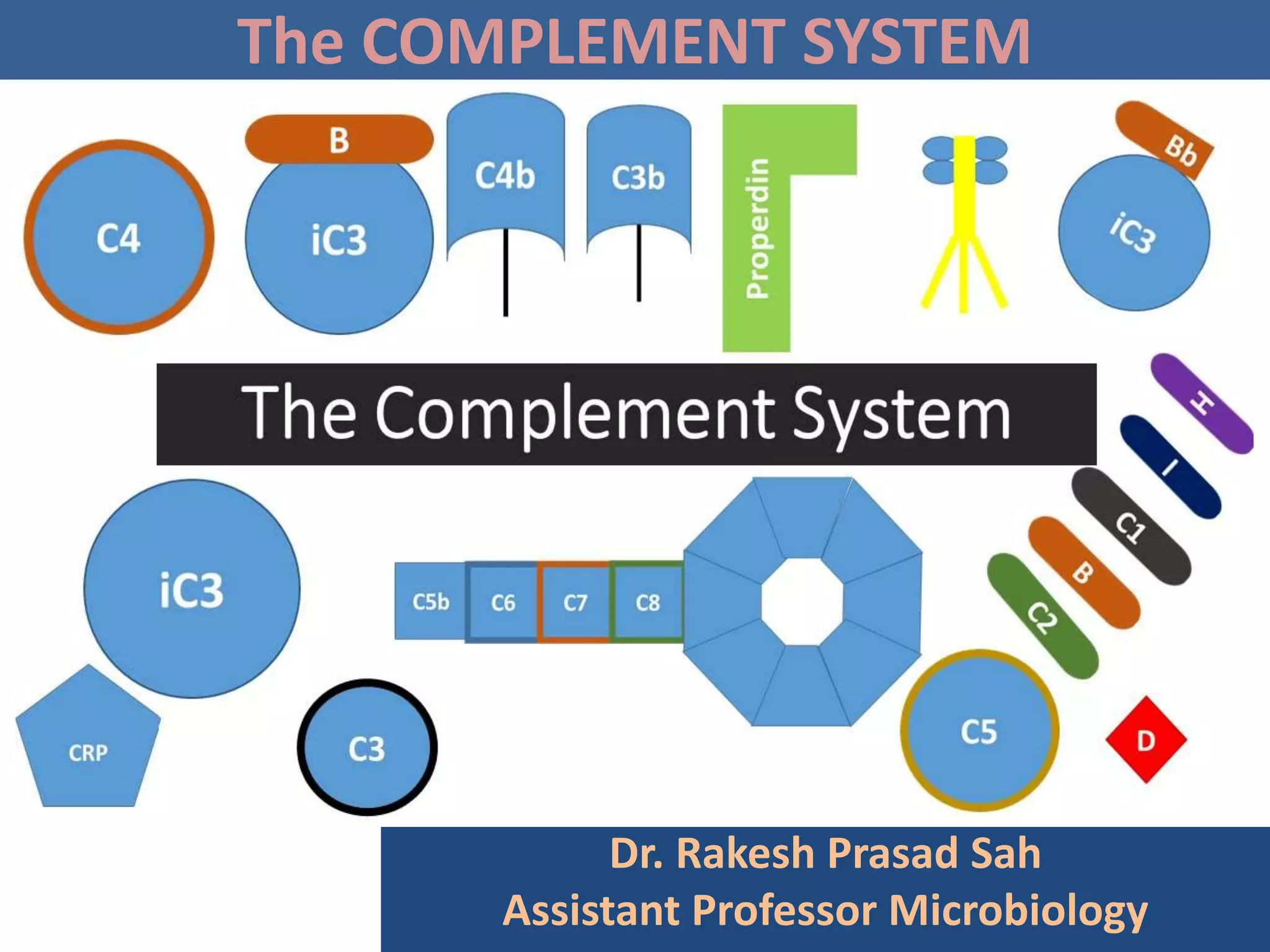
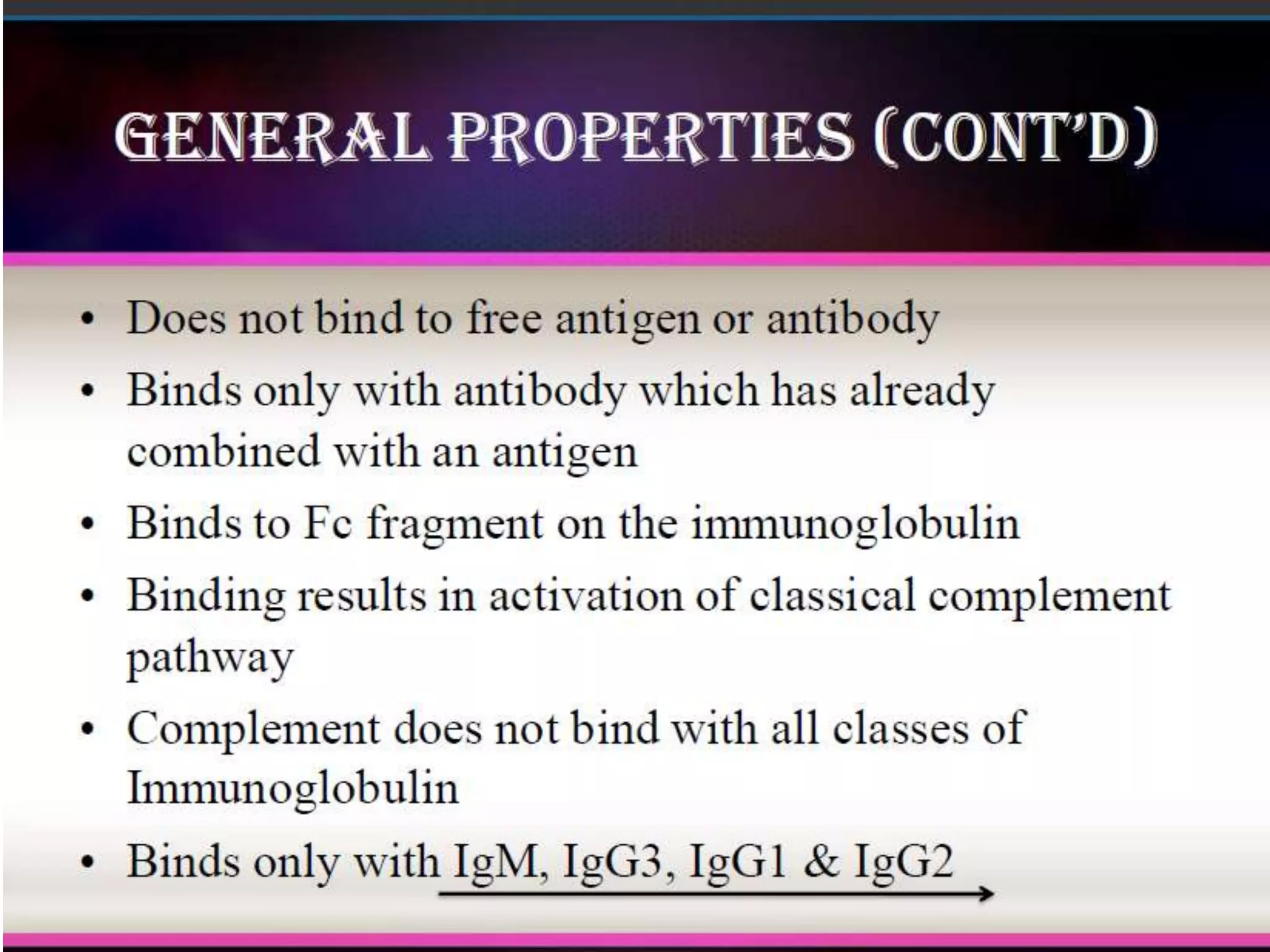
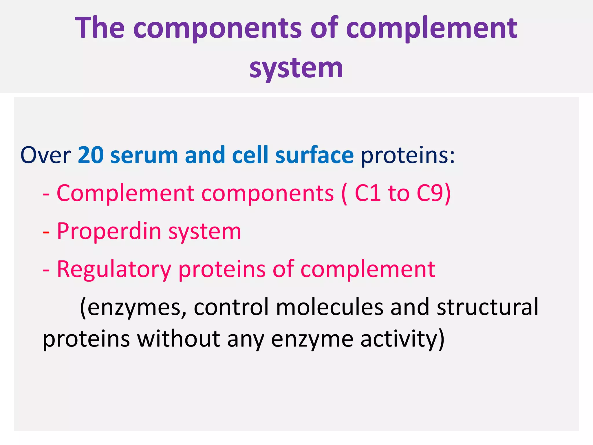
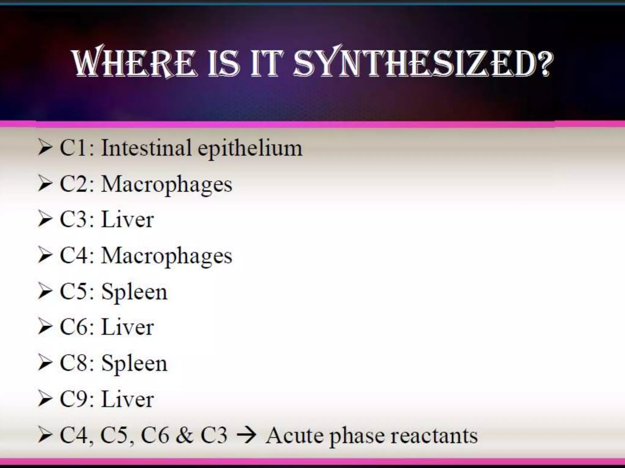
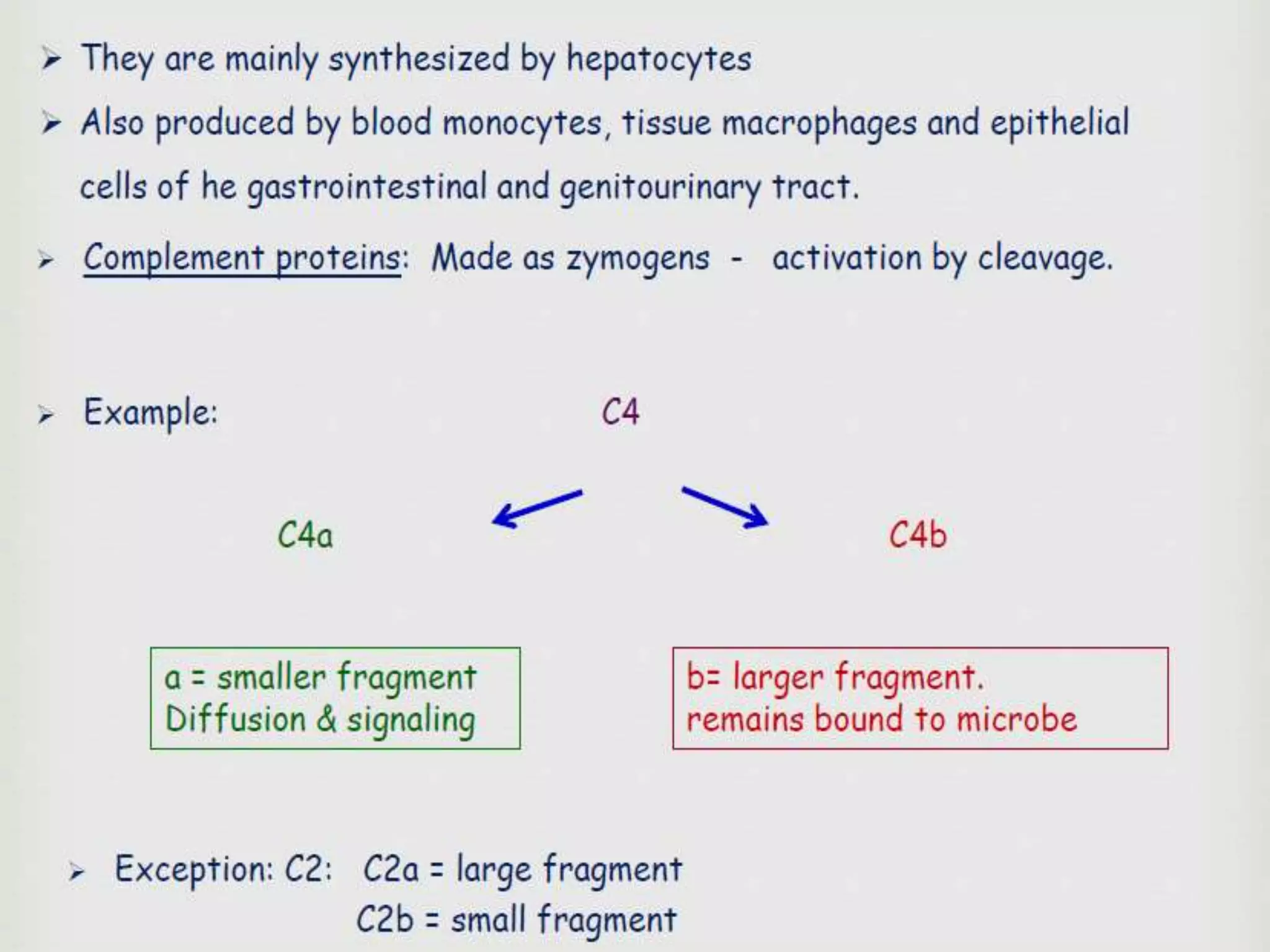
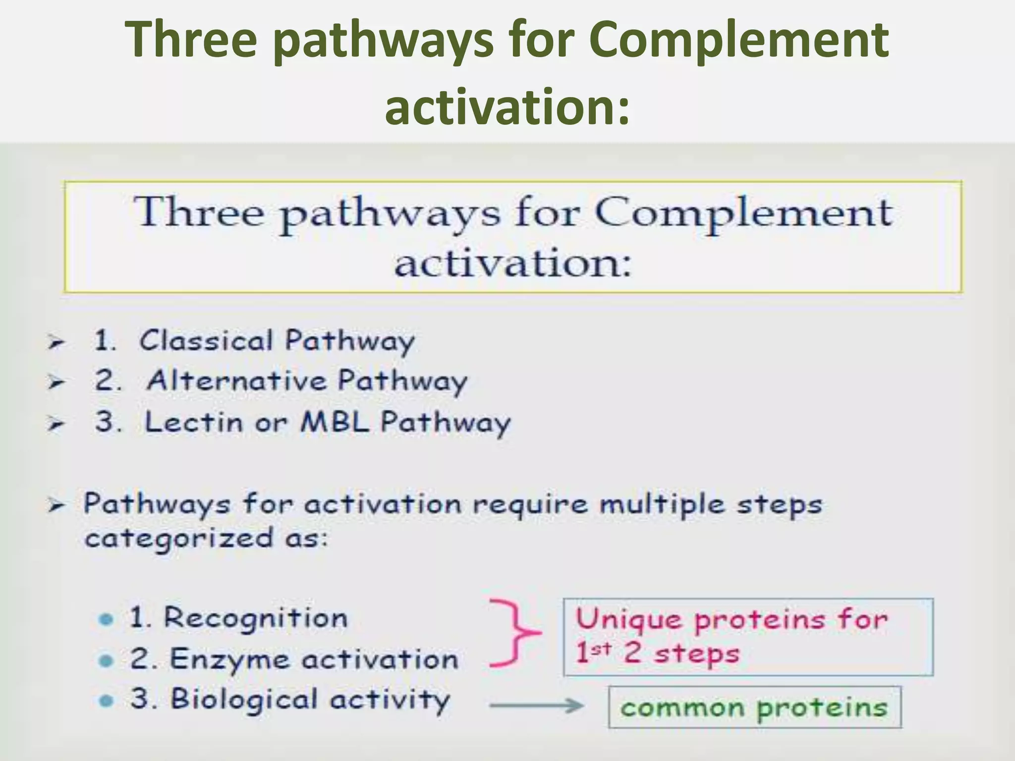
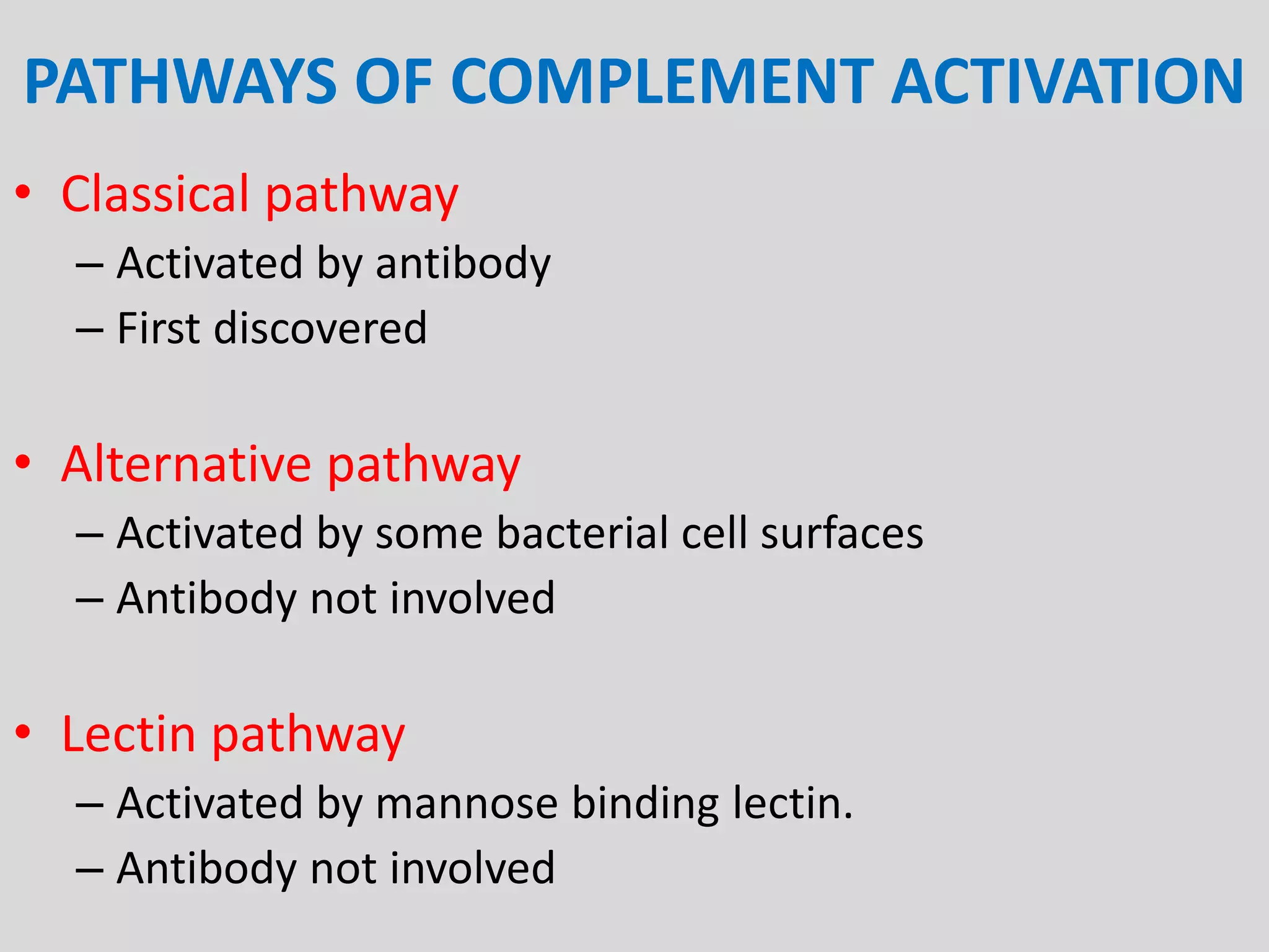
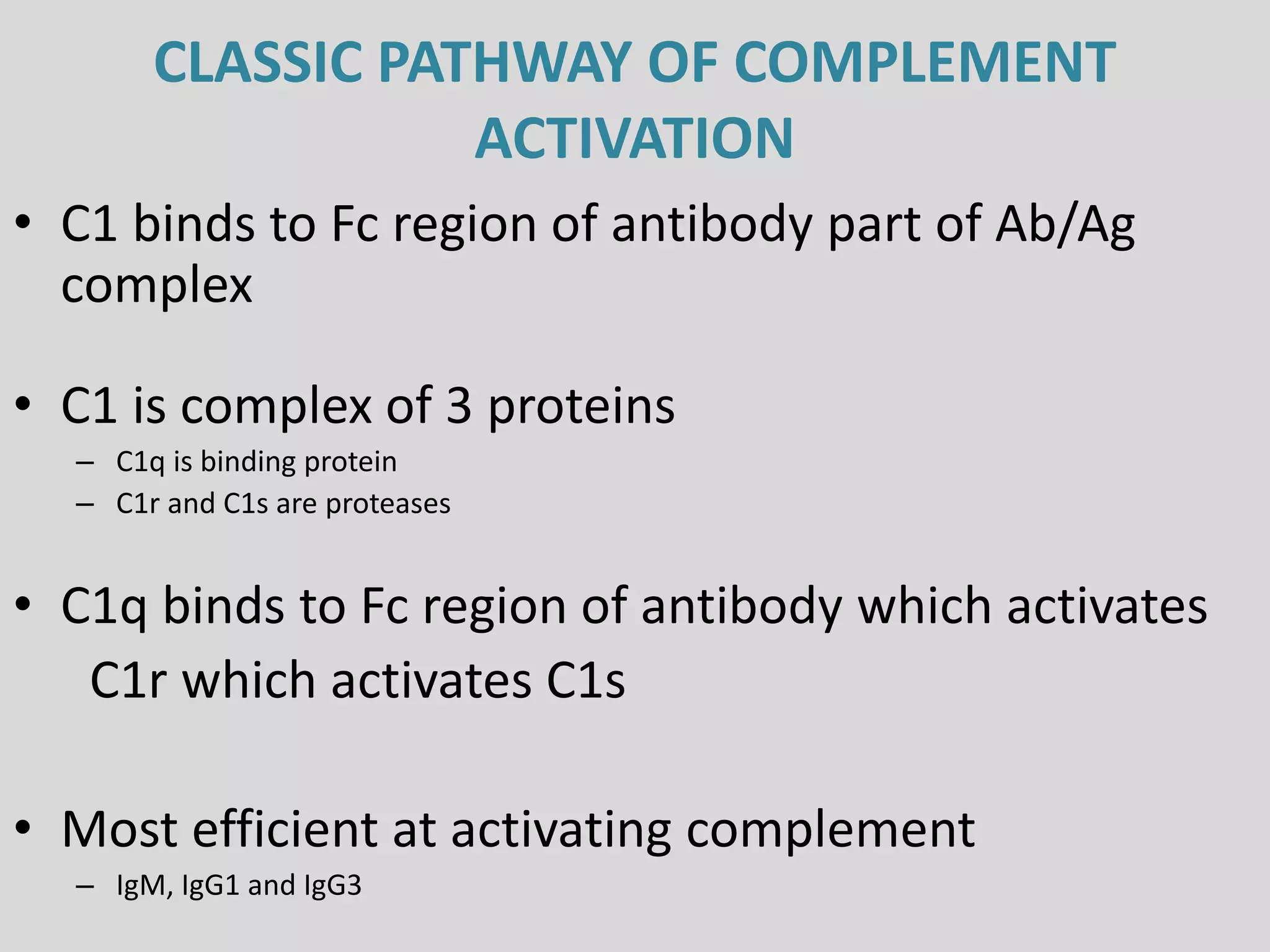
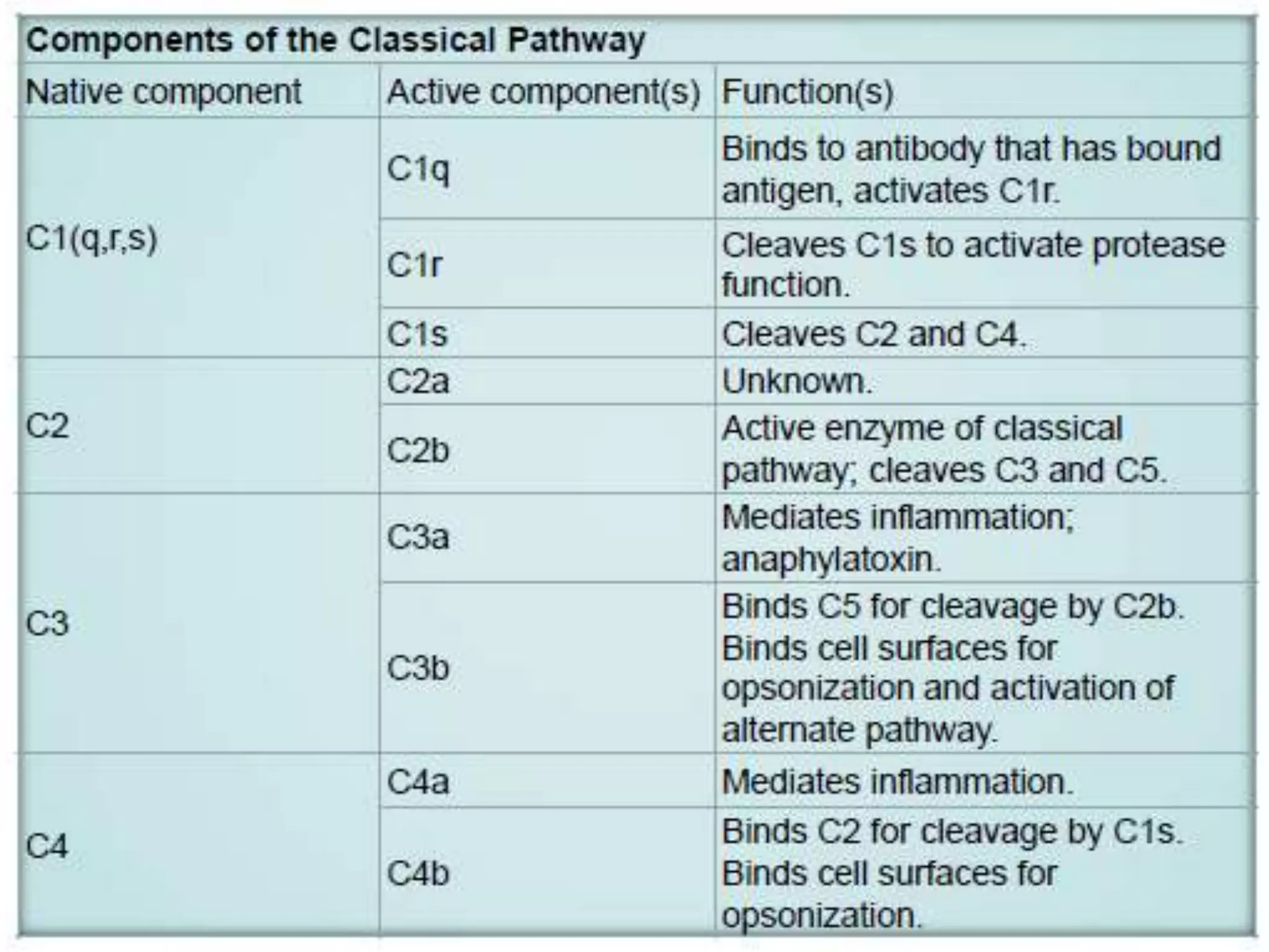
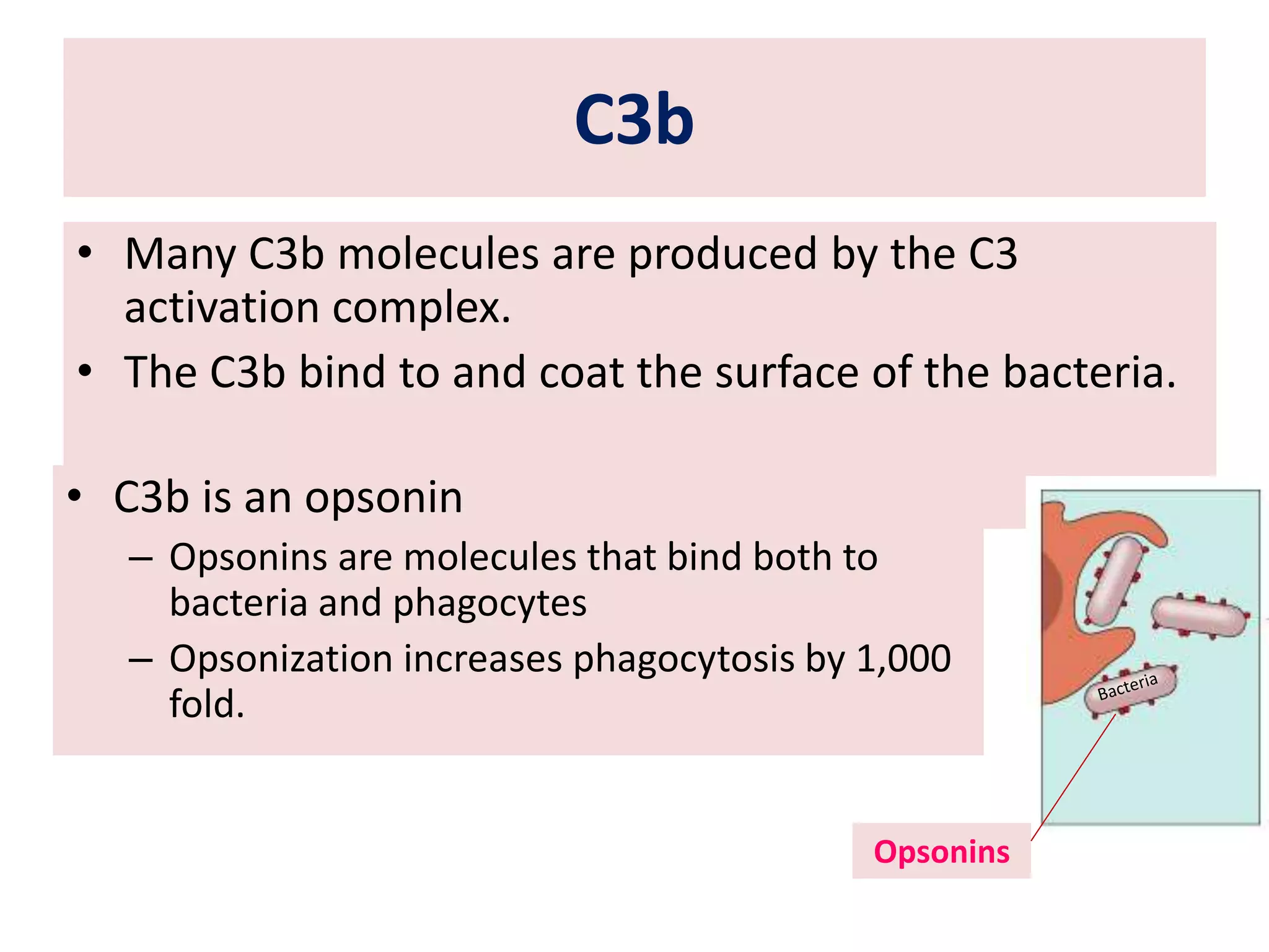
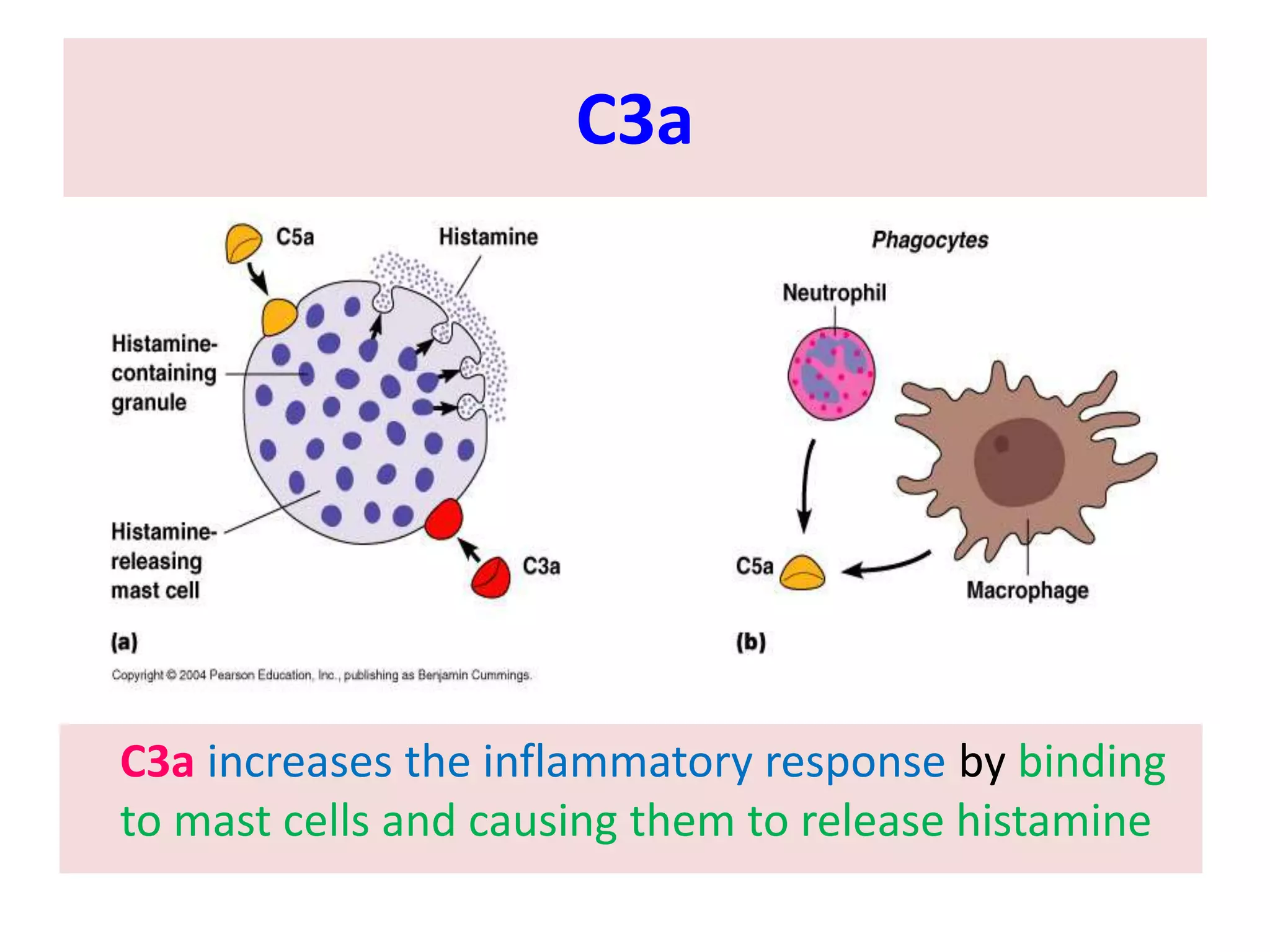
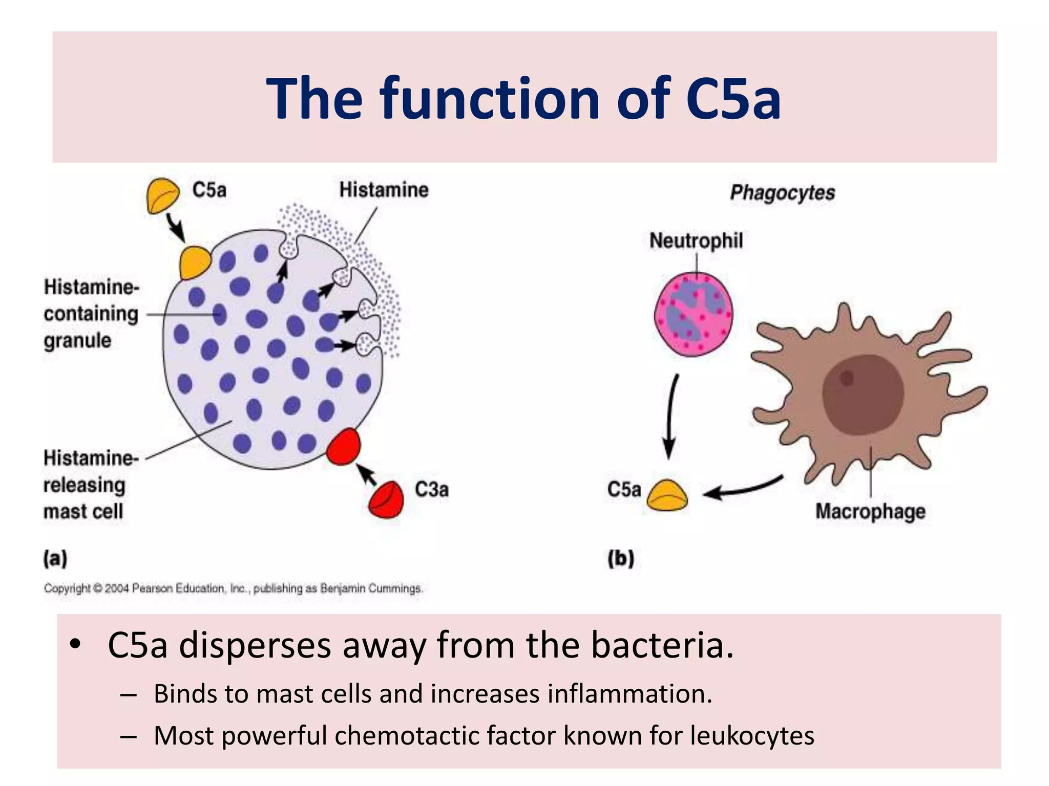
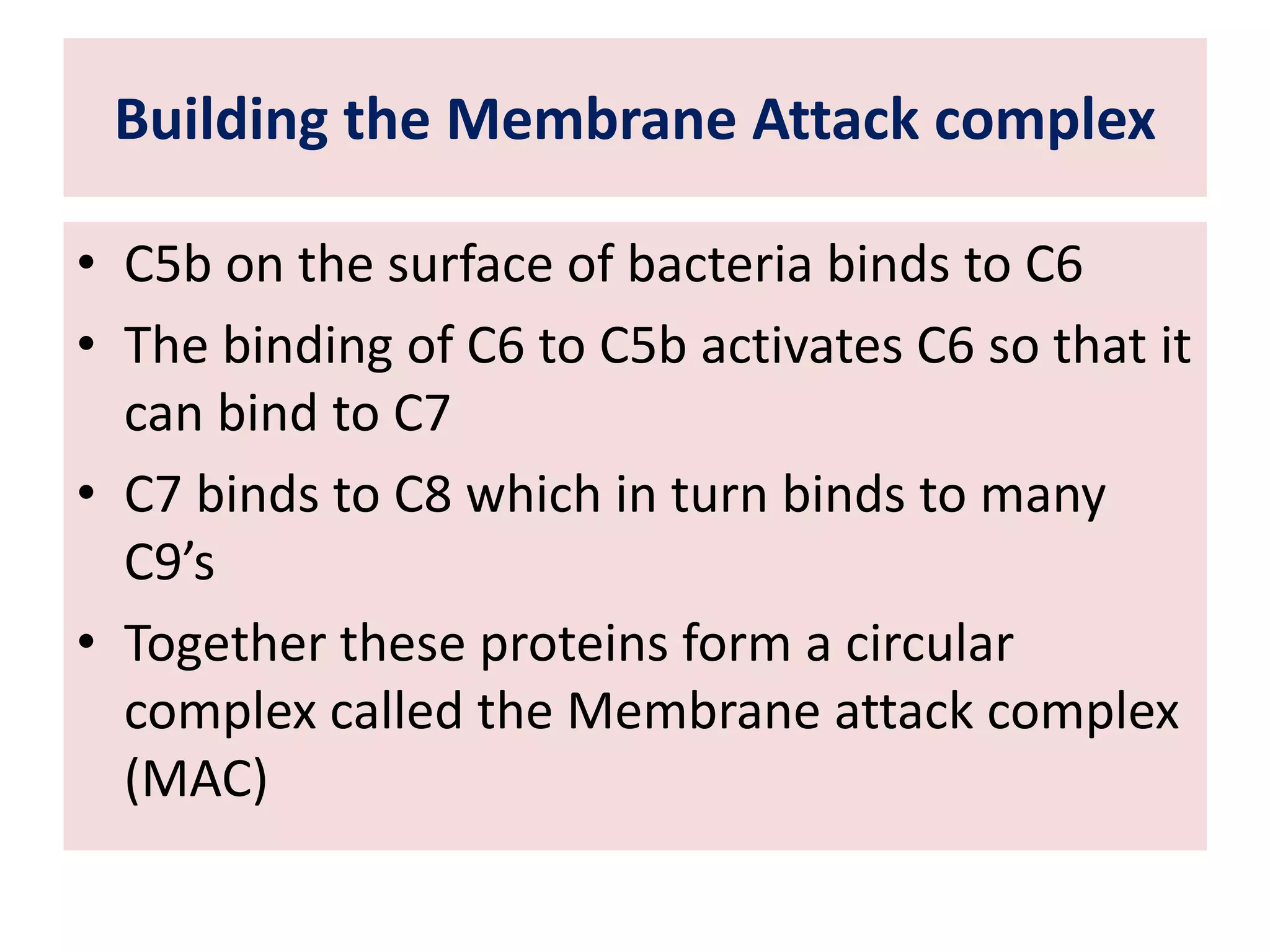
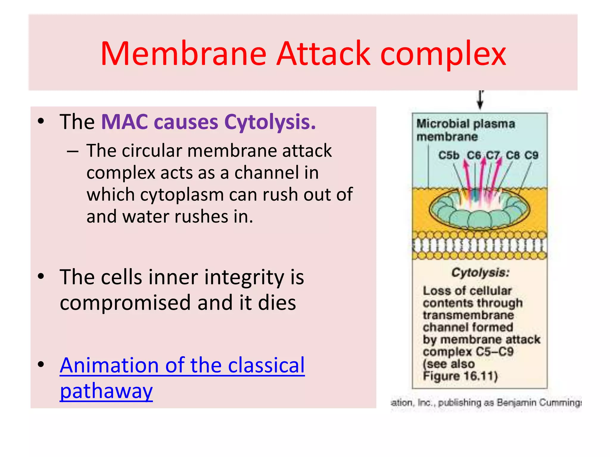
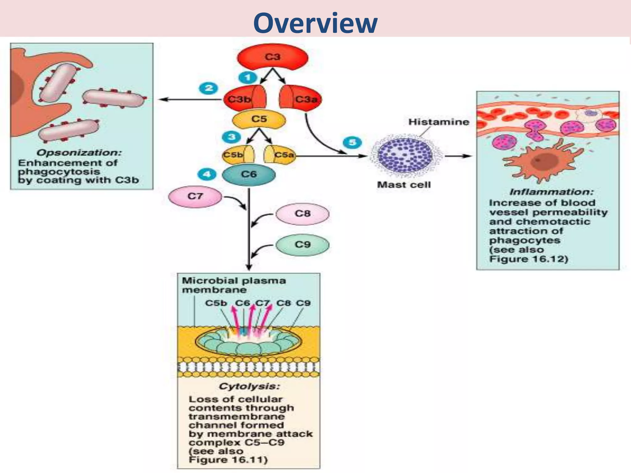
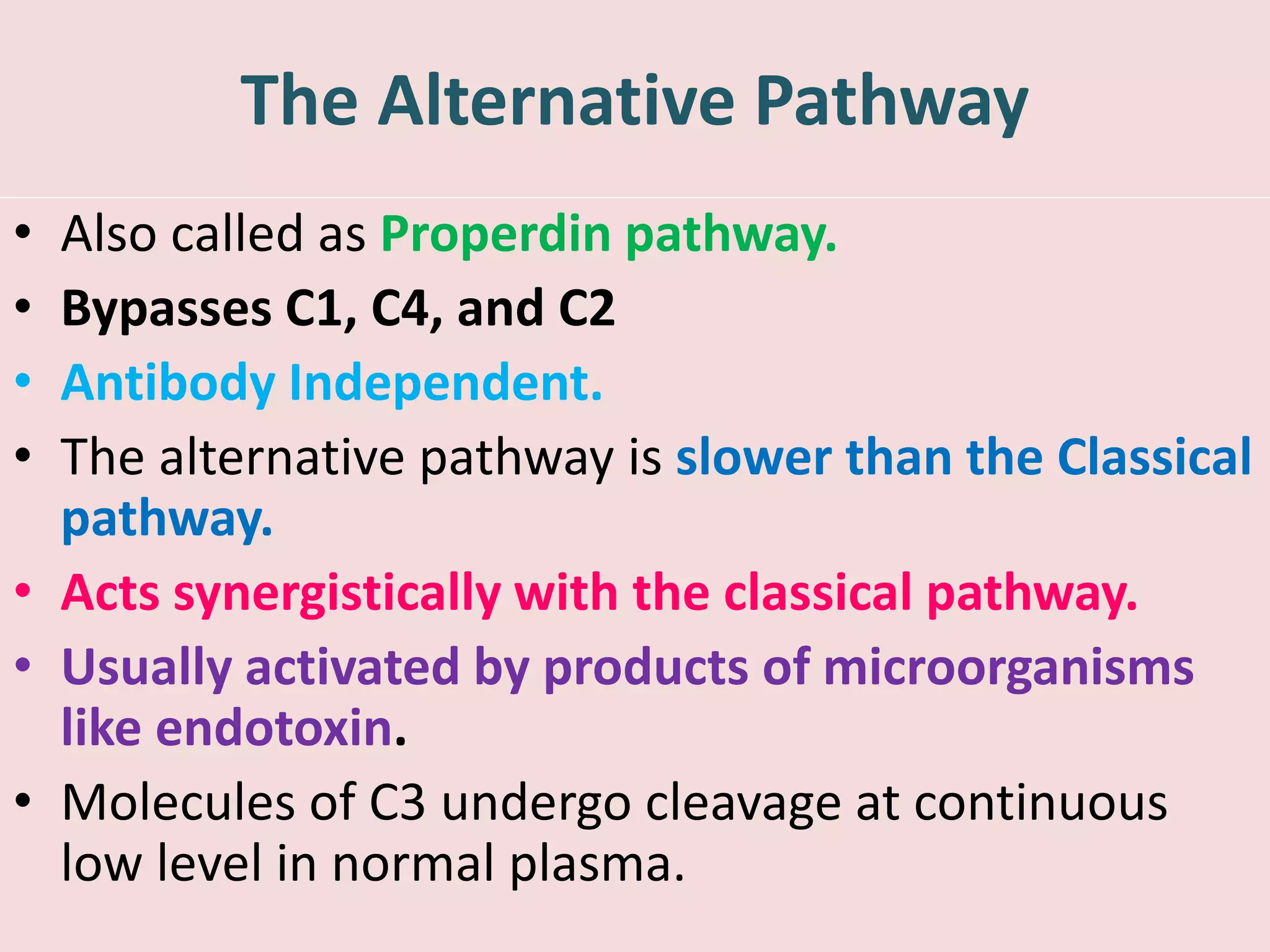
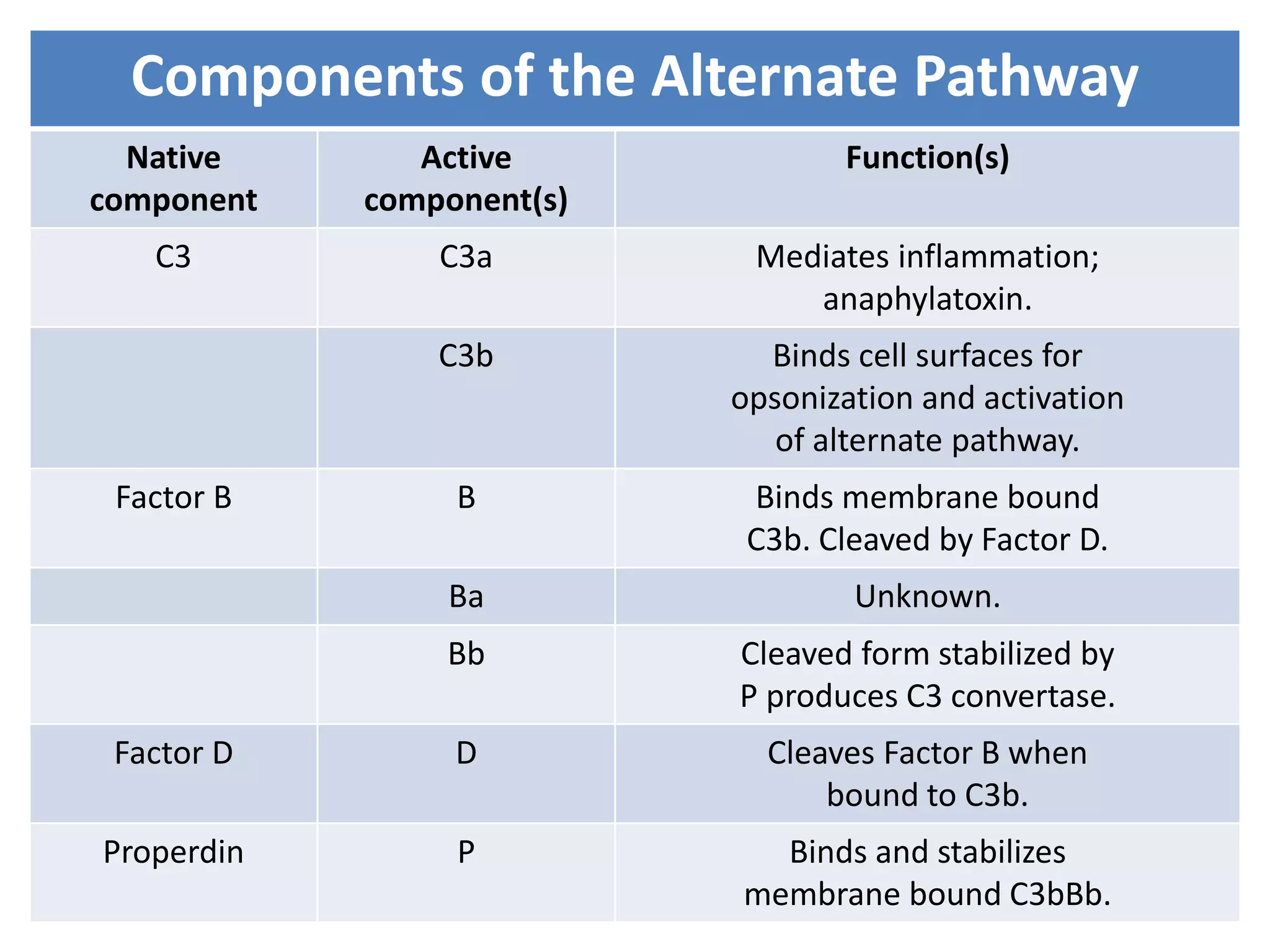
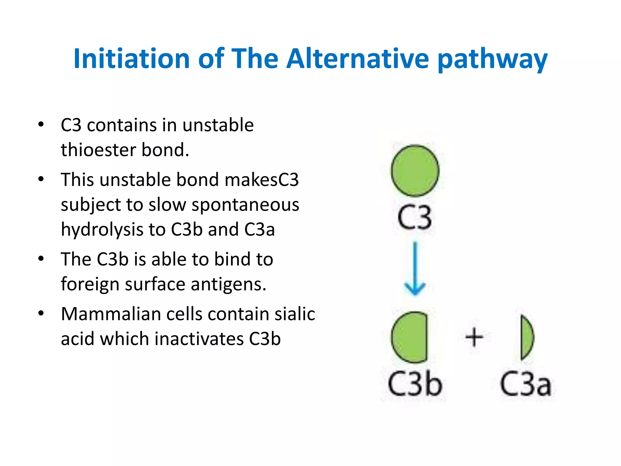
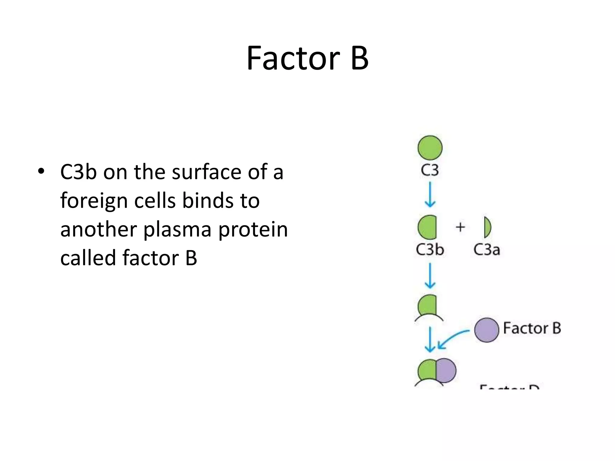
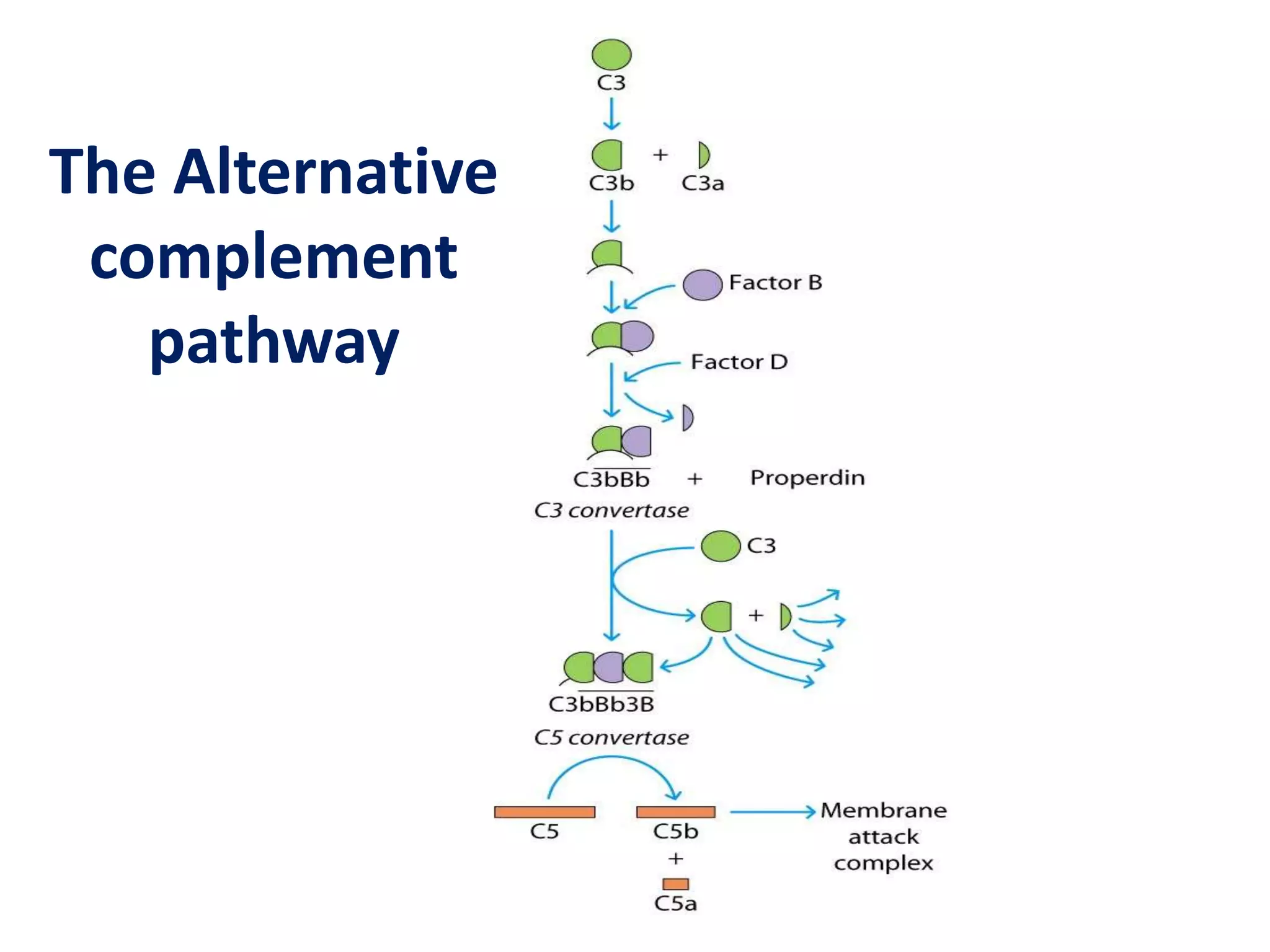
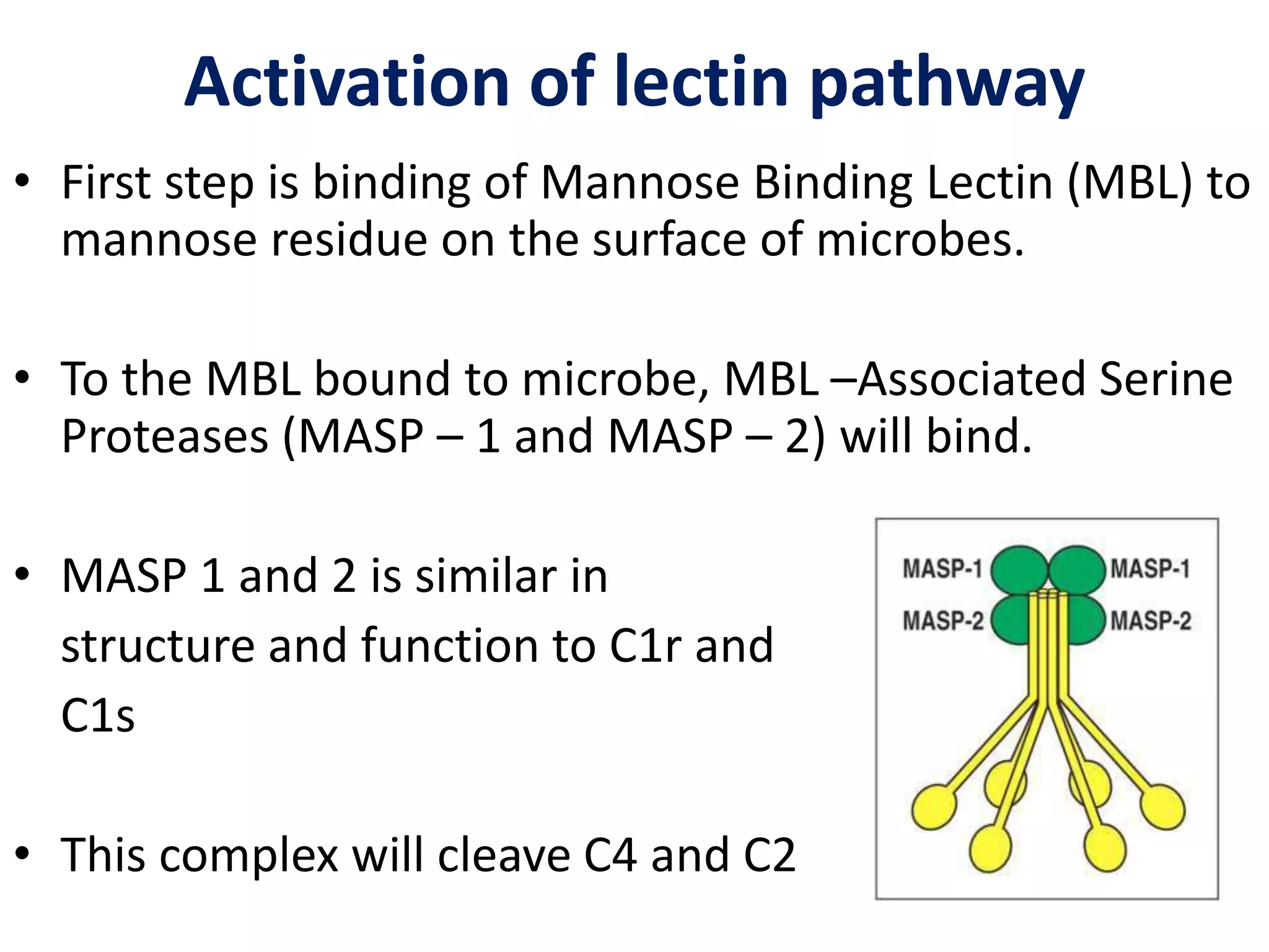
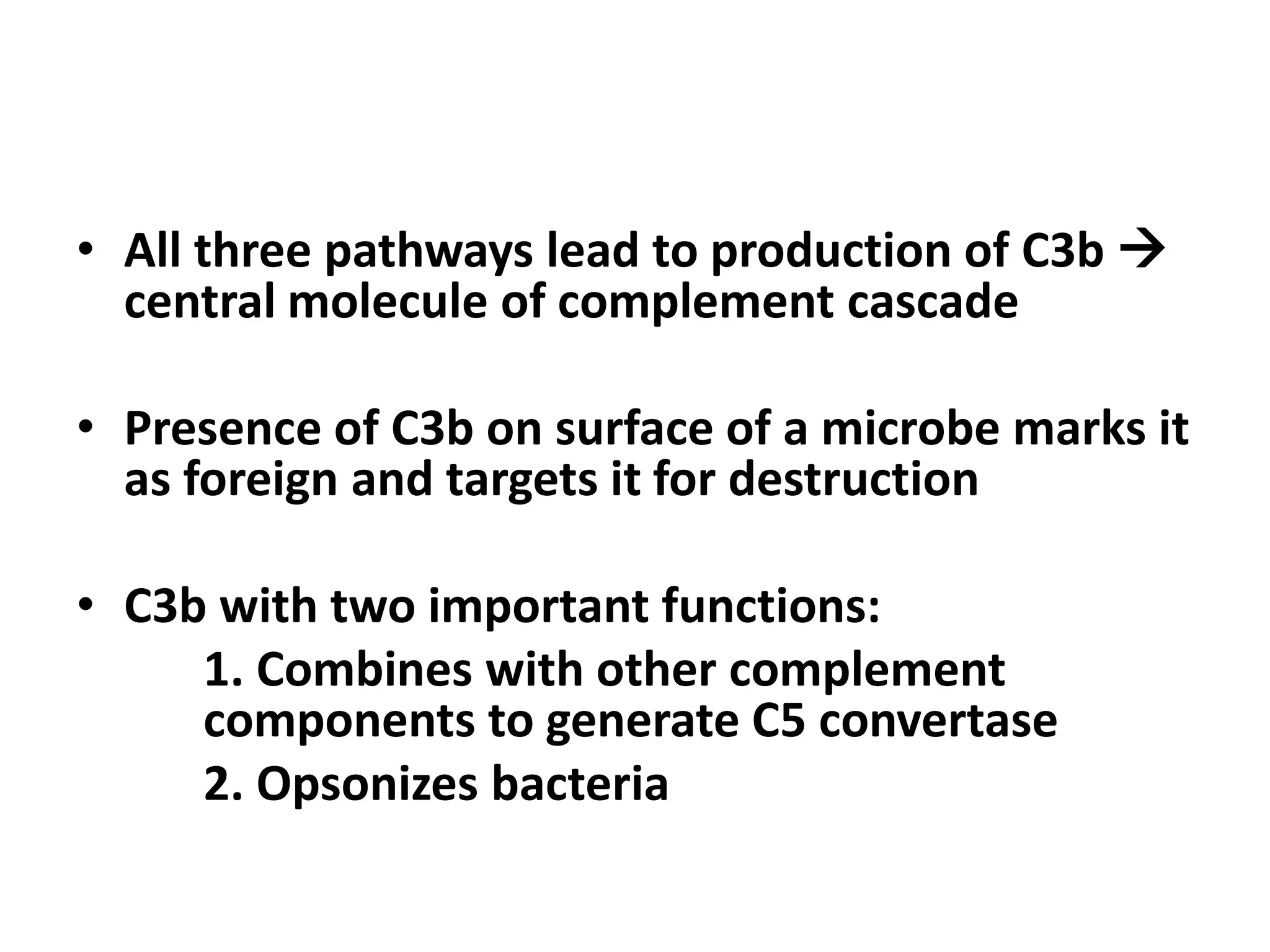
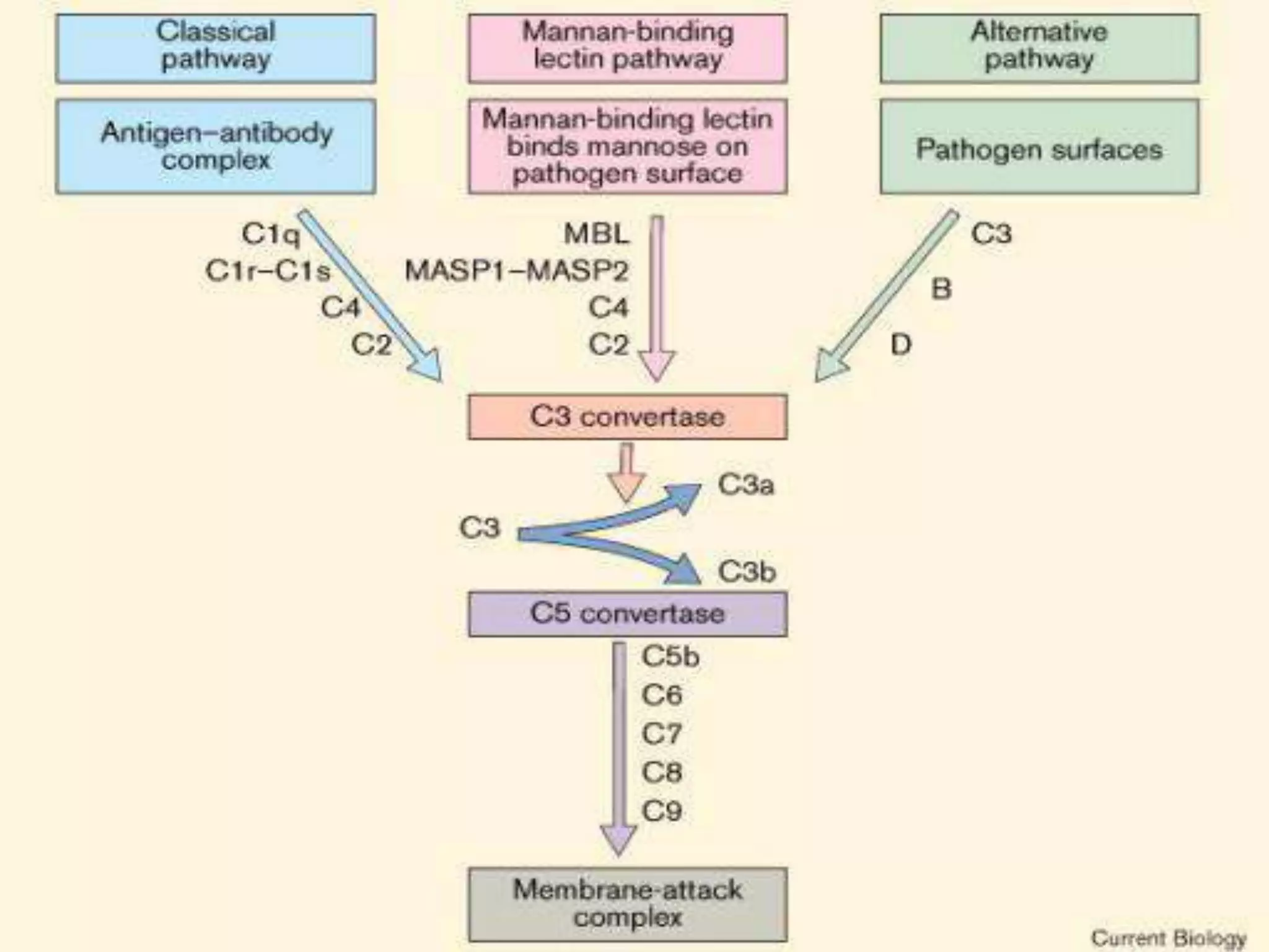
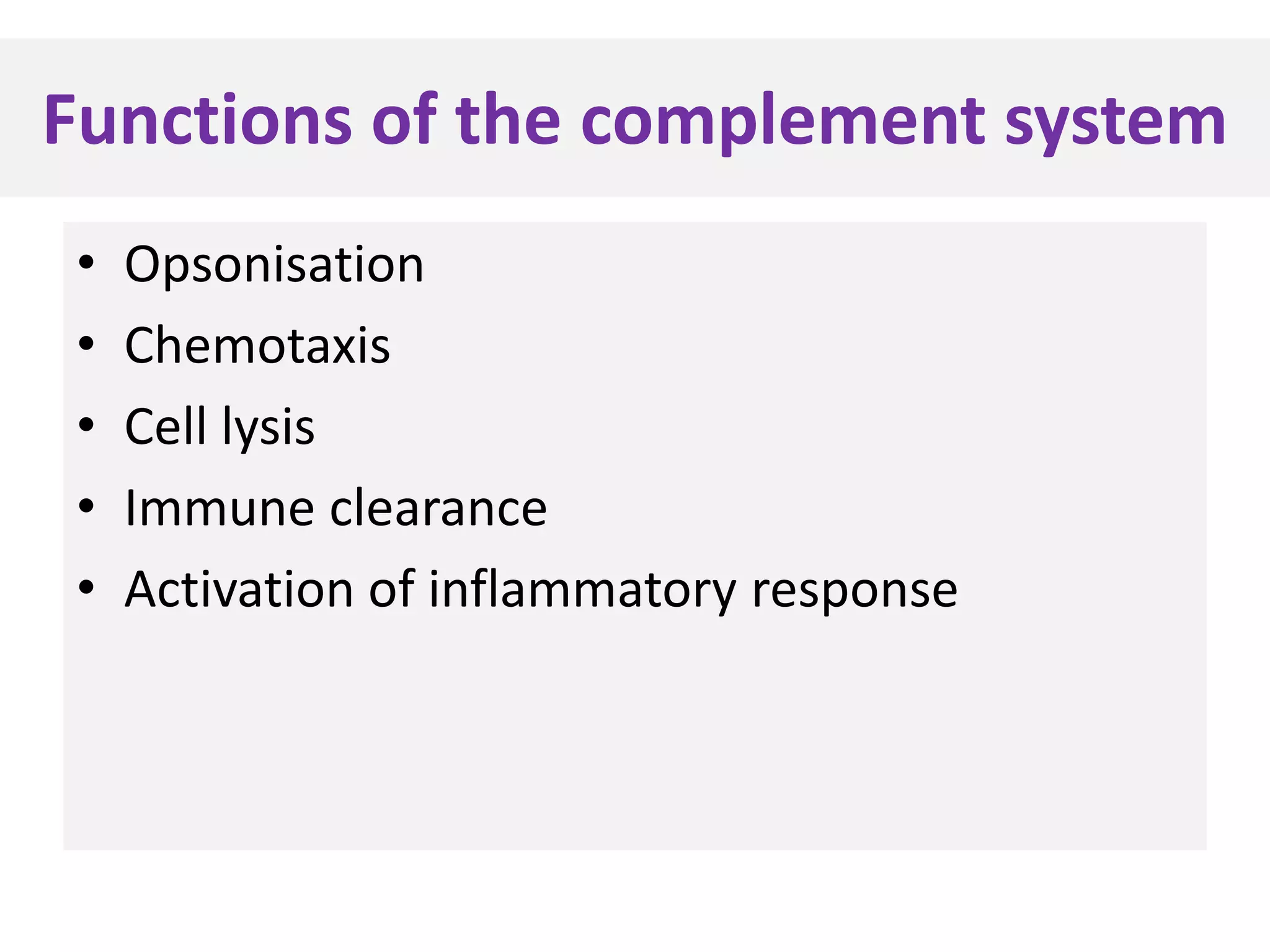
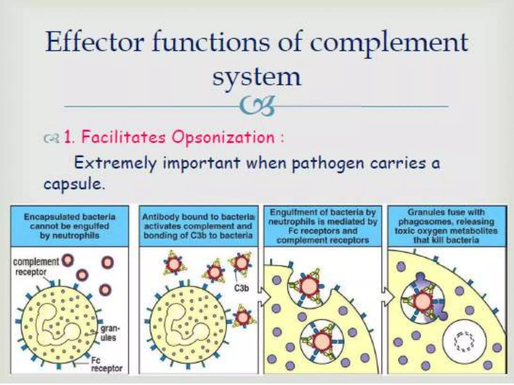
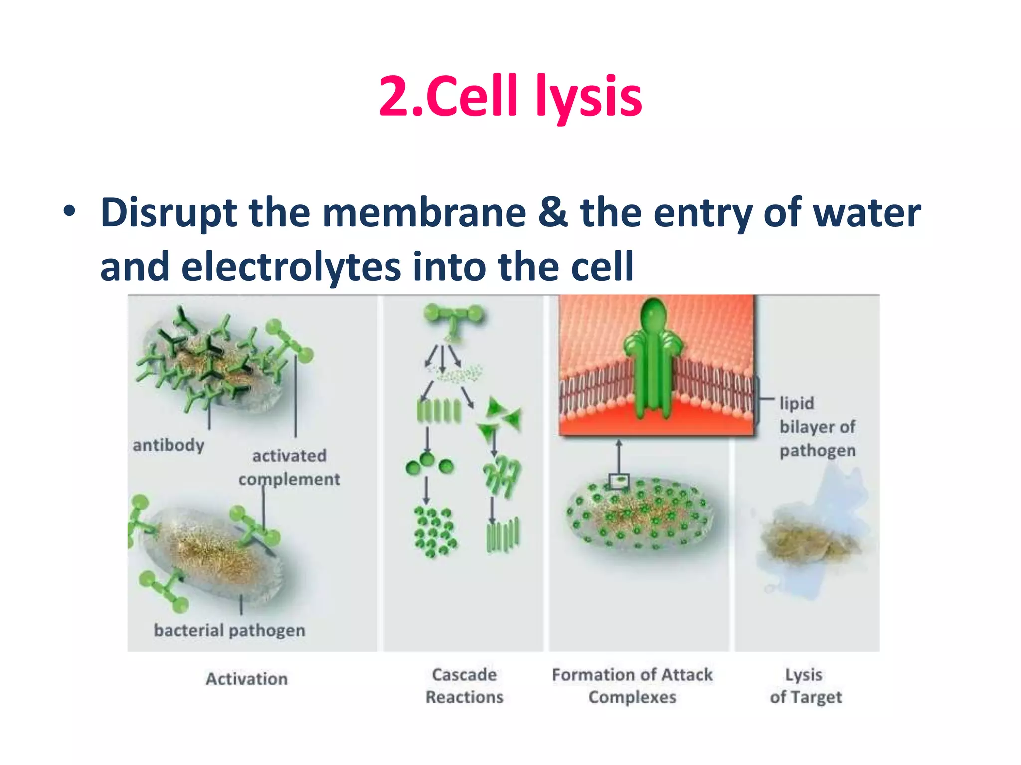
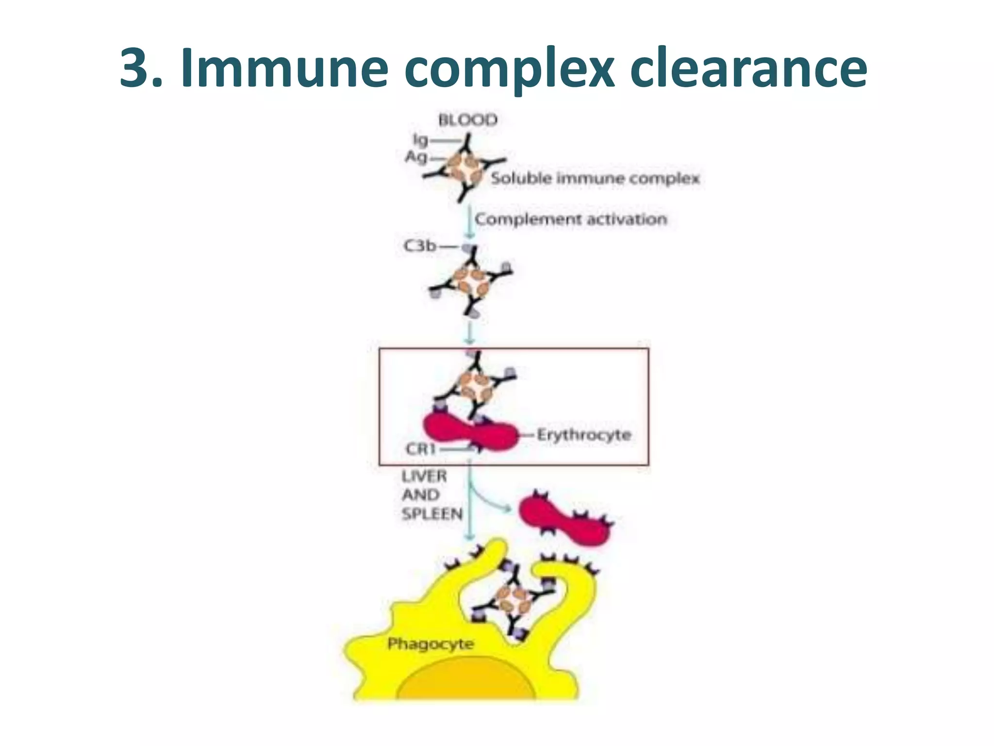
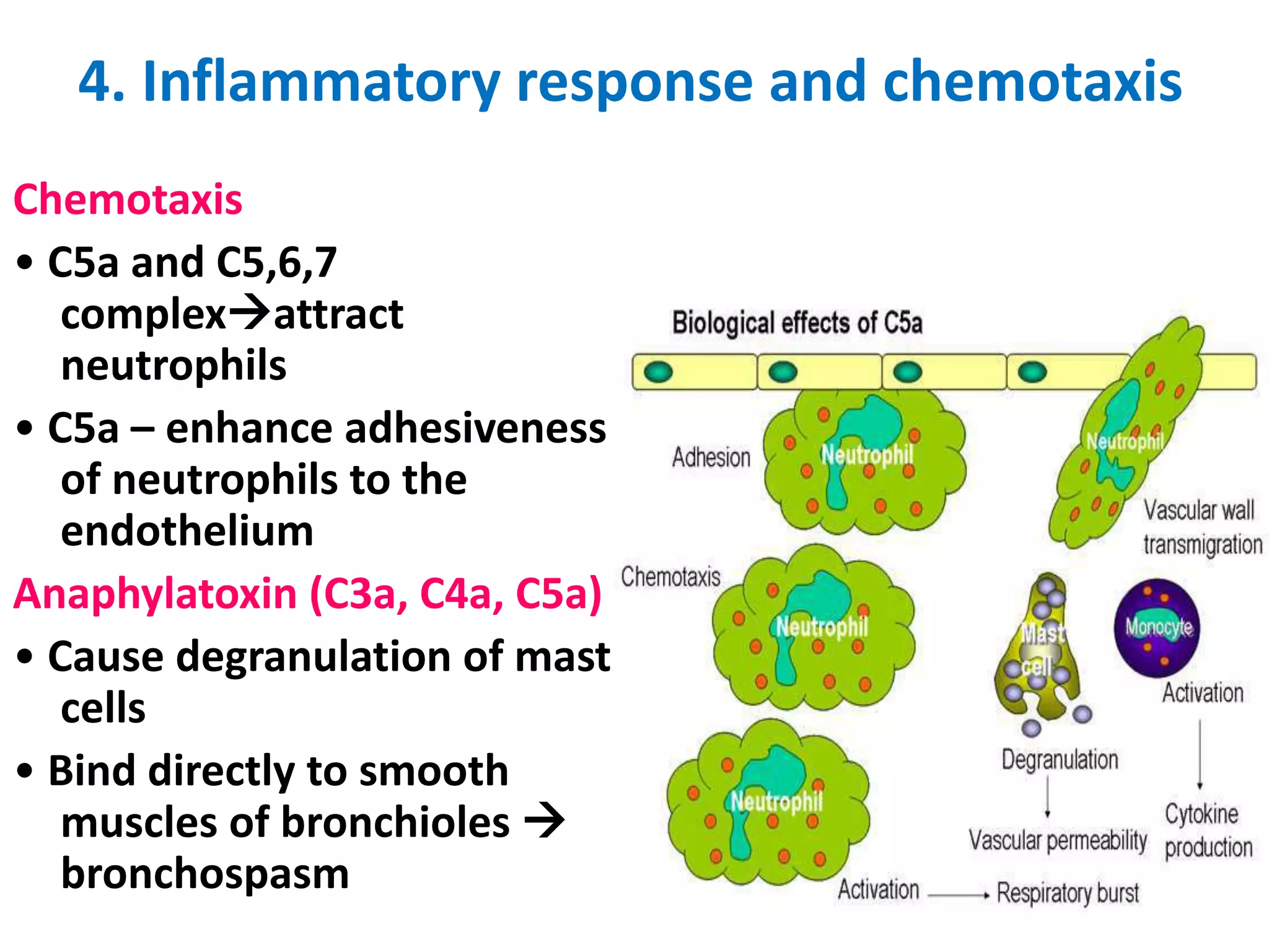
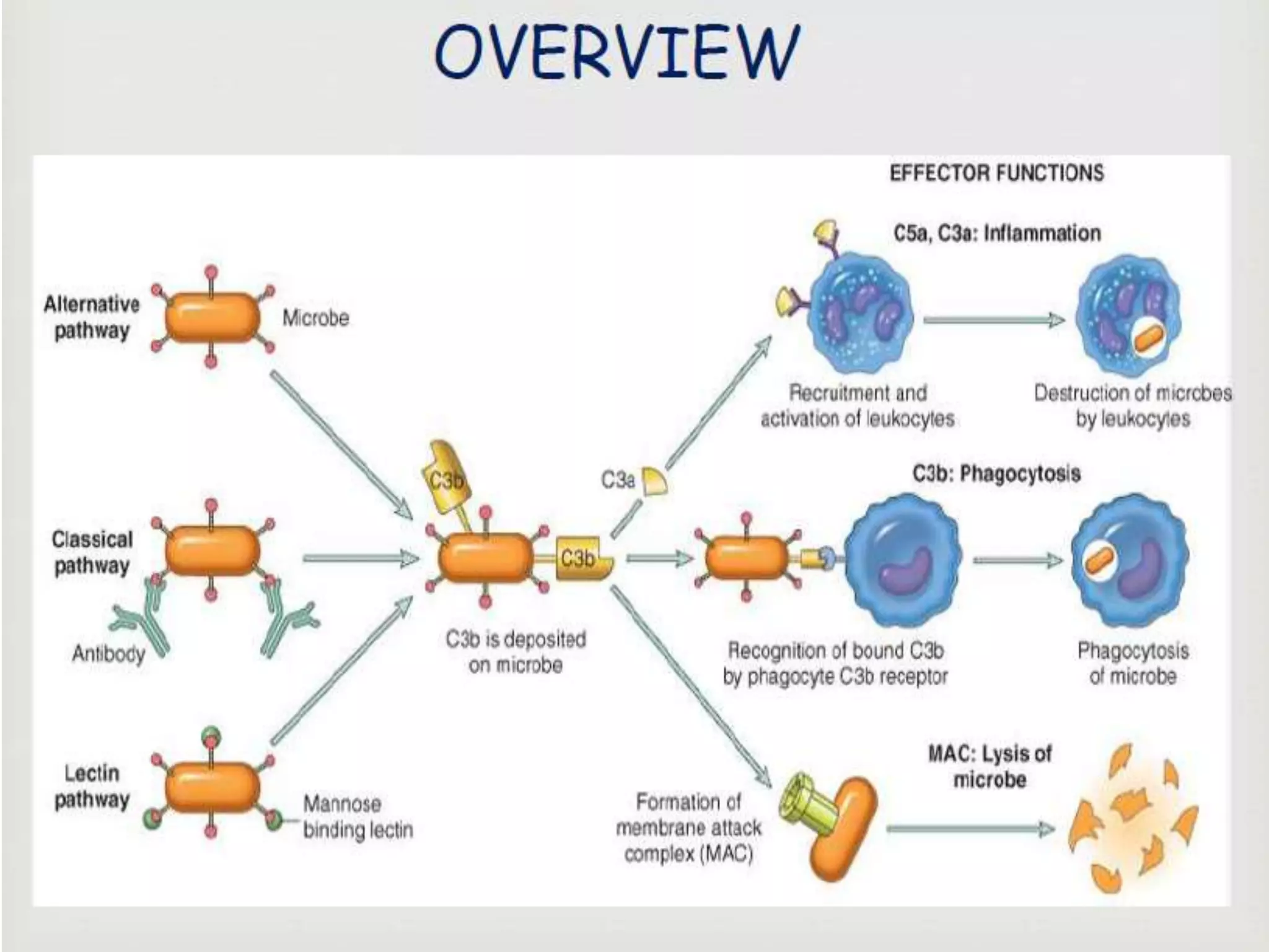
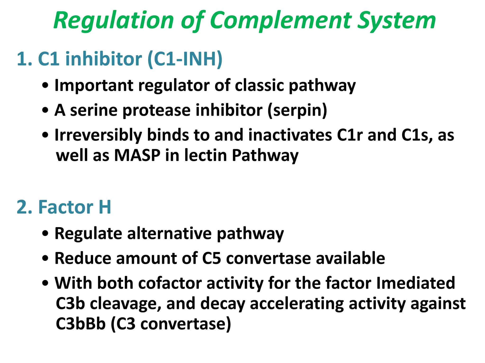
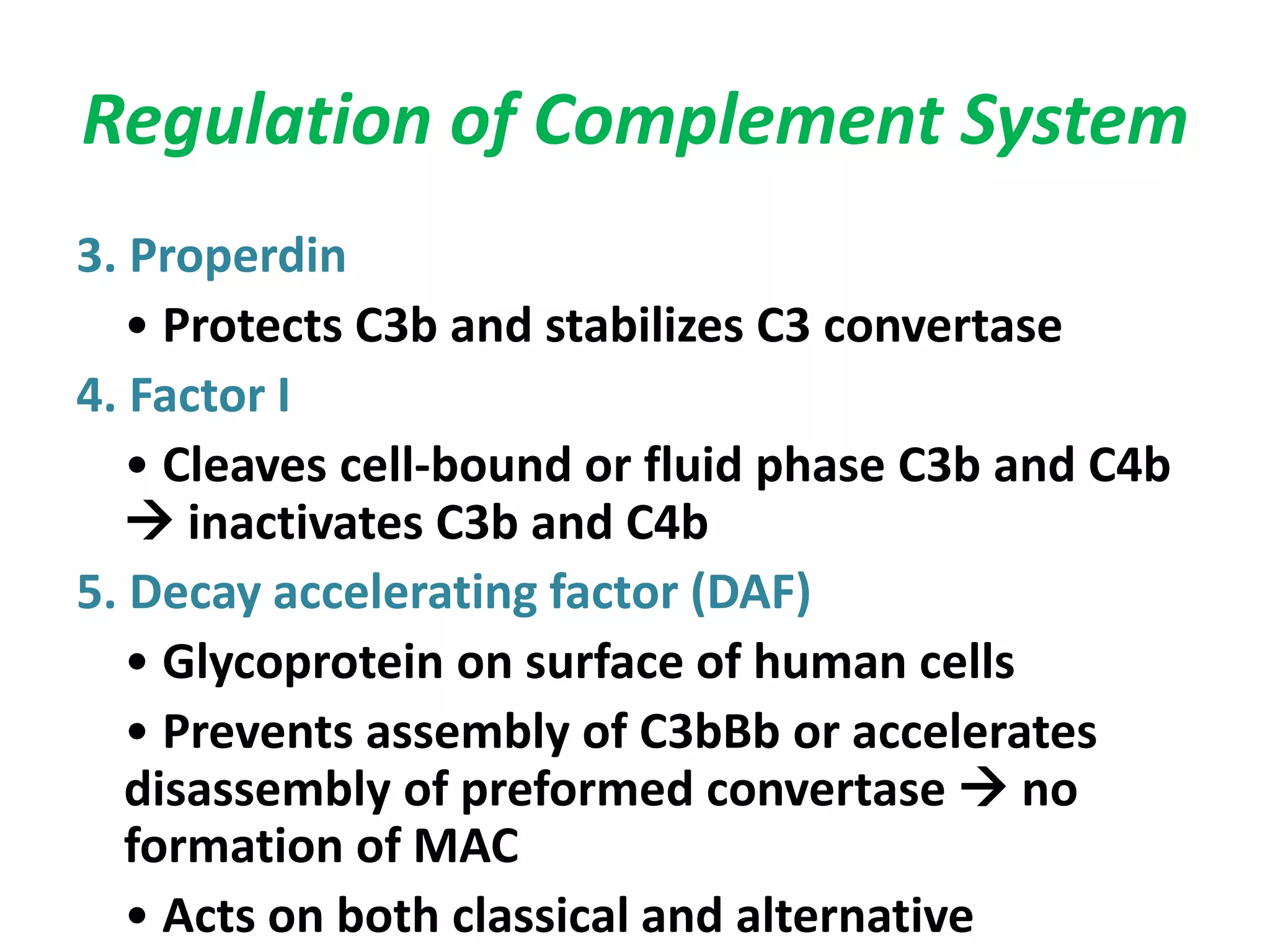
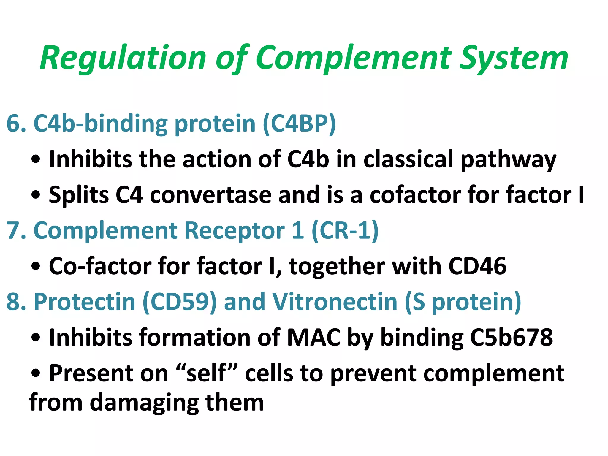
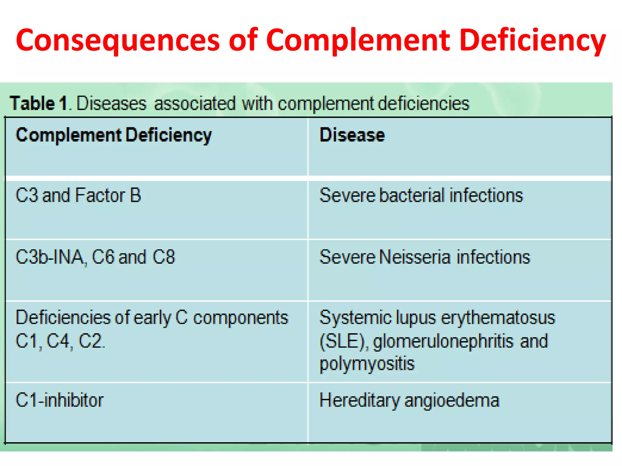
The COMPLEMENT SYSTEM
Dr. Rakesh Prasad Sah
Assistant Professor Microbiology
HISTORY
• Research in complement started in 1890s when
Jules Bordet at the Institute Pasteur of Paris
conducted experiment using sheep antiserum.
• He named those substances as Alexins.
• Paul Ehrlich coined the term
complement.
INTRODUCTION
• It is named “complement system” because it was first
identified as a heat-labile component of serum that
“complemented” antibodies in the killing of bacteria.
• Consists of serum and cell surface proteins involved in
defense against pathogens and tissue damage
mediated by antibodies
• The Complement system is the major effector of
humoral branch of immune system.
• Plays major role in both innate and adaptive immunity.
The components of complement
system
Over 20 serum and cell surface proteins:
- Complement components ( C1 to C9)
- Properdin system
- Regulatory proteins of complement
(enzymes, control molecules and structural
proteins without any enzyme activity)
Three pathways for Complement
activation:
PATHWAYS OF COMPLEMENT ACTIVATION
• Classical pathway
– Activated by antibody
– First discovered
• Alternative pathway
– Activated by some bacterial cell surfaces
– Antibody not involved
• Lectin pathway
– Activated by mannose binding lectin.
– Antibody not involved
CLASSIC PATHWAY OF COMPLEMENT
ACTIVATION
• C1 binds to Fc region of antibody part of Ab/Ag
complex
• C1 is complex of 3 proteins
– C1q is binding protein
– C1r and C1s are proteases
• C1q binds to Fc region of antibody which activates
C1r which activates C1s
• Most efficient at activating complement
– IgM, IgG1 and IgG3
Components of the Membrane-Attack Complex
Native component Active
component(s)
Function(s)
C5 C5a Mediates inflammation;
anaphylatoxin, chemotaxin.
C5b Initiates assembly of the
membrane-attack complex
(MAC).
C6 C6 Binds C5b, forms acceptor
for C7.
C7 C7 Binds C5b6, inserts into
membrane, forms acceptor
for C8.
C8 C8 Binds C5b67, initiates C9
polymerization.
C9 C9n Polymerizes around
C5b678 to form channel
that causes cell lysis.
The Classical Pathway
• The classical pathway is
considered to be part of the
specific immune response
because it relies on
antibodies to initiate it.
• C1 becomes activated when
it binds to the ends of
antibodies
The building of a C3 activation complex
• Once C1 is activated, it activates 2
other complement proteins, C2
and C4 by cutting them in half
• C2 is cleaved into C2a and C2b
• C4 is cleaved into C4a and C4b
• Both C2b and C4b bind together
on the surface of the bacteria
• C2a and C4a diffuse away
C3 Activation complex
• C2b and C4b bind
together on the surface
to form a C3 activation
complex
• The function of the C3
activation complex is to
activate C3 proteins.
– This is done by cleaving C3
into C3a and C3b
C3b
• Many C3b molecules are produced by the C3
activation complex.
• The C3b bind to and coat the surface of the bacteria.
• C3b is an opsonin
– Opsonins are molecules that bind both to
bacteria and phagocytes
– Opsonization increases phagocytosis by 1,000
fold.
Opsonins
C3a
C3a increases the inflammatory response by binding
to mast cells and causing them to release histamine
Building the C5 activation complex
• Eventially enough C3b is cleaved that the
surface of the bacteria begins to become
saturated with it.
• C2b and C4b which make up the C3 activation
complex has a slight affinity for C3b and C3b
binds to them
• When C3b binds to C2b and C4b it forms a
new complex referred to as the C5 activation
complex
The C5 activation complex
• The C5 activation complex (C2b, C4b, C3b)
activates C5 proteins by cleaving them into
C5a and C5b
• Many C5b proteins are produced by the
C5activation complex. These C5b begin to
coat the surface of the bacteria.
The function of C5a
• C5a disperses away from the bacteria.
– Binds to mast cells and increases inflammation.
– Most powerful chemotactic factor known for leukocytes
Building the Membrane Attack complex
• C5b on the surface of bacteria binds to C6
• The binding of C6 to C5b activates C6 so that it
can bind to C7
• C7 binds to C8 which in turn binds to many
C9’s
• Together these proteins form a circular
complex called the Membrane attack complex
(MAC)
Membrane Attack complex
• The MAC causes Cytolysis.
– The circular membrane attack
complex acts as a channel in
which cytoplasm can rush out of
and water rushes in.
• The cells inner integrity is
compromised and it dies
• Animation of the classical
pathaway
Overview
The Alternative Pathway
• Also called as Properdin pathway.
• Bypasses C1, C4, and C2
• Antibody Independent.
• The alternative pathway is slower than the Classical
pathway.
• Acts synergistically with the classical pathway.
• Usually activated by products of microorganisms
like endotoxin.
• Molecules of C3 undergo cleavage at continuous
low level in normal plasma.
Components of the Alternate Pathway
Native
component
Active
component(s)
Function(s)
C3 C3a Mediates inflammation;
anaphylatoxin.
C3b Binds cell surfaces for
opsonization and activation
of alternate pathway.
Factor B B Binds membrane bound
C3b. Cleaved by Factor D.
Ba Unknown.
Bb Cleaved form stabilized by
P produces C3 convertase.
Factor D D Cleaves Factor B when
bound to C3b.
Properdin P Binds and stabilizes
membrane bound C3bBb.
Initiation of The Alternative pathway
• C3 contains in unstable
thioester bond.
• This unstable bond makesC3
subject to slow spontaneous
hydrolysis to C3b and C3a
• The C3b is able to bind to
foreign surface antigens.
• Mammalian cells contain sialic
acid which inactivates C3b
Factor B
• C3b on the surface of a
foreign cells binds to
another plasma protein
called factor B
Factor D
• The binding of C3b to
factor B allows a protein
enzyme called Factor D
to cleave Factor B to Ba
and Bb.
• Factor Bb remains
bound to C3b while Ba
and Factor D disperse
away.
The C3 activation complex
• Properdin, also called factor P, binds to the C3bBb
complex to stabilize it.
• C3bBbP make up the C3 activation complex for the
alternative pathway
The C3 activation Complex
• The C3 activation
complex causes the
production of more
C3b.
• This allows the initial
steps of this pathway to
be repeated and
amplified
• 2X10⁶ molecules can be
generated in 5 minutes
C5 activation complex
• When an additional C3b
binds to the C3 activation
complex it converts it into a
C5 activation complex.
• The C5 activation complex
cleaves C5 into C5a and
C5b.
• C5b begins the production
of the MAC.
The Alternative
complement
pathway
Lectin Binding Pathway
• Also known as the MBL Pathway
• Independent of antibodies.
• Lectins are proteins that bind to specific carbohydrate
targets.
• Activated by Mannose binding lectin (MBL) – lectin that
binds to mannose residues on the microbes.
• MBL is similar to C1q in structure and function.
Activation of lectin pathway
• First step is binding of Mannose Binding Lectin (MBL) to
mannose residue on the surface of microbes.
• To the MBL bound to microbe, MBL –Associated Serine
Proteases (MASP – 1 and MASP – 2) will bind.
• MASP 1 and 2 is similar in
structure and function to C1r and
C1s
• This complex will cleave C4 and C2
• All three pathways lead to production of C3b 
central molecule of complement cascade
• Presence of C3b on surface of a microbe marks it
as foreign and targets it for destruction
• C3b with two important functions:
1. Combines with other complement
components to generate C5 convertase
2. Opsonizes bacteria
General Functions of Complement
Functions of the complement system
• Opsonisation
• Chemotaxis
• Cell lysis
• Immune clearance
• Activation of inflammatory response
2.Cell lysis
• Disrupt the membrane & the entry of water
and electrolytes into the cell
3. Immune complex clearance
4. Inflammatory response and chemotaxis
Chemotaxis
• C5a and C5,6,7
complexattract
neutrophils
• C5a – enhance adhesiveness
of neutrophils to the
endothelium
Anaphylatoxin (C3a, C4a, C5a)
• Cause degranulation of mast
cells
• Bind directly to smooth
muscles of bronchioles 
bronchospasm
Regulation of Complement System
1. C1 inhibitor (C1-INH)
• Important regulator of classic pathway
• A serine protease inhibitor (serpin)
• Irreversibly binds to and inactivates C1r and C1s, as
well as MASP in lectin Pathway
2. Factor H
• Regulate alternative pathway
• Reduce amount of C5 convertase available
• With both cofactor activity for the factor Imediated
C3b cleavage, and decay accelerating activity against
C3bBb (C3 convertase)
Regulation of Complement System
3. Properdin
• Protects C3b and stabilizes C3 convertase
4. Factor I
• Cleaves cell-bound or fluid phase C3b and C4b
 inactivates C3b and C4b
5. Decay accelerating factor (DAF)
• Glycoprotein on surface of human cells
• Prevents assembly of C3bBb or accelerates
disassembly of preformed convertase  no
formation of MAC
• Acts on both classical and alternative
Regulation of Complement System
6. C4b-binding protein (C4BP)
• Inhibits the action of C4b in classical pathway
• Splits C4 convertase and is a cofactor for factor I
7. Complement Receptor 1 (CR-1)
• Co-factor for factor I, together with CD46
8. Protectin (CD59) and Vitronectin (S protein)
• Inhibits formation of MAC by binding C5b678
• Present on “self” cells to prevent complement
from damaging them
Consequences of Complement Deficiency
Complement system by Dr. Rakesh Prasad Sah

Complement system by Dr. Rakesh Prasad Sah

  • 1.
    The COMPLEMENT SYSTEM Dr.Rakesh Prasad Sah Assistant Professor Microbiology
  • 2.
    HISTORY • Research incomplement started in 1890s when Jules Bordet at the Institute Pasteur of Paris conducted experiment using sheep antiserum. • He named those substances as Alexins. • Paul Ehrlich coined the term complement.
  • 3.
    INTRODUCTION • It isnamed “complement system” because it was first identified as a heat-labile component of serum that “complemented” antibodies in the killing of bacteria. • Consists of serum and cell surface proteins involved in defense against pathogens and tissue damage mediated by antibodies • The Complement system is the major effector of humoral branch of immune system. • Plays major role in both innate and adaptive immunity.
  • 6.
    The components ofcomplement system Over 20 serum and cell surface proteins: - Complement components ( C1 to C9) - Properdin system - Regulatory proteins of complement (enzymes, control molecules and structural proteins without any enzyme activity)
  • 9.
    Three pathways forComplement activation:
  • 10.
    PATHWAYS OF COMPLEMENTACTIVATION • Classical pathway – Activated by antibody – First discovered • Alternative pathway – Activated by some bacterial cell surfaces – Antibody not involved • Lectin pathway – Activated by mannose binding lectin. – Antibody not involved
  • 11.
    CLASSIC PATHWAY OFCOMPLEMENT ACTIVATION • C1 binds to Fc region of antibody part of Ab/Ag complex • C1 is complex of 3 proteins – C1q is binding protein – C1r and C1s are proteases • C1q binds to Fc region of antibody which activates C1r which activates C1s • Most efficient at activating complement – IgM, IgG1 and IgG3
  • 13.
    Components of theMembrane-Attack Complex Native component Active component(s) Function(s) C5 C5a Mediates inflammation; anaphylatoxin, chemotaxin. C5b Initiates assembly of the membrane-attack complex (MAC). C6 C6 Binds C5b, forms acceptor for C7. C7 C7 Binds C5b6, inserts into membrane, forms acceptor for C8. C8 C8 Binds C5b67, initiates C9 polymerization. C9 C9n Polymerizes around C5b678 to form channel that causes cell lysis.
  • 14.
    The Classical Pathway •The classical pathway is considered to be part of the specific immune response because it relies on antibodies to initiate it. • C1 becomes activated when it binds to the ends of antibodies
  • 15.
    The building ofa C3 activation complex • Once C1 is activated, it activates 2 other complement proteins, C2 and C4 by cutting them in half • C2 is cleaved into C2a and C2b • C4 is cleaved into C4a and C4b • Both C2b and C4b bind together on the surface of the bacteria • C2a and C4a diffuse away
  • 16.
    C3 Activation complex •C2b and C4b bind together on the surface to form a C3 activation complex • The function of the C3 activation complex is to activate C3 proteins. – This is done by cleaving C3 into C3a and C3b
  • 17.
    C3b • Many C3bmolecules are produced by the C3 activation complex. • The C3b bind to and coat the surface of the bacteria. • C3b is an opsonin – Opsonins are molecules that bind both to bacteria and phagocytes – Opsonization increases phagocytosis by 1,000 fold. Opsonins
  • 18.
    C3a C3a increases theinflammatory response by binding to mast cells and causing them to release histamine
  • 19.
    Building the C5activation complex • Eventially enough C3b is cleaved that the surface of the bacteria begins to become saturated with it. • C2b and C4b which make up the C3 activation complex has a slight affinity for C3b and C3b binds to them • When C3b binds to C2b and C4b it forms a new complex referred to as the C5 activation complex
  • 20.
    The C5 activationcomplex • The C5 activation complex (C2b, C4b, C3b) activates C5 proteins by cleaving them into C5a and C5b • Many C5b proteins are produced by the C5activation complex. These C5b begin to coat the surface of the bacteria.
  • 21.
    The function ofC5a • C5a disperses away from the bacteria. – Binds to mast cells and increases inflammation. – Most powerful chemotactic factor known for leukocytes
  • 22.
    Building the MembraneAttack complex • C5b on the surface of bacteria binds to C6 • The binding of C6 to C5b activates C6 so that it can bind to C7 • C7 binds to C8 which in turn binds to many C9’s • Together these proteins form a circular complex called the Membrane attack complex (MAC)
  • 23.
    Membrane Attack complex •The MAC causes Cytolysis. – The circular membrane attack complex acts as a channel in which cytoplasm can rush out of and water rushes in. • The cells inner integrity is compromised and it dies • Animation of the classical pathaway
  • 24.
  • 25.
    The Alternative Pathway •Also called as Properdin pathway. • Bypasses C1, C4, and C2 • Antibody Independent. • The alternative pathway is slower than the Classical pathway. • Acts synergistically with the classical pathway. • Usually activated by products of microorganisms like endotoxin. • Molecules of C3 undergo cleavage at continuous low level in normal plasma.
  • 26.
    Components of theAlternate Pathway Native component Active component(s) Function(s) C3 C3a Mediates inflammation; anaphylatoxin. C3b Binds cell surfaces for opsonization and activation of alternate pathway. Factor B B Binds membrane bound C3b. Cleaved by Factor D. Ba Unknown. Bb Cleaved form stabilized by P produces C3 convertase. Factor D D Cleaves Factor B when bound to C3b. Properdin P Binds and stabilizes membrane bound C3bBb.
  • 27.
    Initiation of TheAlternative pathway • C3 contains in unstable thioester bond. • This unstable bond makesC3 subject to slow spontaneous hydrolysis to C3b and C3a • The C3b is able to bind to foreign surface antigens. • Mammalian cells contain sialic acid which inactivates C3b
  • 28.
    Factor B • C3bon the surface of a foreign cells binds to another plasma protein called factor B
  • 29.
    Factor D • Thebinding of C3b to factor B allows a protein enzyme called Factor D to cleave Factor B to Ba and Bb. • Factor Bb remains bound to C3b while Ba and Factor D disperse away.
  • 30.
    The C3 activationcomplex • Properdin, also called factor P, binds to the C3bBb complex to stabilize it. • C3bBbP make up the C3 activation complex for the alternative pathway
  • 31.
    The C3 activationComplex • The C3 activation complex causes the production of more C3b. • This allows the initial steps of this pathway to be repeated and amplified • 2X10⁶ molecules can be generated in 5 minutes
  • 32.
    C5 activation complex •When an additional C3b binds to the C3 activation complex it converts it into a C5 activation complex. • The C5 activation complex cleaves C5 into C5a and C5b. • C5b begins the production of the MAC.
  • 33.
  • 34.
    Lectin Binding Pathway •Also known as the MBL Pathway • Independent of antibodies. • Lectins are proteins that bind to specific carbohydrate targets. • Activated by Mannose binding lectin (MBL) – lectin that binds to mannose residues on the microbes. • MBL is similar to C1q in structure and function.
  • 35.
    Activation of lectinpathway • First step is binding of Mannose Binding Lectin (MBL) to mannose residue on the surface of microbes. • To the MBL bound to microbe, MBL –Associated Serine Proteases (MASP – 1 and MASP – 2) will bind. • MASP 1 and 2 is similar in structure and function to C1r and C1s • This complex will cleave C4 and C2
  • 36.
    • All threepathways lead to production of C3b  central molecule of complement cascade • Presence of C3b on surface of a microbe marks it as foreign and targets it for destruction • C3b with two important functions: 1. Combines with other complement components to generate C5 convertase 2. Opsonizes bacteria
  • 38.
  • 39.
    Functions of thecomplement system • Opsonisation • Chemotaxis • Cell lysis • Immune clearance • Activation of inflammatory response
  • 41.
    2.Cell lysis • Disruptthe membrane & the entry of water and electrolytes into the cell
  • 42.
  • 43.
    4. Inflammatory responseand chemotaxis Chemotaxis • C5a and C5,6,7 complexattract neutrophils • C5a – enhance adhesiveness of neutrophils to the endothelium Anaphylatoxin (C3a, C4a, C5a) • Cause degranulation of mast cells • Bind directly to smooth muscles of bronchioles  bronchospasm
  • 45.
    Regulation of ComplementSystem 1. C1 inhibitor (C1-INH) • Important regulator of classic pathway • A serine protease inhibitor (serpin) • Irreversibly binds to and inactivates C1r and C1s, as well as MASP in lectin Pathway 2. Factor H • Regulate alternative pathway • Reduce amount of C5 convertase available • With both cofactor activity for the factor Imediated C3b cleavage, and decay accelerating activity against C3bBb (C3 convertase)
  • 46.
    Regulation of ComplementSystem 3. Properdin • Protects C3b and stabilizes C3 convertase 4. Factor I • Cleaves cell-bound or fluid phase C3b and C4b  inactivates C3b and C4b 5. Decay accelerating factor (DAF) • Glycoprotein on surface of human cells • Prevents assembly of C3bBb or accelerates disassembly of preformed convertase  no formation of MAC • Acts on both classical and alternative
  • 47.
    Regulation of ComplementSystem 6. C4b-binding protein (C4BP) • Inhibits the action of C4b in classical pathway • Splits C4 convertase and is a cofactor for factor I 7. Complement Receptor 1 (CR-1) • Co-factor for factor I, together with CD46 8. Protectin (CD59) and Vitronectin (S protein) • Inhibits formation of MAC by binding C5b678 • Present on “self” cells to prevent complement from damaging them
  • 48.