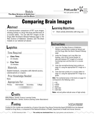
This document provides instructions for an activity comparing brain images from PET scans of a normal, drug-free brain and the brain of a cocaine addict. Students will color-code numbered areas on images based on the level of activity seen in the PET scans, with red and yellow indicating highest activity. Doing this activity allows students to visually see how drug use diminishes brain activity over time in the regions associated with reward and pleasure.
Breakthrough Method for Mapping Brain Epigenomes | The Lifesciences MagazineThe Lifesciences Magazine
Researchers at the College of Engineering have made significant strides in brain research by developing a cost-effective method for spatially characterizing and mapping brain epigenomes.
Breakthrough Method for Mapping Brain Epigenomes | The Lifesciences MagazineThe Lifesciences Magazine
Researchers at the College of Engineering have made significant strides in brain research by developing a cost-effective method for spatially characterizing and mapping brain epigenomes.
Lecture for Reinforcement Learning study group held on August 19th, 2017.
Reference book: http://incompleteideas.net/book/the-book.html
Video: https://youtu.be/xv5ZsOSf6ZQ
Initiated by Taiwan AI Group (https://www.facebook.com/groups/Taiwan.AI.Group/permalink/1796526840669749/)
Contributions of Neuroscience toOur Understanding of CognitiAlleneMcclendon878
Contributions of Neuroscience to
Our Understanding of Cognitive
Development
Adele Diamond1 and Dima Amso2
1
Department of Psychiatry, University of British Columbia, and Department of Child and Adolescent Psychiatry,
BC Children’s Hospital, Vancouver, Canada; and
2
Sackler Institute for Developmental Psychobiology,
Weill Medical College of Cornell University
ABSTRACT—One major contribution of neuroscience to
understanding cognitive development has been in demon-
strating that biology is not destiny—that is, demonstrating
the remarkable role of experience in shaping the mind,
brain, and body. Only rarely has neuroscience provided
wholly new insights into cognitive development, but often
it has provided evidence of mechanisms by which obser-
vations of developmental psychologists could be explained.
Behavioral findings have often remained controversial
until an underlying biological mechanism for them was
offered. Neuroscience has demonstrated promise for de-
tecting cognitive problems before they are behaviorally
observable—and, hence, promise for early intervention. In
this article, we discuss examples drawn from imitation and
mirror neurons, phenylketonuria (PKU) and prefrontal
dopamine, maternal touch and stress reactivity, and non-
genetic (behavioral) intergenerational transmission of bi-
ological characteristics.
KEYWORDS—plasticity; epigenesis; mothering; executive
functions; animal models; molecular genetics; memory
Neuroscience research has made its greatest contributions to the
study of cognitive development by illuminating mechanisms
(providing a ‘‘how’’) that underlie behavioral observations made
earlier by psychologists. It has also made important contribu-
tions to our understanding of cognitive development by dem-
onstrating that the brain is far more plastic at all ages than
previously thought—and thus that the speed and extent by which
experience and behavior can shape the brain is greater than al-
most anyone imagined. In other words, rather than showing that
biology is destiny, neuroscience research has been at the fore-
front of demonstrating the powerful role of experience throughout
life. Besides the surprising evidence of the remarkable extent
of experience-induced plasticity, rarely has neuroscience given
us previously unknown insights into cognitive development, but
neuroscience does offer promise of being able to detect some
problems before they are behaviorally observable.
PROVIDING MECHANISMS THAT CAN ACCOUNT FOR
BEHAVIORAL RESULTS REPORTED BY
PSYCHOLOGISTS
Here we describe two examples of behavioral findings by psy-
chologists that were largely ignored or extremely controversial
until underlying biological mechanisms capable of accounting
for them were provided by neuroscience research. One such
example concerns cognitive deficits documented in children
treated early and continuously for phenylketonuria (PKU). The
second example involves neonatal imitation observed b ...
Introducing the brain
Ethics and the brain initiative
Neurotransmitters, Action Potential, Information Coding, Grey and White Matter, Cerebrospinal Fluid, Central Nervous System, Cerebral Cortex, Subcortex, Limbic System, Midbrain and Hindbrain.
Jorge Alberto Costa e Silva-Psiquiatría: situación actual y perspectivas de f...Fundación Ramón Areces
'Psiquiatría: situación actual y perspectivas de futuro'. Este es el título del simposio internacional que organizamos el 20 de junio de 2016 en la Fundación Ramón Areces con las fundaciones Juan José López-Ibor y Lilly en homenaje al doctor Juan José López-Ibor, fallecido en enero de 2015. Durante esta jornada, expertos internacionales abordaron la profunda crisis que atraviesa la psiquiatría como disciplina científica y especialidad médica. Además, se presentó el libro con el mismo título del simposio, también en recuerdo del doctor López-Ibor.
You wrote this scenario from the perspective of Behaviorism learni.docxrosemarybdodson23141
You wrote this scenario from the perspective of Behaviorism learning theory Now I want two scenarios same this scenario but from two different perspectives that they are Cognitivism Learning theory and Social learning theory
For further clarification see attached example
Learning Situation from Behaviorism Learning Theory
The class of 20 students is divided into two teams, having 10 students in each team. The teacher makes two columns on the board for team A and team B. Teacher points out, Yesterday in our history class we studied about the civil rights movement I hope you have well-prepared that topic. Let’s start an informal quiz based on yesterday’s topic. Are you guys ready? Students say, “Yes”! Teacher starts asking questions. Team A! Which sports Jackie Robinson played? Students raised their hands. Robert? Can you give the answer? Robert says soccer. Teacher appreciating Robert’s effort says very good Robert and write 10 under the column of Team A. Next question for Team B, Dr. Martin Luther King Jr. went to the college to become? Students raise their hands. James, can you answer? James says, “Minister”. Teacher appreciates the attempt but the answer is not correct. Ok! Now, what you guys think what was the main contribution of Abraham Lincoln?Timothy raised his hand and replied, he brought freedom and abolish slavery. Rosie raised her hand and replied, he ran the country being a president of the country. Teacher says, when we freedom was attained by the African American it was not solely due to Abraham Lincoln. Who played the actual role? Joseph replies, African Americans themselves. Teacher appreciated Joseph’s answer saying absolutely right. No leader can bring freedom from slavery or racism until its people are themselves not ready to put their efforts. Nation needs to be united to get rid of inequality.
Learning Situation from Cognitivism Learning Theory:
Learning Situation from Social Learning Theory:
3 | Page
Chapter 2 terminology
Psych260
Nervous System-
A network of billions of cells in the brain and the body responsible for all aspects of what we feel, think, and do.
Central nervous system-
The part of the nervous system that consists of the brain and the spinal cord.
Peripheral nervous system-
The part of the nervous central nervous system with the muscles, organs and glands.
Neurons-
The basic units of the nervous system cells that receive integrate and transmit information in the nervous system. Neurons operate through electrical impulses communicate with other neurons through electrical impulses communicate with other neurons through chemical signals and form neural networks.
Dendrites –
Branchlike extensions of the neuron with receptors that detect information from other neurons.
Cell Body-
Part of the neuron where information from thousands of other neurons is collected and integrated.
Axon-
A long narrow outgrowth of a neuron that enables the neuron to transmit information to other neurons..
More advanced thinkers like Eleanor A. Maguire, a professor of Cognitive Neuroscience
at the University of London mentions in her study of taxi drivers that the hippocampus is
the seat of spatial reasoning, memory planning for the future and is located in
posterior hippocampus-the spatial processing center.
Mani Pavuluri - Discussion on Role of Nucleus Accumbens in Reward & Emotional...drmanipavuluri
1. Dr. Mani Pavuluri is a renowned psychologist, who has acquired widespread recognition all across the globe. She is lately sharing wonderful stories that could enable modern-day individuals to lead a healthy life. View the document and know about the interesting facts on Role of Nucleus Accumbens in Reward & Emotional Circuitry.
Dr Mani Pavuluri - Integrating Pharmacotherapy and Psychotherapy for PBDdrmanipavuluri
Dr Mani Pavuluri is the professor of psychiatry. She is MD and PhD. She is one of the recognized scientists who have developed several programs throughout the world for helping the patients, their families and teachers.