
Recommended
More Related Content
Similar to clinico_pathological conference on cervical cancer
Similar to clinico_pathological conference on cervical cancer (20)
More from RaishemAli1
Recently uploaded
Recently uploaded (20)
clinico_pathological conference on cervical cancer
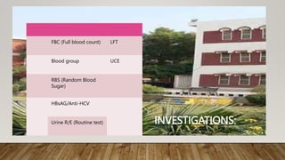
- 1. INVESTIGATIONS: FBC (Full blood count) LFT Blood group UCE RBS (Random Blood Sugar) HBsAG/Anti-HCV Urine R/E (Routine test)
- 2. FULL BLOOD COUNT: • RBC count: 4.2 million/mm3 • Hb level: 9.1g/dl • Hematocrit: 34% • Mean corpuscular volume: 65fl • Mean corpuscular hemoglobin: 28pg • Mean corpuscular hemoglobin concentration: 30g/dl • WBC count: 7100/mm3 • Platelet count: 250000/mm3
- 4. INVESTIGATIONS: Random Blood sugar: 114mg/dl Urea : 28mg Creatinine: 0.9mg Na+ : 134meq/L K+ : 3.6meq/L Cl- : 99meq/L HCO3: 21meq/L
- 5. LIVER FUNCTION TEST •Bilirubin: 0.5mg •Direct bilirubin: 0.21mg •Indirect bilirubin: 0.29mg •ALP: 150U/L •GGT: 14U/L •SGPT: 35U/L
- 7. COAGULATION PROFILE • PT= 13 SECONDS CONTROL 12 SECONDS. • APTT= 28 SECONDS CONTROL 28 SECONDS.
- 9. MRI PELVIS: The small well-defined cystic area noted in the right adnexal region appears hypointense on T1 and hyperintense T2W images. It measures approx. 2.2* 1.9 cm. Findings likely represent dominant follicle/ follicular retention cyst.
- 10. MRI PELVIS: A small abnormal signal intensity area seen in the fundus of the uterus appears hypointense, showing no significant postcontrast enhancement. Likely represents a small intramural fibroid. Thickening of cervical canal noted causing dilatation of uterine cavity. The rest of the scan is unremarkable.
- 11. .
- 12. MANAGEMENT: • Total abdominal hysterectomy bilateral Salpingo Ooophorectomy done and sample send for Histopathology
- 13. HISTO PATHOLOGY SHOWED: Simple endometrial hyperplasia without atypia & and chronic nonspecific endometritis. Small intramural leiomyoma, uterus. Minimally invasive moderately differentiated nonkeratinizing squamous cell carcinoma of cervix Figo Stage 1A Haemorrhagic cystic corpus luteum, one ovary.
- 14. HISTO PATH BLOCKS WERE REVIEWED FOR SECOND OPINION BY AKUH: Cervix: Moderately differentiated Non keratinizing squamous cell carcinoma HPV Stromal invasion 0.1cm, and horizontal extent 0.9cm Lvi : identified Margins: ectocervical resection margin : 0.1cm, deep circumferential margin : 1.2cm Figo stage 1A1
- 15. CONTINUED Endometrium: Autolysed endometrium Myometrium: Histologically unremarkable Ovary: corpus luteum. No comment was done on vaginal & adnexal involvement due to limited specimen Lymph nodes : Not received
- 16. POST SURGICAL MRI PELVIS: • Showing post-surgical changes. • No abnormal intensity enhancing lesion was seen to suggest recurrent/ residual disease • Patient referred to NORIN Hospital
- 17. • Then patient underwent EBRT 45 Gy in 25 Fx with weekly Cisplatin • vaginal brachtherapy • HDR Brachytherapy total of 18Gy in 3 Fractions
- 18. 1ST FOLLOW UP: • 1ST follow up done after 3months • MRI pelvis was done • That showed no recurrent/ residual disease noted in surgical bed. Patient was also doing well with no active complains
- 19. 2ND FOLLOW UP • After 3month of 1st follow-up patient complained of constant lower abdominal and back pain • Excessive watery discharge with occasional spotting. • On vaginal examination: thickening of the stump was palpable with mild PV bleeding • Patient was then again advised MR pelvis with contrast.
- 20. CT SCAN DONE • That showed Ill defined heterogeneously enhancing lesion seen in the right side of the pelvis at the site of the stump causing indentation of the posterior wall of the bladder with loss of fat planes. Likely representing recurrence of disease at the surgical bed. It measures 3.2 * 2.8cm.on
- 21. Ct scan
- 22. . Ct scan
- 23. • She presented to us with blood- stained vaginal discharge • we planned for •EUA and biopsy.
- 24. • THE HISTOPATHOLOGY REPORT OF THE BIOPSY DONE : • MODERATELY DIFFERENTIATED NONKERATINIZING SQUAMOUS CELL CARCINOMA. GRADE 2.
- 25. • Now the question is how to proceed with this case. • Chemotherapy • Exenteration • Re-Rt.
- 26. .