Downloaded 227 times
![Antitoxin
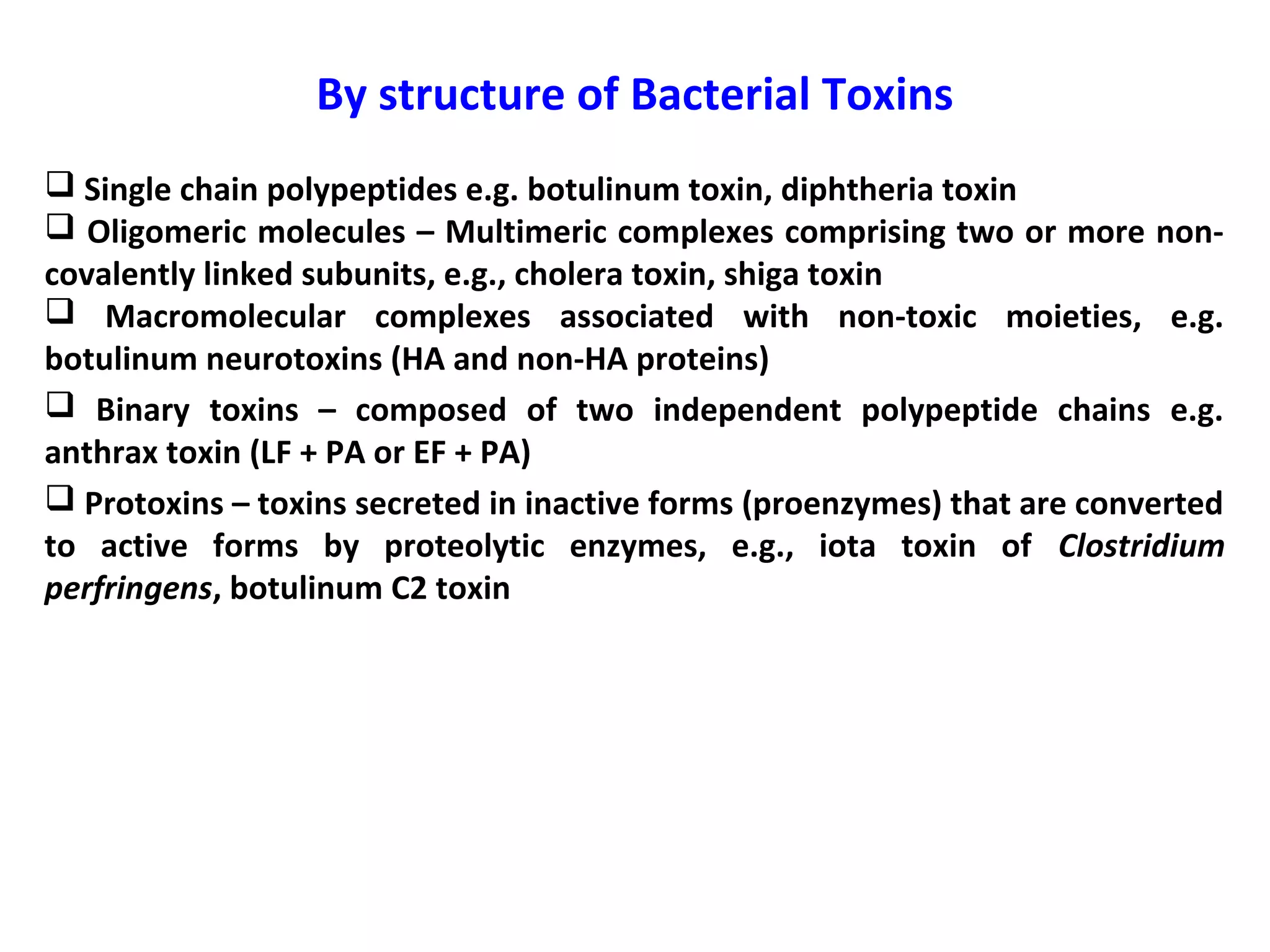
• An antitoxin is an antibody with the ability to neutralize a specific toxin.
• This procedure involves injecting an animal with a safe amount of a
particular toxin. Then, the animal’s body makes the antitoxin needed to
neutralize the toxin. Later, the blood is withdrawn from the animal. When
the antitoxin is obtained from the blood, it is purified and injected into a
human or other animal, inducing passive immunity.
• Antitoxin, antibody, formed in the body by the introduction of a bacterial
poison, or toxin, and capable of neutralizing the toxin. People who have
recovered from bacterial illnesses often develop specific antitoxins that
confer immunity against recurrence.
• To prevent serum sickness, it is often best to use antitoxin generated from
the same species . (e.g. use human antitoxin to treat humans).
• Antitoxins to diphtheria and tetanus toxins were produced by
Emil Adolf von Behring and his colleagues from 1890 onwards.
• The use of diphtheria antitoxin for the treatment of diphtheria was
regarded by the Lancet as the "most important advance of the [19th]
Century in the medical treatment of acute infectious disease".
• For medical use in treating human infectious diseases, antitoxins are
produced by injecting an animal with toxin; the animal, most commonly a
horse, is given repeated small doses of toxin until a high concentration of
the antitoxin builds up in the blood. The resulting highly concentrated
preparation of antitoxins is called an antiserum.](https://image.slidesharecdn.com/lecture-1classificationandnomenclatureofbacterialtoxinsfinal-190604024632/75/Classification-and-nomenclature-of-bacterial-toxins-8-2048.jpg)
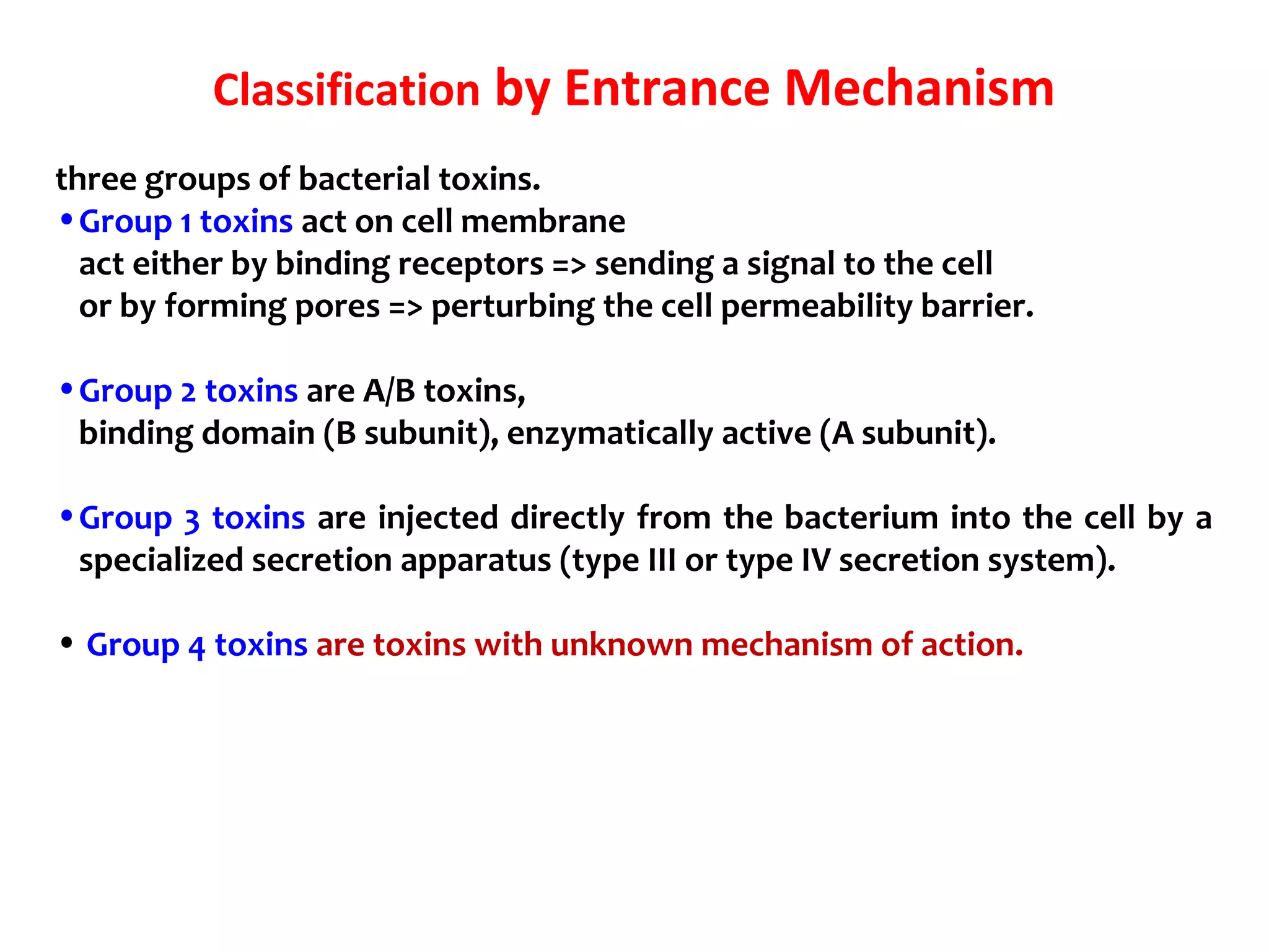
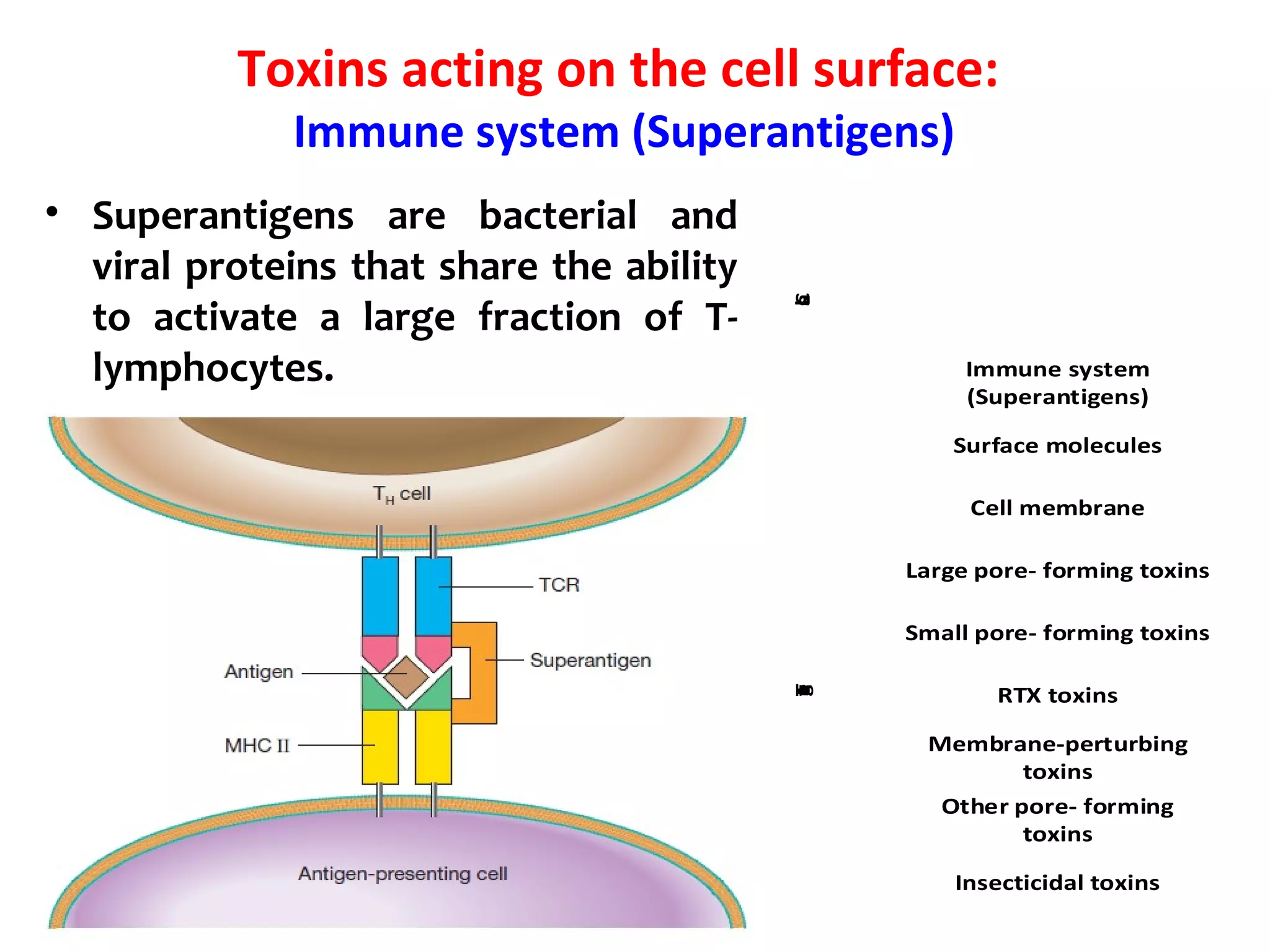
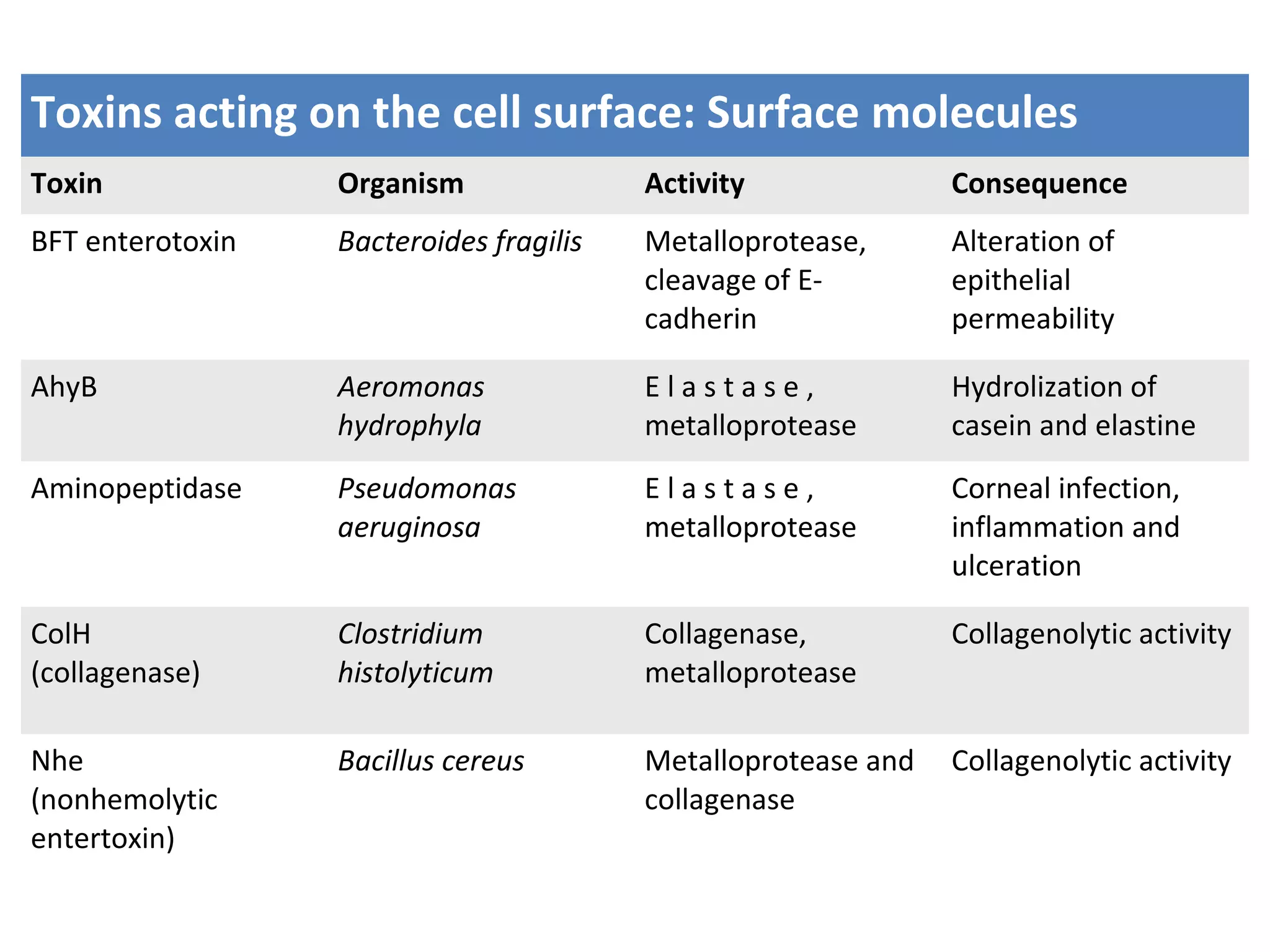
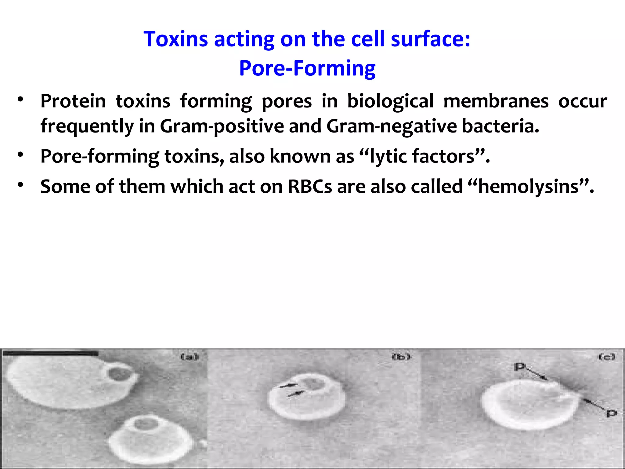
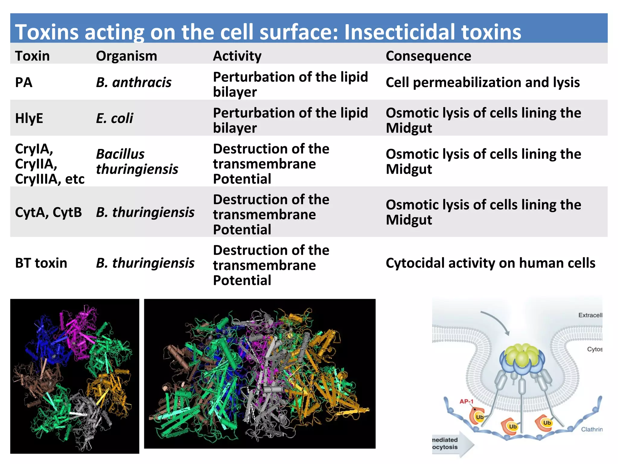
![Toxins acting on the cell surface:
Surface molecules
BFT enterotoxin: The pathogenicity of ETBF is ascribed to a
heat-labile 20-kDa toxin (∼ B. fragilis toxin [BFT], also called
fragilysin).
This toxin binds to a specific intestinal epithelial cell receptor
and stimulates cell proliferation.](https://image.slidesharecdn.com/lecture-1classificationandnomenclatureofbacterialtoxinsfinal-190604024632/75/Classification-and-nomenclature-of-bacterial-toxins-28-2048.jpg)


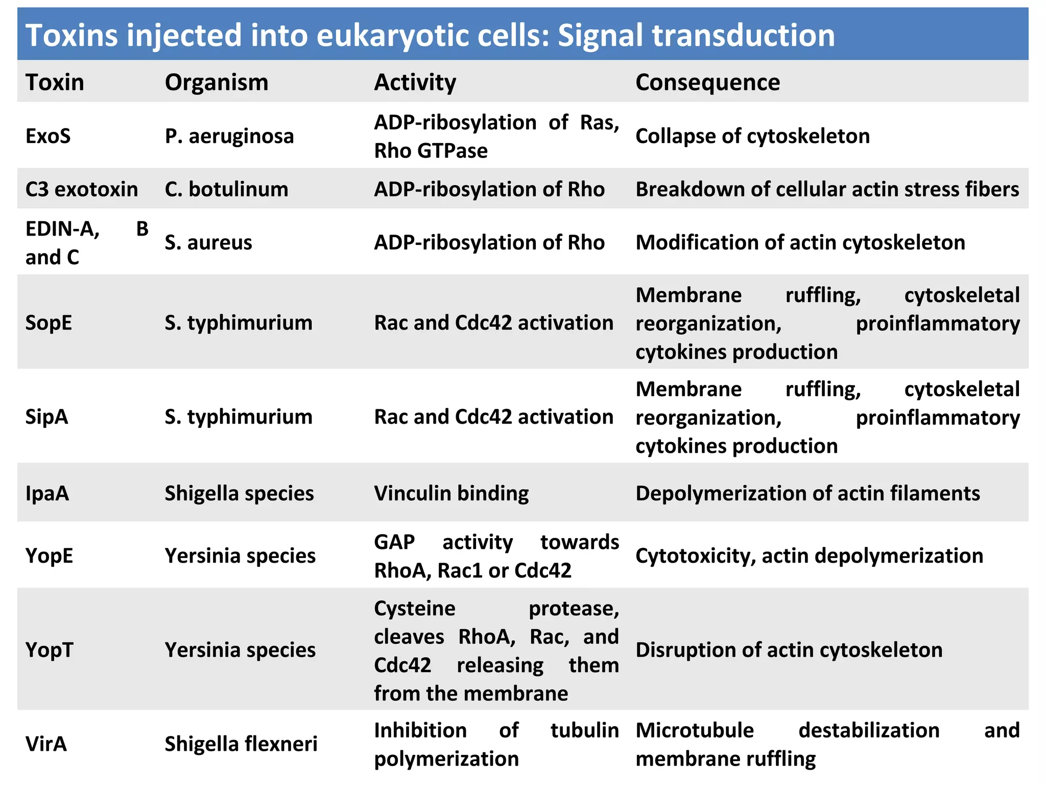
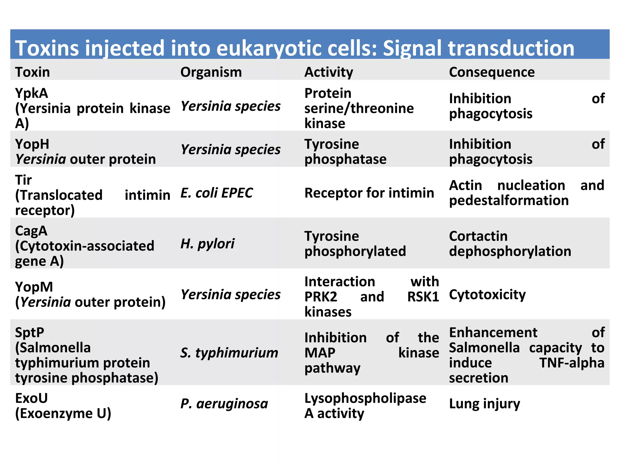
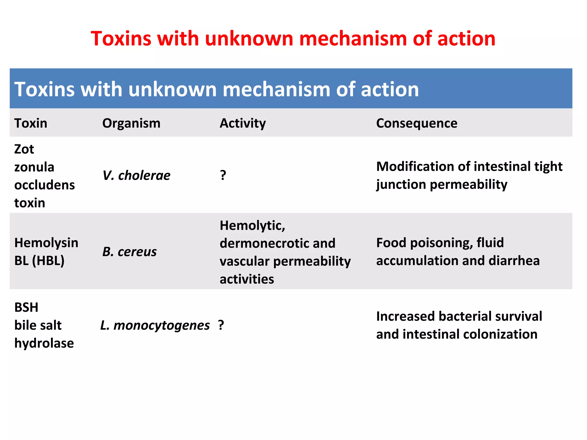
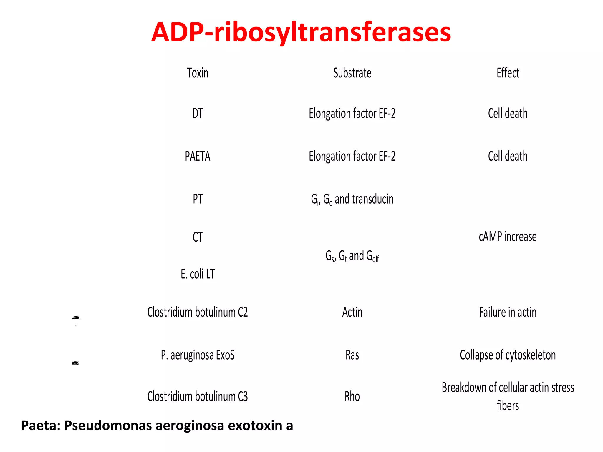
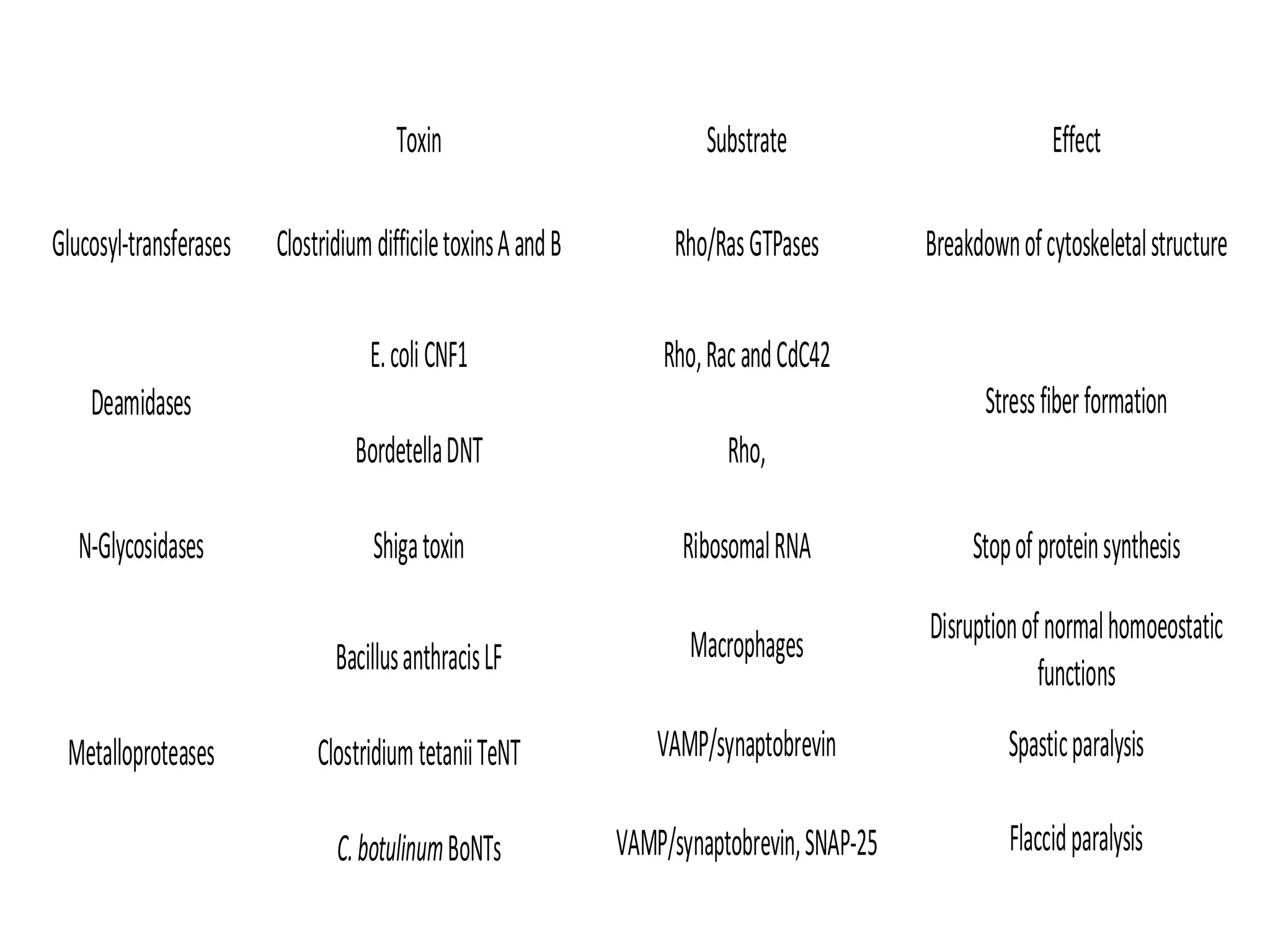


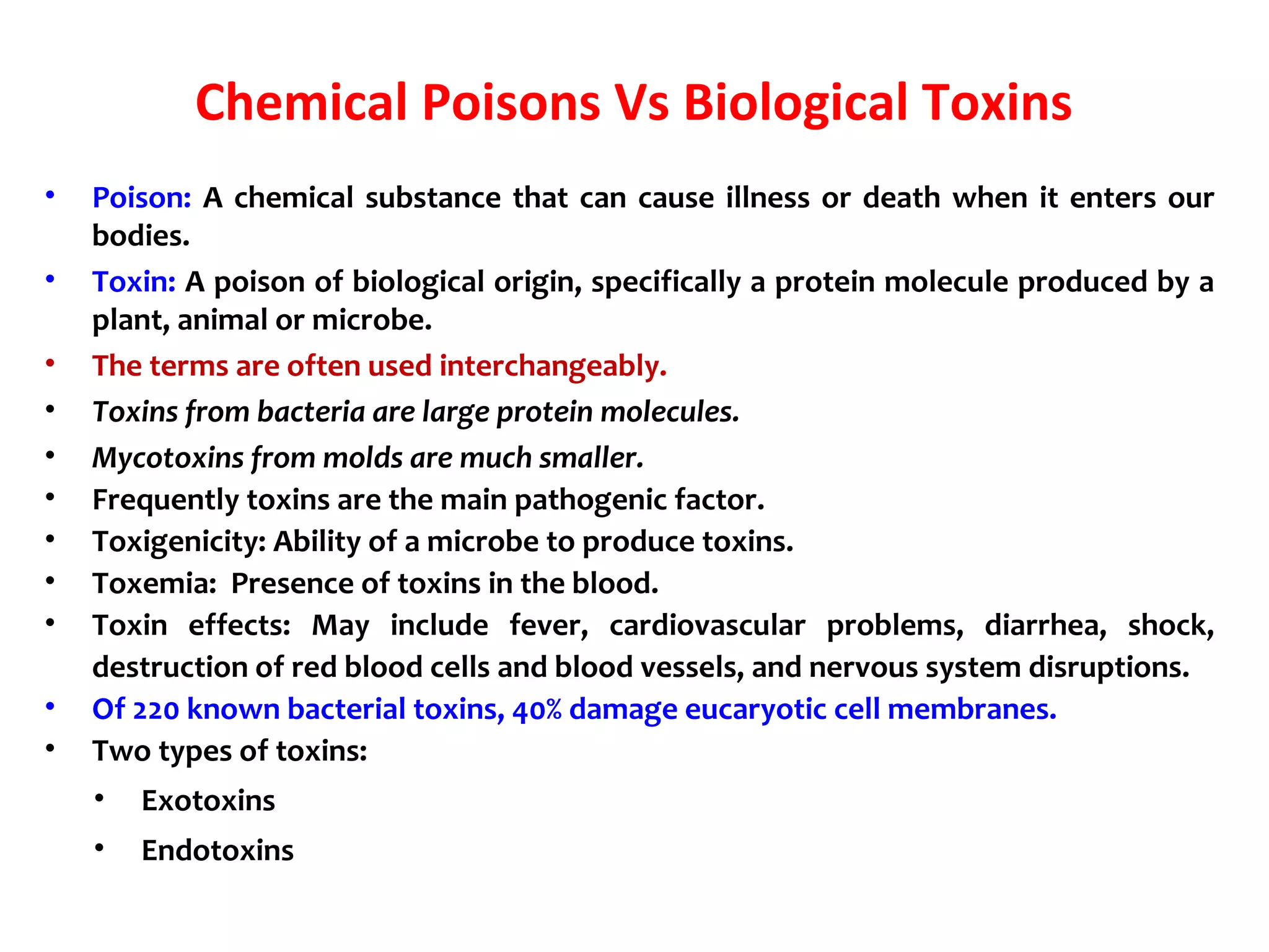
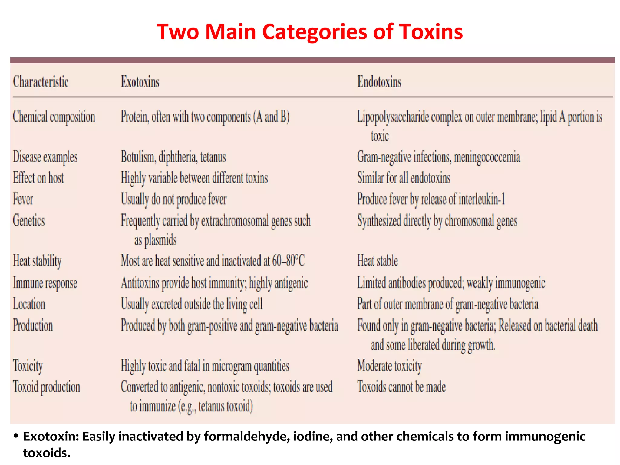
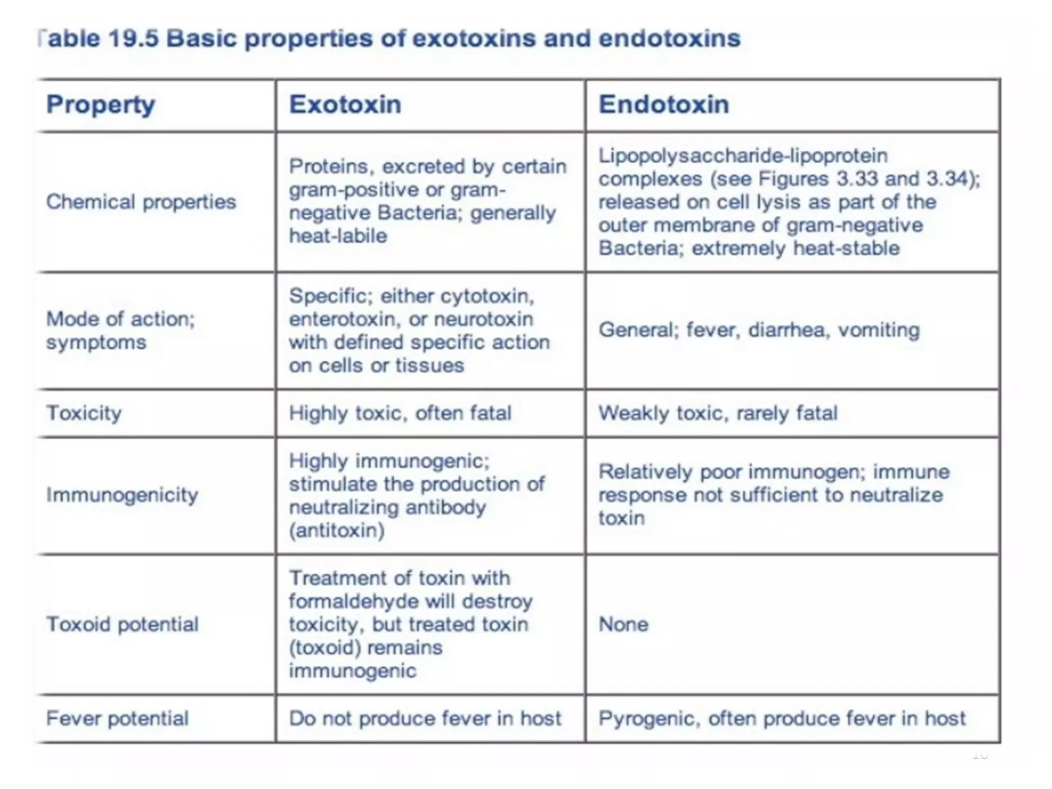
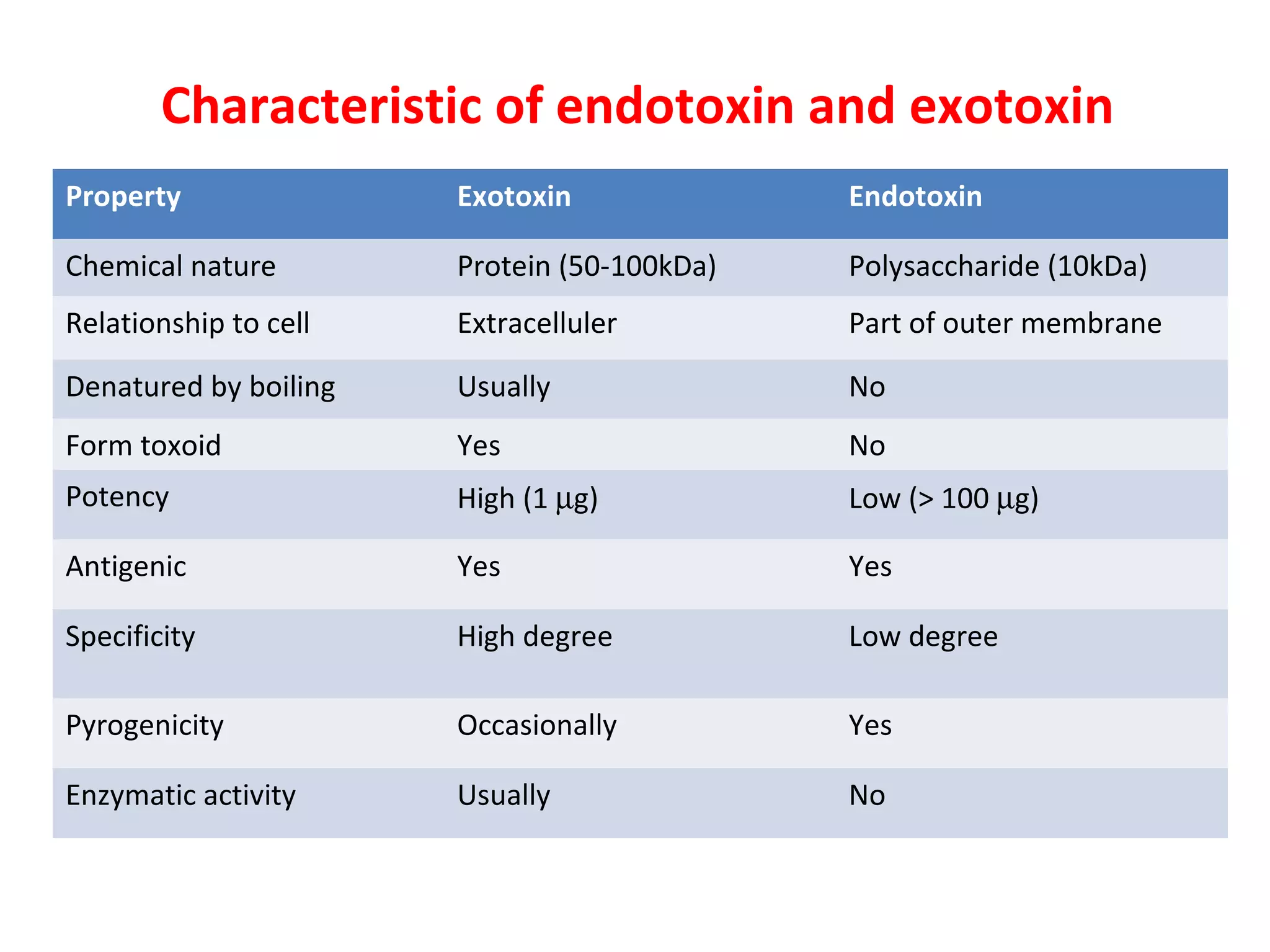
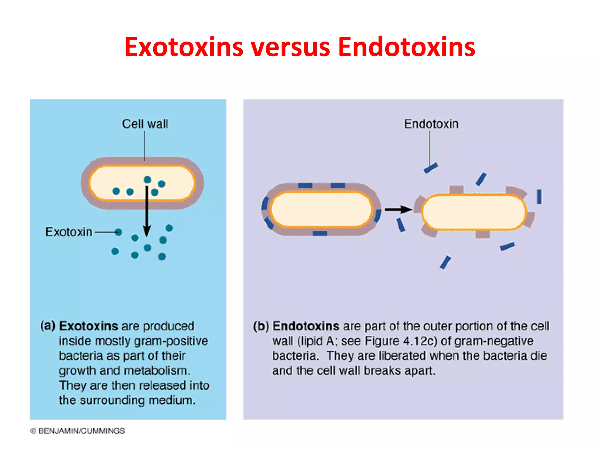
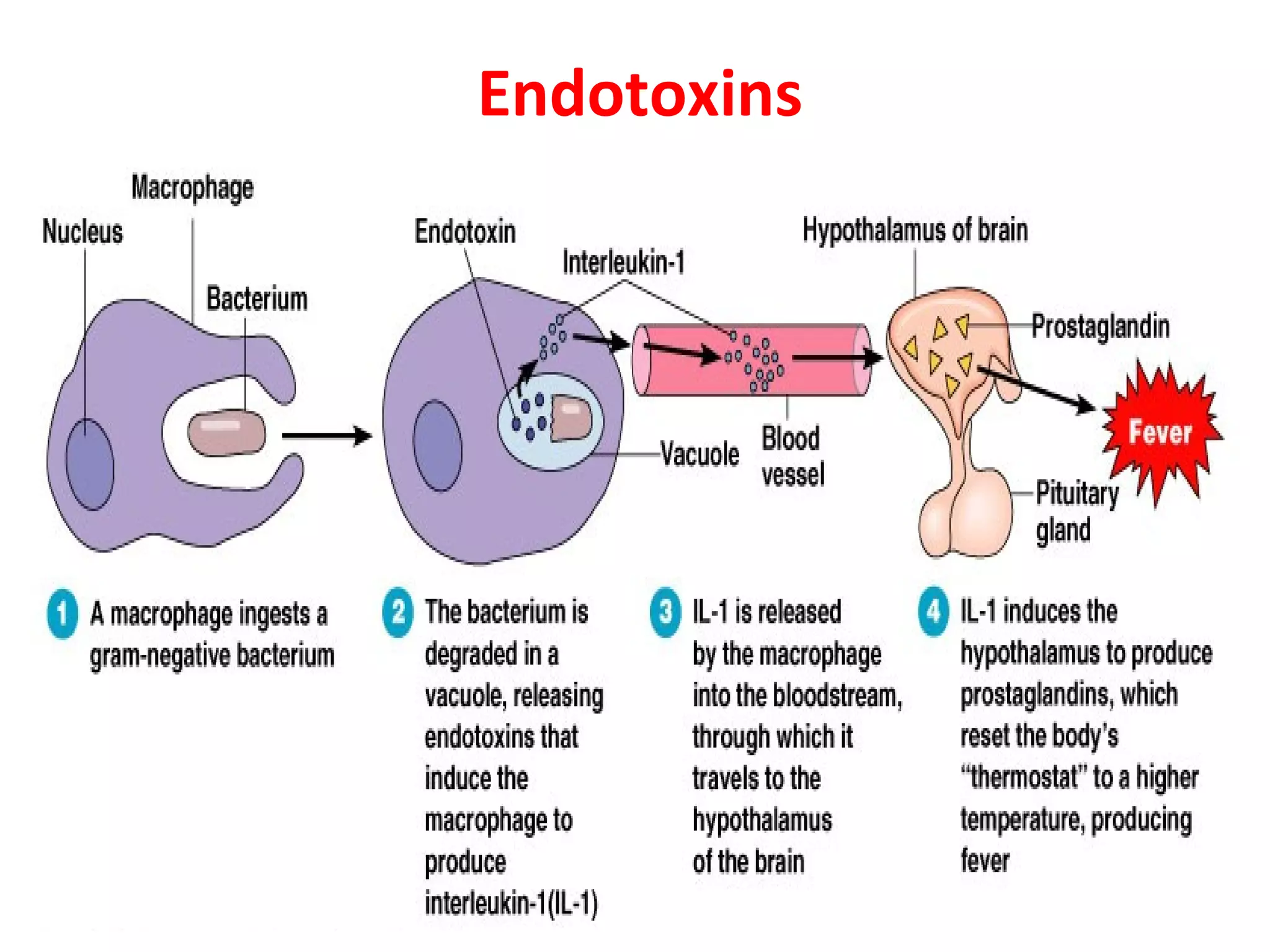
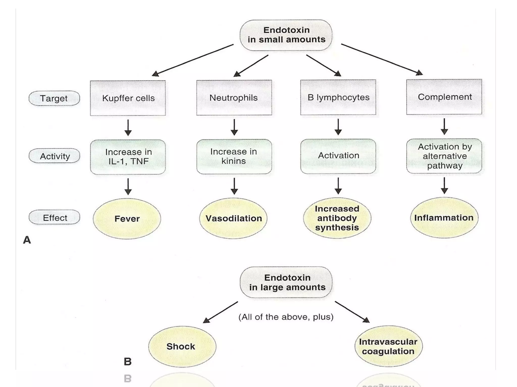
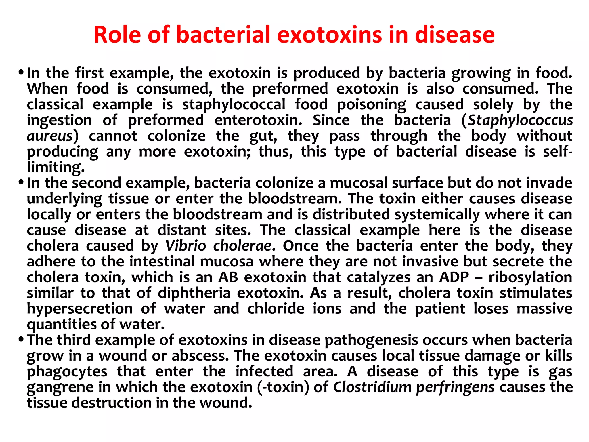
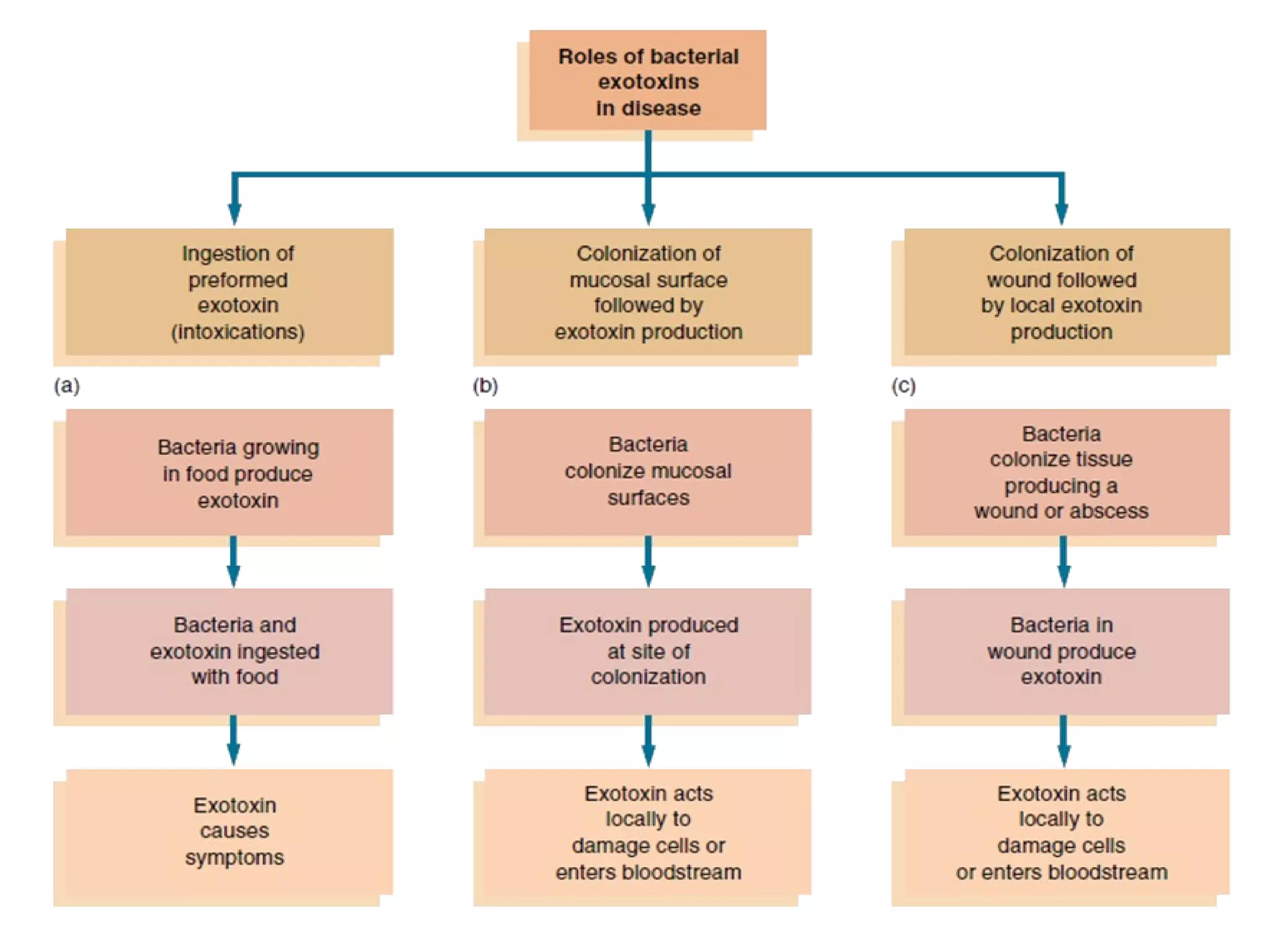
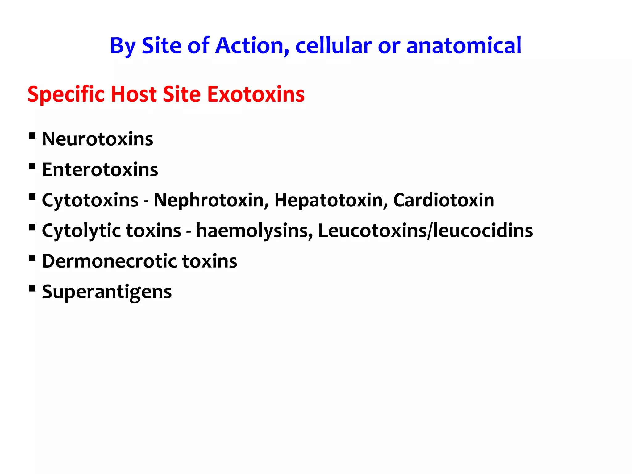
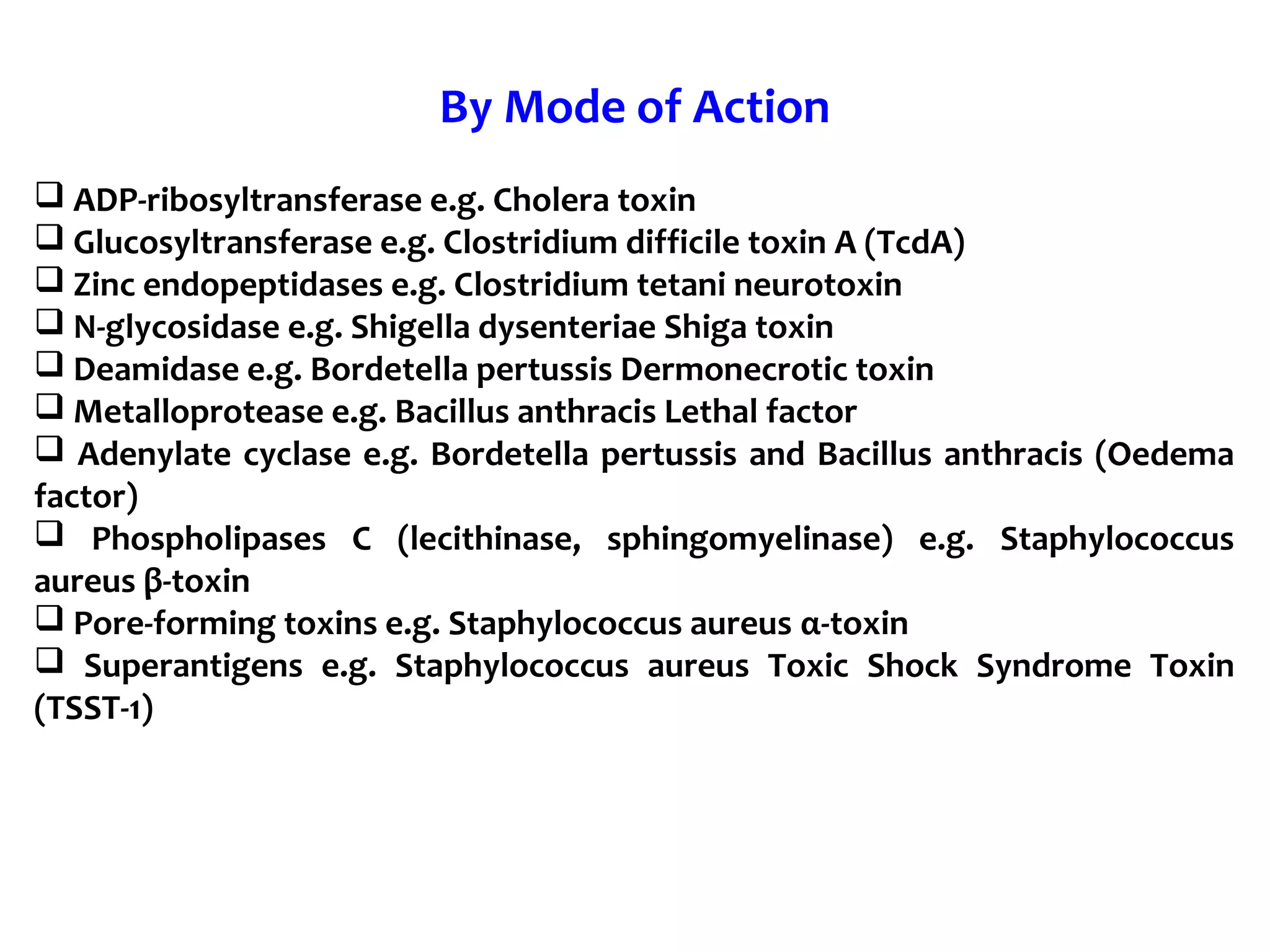
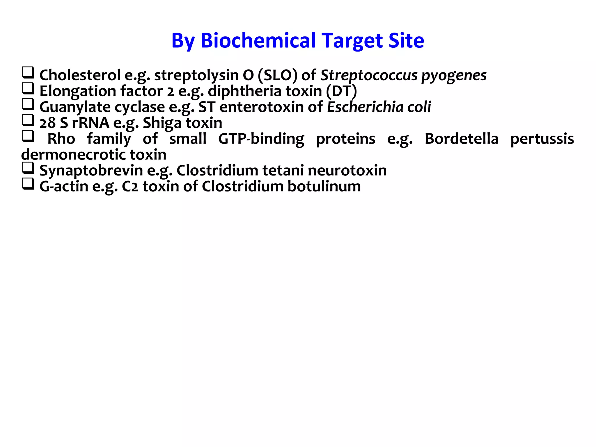
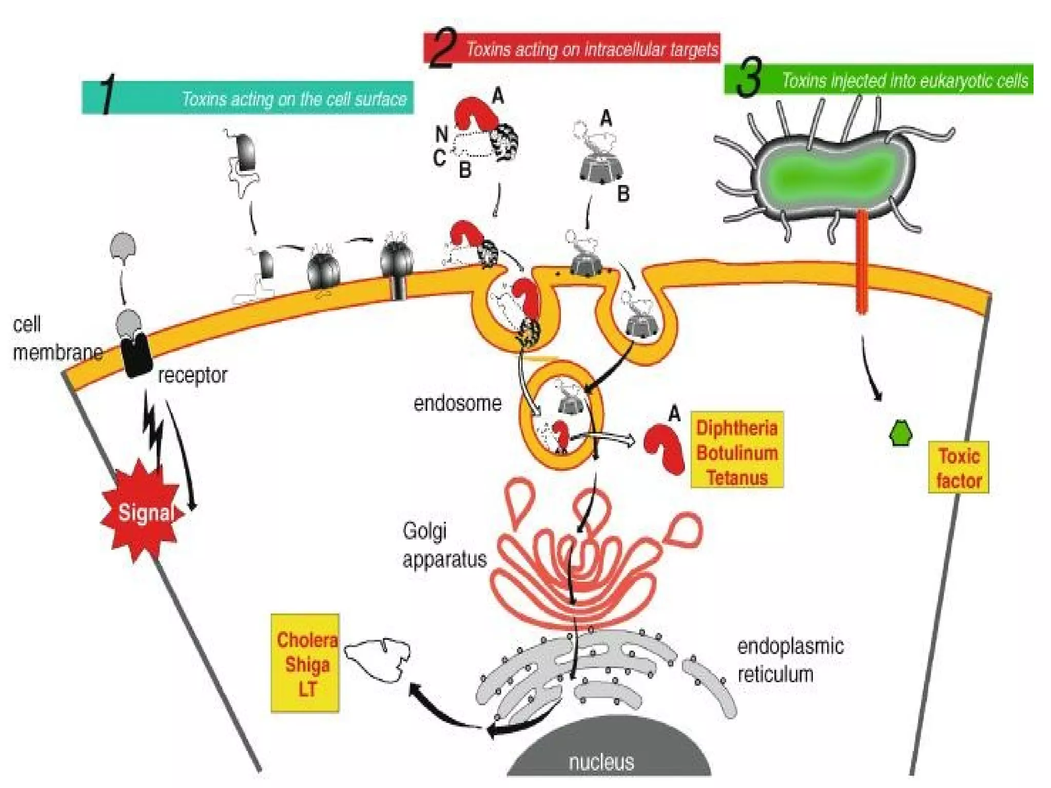
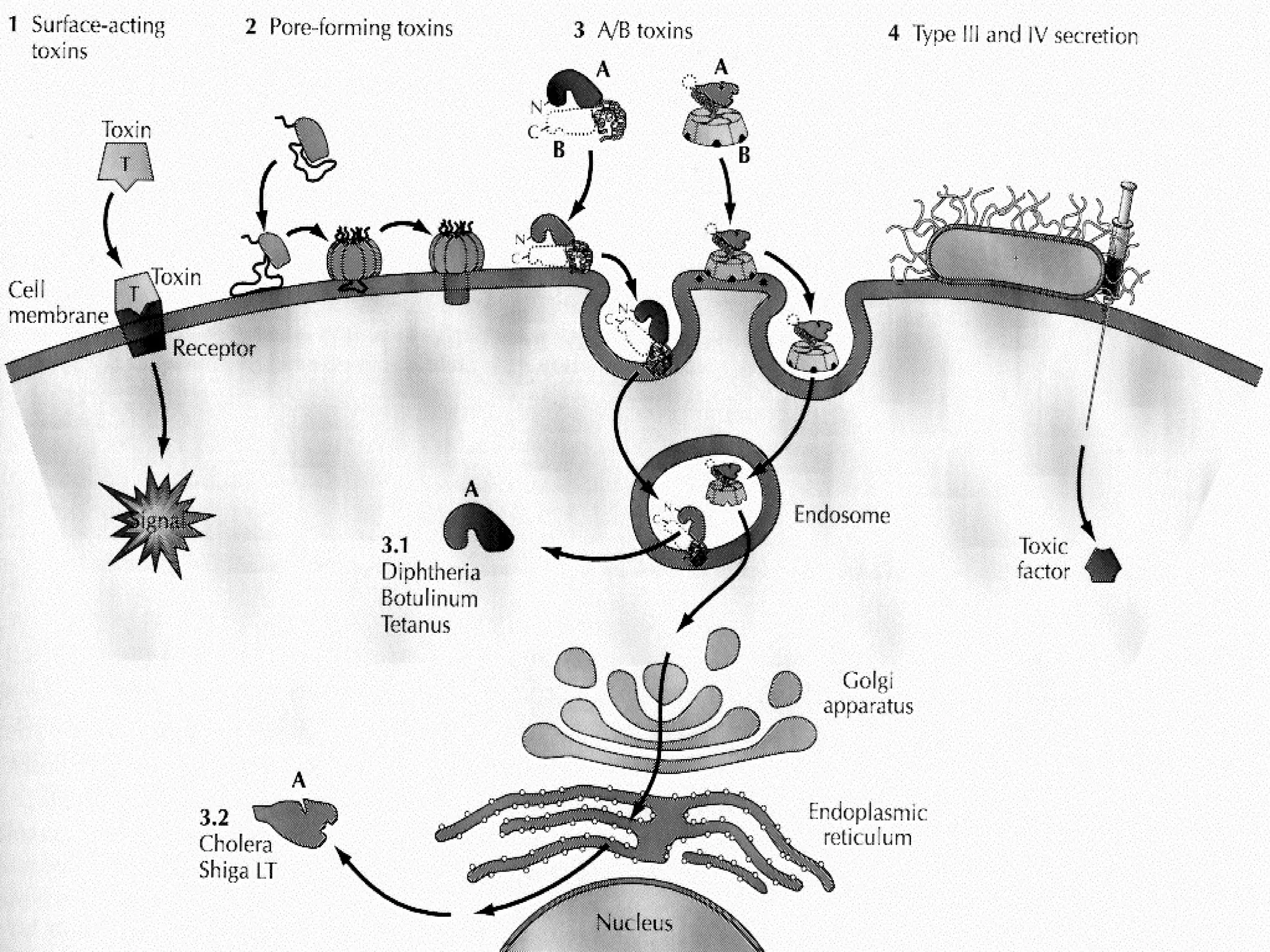
Bacterial toxins can be classified as exotoxins or endotoxins. Exotoxins are protein toxins secreted by bacteria, while endotoxins are structural components of the outer membrane of gram-negative bacteria. Exotoxins can be inactivated by heat or chemicals to form immunogenic toxoids, whereas endotoxins cannot. Exotoxins play an important role in several diseases by directly damaging host cells or tissues both locally and systemically.