Ck19 and babb faseb aug 2010 slide 1 2
•Download as PPTX, PDF•
0 likes•123 views
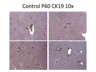
The document discusses a patient with a diagnosis of P51 DKO and a total bilirubin level of 25.1. A control sample labeled P60 CK19 was examined at 10x and 20x magnification under a microscope.
Report
Share
Report
Share

Recommended
Herramientas del software libre para el diseño
Este documento presenta varias herramientas de software libre para el diseño y la organización del conocimiento como Gimp, Inkscape, Blender, Scribus, Ktoon, Kompozer, Cmap Tools, wikis y redes semánticas. Explica cómo usar estas herramientas de manera colaborativa para editar imágenes, crear animaciones 3D, diseñar páginas web, organizar mapas conceptuales y compartir información a través de wikis.
Vanderpool supplemental fig hnf6 stain cont rbp
The document discusses a knockout experiment in mice where the RBP gene was deleted. It appears to report results from two experimental conditions (A and B) observed at embryonic day 16.5, comparing the knockout to a control group. The high-level results are not provided in the given text.
Vanderpool supplemental figure 1 ver 2
The document presents data on gene expression levels in control and knockout mouse models at different developmental stages. Gene expression is measured through a ratio and shown to differ between control mice and those missing HNF-6, RBP, or both genes (DKO). Levels change over time from E16.5 to P0 in various tissues including pancreatic vesicles.
Vanderpool fig 4 ver 2
This document appears to be laboratory results or notes that contain abbreviations and codes that are not defined in the given text. It references control samples, a sample labeled DKO, mentions several biomarkers including Cytokeratin 19, Ki67 and bisBenzimide, and indicates a ratio should be calculated between Ki67 and CK19 expression levels.
Hnf1b figure
The document presents images of control, HNF-6 KO, RBP KO, and DKO (double knockout) mouse pancreases stained with different markers at embryonic day E16.5, postnatal day P3, and postnatal day P60. The images are labeled from A-C with A showing E16.5, B showing P3, and C showing P60 timepoints for each genotype.
Vanderpool fig 2 ver2
This document appears to be reporting on experiments knocking out genes in mice and examining the effects on pancreatic development at different embryonic and postnatal time points. It finds that knocking out genes like DKO, HNF-6, and RBP lead to abnormalities in pancreatic development, with the pancreas failing to form or differentiate properly into exocrine and endocrine tissues at stages E16.5, P3, and P15.
Aasld boston nov 2010 4
This document summarizes research on the genetic interaction between hepatocyte nuclear factor 6 (HNF-6) and Notch signaling during liver development. The researchers found that deleting both HNF-6 and RBP-jκ, a key mediator of Notch signaling, in mouse hepatoblasts led to more severe hepatic fibrosis, cholestasis and impairment of intrahepatic bile duct development compared to deleting either gene alone. Loss of both genes also altered expression of the hepatocyte transcription factor HNF-1β. The results suggest HNF-6 and Notch signaling may compensate for each other during bile duct morphogenesis and that disrupting both pathways heightens phenotypic severity through impaired redundancy.
Ck19 and babb faseb aug 2010 slide 6
Se realizó un procedimiento médico en el lóbulo medio del cerebro de un paciente de 18 años. Se utilizó un dispositivo médico para acceder a una parte del cerebro a través de un orificio en el cráneo.
Ck19 and babb faseb aug 2010 slide 5
The document contains medical codes and abbreviations referring to a procedure on the left lobe of the liver. EES likely refers to a medical procedure, "18" possibly the patient's age, RBP a blood test, P60 a code, BABB an abbreviation, and LEFT LOBE indicates the location of the liver that was involved.
Ck19 and babb faseb aug 2010 slide 3 4
The document describes two patient cases. The first is a 1339 year old male with a double knockout mutation and total bilirubin of 25.1 who underwent microscopic examination at 10x and 20x magnification. The second is a 1381 year old female with a double knockout mutation, total bilirubin of 46.1, and CK19 marker who also underwent microscopic examination at 10x and 20x magnification.
Ck19 slide 1 3 (fo reals)
This document provides instructions for creating a storyboard using slides with color matched pictures from previous talks and posters, as well as new data pictures for control, HNF-6 knockout, RBP knockout, and HNF-6/RBP double knockout genotypes at embryonic day 16.5 and postnatal day 15 and 30 timepoints. The author does not have additional pictures from postnatal day 30 or 60 timepoints to choose from and can take a new picture if an absolute picture is disliked, though options should have already been discussed.
Ck19 cfda august 2010
The document summarizes immunofluorescence staining results for CFDA and CK19 in liver samples from various mouse models and timepoints. In DKO P58 mice, hilar ducts typically co-stained for CFDA and CK19 while peripheral areas showed disorganized CK19-positive cells lacking CFDA. A sicker DKO P58 mouse had fewer disorganized CK19 cells and some hilar ducts with weak CFDA staining. A P70 HNF-6 KO mouse showed normal duct staining. P70 NICD/HNF-6 mice had well-formed ducts positive for CFDA but multiple peripheral and hilar areas lacking CFDA, associated with disorganized CK19-
Ck19 slides 8 9
The document discusses a difficult staining procedure for peripheral blood dendritic cells where only 5 dendritic cells could be found after repeating cut slides, though the tissue looked in good condition.
Ck19 slides 6 7
The document compares CK19 staining patterns in the hilar and peripheral regions of control tissue and P15 HNF-6 KO tissue. CK19 staining was seen in the hilar and peripheral regions of control tissue. In P15 HNF-6 KO tissue, CK19 staining was also seen in the hilar and peripheral regions.
Ck19 slides 4 5
N=1 animal showed some questionable cystic changes in the hilar region that may have been due to tissue and sectioning issues rather than pathology. The peripheral region also had questionable cystic areas but similar areas were seen in control animals. Ductal plate formation was reduced or absent in the hilar region of an E16.5 double knockout animal. Peripherally, ductal plates were difficult to identify and there may have been increased background staining of cells rather than disorganized CK19-positive cells. Further study is needed to identify upregulated pathways leading to changes seen at P60 and P30 if not due to proliferation.
More Related Content
More from cpbv13
Ck19 and babb faseb aug 2010 slide 5
The document contains medical codes and abbreviations referring to a procedure on the left lobe of the liver. EES likely refers to a medical procedure, "18" possibly the patient's age, RBP a blood test, P60 a code, BABB an abbreviation, and LEFT LOBE indicates the location of the liver that was involved.
Ck19 and babb faseb aug 2010 slide 3 4
The document describes two patient cases. The first is a 1339 year old male with a double knockout mutation and total bilirubin of 25.1 who underwent microscopic examination at 10x and 20x magnification. The second is a 1381 year old female with a double knockout mutation, total bilirubin of 46.1, and CK19 marker who also underwent microscopic examination at 10x and 20x magnification.
Ck19 slide 1 3 (fo reals)
This document provides instructions for creating a storyboard using slides with color matched pictures from previous talks and posters, as well as new data pictures for control, HNF-6 knockout, RBP knockout, and HNF-6/RBP double knockout genotypes at embryonic day 16.5 and postnatal day 15 and 30 timepoints. The author does not have additional pictures from postnatal day 30 or 60 timepoints to choose from and can take a new picture if an absolute picture is disliked, though options should have already been discussed.
Ck19 cfda august 2010
The document summarizes immunofluorescence staining results for CFDA and CK19 in liver samples from various mouse models and timepoints. In DKO P58 mice, hilar ducts typically co-stained for CFDA and CK19 while peripheral areas showed disorganized CK19-positive cells lacking CFDA. A sicker DKO P58 mouse had fewer disorganized CK19 cells and some hilar ducts with weak CFDA staining. A P70 HNF-6 KO mouse showed normal duct staining. P70 NICD/HNF-6 mice had well-formed ducts positive for CFDA but multiple peripheral and hilar areas lacking CFDA, associated with disorganized CK19-
Ck19 slides 8 9
The document discusses a difficult staining procedure for peripheral blood dendritic cells where only 5 dendritic cells could be found after repeating cut slides, though the tissue looked in good condition.
Ck19 slides 6 7
The document compares CK19 staining patterns in the hilar and peripheral regions of control tissue and P15 HNF-6 KO tissue. CK19 staining was seen in the hilar and peripheral regions of control tissue. In P15 HNF-6 KO tissue, CK19 staining was also seen in the hilar and peripheral regions.
Ck19 slides 4 5
N=1 animal showed some questionable cystic changes in the hilar region that may have been due to tissue and sectioning issues rather than pathology. The peripheral region also had questionable cystic areas but similar areas were seen in control animals. Ductal plate formation was reduced or absent in the hilar region of an E16.5 double knockout animal. Peripherally, ductal plates were difficult to identify and there may have been increased background staining of cells rather than disorganized CK19-positive cells. Further study is needed to identify upregulated pathways leading to changes seen at P60 and P30 if not due to proliferation.
More from cpbv13 (7)
Ck19 and babb faseb aug 2010 slide 1 2
- 1. Control P60 CK19 10x
- 2. 1339 P51 DKO, T bili 25.1 10x 20x