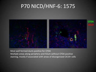
The document summarizes immunofluorescence staining results for CFDA and CK19 in liver samples from various mouse models and timepoints. In DKO P58 mice, hilar ducts typically co-stained for CFDA and CK19 while peripheral areas showed disorganized CK19-positive cells lacking CFDA. A sicker DKO P58 mouse had fewer disorganized CK19 cells and some hilar ducts with weak CFDA staining. A P70 HNF-6 KO mouse showed normal duct staining. P70 NICD/HNF-6 mice had well-formed ducts positive for CFDA but multiple peripheral and hilar areas lacking CFDA, associated with disorganized CK19-