Call Girls Bhubaneswar Just Call 9907093804 Top Class Call Girl Service Avail...
Â
Chyluria -an_overview
1. 14
Chyluria - An Overview
Sachit Sharma, Ashok Kumar Hemal*
Department of Urology, All India Institute of Medical Sciences, New Delhi, India
Abstract
Chyluria, recognized as a urological manifestation of lymphatic system abnormality has been a curious entity
since long. With varied options for management, this tropical, debilitating disease is known for recurrence. The
article provides an in-depth review into the subject with the authorâs experience in the field along with the recent
advances in management. This article also describes the entity from its historical background, etiopathogenesis,
investigative and management protocols including complications. Recent treatment modalities reported in
literature are reviewed with authorâs innovative contributions in the form of applying MR scan for diagnosing
and robotics for treating chyluria.
Keywords: Chyluria, Laparoscopy, Recurrence, Sclerotherapy, Surgery
Review Article
Chyluria, recognized since the time of Hippocrates
(400BC) is described as the passage of milky
appearing white urine due to the presence of chyle
composed of albumin, emulsified fat and fibrin in
varying proportions that are absorbed by intestinal
lacteals. It is associated with spontaneous remissions
and exacerbations. Although rare in the West, it is
not uncommon in Asia. It has been recognized as
a tropical disease more prevalent in the rural and
poverty stricken population. This review article
provides an in-depth into the subject with the authorâs
experience in this field along with the recent advances
in management (1-4).
History of Hypothesis
The following were the explanations in the past for
causation of chyluria:
Prout (1841): Secretion of fat from blood through
kidneys (5).
Ackerman (1863): Blockage of major lymphatics
or thoracic duct causing retrograde flow of lymph
from the gut and pelvis into lumen of GIT (6).
Wucherer (1869): Presence of microfilaria in
blood/urine of chylurics (7).
Lazarus and Marks (1946): Presence of fistulous
openings in bladder causing chylous efflux from
ureteric orifices (8).
Manson-Bahr (1954): Rupture of lymph varix
anywhere in urinary tract leading to escape of lymph
in urine (9).
Sen and Elappan (1968): Parasitic / bacterial
infections Ă fibrosis Ă reduced lymphatic
bedĂ lymphatic HTĂ lymph stasis & shunt formation
(10).
* Correspondence:
Ashok Kumar Hemal, M.D
Department of Urology, All India Institute of Medical Sciences,
Ansari Nagar, New Delhi, India -110029.
Tel: +91-9868397431
Fax: +91-11-26588641
Email: ahemal@wfubmc.edu
Received: 27 Mar 2009
Accepted: 20 Apr 2009
Int J Nephrol Urol, 2009; 1(1): 14 - 26
International Journal of Nephrology & Urology, 2009; 1(1): 14 - 26
2. 15
Recently the following theories have been
implicated as causative factors:
Obstructive theory (Aye and Aung, 1975):
Inflammatory reactions due to parasitic infestation
lead to obliterative lymphangitis, lymphatic HT,
valvular incompetence and retrograde flow of chyle
producing a lymphatico-renal shunt (11).
Regurgitative theory (Ngan and Leong, 1977):
Toxic metabolites or immune reaction to parasitic
infestation leads to lymphatic ectasia and varicosities,
that rupture, releasing chyle from dilated intestinal
lymphatics into the renal system (12).
Etiopathogenesis
Chyluria is associated with abnormal retrograde or
lateral flow of lymph from the intestinal lymphatics
of the kidney, ureter or bladder allowing chylous
material to be discharged into the urinary collecting
system (1). Various causative factors have been
implicated. Passage of chyle into the urine has been
related to a consequence of the rupturing of the
lymphatic varices leading to the aperture of one or
more perirenal lymphatic vessels to the pyelocaliceal
system, characterized by milky urine, rich in protein,
lipids, cholesterol and triglycerides. A malformation
of the chyliferous vessels of the small intestine
associated to hypoplasia of Pecquet's cistern has also
been suggested.
On anatomical basis, the renal lymphatics follow
the renal vein and end in lateral aortic glands;
efferents from which flow to the lumbar trunks.
The intestinal trunks receive lymph from stomach,
intestine, pancreas, spleen and liver. Physiologically
chyle travels from the lacteals to the cisterna chyli
or thoracic duct. Pathological obstruction and/
or insufficiency of the valvular system of lymph
channels leads to retrograde flow to lumbar lymph
glands draining renal lymphatics. Thus there is a short
circuiting of chyle drainage from intestinal lacteals to
renal lymphatics.
Classification
The classification is based on the etiological
factors- broadly divided into two groups, parasitic
and non parasitic (Table 1). Although rarely seen in
some countries, however its prevalence is high in
areas where infections with Wuchereria bancrofti are
endemic. Chyluria is extremely rare in Europe, but is
frequently reported from Asian countries like India,
China, Taiwan and Japan (13-17). Epidemiological
surveillance since 1984 indicates that the transmission
of infection has been interrupted (18). Approximately
120 million people worldwide are affected by
lymphatic filariasis.
Chyluria can also have non-parasitic causes of
which only a few cases have been described. Chyluria
due to a congenital fistulous communication between
the lymphatic system and the bladder has been
described in children (19). Chyluria may occur as an
exceptional complication of retroperitoneal lesions in
lymphangiomatosis in rare cases (20, 21).
Chylous ascites and chyluria have been reported
in nephrotic syndrome with focal segmental
glomerulosclerosis and nephrotic syndrome due to
retroperitoneal lymphangioma. Hypoalbuminemia-
induced bowel edema may predispose to change in
the permeability of mucosal or serosal lymphatics
resulting in chylous ascites (22, 23).
Clinical features
The most common clinical symptom is passage of
milky urine in up to 70% cases (24). Other symptoms
include chylous clot, bloody and milky urine, dysuria,
frequency, urgency and urinary retention in decreasing
order of presentation. Constitutional symptoms
include weight loss, low-back ache, fever, chills
and hydrocele. The duration of symptoms may be as
long as 2- 11 years (1). The disease is significantly
higher in males (86%) than in females (14%) (25).
It has been more frequently reported on the left side
(26). Majority of the cases present in the second and
Ashok Kumar Hemal et al
International Journal of Nephrology & Urology, 2009; 1(1): 14 - 26
3. 16
third decade of life. About 70% cases have genital
manifestations, 25% lymphatic obstruction in limbs
and 5% suffer from manifestations like cellulitis,
abscesses, hematuria and chyluria. Previous history
of filariasis or its complication has been documented
in 19% cases (27).
Grading
The severity of the disease can be graded into (1):
Mild chyluria (34-50%): Intermittent milky urine;
no clot colic/chylous coagulum/urinary retention/
weight loss; involvement of single calyx on RGP.
Moderate chyluria (33- 40%): Intermittent
continuous milky urine with occasional clot colic/
chylous coagulum; no urinary retention/weight loss;
involvement of 2 or more calices on RGP.
Severe chyluria (15- 26%): Continuous milky
urine with clot colic/chylous coagulum/urinary
retention /weight loss; involvement of most of calices
with/without involvement of ureter on RGP (1).
Investigations
Urinary Examination
Afatty diet a day or night before enhances chyluria
and helps in its diagnosis. A fresh sample of urine is
grossly inspected. The classical urine in chyluria is
like milk, frequently containing a semisolid gel, blood
and fibrin clots. On standing in test tubes, it separates
into a top fat layer, middle fibrin and bottom layer
of cells and debris. Microscopic examination of the
sediment comprises of erythrocytes and lymphocytes.
In the presence of symptoms like milky urine,
proteinuria and leukocyturia in the urinary sediment
and a negative urine leukocyte stick test and absence
of infectious signs, chyluria must be suspected (13).
Ether test
Vigorous shaking (few minutes) with equal amount
of ether clears opacity.
Methylene blue test
Fresh stain with one/two drops of 1:1500 reveals
small lymphocytes (single/clump).
Sudan III test
Oral ingestion of fat labelled with Sudan III (10
gm butter + 100 mg Sudan red III) causes orange pink
colouration of urine in chylurics within 2-6 hours.
Biochemistry
Urinary triglycerides are invariably present
in morning sample, 100% sensitive/specific test.
Postprandial urine lipids, especially triglycerides
(TGs) ranging from 10 to 1955 mg/dl, might be used as
markers for the clinical evaluation of chyluria. Urine
albumin is abnormally high in most cases (16).
Immunoelectrophoresis
Shows globulins of various types of apolipoprotein
A 48 (of intestinal origin).
Cystourethroscopy
Helps to localize the side of milky efflux from one/
both ureteric orifices. Rarely chylous efflux may be
seen from bladder or even posterior urethra. A fatty
diet previous day/night enhances results. Ureteric
catheterisation (5-6 F) and split urinanalysis may be
Table1. Classification of chyluria based on etiological factors
Parasitic (primary-tropical) Non-parasitic (secondary-nontropical)
Wuchereria bancrofti (90%)
Taenia echinococcus
Taenia nana
Ankylostomiasis
Trichiniasis
Malarial parasites
Congenital
Lymphangioma of urinary tract
Megalymphatics & urethral/vesical fistulae
Stenosis of thoracic duct
Retroperitoneal lymphangiecatasia
Traumatic lymphangiourinary fistulae
Obstruction of thoracic duct/ lymphatics (tumor, granulomas, aortic aneurysm)
Other causes (pregnancy, diabetes, abscess)
Nephrotic syndrome
Chyluria
International Journal of Nephrology & Urology, 2009; 1(1): 14 - 26
4. 17
done simultaneously.
Retrograde pyelography (RGP)
With help of fluoroscopy and spot films it
demonstrates pyelolymphatic backflow. RGP is
not specific as the same is also seen in normal
kidneys if contrast is injected under pressure. 20°
Trendelenberg position is used under fluoroscopy
for ureteric catheterization to reach renal pelvis
after cystoscopically identifying laterality of efflux.
Gravity propagated contrast instillation prevents
sudden distension of renal pelvis which may lead to
loin pain due to opening up of pyelovenous/pyelosinus
channels thus causing inadvertent reactions.
Lymphangiography
Lymphangiography is the preoperative imaging
procedure of choice and the most powerful diagnostic
tool as it demonstrates the site, the caliber and the
number of the fistulous communications. It can
also diagnose vascular dysplasias of the lymphatic
vessels (28). Findings generally include lymphatico-
urinary fistulae at level of kidney, ureter or bladder;
tortuous dilated lymphatics around hilar region
(lymphangiectasia)communicatingwithparavertebral
lymphatics; contrast outlining major/minor calyces.
Other findings that may be seen include tortuosity
and beading of thoracic duct, round granular enlarged
nodes in paraortic area, skipping of lymphatic chain,
dilated cisterna chylii and abnormal lymphatics
coursing down along the ureters. In 40% cases
contrast may enter PCS and drain via bladder.
It is not routinely done as it is time consuming,
technically demanding and invasive (29).
Unilateral pedal lymphangiography can detect
lymphaticorenal fistulae via lymphatic crossover even
when it is on the side opposite from which contrast
agent has been injected. The advantages of unilateral
lymphangiography over bilateral procedures are
that it is easy to identify crossover channels, and the
discomfort for the patient is reduced because there are
fewer incisions and it is quicker (29, 30).
Lymphangioscintigraphy
Though useful in delineating the site of the fistula
it is not as precise as lymphangiography. However, it
is a useful, noninvasive, safe, simple technique using
Tc99 human serum albumin sulphur colloid, Tc-99m
diethylenetriamine pentaacetic acid, 99mTc-antimony
sulfide colloid or dextran. It is the investigation of
choice (if available) to localize, lateralize and to
know the functional extent of reflux and to detect
recurrence on follow-up. It can demonstrate abnormal
lymphatic drainage in chylurics and allows functional
assessment of lymphatic transport and depiction of
regional lymph nodes. A rapid and non-traumatic
investigative procedure, it has no known side effects
(31-33).
Immunological Studies
ELISA Test: The test is useful in diagnosing cases
where causative agent is suspected to be filarial and is
based on humoral immune response of host to filarial
antigen. Filarial IgG antibodies against microfilarial
excretory-secretory (mf ES) antigen have been
detected in 89% of cases with genital manifestations,
87% with lymphoedema, 67% with lymphadenitis
and 60% with other clinical manifestations. With a
85% specificity and 95% sensitivity, it is positive in
3% of endemic normals (25).
Immune function of the patients who present with
filarial chyluria is impaired in terms of the changes in
T-lymphocytesubsets.ThepercentageofCD3+,CD4+
cell is significantly decreased, the ratio of CD4+/
CD8+ being under 1.0 (34). Both cell-mediated and
humoral immunity are low in chyluric cases. OKT3
and OKT4 levels are commonly low, and OKT8 is
higher than normal value with the OKT4/OKT8 ratio
inverted (35).
Ultrasonography : Echolucent areas may be seen
within blood clot due to the chylous component;
leading to heterogenous character of clot. This needs
to be differentiated from carcinoma bladder.
Intravenous pyelography : Rarely used to
demonstrate dilated para-calyceal lymphatics
(need for application of ureteric pressure), it may
delineate increased renal size in severe disease state
due to obstruction. Overall it is not a cost effective
Ashok Kumar Hemal et al
International Journal of Nephrology & Urology, 2009; 1(1): 14 - 26
5. 18
investigation.
CT scan : Fat in the bladder secondary to chyluria
can be picked up on CT (36). Dilated lymphatic
channels may mimic a mass of confluent low-density
lymph nodes (37).
MR scan (Figure 1) : A non-invasive and less
time consuming study -best to delineate anatomy (but
no functional assessment). It is most useful in cases
where site of obstruction is lower ureter/bladder. On
MR urography, the lymphatic channels are seen as a
meshwork of multiple tubular, tortuous, fluid filled
structures in the retroperitoneum of the abdomen
and pelvis. On axial T1W images, these channels
are seen as numerous, interconnected small, nodular
and streaky intensities and as a cloak of diffuse
homogenous hyperintensity on T2W axial images
(37). An innovation of the author, the results of this
useful modality of investigation are being evaluated.
Renal biopsy : Mainly undertaken for academic
research generally shows immune complex type of
glomerulonephritis (38).
Others : Serum type I collagen and type III
procollagen are decreased in patients with filariasis. In
contrast, serum hyaluronan (linked to perilymphatic
granulomatous inflammation) and serum eosinophil
cationic protein is significantly increased in patients
of fileria exhibiting chyluria (39).
Management
Non surgical management
Dietary modifications: As absorption of dietary
fats leads to chyle formation, hence a fat restricted diet
is recommended (<25 g/day). Fats containing MCT
(<12 C atoms) are absorbed directly through portals-
hence use of coconut oil recommended (40). In heavy
chyluria, parenteral administration containing fat as
MCT with albumin is given. TPN with enteric rest is
advised in intractable chyluria (41).
Diethyl carbamazine (DEC/ Hetrazan /
Benocide): The dose is 6mg/kg in 3 divided doses
after food for 10-14 days (42). Ivermectin 400mg/
kg (single dose) and/or albendazole 400mg may be
given along with DEC as symptomatic treatment
where filarial infection seems to be the cause.
Supportive treatment: High protein diet is advised
to make up for the albumin lost in the form of chyle.
Hematinics, multivitamins and green leafy vegetables
are recommended in hematochyluria. Bed rest, anti-
inflammatory, analgesics and antipyretics are useful
in managing in associated lymphadenitis. Abdominal
binders may be applied during acute attacks of
chyluria to reduce loss of chyle. Cytoscopic bladder
wash is recommended in cases of clot/chylous urine
retention and/or recurrent urinary tract infections.
Sclerotherapy (Table 2): Comprises of instillation
of renal pelvis with chemicals - silver nitrate (0.1-
3.0%), 0.2% povidone iodine, 15-25% sodium
iodide, 10-25% potassium iodide, 50% dextrose, 76%
hypertonic saline, combination therapy using 5/6 F
ureteric catheter after localizing site on cystoscopy
in cases of failure of conservative management i.e.
dietarymodificationsandasprophylaxis.Theseagents
bring about relief by causing chemical lymphangitis
(Diagram 1).
Permanent relief: The procedure is performed
under LA/ sedation with aseptic precaution keeping
the head down (200
) and instilling 7-10ml sclerosant
Figure1.Non-invasiveMRscanshownextensive
involvement of bilateral lymphatics with good
anatomical background
Chyluria
International Journal of Nephrology & Urology, 2009; 1(1): 14 - 26
6. 19
under gravity (fluoroscopy assisted). Analgesic /
antibiotic cover is given for at least 5 days. Dosage
schedule is 8 hourly for 3 days or 12 hourly for 2 days
or weekly for 6-8 weeks. Recurrence after 1st course
can be treated for second time with high success rate.
Patients with shorter as disease free duration fare
poorly with second course. In bilateral efflux gap
period of minimum 6-8 weeks between 2 sittings is
recommended (43).
Silver Nitrate (0.1-3.0%): It is most commonly
used sclerosant till date. 2gm silver nitrate powder is
dissolvedin200mlofwaterinabottle(blackpaper/dark
room). The need for sterilization (autoclaving) results
in a 8-24 hr undesirable delay. Other disadvantages
include procuring and precisely weighing good
quality silver nitrate, water insolubility, susceptibility
to light, and precipitation with normal saline to form
insoluble silver chloride salts that may cause uretric
obstruction. As even mortality has been reported
due to acute tubular necrosis (ATN) on instillation
of silver nitrate in both ureters simultaneously, care
needs to be observed (44, 45).
Povidone Iodine (0.2%): It is a water soluble, non-
ionic surfactant polymer that releases iodine slowly.
Prepared by mixing 2ml 5% povidone iodine to 8ml
distilled water it is to be used as a fresh solution. It is
as effective as 1% silver nitrate with no side effects
like flank pain, nausea, vomiting, interstitial nephritis,
chemical cystitis, papillary necrosis and ATN.
Also having antiseptic antibacterial and antifungal
properties (46) it is considered safe and effective for
managing chyluria (15).
Combination therapy: Combination sclerotherapy
using a 5% povidone iodine and 50% dextrose
solution has been found to be a safe and cost-effective
minimally invasive therapy for chyluria refractory
to conservative treatment. 5ml of 50% dextrose
combined with 5ml 0.2% povidone iodine provides
stronger and better fibrotic response (24).
Heat-clearing and hemostatic treatment: 26
out of 30 patients of chyluria have been reported to
be cured completely with a basic heat-clearing and
hemostatic prescription (35).
Surgical management: Surgery is the treatment
of choice in severe forms of chyluria i.e. significant
weight loss; hypoproteinemia, anasarca, and/
or severe anemia; recurrent clot retention and
hematochyluria; recurrent UTI; refractory chyluria
(failure of conservative treatment with adequate
dietary modification, medical management and
two or more instillations of sclerosants); clinically
significant chyluria (associated with chylous clot or
Table 2. Results of sclerotherapy of different series
Study Year Response (%) FU (months) Recurrence (%)
Silver Nitrate
Yu et al (70)
Ohyama et al (71)
Okamoto and Ohi (72)
Tan et al (73)
Sabnis et al (74)
1978
1979
1983
1990
1992
119/161 (74%)
- (62%)
129/217 (59%)
46/55 (83%)
51/62 (82%)
-
2 â115
24
24
24 â 84
26%
37%
40%
16%
18%
Povidone Iodine
Shanmugam et al (75)
Goel et al (46)
1998
2004
5/5 (100%)
36/41 (88%)
6
24
-
12%
50% Dextrose
Goel et al (46) 2004 20/21 (95%) 5.2 98%
Povidone Iodine + 50%
dextrose
Nandy et al (24)
2004 40/46 (87%) 24 13%
Ashok Kumar Hemal et al
International Journal of Nephrology & Urology, 2009; 1(1): 14 - 26
7. 20
hematochyluria or duration of more than 1 year or
failure to respond to conservative medical measures)
(47); altered immune status; marked psychological
disturbance.
Open surgical techniques
Lympho-venous disconnection: First described
by Katamine in 1952 it is the most commonly
performed procedure. A fatty diet 24-36 hr prior to
surgery facilitates detection of lymphatics. Pedal
lymphangiography and/or methylene blue injection
delineates the lymph channels. Reno-lymphatic
disconnection operation is performed via a lumbar
incision. The kidney is freed, lymphatics in hilar area
coursing along the renal vein are dissected, ligated
& cut. Areolar tissue containing dilated lymphatics
travelling to the kidney in the perirenal and hilar
region are dissected and divided between ligatures
- thus stripping these structures completely. Hilar
stripping is continued up to proximal 3-4cm ureter,
the site for majority of shunts. Recurrence or bladder
leak on imaging, mandates stripping of the entire
ureter (48).
Patna operation (1977): It is a modified procedure
of peri-ureteric lymphovenous stripping. Disconnection
of only abdominal ureter is done without hilar
disconnection. âUreter in Lymphatic Tunnelâ i.e.
pampniform plexus in front and pre/para aortic plexus
behind is the basic concept followed (49).
Omental wrapping (2004): Omentum is used to
wrap the renal vessels after stripping both anteriorly
and posteriorly. This reduces the incidence of
postoperative lymphatic drainage, lymphocele
formation, recurrence, fibrosis and postoperative
adhesions (47).
Renalautotransplantation:Thisisrarelyindicated
for fistulas in the lower portion of ureter /bladder or
when renal pedicle stripping has failed (incomplete
procedure/ recanalization) or there is formation of
newer fistulas (50).
Nephrectomy: It is used only for kidneys
rendered non-functional following lymphovenous
disconnection and serves as a life saving measure in
severely refractory chyluria (51).
Diagram 1. Sclerotherapy â Mechanism of Action
Instillation of sclerosant
Reaches lymphatic through pyelolymphatic fistulae
Chemical lymphangitis
Blockage of lymphatic due to edema Healing by fibrosis
Immediate relief Permanent relief
Chyluria
International Journal of Nephrology & Urology, 2009; 1(1): 14 - 26
8. 21
Cockett and Goodwin procedure (1962): The
procedureisbasedondiversionofasinglehilarlymphatic
to a spermatic/gonadal vein in end-to-side fashion. It
provides a safety valve mechanism for renal lymphatic
hypertension-the basis of chyluria. It obviates need for
complete stripping and there is a decreased incidence of
lymphocutaneous fistula. However, the demerits include
increased operative time, technical difficulty and need
for magnification (52).
Microsurgical procedures: Recommended in the
old and debilitated patients unable to tolerate ligation
/ division of lymphatics this technique has been found
useful in persistent or recurrent chyluria. Aiming to
physiologically decrease the intra lymphatic pressure
by increasing lymph drainage into venous system it
reduces incidence of lymphangiectasia and facilitates
healing of lymphatic fistula in the renal papilla. A
simple, easy relatively superficial procedure â less
traumatic to surrounding tissues it obviates the use
of CPE / lymphangiography as there is no correlation
between site of operation or side of lympho-renal
fistula. Technically demanding - magnification is very
essential as 90% of lymphatic vessels have diameter
<1mm making it difficult to finding vein of adequate
diameter and in proper place (for anastomosis).
Lymphangiovenous anastomosis: An end-to-end
anastomosis is made in the inguinal region in men
(Figure 2) and either in dorsum of foot/leg/thigh in
women. 3 to 4 anastomoses per side are made using 4
to 6 interrupted (9-11) 0 nylon sutures (53).
Lymph node â Saphenous vein anastomosis:
An end-to-side anastomosis of inguinal lymph
node to generally a tributary of saphenous vein (the
distal end of which is ligated) is made. Generally a
single anastomosis using 6 to 7 interrupted sutures is
sufficient (54).
Laparoscopic procedures: The objectives of open
surgicaltreatmentofintractablechyluriacanbeachieved
by the minimally invasive laparoscopic techniques
with the added advantages of magnified view, better
identification of lymphatics, better anastomatic results,
minimal morbidity, shorter hospital stay, excellent
cosmesis and early return to work. Of the two main
routes i.e. transperitoneal (peritoneum transgressed) and
retroperitoneal (easy and straight access to kidney) the
latter approach significantly reduces incision â related
morbidity without compromising the principles of open
surgery (2).
Retroperitoneoscopy: The technique of
retroperitoneoscopic management of chyluria
comprise of nephrolympholysis, ureterolympholysis,
hilar vessel stripping, fasciectomy or nephropexy
out of which the first three are mandatory while
fasciectomy and nephropexy (to prevent renal
pedicle torsion and nephroptosis) are not routinely
recommended (1).
Retroperitoneoscopic renal pedicle lymphatic
disconnection for chyluria completely ligates the
lymphatic vessels and is a safe, effective and efficient
surgical procedure with minimal invasion, less pain, less
blood loss, lower morbidity, short hospital stay and rapid
recovery (55, 56). The procedure is recommended for
treatment of recalcitrant chyluria (57).
Robotics: The author has been a pioneer in the
use of this modality of management for chyluria.
Nephrolympholysis, ureterolympholysis, hilar vessel
stripping, fasciectomy and nephropexy can all be done
as in conventional laparoscopy. This state-of-the-art
Master-Slave system provides 3-dimensional vision,
seven degress of freedom, 540 wristed movements,
elimination of hand tremors, accurate scaling of
movements and ergonomic comfort in addition to the
advantages and objectives of open surgery (58).
Other therapies
Medical therapy: Posttraumatic chyluria due to
lymphorenal fistula has been found to regress after
somatostatin therapy (59). Chyluria has been reported
to disappear following percutaneous sclerotherapy
to the left inguinal lymph nodes with doxycycline
in a postoperative case of lymphangiomatosis (60).
Chyluria (after radical nephrectomy) has o been
treated with N-butyl-2-cyanoacrylate (61).
Gerota's fasciectomy: Bilateral excision of the
Ashok Kumar Hemal et al
International Journal of Nephrology & Urology, 2009; 1(1): 14 - 26
9. 22
perinephric fascia and fat (Gerota's fasciectomy)
in 2 stages has been reported to result in complete
resolution of chyluria (62).
Endoscopic therapy: Chyluria has been
successfully treated in 5 patients with endoscopic
coagulation using guide tube methods (63).
Recurrence
Incomplete stripping, recanalization, reflux
from contralateral side, reflux from terminal ureter
or bladder are important causes of recurrence
generally diagnosed by radio imaging especially
lymphangioscintigraphy, lymphangiography (detects
leaks at bladder level) and cystoscopy (blebs under
mucosa / loosely hanging chylous clots from bladder
wall). This can however be prevented by use of
magnifying loops, operating microscope, omental
wrap technique or the use of laparoscope.
Complications
Although, Sclerotherapy is described as a
relatively safe procedure, few complications have
been reported with it. These include anuria with
pelvi-calyceal cast formation, acute necrotizing
ureteritis and fatal renal and hepatic failure (64-67).
Complications of retroperitoneoscopic surgery are
few - inferior vena cava injury, renal segmental artery
injury, lymphatic leak through the drain, and delayed
wound healing. Advancements in laparoscopy with
the advent of robotics is a step forward to minimize
the complication rate (58).
Conclusions
Acute manifestations of filaria and chyluria have
been reported to disappear or greatly improve after
filariasis eradication by a low dose of DEC medicated
salt for a period of 6 months while chronic ones
remained unchanged (68).
Figure 2. End-to-end lymphatico-venous
anastomosis using spermatic cord (males)
Chyluria
Table 3. Follow up results of operative procedures for chyluria in various series
Study Year Technique Complete cure Improved No change
Okamoto et al (72) 1983 LVD 375 (97.7%) 5 (1.3%) 3 (0.8%)
Punekar et al (76) 1997 LVD 69 (88.0%) - 9 (12.0%)
Prasad et al (49) 1977 P 14 (93.3%) 1 (6.7%) Nil
Dalela et al (47) 2004 LVD +0 4 (100.0%) Nil Nil
Brunkwall et al (50) 1990 RA 1 (100.0%) Nil Nil
Cockett et al (52) 1962 HLVD 1 (100.0%) Nil Nil
Xu YM et al (54) 1991 LVA 30 (60.0%) 6 (12.0%) 14 (28.0%)
Xu YM et al (54) 1991 LNSVA 6 (50.0%) 2 (16.7%) 4 (33.3%)
LVD,lymphovenous disconnection; P, Patna operation; O, Omental wrap; RA, Renal autotransplant; HLVD, Hilar LV
disconnection; LVA, Lymphangiovenous anastomosis; LNSVA, Lymphnode saphenous vein anastomosis
International Journal of Nephrology & Urology, 2009; 1(1): 14 - 26
10. 23
The prognosis of non-parasitic chyluria is usually
very good and the treatment is mostly conservative
(19). A long-term remission rate of 62% in the
conservatively managed group (DEC + fat restricted
diet), and a cure rate of 90% of patients in the operated
group has been reported. Postoperative recurrence rate
was 10%. There was more weight gain and dietary
freedom along with a longer chyluria free period
in the operated group relative to the conservatively
managed group (27).
Though pure dextrose treatment has been
discontinued due to poor success, povidone iodine
0.2% has been found to be as effective as 1% silver
nitrate [82% (silver nitrate) and 83% (povidone;
P=1.0)] (45). Definitive surgical ablation of lymphatic
urinary fistula is better than conservative medical
management because it has a higher success rate,
more dietary freedom and better patient acceptability
(27). Over 95% success rate has been described
with a follow up of 1 to 9 years following a single
microsurgical operation (54).
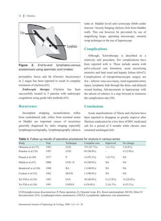
Reno-lymphatic disconnection is the reference
procedure, with long-term success rates of 99% (69).
The follow up results of various series are depicted
(Table 3).
References
1. Hemal AK, Gupta NP. Retroperitoneoscopic
lymphatic management of intractable chyluria. J
Urol. 2002 Jun;167(6):2473-6.
2. Hemal AK, Kumar M, Wadhwa SN.
Retroperitoneoscopic nephrolympholysis and
ureterolysis for management of intractable filarial
chyluria. J Endourol. 1999 Sep;13(7):507-11.
3. HemalAK, RS KMP, Gupta NP. The laparoscopic
management of chyluria by retroperitoneal access.
BJU Int. 2000;86(3):402.
4. Kumar M, Kumar R, Hemal AK, Gupta NP.
Complications of retroperitoneoscopic surgery at
one centre. BJU Int. 2001 May;87(7):607-12.
5. Prout W. On the nature and treatment of stomach
and urinary diseases. third ed: Churchill, London
120; 1841.
6. Maget A. Renal chyluria. Br J Urol.
1967;39:555.
7. Choi JK, Wiedemer HS. Chyluria:
Lymphangiographic study and review of
literature. J Urol. 1964 Dec;92:723-7.
8. Lazarus JA, Marks MS. Non-parasitic chyluria
with special reference to traumatic chyluria. J
Urol. 1946;56:246.
9. MansonBahr PH. Tropical diseases sixteen ed.
London Balliere Tindall and Cassel; 1954.
10. Sen SB, Ellappan S. Chylous manifestations
of filariasis: a clinical and lymphographic
study: Part I: Filarial chyluria. Ind J Med Res.
1968;56(10):1535 - 45.
11. Aye UT, Aung STT. Chyluria. Clin Rad.
1975;26:237.
12. Ngan H, Leong CH. A lymphographic study of
chyluria. Br J Radiol. 1977 Dec;50(600):863-70.
13. Gluck Z, Ruegger S, Sulser T. A rare cause of
nephrotic syndrome in a young woman with flank
pain, milky urine and leukocyturia. Ther Umsch.
2002 Mar;59(3):144-7.
14. Gupta NP. Retroperitoneoscopic management of
intractable chyluria. Indian Journal of Urology
2005;21(1):63-5.
15. Lin TP, Hsu YS, Chen KK, Lin AT, Chang YH,
Wu HH, et al. Chyluria--experience of Taipei
Veterans General Hospital. J Chin Med Assoc.
2003 Feb;66(2):109-12.
16. Peng HW, Chou CF, Shiao MS, Lin E, Zheng
HJ, Chen CC, et al. Urine lipids in patients with
a history of filariasis. Urol Res. 1997;25(3):217-
21.
17. Muguruma K, Matsuda T, Koyama Y, Komatz Y.
Chyluria treated with inguinal lymphangiovenous
and lymph node-venous anastomosis: a case
report. Nippon Hinyokika Gakkai Zasshi. 1994
Oct;85(10):1571-4.
18. Cao W, Van der Ploeg CP, Ren Z, Habbema JD.
Success against lymphatic filariasis. World Health
Forum. 1997;18(1):17-20.
19. Stalens JP, Falk M, Howmann-Giles R, Roy
Ashok Kumar Hemal et al
International Journal of Nephrology & Urology, 2009; 1(1): 14 - 26
11. 24
LP. «Milky» urine--a child with chyluria. Eur J
Pediatr. 1992 Jan;151(1):61-2.
20. Guillonneau B, Bouchot O, Buzelin F, Dupas B,
Barrier JH, Buzelin JM. Lymphangiomyomatosis:
an exceptional cause of chyluria. Report of a case.
Prog Urol. 1993 Jun;3(3):484-9.
21. Kekre NS,Arun N, DateA. Retroperitoneal cystic
lymphangioma causing intractable chyluria. Br J
Urol. 1998 Feb;81(2):327-8.
22. Lewsuwan S, Kanjanabuch T, Avihingsanon Y,
Praditpornsilpa K, Eiam-Ong S. A rare case of
chylous ascites and chyluria in an adult nephrotic
syndromewithfocalsegmentalglomerulosclerosis.
J Med Assoc Thai. 2006 Aug;89 Suppl 2:S253-6.
23. KanoK,ArisakaO.Chyluriaduetoretroperitoneal
lymphangioma producing nephrotic syndrome. J
Pediatr. 2003 Nov;143(5):685.
24. Nandy PR, Dwivedi US, Vyas N, Prasad M,
Dutta B, Singh PB. Povidone iodine and dextrose
solution combination sclerotherapy in chyluria.
Urology. 2004 Dec;64(6):1107-9; discussion 10.
25. Mehta VK, Lohar H, Banerjee GK, Reddy MV,
HarinathBC.Surgicalfilariasis:immunoscreening
for filarial IgG antibodies using Wuchereria
bancrofti microfilarial excretory-secretory
antigen. J Commun Dis. 1999 Mar;31(1):35-40.
26. Zhang X, Ye ZQ, Chen Z, Chen ZQ, Zhu QG,
Xin M, et al. Comparison of open surgery versus
retroperitoneoscopic approach to chyluria. J Urol.
2003 Mar;169(3):991-3.
27. Tandon V, Singh H, Dwivedi US, Mahmood M,
Singh PB. Filarial chyluria: long-term experience
of a university hospital in India. Int J Urol. 2004
Apr;11(4):193-8; discussion 9.
28. Grunert JH, Hendrickx P, Wagner HH. The
diagnosis of lymphatic vascular dysplasia
and lymphatic fistula formation. Rofo. 1992
Mar;156(3):282-5.
29. Koga S, Nagata Y, Arakaki Y, Matsuoka M,
Ohyama C. Unilateral pedal lymphography in
patients with filarial chyluria. BJU Int. 2000
Feb;85(3):222-3.
30. Koga S, Nagata Y, Arakaki Y, Matsuoka M,
OhyamaC.Lymphangiographyoffilarialchyluria:
injections via right and left feet at different times.
Br J Urol. 1992 Mar;69(3):318.
31. Pui MH, Yueh TC. Lymphoscintigraphy in
chyluria, chyloperitoneum and chylothorax. J
Nucl Med. 1998 Jul;39(7):1292-6.
32. Thet Thet L, Takeda T, Kuramochi M, Sato M,
Wu J, Myo M, et al. Tc-99m diethylenetriamine
pentaacetic acid (DTPA)-human serum albumin
(HSA) radionuclide lymphography for detecting
the location of chyluria. Ann Nucl Med. 1998
Aug;12(4):205-7.
33. Nishiyama Y, Yamamoto Y, Mori Y, Satoh K,
Takashima H, Ohkawa M, et al. Usefulness
of Technetium-99m human serum albumin
lymphoscintigraphy in chyluria. Clin Nucl Med.
1998 Jul;23(7):429-31.
34. Gu XT, Gao ZG, Shen BR, Hu GZ, Tang XL, Ma
Q.[ChangesinT-lymphocytesubsetsofperipheral
blood in patients with filarial chyluria]. Zhongguo
Ji Sheng Chong Xue Yu Ji Sheng Chong Bing Za
Zhi. 2000;18(2):103-5.
35. Xing C, Xing XM, Xing L. Immunological
observation on chyluric patients with heat-
clearing and hemostatic drugs. Zhongguo Zhong
Xi Yi Jie He Za Zhi. 1994 Oct;14(10):601-3.
36. Miller FH, Keppke AL, Yaghmai V, Gabriel
H, Hoff F, Chowdhry A, et al. CT diagnosis of
chyluria after partial nephrectomy. AJR Am J
Roentgenol. 2007 Jan;188(1):W25-8.
37. Govil S, Justus A, Lakshminarayanan R, Nayak
S, Devasia A, Gopalakrishnan G. Retroperitoneal
lymphatics on CT and MR.Abdom Imaging. 2007
Jan-Feb;32(1):53-5.
38. Date A, Shastry JC, Johny KV. Ultrastructural
glomerular changes in filarial chyluria. J Trop
Med Hyg. 1979 Jul;82(7):150-4.
39. Esterre P, Plichart C, Huin-Blondey MO, Nguyen
LN, Hartmann D, Guerret S, et al. Circulating
fibrosis markers, eosinophil cationic protein and
eosinophil protein X in patients with Wuchereria
bancrofti infection: association with clinical
status. Parasite. 2006 Jun;13(2):165-70.
40. Geliebter A, Torbay N, Bracco EF, Hashim SA,
Van Itallie TB. Overfeeding with medium-chain
triglyceride diet results in diminished deposition
of fat. Am J clin Nutr. 1983; 37(1):1-4.
41. Buttiker V, Fanconi S, Burger R. Chylothorax
in children: guidelines for diagnosis and
Chyluria
International Journal of Nephrology & Urology, 2009; 1(1): 14 - 26
12. 25
management. Chest. 1999 Sep;116(3):682-7.
42. Panicker KN, Arunachalam N, Kumar
NP, Prathibha J, Sabesan S. Efficacy of
diethylcarbamazine-medicated salt for
microfilaraemia of Brugia malayi. Natl Med J
India. 1997 Nov-Dec;10(6):275-6.
43. Pandey AP. Chyluria. In: Morris PJ, Wood
WC, editors. Oxford Text of Surgery. second
ed. Oxford: Oxford University Press; 2000 p.
3321-3.
44. Mandhani A, Kapoor R, Gupta RK, Rao
HS. Can silver nitrate instillation for the
treatment of chyluria be fatal? Br J Urol. 1998
Dec;82(6):926-7.
45. Dalela D, Rastogi M, Goel A, Gupta VP,
Shankhwar SN. Silver nitrate sclerotherapy for
'clinically significant' chyluria: a prospective
evaluation of duration of therapy. Urol Int.
2004;72(4):335-40.
46. Goel S, Mandhani A, Srivastava A, Kapoor R,
Gogoi S, Kumar A, et al. Is povidone iodine
an alternative to silver nitrate for renal pelvic
instillation sclerotherapy in chyluria? BJU Int.
2004 Nov;94(7):1082-5.
47. Dalela D, Gupta VP, Goel A, Singh KM. Omental
wrap around the renal pedicle: an adjunctive
step to minimize morbidity and recurrence after
lymphorenal disconnection for chyluria. BJU Int.
2004 Sep;94(4):673-4.
48. Viswaroop B, Gopalakrishnan G. Open surgery
for chyluria. Indian Journal of Urology.
2005;21(1):31-4.
49. Prasad PB, Choudhary DK, Barnwal SN, Jha
S, Bharthuar A. Periureteric lympho-venous
stripping in cases of chylohaematuria-report of
15 cases. IJS. 1977;39:607-12.
50. Brunkwall J, Simonsen O, Bergqvist D, Jonsson
K, Bergentz SE. Chyluria treated with renal
autotransplantation: a case report. J Urol. 1990
Apr;143(4):793-6.
51. Chang CY, Lue YB, Lapides J. Surgical treatment
for chyluria. J Urol. 1973 Feb;109(2):299-301.
52. Cockett AT, Goodwin WE. Chyluria: attempted
surgical treatment by lymphatic-venous
anastomosis. J Urol. 1962 Oct;88:566-8.
53. Ji YZ, Zheng JH, Chen JN, Wu ZD. Microsurgery
inthetreatmentofchyluriaandscrotallymphangial
fistula. Br J Urol. 1993 Dec;72(6):952-4.
54. Xu YM, Ji RJ, Chen ZD, Qiao Y, Jin NT.
Microsurgical treatment of chyluria: a preliminary
report. J Urol. 1991 Jun;145(6):1184-5.
55. Jiang J, Zhu F, Jin F, Jiang Q, Wang L.
Retroperitoneoscopic renal pedicle lymphatic
disconnection for chyluria. Chin Med J (Engl).
2003 Nov;116(11):1746-8.
56. Zhang X, Zhu QG, Ma X, Zheng T, Li HZ, Zhang
J, et al. Renal pedicle lymphatic disconnection
for chyluria via retroperitoneoscopy and open
surgery: report of 53 cases with followup. J Urol.
2005 Nov;174(5):1828-31.
57. Chiu AW, Chen MT, Chang LS. Laparoscopic
nephrolysis for chyluria: case report of long-term
success. J Endourol. 1995 Aug;9(4):319-22.
58. Badani KK, Hemal AK, Peabody JO, Menon
M. Robotic radical prostatectomy: the Vattikuti
Urology Institute training experience. World J
Urol. 2006 Jun;24(2):148-51.
59. Campieri C, Raimondi C, Dalmastri V,
Sestigiani E, Neri L, Giudicissi A, et al.
Posttraumatic chyluria due to lymphorenal fistula
regressed after somatostatin therapy. Nephron.
1996;72(4):705-7.
60. Giordano M, Crillo D, Baron I, Sorrentino F,
Tomasino G, Cicchella T, et al. Treatment of post-
traumatic chyluria with subcutaneous octreotide
administration. Nephrol Dial Transplant. 1996
Feb;11(2):365-7.
61. Maeda Y, Arima K, Matsuura H, Hayashi N,
Yanagawa M, Kawamura J. Percutaneous
sclerotherapy for chyluria with leg edema: a case
report. Hinyokika Kiyo. 1998 Jan;44(1):25-7.
62. Tuck J, Pearce I, Pantelides M. Chyluria after
radical nephrectomy treated with N-butyl-
2-cyanoacrylate. J Urol. 2000 Sep;164(3 Pt
1):778-9.
63. Pool MO, van der Hem KG, Stel HV, Luth WJ,
Rasker FM, Gans RO. Bilateral excision of
perinephric fat and fascia (Gerota's fasciectomy)
in the treatment of intractable chyluria. J Urol.
1991 Nov;146(5):1374-6.
64. Yagi S, Goto T, Kawamoto K, Miyawaki I,
Ashok Kumar Hemal et al
International Journal of Nephrology & Urology, 2009; 1(1): 14 - 26
13. 26
Tanaka I, Mori K, et al. Endoscopic treatment of
refractory filarial chyluria: a preliminary report. J
Urol. 1998 May;159(5):1615-8.
65. Gulati MS, Sharma R, Kapoor A, Berry M. Pelvi-
calyceal cast formation following silver nitrate
treatment for chyluria. Australas Radiol. 1999
Feb;43(1):102-3.
66. Su CM, LeeYC,WuWJ, Ke HL, ChouYH, Huang
CH. Acute necrotizing ureteritis with obstructive
uropathy following instillation of silver nitrate
in chyluria: a case report. Kaohsiung J Med Sci.
2004 Oct;20(10):512-5.
67. Dash SC, Bhargav Y, Saxena S, Agarwal SK,
Tiwari SC, Dinda A. Acute renal failure and renal
papillary necrosis following instillation of silver
nitrate for treatment of chyluria. Nephrol Dial
Transplant. 1996 Sep;11(9):1841-2.
68. Kulkarni AA, Pathak MS, Sirsat RA. Fatal
renal and hepatic failure following silver nitrate
instillation for treatment of chyluria. Nephrol
Dial Transplant. 2005 Jun;20(6):1276-7.
69. Moufid K, Mejean A, Balian C, Bazin JP,
Dufour B, Eiss D, et al. Laparoscopic kidney
release for refractory chyluria. Prog Urol. 2005
Feb;15(1):72-4; discussion 4.
70. Yu HH, Ngan H, Leong CH. Chyluria--a 10 year
follow-up. Br J Urol. 1978 Apr;50(2):126-33.
71. Ohyama C, Saita H, Miyasato N. Spontaneous
remission of chyluria. J Urol. 1979
Mar;121(3):316-7.
72. Okamoto K, Ohi Y. Recent distribution and
treatment of filarial chyluria in Japan. J Urol.
1983 Jan;129(1):64-7.
73. Tan LB, Chiang CP, Huang CH, Chou YH, Wang
CJ. Experiences in the treatment of chyluria in
Taiwan. J Urol. 1990 Sep;144(3):710-3.
74. Sabnis RB, Punekar SV, Desai RM, Bradoo
AM, Bapat SD. Instillation of silver nitrate
in the treatment of chyluria. Br J Urol. 1992
Dec;70(6):660-2.
75. Shanmugam TV, Prakash JV, Sivashankar G.
Povidone iodine used as a sclerosing agent
in the treatment of chyluria. Br J Urol. 1998
Oct;82(4):587.
76. Punekar SV, Kelkar AR, Prem AR, Deshmukh
HL, Gavande PM. Surgical disconnection of
lymphorenal communication for chyluria: a 15-
year experience. Br J Urol. 1997 Dec;80(6):858-
63.
Chyluria
International Journal of Nephrology & Urology, 2009; 1(1): 14 - 26