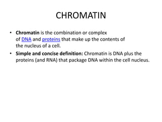
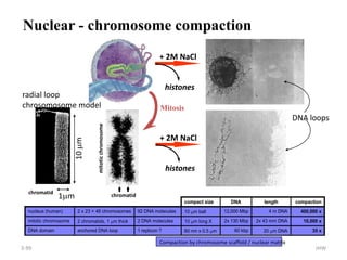
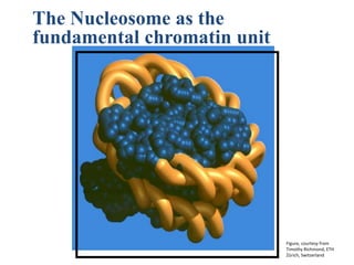
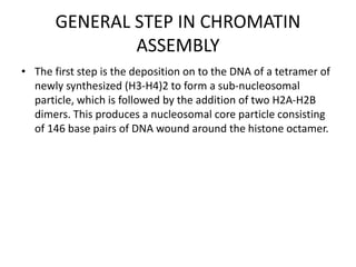
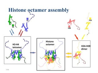
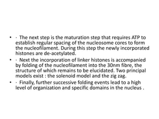
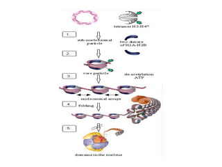
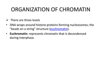
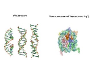
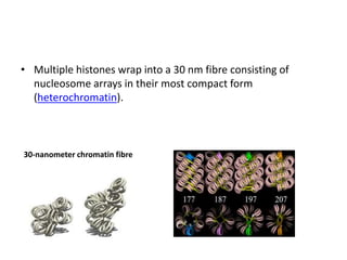
By
KAUSHAL KUMAR SAHU
Assistant Professor (Ad Hoc)
Department of Biotechnology
Govt. Digvijay Autonomous P. G. College
Raj-Nandgaon ( C. G. )
• MICROMOLECULES
• TYPES OF MICROMOLECULES
• CHROMATIN
• NUCLEOOME
• HISTONE
• GENERAL STEP IN CHROMATIN ASSEMBLY
• ORGANIZATION OF CHROMATIN
• CONCLUSIONS
• REFERENCE
Synopsis
MICROMOLECULES
• ‘A macromolecule is a very large molecule commonly created
by polymerization of smaller subunits. In biochemistry, the
term is applied to the four conventional biopolymers (nucleic
acids, proteins, carbohydrates, and lipids), as well as non-
polymeric molecules with large molecular mass such as macro
cycles.’
TYPES OF MICROMOLECULES
There are four macromolecules essential to living matter
containing C, H, O, N and sometimes S.
• Proteins
• Carbohydrates
• Nucleic Acids
• Lipids.
CHROMATIN
• Chromatin is the combination or complex
of DNA and proteins that make up the contents of
the nucleus of a cell.
• Simple and concise definition: Chromatin is DNA plus the
proteins (and RNA) that package DNA within the cell nucleus.
3-99 JHW
Nuclear - chromosome compaction
compact size DNA length compaction
nucleus (human) 2 x 23 = 46 chromosomes 92 DNA molecules 10 m ball 12,000 Mbp 4 m DNA 400,000 x
mitotic chromosome 2 chromatids, 1 m thick 2 DNA molecules 10 m long X 2x 130 Mbp 2x 43 mm DNA 10,000 x
DNA domain anchored DNA loop 1 replicon ? 60 nm x 0.5 m 60 kbp 20 m DNA 35 x
chromatin fiber approx. 6 nucleosomes per ‘turn’ of 11 nm 30 nm diameter 1200 bp 400 nm DNA 35 x
nucleosome disk 1 ¾ turn of DNA (146 bp) + linker DNA 6 x 11 nm 200 bp 66 nm DNA 6 - 11 x
base pair 0.33 x 1.1 nm 1 bp 0.33 nm DNA 1 x
Compaction by chromosome scaffold / nuclear matrix
radial loop
chrosomosome model
1m
10m
chromatid
chromatid
mitoticchromosome
+ 2M NaCl
histones
+ 2M NaCl
histones
Mitosis
DNA loops
• The primary functions of chromatin are
• 1) To package DNA into a smaller volume to fit in the cell,
• 2) To strengthen the DNA to allow mitosis,
• 3) To prevent DNA damage, and
• 4) To control gene expression and DNA replication.
NUCLEOSOME
• The fundamental unit of chromatin, termed
the nucleosome, is composed of DNA and histone proteins.
This structure provides the first level of compaction of DNA
into the nucleus.
3-99 JHW
The Nucleosome as the
fundamental chromatin unit
Figure, courtesy from
Timothy Richmond, ETH
Zürich, Switzerland
 The nucleosome is the fundamental unit of chromatin. It is
composed of:
• a core particle and
• a linker region (or internucleosomal region) that joins
adjacent core particles
• The core particle is highly conserved between species and is
composed of 146 base pairs of DNA wrapped 1.7 turns around
a protein octamer of two each of the core histones H3, H4,
H2A and H2B.
• The length of the linker region, however, varies between
species and cell type. It is within this region that the variable
linker histones are incorporated.
HISTONE
• Histone are highly conserved small basic protein
• 1) Core histone
• 2) Linker histone
 Core histone
• The core histones, H3, H4, H2A and H2B, are small, basic
proteins highly conserved in evolution.
• The most conserved region of these histones is their central
domain structurally composed of the "histone fold domain"
consisting of three a-helicies separated by two loop regions.
• Linker histone
• Linker histones associate with the linker region of DNA between two
nucleosome cores and, unlike the core histones, they are not well
conserved between species.
• In higher eukaryotes, they are composed of three domains: a globular,
non-polar central domain essential for interactions with DNA and two
non-structured N- and C- terminal tails that are highly basic and proposed
to be the site of post translational modifications
• The linker histones have a role in spacing nucleosomes and can modulate
higher order compaction by providing an interaction region between
adjacent nucleosomes.
3-99 JHW
HISTONES
are
highly conserved,
small, basic proteins
H1
H2B
H2A
H3
H4
helix
variable
conserved
Linker histone
Core histones
N
GENERAL STEP IN CHROMATIN
ASSEMBLY
• The first step is the deposition on to the DNA of a tetramer of
newly synthesized (H3-H4)2 to form a sub-nucleosomal
particle, which is followed by the addition of two H2A-H2B
dimers. This produces a nucleosomal core particle consisting
of 146 base pairs of DNA wound around the histone octamer.
3-99 JHW
Histone octamer assembly
H3-H4
tetramer
H2A-H2B
dimer
Histone
octamer
• · The next step is the maturation step that requires ATP to
establish regular spacing of the nucleosome cores to form
the nucleofilament. During this step the newly incorporated
histones are de-acetylated.
• · Next the incorporation of linker histones is accompanied
by folding of the nucleofilament into the 30nm fibre, the
structure of which remains to be elucidated. Two principal
models exist : the solenoid model and the zig zag.
• · Finally, further successive folding events lead to a high
level of organization and specific domains in the nucleus .
ORGANIZATION OF CHROMATIN
 There are three levels
• DNA wraps around histone proteins forming nucleosomes; the
"beads on a string" structure (euchromatin).
• Euchromatin: represents chromatin that is decondensed
during interphase.
DNA structure The nucleosome and "beads-on-a-string"[
• Multiple histones wrap into a 30 nm fibre consisting of
nucleosome arrays in their most compact form
(heterochromatin).
30-nanometer chromatin fibre
• Heterochromatin was defined as a structure that does not
alter in its condensation throughout the cell cycle whereas
euchromatin is decondensed during interphase.
Heterochromatin is localized principally on the periphery of
the nucleus and euchromatin in the interior of the
nucleoplasm. We can distinguish:
• constitutive heterochromatin, containing few genes and
formed principally of repetitive sequences located in large
regions coincident with centromeres and telomeres, from
• facultative heterochromatin composed of transcriptionally
active regions that can adopt the structural and functional
characteristics of heterochromatin, such as the inactive X
chromosome of mammals.
• Higher-level DNA packaging of the 30 nm fibre into the
metaphase chromosome (during mitosis and meiosis).
• There are, however, many cells that do not follow this
organisation. For example, spermatozoa and avian red blood
cells have more tightly packed chromatin than most
eukaryotic cells, and trypanosomatid protozoa do not
condense their chromatin into visible chromosomes for
mitosis.
CONCLUSIONS
• Chromatin is packaged in a hierarchy of structures.
Each of these levels of packaging has regulatory roles in
the genome
• The level of packaging we know best, also thanks to
formidable tools & technology development in the
recent years, is the nucleosome.
• In particular, post-translational histone modifications
play key roles in regulation of genome function, and
the combinatorial power of these modifications is only
beginning to be unraveled.
REFERENCE
• Principle of Biochemistry- Nelson & Cox 5th edition
• Internet
• es.wikipedia.org/wiki/Macromolécula
• bibliotecadigital.ilce.edu.mZ

Chromatin, Organization macromolecule complex

  • 1.
    By KAUSHAL KUMAR SAHU AssistantProfessor (Ad Hoc) Department of Biotechnology Govt. Digvijay Autonomous P. G. College Raj-Nandgaon ( C. G. )
  • 2.
    • MICROMOLECULES • TYPESOF MICROMOLECULES • CHROMATIN • NUCLEOOME • HISTONE • GENERAL STEP IN CHROMATIN ASSEMBLY • ORGANIZATION OF CHROMATIN • CONCLUSIONS • REFERENCE Synopsis
  • 3.
    MICROMOLECULES • ‘A macromoleculeis a very large molecule commonly created by polymerization of smaller subunits. In biochemistry, the term is applied to the four conventional biopolymers (nucleic acids, proteins, carbohydrates, and lipids), as well as non- polymeric molecules with large molecular mass such as macro cycles.’
  • 4.
    TYPES OF MICROMOLECULES Thereare four macromolecules essential to living matter containing C, H, O, N and sometimes S. • Proteins • Carbohydrates • Nucleic Acids • Lipids.
  • 5.
    CHROMATIN • Chromatin isthe combination or complex of DNA and proteins that make up the contents of the nucleus of a cell. • Simple and concise definition: Chromatin is DNA plus the proteins (and RNA) that package DNA within the cell nucleus.
  • 6.
    3-99 JHW Nuclear -chromosome compaction compact size DNA length compaction nucleus (human) 2 x 23 = 46 chromosomes 92 DNA molecules 10 m ball 12,000 Mbp 4 m DNA 400,000 x mitotic chromosome 2 chromatids, 1 m thick 2 DNA molecules 10 m long X 2x 130 Mbp 2x 43 mm DNA 10,000 x DNA domain anchored DNA loop 1 replicon ? 60 nm x 0.5 m 60 kbp 20 m DNA 35 x chromatin fiber approx. 6 nucleosomes per ‘turn’ of 11 nm 30 nm diameter 1200 bp 400 nm DNA 35 x nucleosome disk 1 ¾ turn of DNA (146 bp) + linker DNA 6 x 11 nm 200 bp 66 nm DNA 6 - 11 x base pair 0.33 x 1.1 nm 1 bp 0.33 nm DNA 1 x Compaction by chromosome scaffold / nuclear matrix radial loop chrosomosome model 1m 10m chromatid chromatid mitoticchromosome + 2M NaCl histones + 2M NaCl histones Mitosis DNA loops
  • 7.
    • The primaryfunctions of chromatin are • 1) To package DNA into a smaller volume to fit in the cell, • 2) To strengthen the DNA to allow mitosis, • 3) To prevent DNA damage, and • 4) To control gene expression and DNA replication.
  • 8.
    NUCLEOSOME • The fundamentalunit of chromatin, termed the nucleosome, is composed of DNA and histone proteins. This structure provides the first level of compaction of DNA into the nucleus.
  • 9.
    3-99 JHW The Nucleosomeas the fundamental chromatin unit Figure, courtesy from Timothy Richmond, ETH Zürich, Switzerland
  • 10.
     The nucleosomeis the fundamental unit of chromatin. It is composed of: • a core particle and • a linker region (or internucleosomal region) that joins adjacent core particles • The core particle is highly conserved between species and is composed of 146 base pairs of DNA wrapped 1.7 turns around a protein octamer of two each of the core histones H3, H4, H2A and H2B. • The length of the linker region, however, varies between species and cell type. It is within this region that the variable linker histones are incorporated.
  • 11.
    HISTONE • Histone arehighly conserved small basic protein • 1) Core histone • 2) Linker histone  Core histone • The core histones, H3, H4, H2A and H2B, are small, basic proteins highly conserved in evolution. • The most conserved region of these histones is their central domain structurally composed of the "histone fold domain" consisting of three a-helicies separated by two loop regions.
  • 12.
    • Linker histone •Linker histones associate with the linker region of DNA between two nucleosome cores and, unlike the core histones, they are not well conserved between species. • In higher eukaryotes, they are composed of three domains: a globular, non-polar central domain essential for interactions with DNA and two non-structured N- and C- terminal tails that are highly basic and proposed to be the site of post translational modifications • The linker histones have a role in spacing nucleosomes and can modulate higher order compaction by providing an interaction region between adjacent nucleosomes.
  • 13.
    3-99 JHW HISTONES are highly conserved, small,basic proteins H1 H2B H2A H3 H4 helix variable conserved Linker histone Core histones N
  • 14.
    GENERAL STEP INCHROMATIN ASSEMBLY • The first step is the deposition on to the DNA of a tetramer of newly synthesized (H3-H4)2 to form a sub-nucleosomal particle, which is followed by the addition of two H2A-H2B dimers. This produces a nucleosomal core particle consisting of 146 base pairs of DNA wound around the histone octamer.
  • 15.
    3-99 JHW Histone octamerassembly H3-H4 tetramer H2A-H2B dimer Histone octamer
  • 16.
    • · Thenext step is the maturation step that requires ATP to establish regular spacing of the nucleosome cores to form the nucleofilament. During this step the newly incorporated histones are de-acetylated. • · Next the incorporation of linker histones is accompanied by folding of the nucleofilament into the 30nm fibre, the structure of which remains to be elucidated. Two principal models exist : the solenoid model and the zig zag. • · Finally, further successive folding events lead to a high level of organization and specific domains in the nucleus .
  • 18.
    ORGANIZATION OF CHROMATIN There are three levels • DNA wraps around histone proteins forming nucleosomes; the "beads on a string" structure (euchromatin). • Euchromatin: represents chromatin that is decondensed during interphase.
  • 19.
    DNA structure Thenucleosome and "beads-on-a-string"[
  • 20.
    • Multiple histoneswrap into a 30 nm fibre consisting of nucleosome arrays in their most compact form (heterochromatin). 30-nanometer chromatin fibre
  • 21.
    • Heterochromatin wasdefined as a structure that does not alter in its condensation throughout the cell cycle whereas euchromatin is decondensed during interphase. Heterochromatin is localized principally on the periphery of the nucleus and euchromatin in the interior of the nucleoplasm. We can distinguish: • constitutive heterochromatin, containing few genes and formed principally of repetitive sequences located in large regions coincident with centromeres and telomeres, from • facultative heterochromatin composed of transcriptionally active regions that can adopt the structural and functional characteristics of heterochromatin, such as the inactive X chromosome of mammals.
  • 22.
    • Higher-level DNApackaging of the 30 nm fibre into the metaphase chromosome (during mitosis and meiosis). • There are, however, many cells that do not follow this organisation. For example, spermatozoa and avian red blood cells have more tightly packed chromatin than most eukaryotic cells, and trypanosomatid protozoa do not condense their chromatin into visible chromosomes for mitosis.
  • 23.
    CONCLUSIONS • Chromatin ispackaged in a hierarchy of structures. Each of these levels of packaging has regulatory roles in the genome • The level of packaging we know best, also thanks to formidable tools & technology development in the recent years, is the nucleosome. • In particular, post-translational histone modifications play key roles in regulation of genome function, and the combinatorial power of these modifications is only beginning to be unraveled.
  • 24.
    REFERENCE • Principle ofBiochemistry- Nelson & Cox 5th edition • Internet • es.wikipedia.org/wiki/Macromolécula • bibliotecadigital.ilce.edu.mZ