Download to read offline

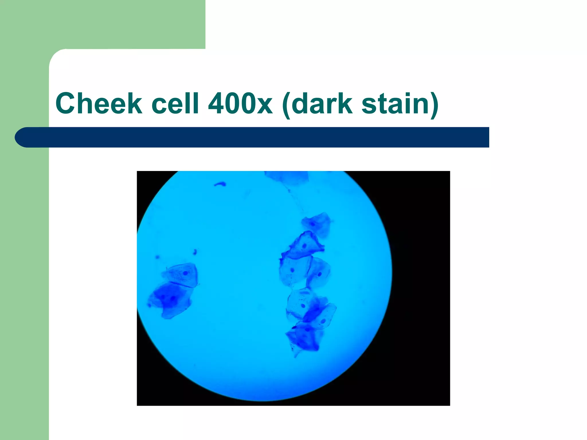
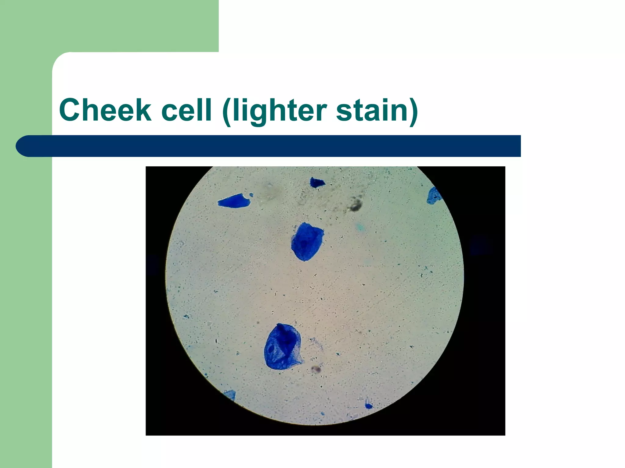

Cells were taken from the inside of the cheek and viewed under a microscope at 400x magnification with both dark and light staining. The images show cheek cells at high magnification with different staining techniques applied.