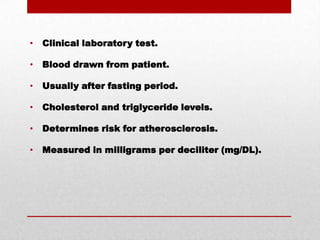
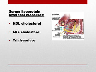
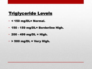
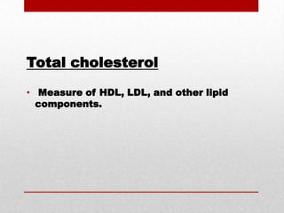
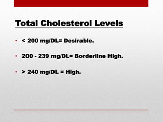
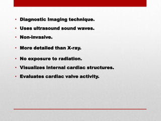
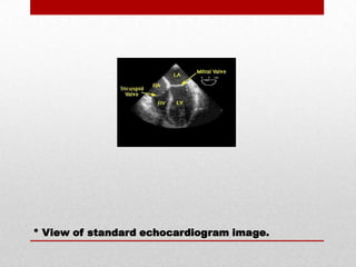
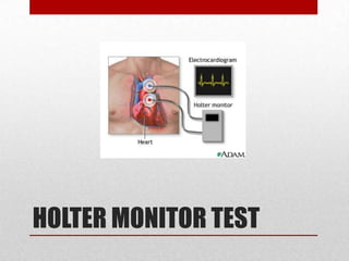
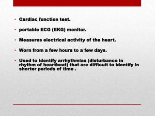
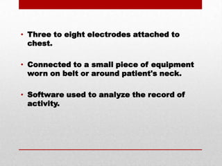
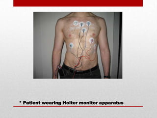
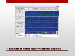
Cardiovascular System Diagnostic ProceduresJeff ButtramBio 120 Medical Terminology
There are many diagnostic procedures to help discover and diagnose various cardiovascular pathologies, falling into three main categories.
Clinical Laboratory TestsDiagnostic ImagingCardiac Function Tests
This presentation will focus on these three tests, one from each category.1. Serum lipoprotein levels (Clinical Lab). 2. Echocardiography (Diagnostic Imaging). 3. Holter monitor (Cardiac Function).
SERUM LIPOPROTEIN LEVEL
 Clinical laboratory test. Blood drawn from patient. Usually after fasting period. Cholesterol and triglyceride levels. Determines risk for atherosclerosis. Measured in milligrams per deciliter (mg/DL).
artherosclerosisfatty substances form plaque deposits on arterial walls.
Serum lipoprotein level test measures: HDL cholesterol LDL cholesterol Triglycerides
HDL high-density lipoprotein. "good" cholesterol.
HDL Levels< 60 mg/DL = low risk for heart disease. > 40 mg/DL in men = risk for heart disease. > 50 mg/DL in women = risk for heart disease.
LDL  Low-density lipoprotein."Bad" cholesterol - responsible for plaque build-up.
LDL Levels< 100 mg/DL = Optimal.100-129 mg/DL= Near optimal.130 - 159 mg/DL = Borderline high.160 - 189 mg/DL = High.> 190 mg/DL = Very High.
TriglyceridesChemical form of fat in foods and body.High-levels considered a predictor of Coronary Artery Disease (CAD).
Triglyceride Levels< 150 mg/DL= Normal.150 - 159 mg/DL= Borderline High.200 - 499 mg/DL = High.> 500 mg/DL = Very High.
Total cholesterol Measure of HDL, LDL, and other lipid components.
Total Cholesterol Levels < 200 mg/DL= Desirable. 200 - 239 mg/DL= Borderline High. > 240 mg/DL = High.
ECHOCARDIOGRAPHY
Diagnostic Imaging technique.Uses ultrasound sound waves.Non-invasive.More detailed than X-ray.No exposure to radiation.Visualizes internal cardiac structures.Evaluates cardiac valve activity.
* View of standard echocardiogram image.
Provides information about:Size and shape of the heart.How well the heart chambers are working.Abnormal muscle contractions in myocardium due to poor blood flow .Blood flow through chambers and valves of the heart (Doppler ultrasound).
Used to identify:Blood clots in the heart.Issues with pericardium (sac around the heart).Issues with aorta.
Used to evaluate: heart murmurs. heart's pumping function. patients with heart attack history.
Abnormal results can mean: heart valve disease.cardiomyopathy (disease of the heart muscle).pericardial effusion (excess fluid around the heart).
HOLTER MONITOR TEST
Cardiac function test.portable ECG (EKG) monitor.Measures electrical activity of the heart.Worn from a few hours to a few days.Used to identify arrhythmias (disturbance in rhythm of heartbeat) that are difficult to identify in shorter periods of time .
Three to eight electrodes attached to chest.Connected to a small piece of equipment worn on belt or around patient's neck.Software used to analyze the record of activity.
* Patient wearing Holter monitor apparatus
* Example of Holter monitor software analysis
Software Analysis:Heartbeat morphology.Beat interval measurement.Variations in heart rate.Overview of heart rhythms.

Chap5 pres jeb