Downloaded 268 times
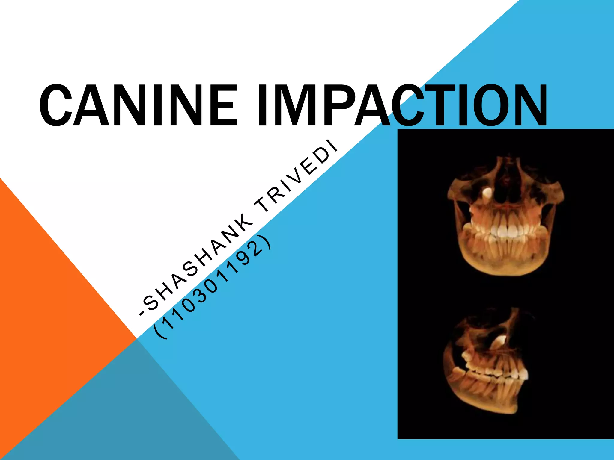


An impacted tooth is one that fails to erupt into the dental arch at the proper time. Wisdom teeth and canines are most commonly impacted. Canine impaction occurs in 2% of people and is more common in females. It is caused by overcrowding or issues during tooth development. Impacted canines can cause problems like cysts, infections, and damage to nearby teeth if not treated. Treatment options include exposing and orthodontically moving the tooth, extraction, or leaving it with monitoring. Proper diagnosis and management of impacted canines is important for dental health and aesthetics.


