Downloaded 971 times
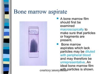

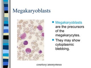
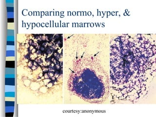
![proerythroblast
Normal proerythroblast
[dark red arrow] in the
bone marrow. This is a
large cell with a round
nucleus and a finely
stippled chromatin
pattern. Nucleoli are
sometimes apparent.
The cytoplasm is
moderately to strongly
basophilic.
There may be a paler
staining area of
cytoplasm surrounding
the nucleus.
courtesy:anonymous
](https://image.slidesharecdn.com/bonemarrowmorphology-140107041748-phpapp01/85/Bone-marrow-morphology-9-320.jpg)
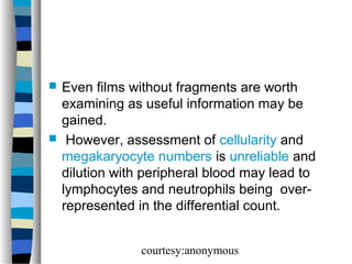
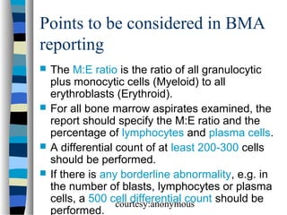
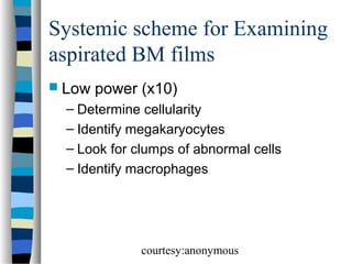
![Normal erythroblasts in the BM
. The early erythroblast [red
arrow] is similar to a
proerythroblast but is smaller
and no longer has visible
nucleoli.
The intermediate
erythroblast [green arrow]
and the late erythroblast
[blue arrow] show a
progressive reduction in cell
size, reduction in
cytoplasmic basophilia and
increase in chromatin
clumping.
The cytoplasm of the late
erythroblast may have a pink
tinge attributable to
haemoglobin.
courtesy:anonymous](https://image.slidesharecdn.com/bonemarrowmorphology-140107041748-phpapp01/85/Bone-marrow-morphology-10-320.jpg)

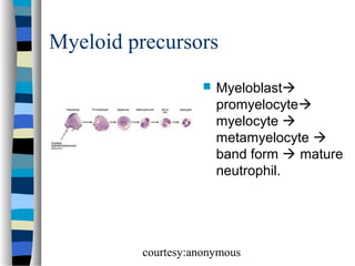
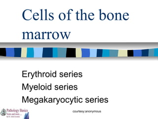
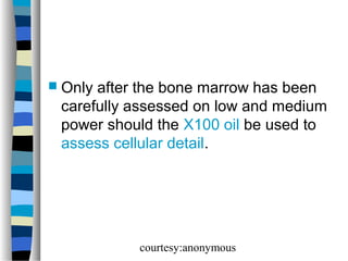
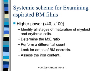
![Normal granulocyte precursors in
the bone marrow
. Note the myeloblast
[dark red arrow] with a
high nucleocytoplasmic
ratio, diffuse chromatin
pattern and nucleolus.
There is a promyelocyte
[green arrow] which is
larger and has a lower
nucleocytoplasmic
ratio and abundant
azurophilic granules.
courtesy:anonymous](https://image.slidesharecdn.com/bonemarrowmorphology-140107041748-phpapp01/85/Bone-marrow-morphology-14-320.jpg)


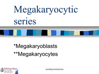
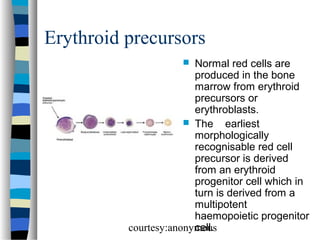
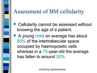
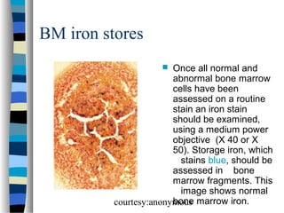
![Megakaryocytes in the BM
. Most megakaryocytes
[arrows] are large cells
which can be identified
with low power. Their
numbers are very
variable in normal bone
marrow films, being
partly related to the
number of fragments
present.
This image shows
increased
megakaryocyte
numbers.
courtesy:anonymous
](https://image.slidesharecdn.com/bonemarrowmorphology-140107041748-phpapp01/85/Bone-marrow-morphology-20-320.jpg)
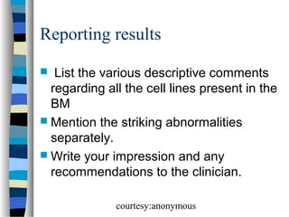
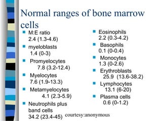

The document provides an overview of bone marrow examination techniques including aspiration sites and sample evaluation for reliable morphological examination. It details the different blood cell precursors in the marrow, including erythroid, myeloid, and megakaryocytic series, emphasizing the importance of assessing cellularity and presenting an appropriate differential count. Furthermore, it outlines reporting procedures and normal ranges for various bone marrow cells.


