Bioinformatics Methods From Omics To Next Generation Sequencing Sujay Datta
Bioinformatics Methods From Omics To Next Generation Sequencing Sujay Datta
Bioinformatics Methods From Omics To Next Generation Sequencing Sujay Datta
Bioinformatics Methods From Omics To Next Generation Sequencing Sujay Datta
Bioinformatics Methods From Omics To Next Generation Sequencing Sujay Datta
1.
Bioinformatics Methods FromOmics To Next
Generation Sequencing Sujay Datta download
https://ebookbell.com/product/bioinformatics-methods-from-omics-
to-next-generation-sequencing-sujay-datta-44079156
Explore and download more ebooks at ebookbell.com
2.
Here are somerecommended products that we believe you will be
interested in. You can click the link to download.
Bioinformatics Methods From Omics To Next Generation Sequencing Shili
Lin
https://ebookbell.com/product/bioinformatics-methods-from-omics-to-
next-generation-sequencing-shili-lin-49133764
From Sequences To Graphs Discrete Methods And Structures For
Bioinformatics Annie Chateau
https://ebookbell.com/product/from-sequences-to-graphs-discrete-
methods-and-structures-for-bioinformatics-annie-chateau-46082730
Computational Methods To Study The Structure And Dynamics Of
Biomolecules And Biomolecular Processes From Bioinformatics To
Molecular Quantum Mechanics 1st Edition Harold A Scheraga Auth
https://ebookbell.com/product/computational-methods-to-study-the-
structure-and-dynamics-of-biomolecules-and-biomolecular-processes-
from-bioinformatics-to-molecular-quantum-mechanics-1st-edition-harold-
a-scheraga-auth-4404508
Computational Methods To Study The Structure And Dynamics Of
Biomolecules And Biomolecular Processes From Bioinformatics To
Molecular Quantum Mechanics 2nd Ed Adam Liwo
https://ebookbell.com/product/computational-methods-to-study-the-
structure-and-dynamics-of-biomolecules-and-biomolecular-processes-
from-bioinformatics-to-molecular-quantum-mechanics-2nd-ed-adam-
liwo-7323868
3.
Bioinformatics Methods Express1st Edition Paul Dear
https://ebookbell.com/product/bioinformatics-methods-express-1st-
edition-paul-dear-2265976
Bioinformatics Methods And Applications Dev Bukhsh Singh Rajesh Kumar
Pathak
https://ebookbell.com/product/bioinformatics-methods-and-applications-
dev-bukhsh-singh-rajesh-kumar-pathak-38251422
Bioinformatics Methods In Clinical Research 1st Edition Ewa Gubb
https://ebookbell.com/product/bioinformatics-methods-in-clinical-
research-1st-edition-ewa-gubb-4288382
Structural Bioinformatics Methods And Protocols 1st Ed 2020 Edition
Zoltn Gspri
https://ebookbell.com/product/structural-bioinformatics-methods-and-
protocols-1st-ed-2020-edition-zoltn-gspri-47706558
Plant Bioinformatics Methods And Protocols 1st Edition Peter Sterk
https://ebookbell.com/product/plant-bioinformatics-methods-and-
protocols-1st-edition-peter-sterk-2387236
6.
Bioinformatics Methods
The pastthree decades have witnessed an explosion of what is now referred to as high-
dimensional “omics” data. Bioinformatics Methods: From Omics to Next Generation
Sequencing describes the statistical methods and analytic frameworks that are best
equipped to interpret these complex data and how they apply to health-related research.
Covering the technologies that generate data, subtleties of various data types, and statisti-
cal underpinnings of methods, this book identifies a suite of potential analytic tools and
highlights commonalities among statistical methods that have been developed.
An ideal reference for biostatisticians and data analysts that work in collaboration with
scientists and clinical investigators looking to ensure the rigorous application of available
methodologies.
Key Features:
• Survey of a variety of omics data types and their unique features
• Summary of statistical underpinnings for widely used omics data analysis methods
• Description of software resources for performing omics data analyses
Shili Lin, PhD is a Professor in the Department of Statistics and a faculty member in the
Translational Data Analytics Institute at the Ohio State University. Her research interests
are in statistical methodologies for high-dimensional and big data, with a focus on their
applications in biomedical research, statistical genetics and genomics, and integration of
multiple omics data.
Denise Scholtens, PhD is a Professor and Chief of the Division of Biostatistics in the
Department of Preventive Medicine at Northwestern University Feinberg School of Medi-
cine. She is interested in the design and conduct of large-scale multi-center prospective
health research studies, and in the integration of high-dimensional omics data analyses
into these settings.
Sujay Datta, PhD is an Associate Professor and the Graduate Program Coordinator in the
Department of Statistics at the University of Akron. His research interests include statisti-
cal analyses of high-dimensional and high-throughput data, graphical and network-based
models, statistical models and methods for cancer data, as well as sequential/multistage
sampling designs.
Preface
The past threedecades have witnessed an explosion of what we now often refer
to as high-dimensional “omics” data. Initial investigations into the sequence of
the human genome and variation in gene expression as evidenced by the assays
of messenger RNA abundance, i.e. genomics, ushered in a paradigm of high-
throughput assay development and data generation for molecular and cellular
biology that has revolutionized the way scientists think about human health.
Emergence of these varied, complex and often related data types demands
development of statistical methodologies and analytic paradigms to support
meaningful interpretation of these data. This book is written to describe a
sampling of statistical methods and analytic frameworks that have been de-
veloped to address the unique features of several types of high-dimensional
molecular omics data. The content describes the technologies that generate
data, subtleties of various data types, basic statistical underpinnings of meth-
ods that are used for their analysis, available software for implementing anal-
yses and discussions of various applications in health related research. The
intended audience is collaborative biostatisticians and data analysts who are
looking to ensure rigorous application of available methodologies as they part-
ner with basic scientists and clinical investigators.
The introductory Chapter 1, The Biology of a Living Organism, summa-
rizes the basic cellular components and molecular biology that the various
omics technologies discussed in this book are designed to assay. This descrip-
tion is included to give context for the resultant data and highlight how, in
many cases, different data types provide multiple points of view on a highly
coordinated physiologic system. An Appendix provides similar foundational
content, but from a statistical point of view. It is expected that most read-
ers of this book will be familiar with basics of probability, random variables,
probability distributions, basics of stochastic processes, hidden Markov mod-
els, and both frequentist and Bayesian inference procedures. Nevertheless,
appendix content is provided to help fill in gaps where needed.
The primary content of the book focuses on various types of omics data.
Chapters 2 and 3 are dedicated to Protein-Protein Interactions and Protein-
Protein Interaction Network Analyses. Chapter 4 summarizes Detection of
Imprinting and Maternal Effects through epigenetic mechanisms. Sequenc-
ing data are the focus of both Chapter 5 Modeling and Analysis of Next-
Generation Sequencing Data and Chapter 6 Sequencing-Based DNA Methyla-
tion Data. Chapter 7 Modeling and Analysis of Spatial Chromatin Interactions
and Chapter 8 Digital Improvement of Single Cell Hi-C Data describe some
xiii
19.
xiv Preface
of themost recently emerging data types and related methods. Finally, Chap-
ter 9 Metabolomics Data Preprocessing and Chapter 10 Metabolomics Data
Analysis are dedicated to a survey of metabolomics data. A strong common
theme in each chapter is the importance of specifying statistical models that
closely reflect the underlying biology that is being queried. Themes of prepro-
cessing to handle for technical variation and artifacts, accounting for testing
multiplicity and cross-feature dependencies, and computational efficiency also
arise.
The content herein is by no means a comprehensive survey of the state-of-
the-art. Given the pace of technology development and data availability, new
techniques, descriptive and inferential procedures, analysis packages and po-
tential applications emerge almost daily. It is our hope that the survey offered
here will identify a suite of potential analytic tools, highlight commonalities
among statistical methods that have been developed, and motivate contin-
ued statistical methods development for meaningful, integrated analyses of
high-dimensional molecular omics data.
20.
1
The Biology ofa Living Organism
CONTENTS
1.1 Cells .............................................................. 2
1.2 Genes, DNA and RNA ........................................... 5
1.3 Proteins .......................................................... 9
1.4 The epigenome ................................................... 11
1.5 Metabolism ....................................................... 12
1.6 Biological regulation and cancer ................................. 14
1.7 Data generating technologies .................................... 16
There is no unique definition of bioinformatics in the scientific literature.
Generally speaking, it is a rapidly evolving field of science that harnesses
the powers of mathematics, statistics and information science to analyze very
large biological datasets using modern-day high-performance computing. It is
all about extracting knowledge from the deluge of information generated by
cutting-edge high-throughput technologies in the biomedical sciences. A great
deal of research in the current century has gone into developing innovative
tools and techniques enabling us to handle the information overload that the
scientific community is currently experiencing. In this book, one of our goals
is to shed some light on those tools and techniques.
Among the natural sciences, biology is the one that studies highly com-
plex systems known as living organisms. Before the era of modern technology,
it was primarily a descriptive science that involved careful observation and
detailed documentation of various aspects of a living being. (e.g. its appear-
ance, behavior, interaction with the surrounding environment, etc.). These
led to a reasonably accurate classification of all visible forms of life on the
earth (the binomial nomenclature by Carolas Linnaeus) and to a theory of
how the diversity of life-forms we see all around us came into being over
billions of years (the theory of evolution by Charles Darwin). However, the
technology available in those days was not good enough for probing the in-
ternal mechanisms that made life possible and sustained it – the complex
biochemistry of metabolism, growth and self-replication. So the biological
datasets that were available for statistical analysis in those days (including
clinical, epidemiological and taxonomic data) were relatively small and man-
ageable. Standard classical statistical procedures such as two-sample t-tests,
ANOVA and least-squares regression were adequate to handle them. But it all
DOI: 10.1201/9781315153728-1 1
21.
2 Bioinformatics Methods:From Omics to Next Generation Sequencing
began to change around the middle of the twentieth century with a series of key
breakthroughs in the biomedical sciences and rapid technological development.
These breakthroughs enabled us to probe the inner sanctum of a living organ-
ism at the molecular level and brought increased sophistication to our concept
of medicine. A series of new discoveries gave us unprecedented insight into the
modus operandi of living systems, the most famous of which was the Franklin-
Watson-Creek double helix model for DNA. Building on these foundations, an
emerging goal of medical scientists became personalized medicine. New ex-
periments powered by advanced technologies were now generating enormous
amounts of data on various aspects of life, and increasingly efficient comput-
ers made it possible to create and query gigantic databases. Statisticians and
information scientists are now inundated with an incredible amount of data
and the demand for methods that can handle huge datasets that often violate
some of the basic assumptions of classical statistics. Enhanced data-storage
capability enables an abundance of prior information on unknown parameters,
and the lack of closed-form analytical solutions matters less given the capabil-
ity for approximate numerical solutions. In summary, modern-day statisticians
almost invariably find themselves navigating an ocean of information where
traditional statistical methods and their underlying assumptions often are of-
ten inappropriate.
Understanding the intricate chemistry by which life survives, propagates,
responds and communicates is the overarching goal of molecular biology –
a field which relies increasingly on automated technologies for performing
biological assays and on computers for analyzing the results. While the com-
plexity and diversity of life on earth are truly awe-inspiring, at the molecular
level there are many commonalities between different forms of life. In the next
section, we briefly review some essential concepts in cellular and molecular
biology, which will provide the necessary background and motivation for the
bioinformatics problems discussed throughout the book. First, we discuss cells
and organelles, followed by an introduction to DNA (the genetic material)
and the chromosomes’ 3D structure. Proteins are considered next. Finally,
biochemical networks are discussed, with a focus on cell metabolism and con-
trol of proliferation. For further details on any of these topics, the reader is
referred to any modern textbook on molecular and cellular biology.
1.1 Cells
The basic structural and functional unit of a living organism is a cell. Robert
Hooke, a British scientist of the seventeenth century, made a chance discovery
of them when he looked at a piece of cork under the crude microscope that was
available at that time. Soon it was hypothesized, and ultimately verified that
every living organism is made of cells. In more than three and a half centuries
22.
The Biology ofa Living Organism 3
since their discovery, thanks to increasingly powerful microscopes and other so-
phisticated technologies, astonishing details are available regarding all aspects
of cells, including their internal structure, day-to-day function, proliferation
and death. Primitive organisms such as bacteria (e.g. Proteobacteria that in-
clude pathogens such as Escherichia coli and Salmonella, Cyanobacteria that
are capable of converting the sun’s electromagnetic energy to chemical energy
through photosynthesis and Archaebacteria that survive in extreme environ-
ments such as the deep-ocean hydrothermal vents) are single-cell organisms
known as prokaryotes. They lack an organelle, called a nucleus, that is present
in the cells of all higher organisms known as eukaryotes. Eukaryotes, which
include everything from a bath sponge to an elephant, may be uni-cellular or
multi-cellular. In multi-cellular organisms, the cells are organized into tissues
or groups of cells that collectively perform specific tasks, such as transport
of oxygen or absorption of nutrients. Several such tissues collectively form an
organ, such as the leaves of a plant or the lungs in our body. All the different
types of cells in a multi-cellular organism originate from the same “mother
cells” in the embryo, known as embryonic stem cells, which are pluripotent
(i.e. capable of transforming into various types of specialized cells through a
process called differentiation). This is why embryonic stem cells are considered
so promising in the biomedical sciences as a possible cure for certain debilitat-
ing and degenerative diseases. The process of differentiation is still not fully
understood, and bioinformatics may play a key role in helping us understand
it.
The human body consists of trillions of eukaryotic cells. There are at least
two hundred different types of cells in our body, and they may be as little as
a few thousandths of a millimeter in diameter or up to a meter in length (e.g.
neurons with long axons). Each cell has an outer membrane which encloses the
cytoplasm and various components called organelles. They include the nucleus,
the endoplasmic reticulum, the Golgi apparatus, the mitochondria, the ribo-
somes and the lysosomes. A plant cell also has large cavities called vacuoles
and different types of plastids (chloroplasts containing chlorophyll for pho-
tosynthesis, leucoplasts and chromoplasts). Each of these organelles has some
specific role to play in the life of a cell. Because mitochondria and chloroplasts
can multiply within a cell and bear structural similarities to certain unicel-
lular prokaryotic organisms, it is hypothesized that a long time ago, some
prokaryotic cells started living inside others, forming a symbiotic or mutually
beneficial relationship with their hosts. This process, called endosymbiosis, is
believed to have led ultimately to the development of eukaryotic cells.
There are four major classes of small organic (carbon-containing) molecules
in cells: sugars, lipids, amino acids and nucleotides. These molecules may exist
on their own in the cell but may also be bound into macromolecules or poly-
mers, large molecules comprising specific patterns of different types of small
organic molecules. For example, simple sugars such as glucose act as a source
of energy for the cell and as a means for storing energy. Glucose can also be
assembled into cellulose, a structural polymer that forms the cell walls of plant
23.
4 Bioinformatics Methods:From Omics to Next Generation Sequencing
cells. Lipids are a crucial component of cellular membranes, including the outer
cell membrane and the membranes of organelles. They are also used for the
storage of energy and act as hormones, transmitting signals between different
cells. Amino acids are the building blocks of proteins which perform numerous
functions in a living organism. The nucleotide adenosine triphosphate (ATP)
is used to transfer energy needed in a variety of reactions, while long chains
of nucleotides form the deoxyribonucleic acids (DNA) and ribonucleic acids
(RNA) that act as the basis of heredity and cellular control. In addition to
these organic molecules, there are a variety of inorganic molecules inside the
cell. By mass, a cell for the most part is water. A few other important inorganic
compounds are oxygen and carbon monoxide (which are usually considered in-
organic despite containing carbon), various ions (the medium for transmitting
electrical signals in neurons and heart cells) and phosphate groups (crucial in
intracellular signaling).
The cell membrane is primarily made of phospholipid, with various mem-
brane proteins and other substances embedded in it. A phospholipid molecule
has a hydrophobic (water-repelling) end and a hydrophilic (water-attracting)
end. A cell membrane consists of a phospholipid bi-layer, with the hydropho-
bic “heads” of the molecules tucked inside and the hydrophilic “tails” forming
surfaces that touch water. In addition to serving as an enclosure for the or-
ganelles, the membrane controls the passage of substances into and out of
the cell, and various receptors embedded in it play a crucial role in signal
transduction. The endoplasmic reticulum primarily serves as a transport net-
work and storage area for various cellular substances. It is continuous with
the membrane of the nucleus. Various nuclear products (e.g. the messenger
RNA produced by transcription) are transported to places where they are
needed through the endoplasmic network. This reticulum has smooth surfaces
and rough surfaces, the rough appearance being the result of numerous ri-
bosomes attached to it. The ribosomes are the cell’s protein-manufacturing
plants where the macro-molecules of proteins are synthesized component by
component according to the instructions in the messenger RNA. The proteins
produced at the ribosomes are transported to the Golgi apparatus (named
after the Italian scientist Camillo Golgi who first noticed them) where they
are further modified as needed and then sent off in vesicles (little bubble-like
sacks) to other organelles or to the cell membrane for secretion outside the
cell. This Golgi apparatus has a cis end that is nearer to the endoplasmic
reticulum and a trans end that is farther from it. The incoming proteins are
received at the cis end and the outgoing vesicles bud off from the trans end.
The lysosomes are the cell’s scavengers or garbage cleaners, facilitating the
disintegration of unwanted or harmful substances in the cell. The mitochon-
dria are the energy-producing plants where the cell’s energy currency (ATP)
is synthesized. A mitochondrion has a membrane quite similar to that of a
prokaryotic bacterium and inside it, there are protruded folds of the mem-
brane called christae. The rest of the space inside it is called the lumen where
ATP synthesis takes place. In a plant cell, a chloroplast also has an outer
24.
The Biology ofa Living Organism 5
membrane resembling that of a prokaryotic bacterium and inside it, the lu-
men contains stacks of flat disc-like structures called thylakoids. This is where
chlorophyll, a magnesium-containing protein, is found that plays a key role
in photosynthesis – the plant’s food production process. Chlorophyll is also
the reason why leaves are green. Chromoplasts contain other pigments such as
carotene and xanthophylls instead of chlorophyll. These are the reason behind
the bright display of yellow, orange and brown in autumn. The remaining ma-
jor organelle in a eukaryotic cell is the nucleus. It is roughly spherical with
an outer membrane that has pores in order to let nuclear products out into
the cytoplasm or let cytoplasmic substances in. The central, denser region of a
nucleus is called the nucleolus. Most importantly, a living organism’s blueprint
of life (i.e. its DNA) is stored inside the nucleus.
1.2 Genes, DNA and RNA
Deoxyribonucleic acid or DNA is present in all living organisms and is of
central importance to regulating cellular function and conveying hereditary
information. It does so primarily through the genes it encodes. All cells in
our body contain DNA in their nuclei (except a few such as the erythrocytes
or red blood cells). As mentioned earlier, DNA is a polymer of nucleotides,
conceptually arranged like a ladder. The functionally interesting component
of a nucleotide is its base. There are four different bases: adenine, guanine,
thymine and cytosine, often referred to by their single-letter abbreviations A,
G, T and C. Adenine and guanine are called purines, while the other two are
called pyrimidines. Pairs of bases, one from each strand of the double-stranded
DNA, bind together to form the steps of the ladder. These base-pairs are held
in place by two backbones of sugar (deoxyribose) and phosphate molecules,
which form the sides of the ladder. Each purine binds with only one kind of
pyrimidine – A with T and G with C. This is known as complementary base
pairing.
In reality, DNA molecules are not shaped like a straight ladder but have
a complex three-dimensional (3D) structure. The two strands of the DNA
(i.e. the two sides of the ladder) are twisted into the well-known double helix
shape, famously discovered in 1953 by James Watson and Francis Crick with
the help of X-ray crystallographic studies done by Rosalind Franklin. At a
larger scale, the DNA double-helix is wrapped around barrel-shaped protein
molecules called histones. The DNA and histones are, in turn, wrapped into
coils and other structures, depending on the state of the cell.
In some cells, most or all of the DNA occurs in the form of a single molecule.
In prokaryotes such as bacteria, most of the DNA resides in a single chromo-
some, a long loop of DNA without beginning or end. Bacteria also often con-
tain plasmids, which are much shorter loops of DNA that can be exchanged
25.
6 Bioinformatics Methods:From Omics to Next Generation Sequencing
between bacteria as a means of sharing beneficial genes. In eukaryotes, the
DNA is divided into several chromosomes, each a linear stretch of DNA with
a definite beginning and end. Many eukaryotes are polyploid, carrying more
than one copy of each chromosome. For example, humans are diploid as most
human cells carry two copies of each chromosome – one inherited from each
parent. Some plants are tetraploid, carrying four copies of each chromosome.
The total length of DNA and the number of chromosomes into which it is
divided vary from one organism to another. The single chromosome of the
bacterium Escherichia coli has about 4.6 million base pairs, while humans
have over 3 billion base pairs divided into 23 pairs of chromosomes. Fruit flies
(Drosophila melanogaster have less than 200 million base pairs divided into 8
pairs of chromosomes, whereas rice has roughly 400 million base pairs divided
into 24 pairs of chromosomes.
When a cell divides to produce two daughter cells, as is necessary for
organism growth and for replacing old or damaged tissue, the DNA in the
parent cell must be replicated to provide each daughter cell with its own copy.
Complementary base-pairing is key to this process. During replication, a group
of proteins including DNA polymerase travels along the DNA strands. The
two strands of DNA are separated, much like a zipper being unzipped, and
complementary base pairs are filled in along both strands, resulting in the two
needed copies.
Less than 5% of the DNA is believed to encode useful information for
protein synthesis. The other 95%, the so-called “junk” DNA, was believed to
have no apparent function until recently. However, cutting-edge research in
the last few years has shed more light on the relevance of this non-protein-
coding DNA in terms of gene regulation and other important things. Of the
5% functional DNA, the majority is part of some gene. Genes are regions
of the DNA comprising two parts – a regulatory region or promoter and a
coding region. The regulatory region is partly responsible for specifying the
conditions under which the gene-product is produced, or the extent to which
it is produced (i.e. the expression of the gene). The coding region specifies
the functional molecular product or products – often a protein but sometimes
an RNA (ribonucleic acid). Proteins, as mentioned earlier, are sequences of
amino acids. For protein-coding genes, the coding region specifies the amino-
acid sequence of the protein. However, the protein is not directly constructed
from the DNA. Instead, the DNA is transcribed into an RNA intermediate,
called a messenger RNA (mRNA), which is then translated into protein at
the ribosome. For RNA-coding genes, the RNA coded by it is itself the final
product and serves some important function instead of being translated into
protein.
RNA consists of a single sequence, or strand, of nucleotides. These nu-
cleotides are similar to the ones in DNA, except that the backbone uses
the sugar ribose, and the base uracil (U) is used instead of thymine (T).
Transcription (i.e. the construction of an RNA chain from the DNA) is sim-
ilar to DNA replication. A group of proteins, collectively known as RNA
26.
The Biology ofa Living Organism 7
polymerases, open up the DNA at the start of the coding region and move
along the DNA until reaching the end of the coding region. The transcript is
produced one nucleotide at a time by complementary base-pairing (A with U,
C with G, G with C and T with A). The resulting RNA molecule does not stay
bound to the DNA template that generated it. As it is constructed, the RNA
nucleotide chain separates from the DNA and the two DNA strands then re-
join, going back to the same state as they were in before transcription. Instead
of forming a stable and neat geometric shape (such as the DNA double-helix),
the nucleotides in an RNA strand bind to each-other forming a complex 3D
shape unique to each RNA sequence. For RNA-coding genes, features of this
3D shape determine its functional properties (for example, the ability to bind
to specific parts of certain protein molecules). For protein-coding genes, the
creation of the RNA is only the first step in protein production.
Proteins are composed of 20 naturally occurring amino acids. The RNA
nucleotide sequence encodes an amino acid sequence in a relatively straight-
forward way. Each triplet of nucleotides, called a codon, specifies one amino
acid. For example, the RNA nucleotide triplet AAA is the codon for the amino
acid lysine, while the triplet AAC codes for asparagine. Since there are 4 nu-
cleotides, there are 43
= 64 distinct possible codons. However, as there are only
20 distinct amino acids, there is quite a bit of redundancy among the codons.
For example, the amino acid leucine is encoded by six different codons: CUU,
CUG, CUC, CUA, UUG and UUA. The entire length of the RNA strand does
not code for amino acids. A small amount of RNA at the start and the end of
the transcript (called untranslated regions or UTRs) are not translated into
amino acids. For some genes, especially in eukaryotes, there are untranslated
gaps in the middle of an RNA transcript, called introns. The portions of the
transcript that are translated are known as exons. In eukaryotes, a process
called splicing removes the introns before the transcript leaves the nucleus
for translation. Splicing can result in the elimination of some exons as well,
leading to different splice-variants of a protein. This is referred to as alter-
native splicing, which allows a cell to produce different versions of a protein
from the same DNA. Ribosomes translate the RNA transcript into the cor-
responding amino acid sequence. In prokaryotes, free-floating ribosomes can
begin translation as soon as the transcript is created, or even while it is be-
ing created. In eukaryotes, the transcripts must be transported outside the
nucleus through the nucleopores and reach a ribosome attached to the rough
endoplasmic reticulum.
Transcription, splicing and translation are regulated in various ways. Char-
acteristic patterns of nucleotides in the DNA indicate where transcription
should begin and end. For example, in many bacteria, transcription begins
just after a stretch of DNA with the sequence TATAAT. However, some vari-
ations on this sequence are allowed and, conversely, not every occurrence of
this sequence in the DNA is immediately followed by the coding region of a
gene. A different pattern indicates the end of the coding region. In eukary-
otes, there is much more variation in these patterns. In either case, even when
27.
8 Bioinformatics Methods:From Omics to Next Generation Sequencing
the DNA of an organism is completely sequenced, there remains uncertainty
about the exact locations and number of the genes.
Variations in transcription initiation patterns have apparently evolved in
order to control the rate or frequency with which the RNA polymerase binds
to the DNA beginning the process of transcription. However, transcription is
primarily regulated by transcription factors. These proteins bind to the regu-
latory region of the gene on the DNA, usually located within a few hundreds
or thousands of base pairs of the transcription initiation site, and influence
the rate or frequency of transcription by one or more of the following mech-
anisms: blocking the binding of RNA polymerase, changing the shape of the
DNA to expose the transcription initiation site and thereby increasing RNA
polymerase binding, attracting RNA polymerase to the region of transcription
initiation, or acting as a dock or blocker for other proteins which themselves
have similar effects. Since these transcription factors are proteins themselves
and thus generated from genes, the logical conclusion is that some genes regu-
late others, giving rise to gene regulatory networks. Transcription factors bind
to the DNA at specific sites known as transcription factor binding sites by
virtue of their 3D shapes. These sites are typically between 8 and 20 nu-
cleotides long. An organism may have hundreds of different proteins acting as
transcription factors, each of which binds to different characteristic patterns
of nucleotides. However, there are variations in these patterns, as was the
case for transcription initiation sites. Some of these variations may serve the
purpose of influencing the frequency or strength with which the transcription
factor binds. Because of the short lengths of these sites, the variability seen
in their patterns, and the virtual “haystack” of DNA in which they can be
situated, it is not easy to identify transcription factor binding sites. Further,
the binding of a transcription factor to a site does not guarantee any influence
on the transcription process. Thus, identifying functional binding sites is an
additional layer of difficulty.
In eukaryotes, and for a few genes in prokaryotes, splicing and alterna-
tive splicing follow transcription. The signals that regulate splicing are partly
in the transcribed RNA sequence itself, although clear signals have not been
identified. RNA from RNA-coding genes also plays a role, especially in alter-
native splicing. However, this depends on a complex interplay between the 3D
structures of the regulatory RNA, the transcript and the splicing machinery.
Translation is not as heavily regulated as transcription, and its regulation is
better understood. A ribosome assembles the sequence of amino acids specified
by the transcript in the order in which they are encountered. A type of RNA,
known as transfer RNA or tRNA, plays an important role in this assembly
process. Any of the three codons UAA, UAG and UGA indicate the end of
the protein and the termination of translation. In eukaryotes, ribosomes bind
to the start of the transcript and move along it until encountering the first
AUG codon, when translation begins. Sometimes the translation machinery
will skip over the first (or even the second) occurrence of AUG and begin at
the next occurrence. This skipping is influenced by the adjacent nucleotides,
28.
The Biology ofa Living Organism 9
and is another mechanism by which different variants of a protein are created
from the same DNA coding region. In bacteria, the ribosomes do not bind
to the start of the transcript. Instead, there is a longer nucleotide sequence
(including an AUG codon) to which they bind. The ribosomes can bind to that
nucleotide sequence anywhere it occurs in the transcript and begin translation.
As a result, it is possible for a single transcript to code for several proteins,
each separated by a region that is not translated. Such a gene is called an
operon and is a common means by which proteins that have related functions
are co-regulated. For example, the well-known lac operon in the bacterium
Escherichia coli includes three genes, one of which helps the organism to
metabolize the sugar lactose and another transports lactose into the cell from
the extracellular environment. Translation of an RNA transcript can also be
regulated by other proteins or RNAs (such as micro-RNAs), which can, for
example, bind with the RNA transcript and block the translation mechanism.
1.3 Proteins
The reason why proteins are called the “work-horses” of a cell is that they
are involved in virtually every biological process in cells, including sensing of
the cell’s environment and communication between cells. Proteins are polymer
chains with amino acids as their building blocks. A backbone comprising one
nitrogen and two carbon atoms is bound to various hydrogen and oxygen
atoms. The central carbon is also bound to a unit (which can be a single
atom or a group of atoms) that distinguishes among different amino acids.
In glycine, the simplest amino acid, that unit is just one hydrogen atom. In
methionine, that unit contains three carbon, seven hydrogen and one sulfur
atoms. In different amino acids, that unit differs in its chemical properties,
most relevantly in size, acidity and polarity. When amino acid molecules bind
together to form polymers, water molecules are released, and the parts of the
amino acids that still remain are called residues. Such a bond formation is
known as peptide bonding. Shorter amino acid sequences are peptides, while
longer ones are proteins (typically containing hundreds or thousands of amino-
acid residues).
As a protein is generated by translation from a transcript, it folds into a
complex 3D shape, which depends on its amino acid sequence as well as the
chemical environment in which it is folding. The structure of a protein can be
described at four different levels. The primary structure of a protein is simply
its amino acid sequence. The secondary and tertiary structures refer to the
3D folded shape of the protein. The tertiary structure is a specification of 3D
coordinates for every atom in the protein in an arbitrary coordinate system,
as well as that of which atoms are chemically bound to each other. Tertiary
structures contain commonly occurring patterns, such as alpha-helices and
29.
10 Bioinformatics Methods:From Omics to Next Generation Sequencing
beta-sheets Alpha-helices are stretches of the protein that wind into a helical
form. A beta-sheet is a set of beta-strands that align with each other length-
wise, forming a sheet-like shape. The secondary structure of a protein assigns
each amino acid to participating in an alpha-helix, participating in a beta-
sheet, participating in a bend between two alpha-helices, and so on. So the
secondary structure of a protein is an abstraction of its tertiary structure
components. Many proteins participate in complexes (groups of proteins and
other molecules that are weakly bound together). A complex may contain
proteins of different types. Conversely, one type of protein can participate
in different complexes. A specification of how proteins and other molecules
organize into complexes is called the quaternary structure.
In addition to serving as the building material for various parts of an
organism’s body, proteins play a number of important roles related to DNA
and RNA. Transcription factors regulate transcription rates, RNA polymerase
creates the transcripts, DNA polymerase opens up the DNA double-strand
like a zipper and then replicates it, histones (little packs of proteins in the
chromatin wrapped by the DNA double-strand) affect the 3D structure of the
DNA and influence transcription. Proteins are also responsible for repairing
damage to DNA (caused by exposure to radiation or certain toxic chemicals,
among other things) and for untangling DNA. Proteins also act as enzymes,
molecules that facilitate the occurrence of chemical reactions (most often very
specific reactions). For instance, the enzyme lactase facilitates the conversion
of the sugar lactose to two other simpler sugars, glucose and galactose. The
roles that DNA polymerase and RNA polymerase play can also be viewed
as enzymatic. The specificity of an enzyme to the reactions it catalyzes (i.e.
accelerates) depends on its active sites (i.e. portions of the tertiary structure
of the enzyme that bind to particular reactants.
Cell membranes, such as the outer membrane of a cell or the nuclear mem-
brane, are impermeable to many types of molecules. Trans-membrane proteins
control the flow of molecules across membranes and convey signals from one
side of the cell to the other. These proteins reside within the membrane and
protrude on either side. Channels allow the transport of molecules (mostly
very specific molecules). For example, ion channels in cardiac muscles or neu-
rons control the flow of ions (such as sodium, potassium, calcium and chlorine)
by opening to allow the flow or closing to stop it, depending on triggers such
as electrical activity. The dynamical properties of these channels (opening
and closing), and the resulting changes in ion flows, drive larger-scale electro-
physiological phenomena, such as the beating of the heart and the transmission
of action potentials down the axon of a nerve cell. Nuclear pore proteins form
complexes that allow molecules such as RNA transcripts and transcription
factors across the nuclear membrane. Signaling is a related task but does not
necessarily involve the transport of a molecule from one side of a membrane
to the other. Often, the binding of a specific molecule to a trans-membrane
protein causes a structural change to the part of the protein on the other side
of the membrane. This structural change then typically sets off a chain of
30.
The Biology ofa Living Organism 11
reactions which convey the signal (about the presence of the molecule on the
other side of the membrane) to its appropriate destination.
Structural proteins provide rigidity to cells and perform various mechanical
functions. The cytoskeleton is made of long, filamentous protein complexes and
maintains the shape of cells and organelles by forming a network beneath the
cellular or organellar membrane. The cytoskeleton is also involved in changes
to cell shape, as in the formation of pseudopods. Microtubules, a part of the cy-
toskeleton, act as conduits or roads, directing the movement of molecules and
organelles within the cell. Structural proteins are also involved in movements
at the cellular and whole-organism levels. The flagella of bacteria and that of
sperms are composed of microtubules. The movement of the protein myosin
along actin filaments (part of the cytoskeleton) generates the contractile force
in animal muscle cells.
Proteins and peptides are found in many other locations and are involved
in many other processes (e.g. antibodies in the immune system, hemoglobin in
the red blood cells, hormones, neuro-transmitters, etc.).
1.4 The epigenome
The epigenome is the answer to questions such as “Why did only one of a pair
of identical twins develop cancer and the other remain healthy?” or “How
did a muscle cell end up being so different from a nerve cell, given that all
cells in the human body have the same genetic blueprint?” The short answer
is that it happens due to DNA modifications that do not change the DNA
sequence but still affect gene activities. An example of such modification is
the addition of a chemical compound to an individual gene. The epigenome
is the totality of all the chemical compounds that have been added to the
entirety of one’s DNA (genome) as a way of regulating the expressions of all
the genes in that genome. Although these chemical compounds are not part
of the DNA sequence, they remain in position as the cell divides and, in some
cases, can be inherited through the generations. This is the mechanism by
which environmental influences such as the experience of an acute famine or
the exposure to toxic chemicals are sometimes stored as genetic “memory”
and handed down to subsequent generations.
Epigenetic changes can help determine whether genes are turned on or
off and can, thereby, have an impact on protein production, ensuring that
only necessary proteins are produced. For example, proteins that are needed
for muscle growth are not produced in nerve cells. Patterns of epigenomic
modification vary among individuals. Among different tissue types within an
individual and even sometimes among different cells within a tissue. A com-
monly encountered epigenomic modification is methylation, which involves the
31.
12 Bioinformatics Methods:From Omics to Next Generation Sequencing
attachment of small molecules called methyl groups (CH3) to DNA segments.
When methyl groups are added to an individual gene, that gene is turned off
or silenced (i.e. its transcription and translation don’t happen). Other types
of epigenomic modification are acetylation, ubiquitination, etc.
The process of epigenomic modification is not error-free. Silencing a wrong
gene or failing to silence a gene that needs to be suppressed can lead to ab-
normalities and cause genetic disorders (including cancers).
1.5 Metabolism
The totality of all biochemical reactions that occur in a cell is known
as metabolism. It involves all the four types of fundamental biochemical
molecules mentioned earlier, plus the cooperative and coordinated action of
various catalysts called enzymes (which are actually proteins). Often a number
of biochemical reactions are linked to one another through cofactors forming
what are known as pathways. A metabolic pathway is, in some sense, like a
roadmap showing the steps in the conversion process of one biochemical com-
pound to another. The important “landmarks” or points of interest on this
map are, for example, enzymes that may be targets for drugs or toxins (i.e.,
poisons), enzymes that are affected by diseases and other regulating factors.
Learning a metabolic pathway basically means being able to identify these
crucial “landmarks,” understanding how metabolic intermediates are related
to each other and how perturbations of one system may affect other systems.
Metabolic pathways can be broadly categorized into three groups:
catabolic, anabolic and central. Catabolism means disassembly of complex
molecules to form simpler products, and its main objectives are to produce
energy or provide raw materials to synthesize other molecules. The energy
produced is temporarily stored in high-energy phosphate molecules (adeno-
sine triphosphate or ATP) and high-energy electrons (NADH). Anabolism
means synthesis of more complex compounds from simpler ingredients, and
it usually needs the energy derived from catabolic reactions. Central path-
ways are usually involved in interconversions of substrates (i.e., substances
on which enzymes act), and these can be regarded as both catabolic and an-
abolic. An example is the citric acid cycle. Catabolic pathways are convergent
because through them, a great diversity of complex molecules is converted to
a relatively small number of simpler molecules and energy-storing molecules.
Anabolic pathways are divergent since through them, a small number of simple
molecules synthesize a variety of complex molecules. Another way of classify-
ing metabolic pathways is to categorize them as linear, branched, looped or
cyclic. A linear pathway can be is the simplest of all; it is a single sequence of
reactions in which a specific initial input is ultimately converted to a specific
32.
The Biology ofa Living Organism 13
end-product with no possibility of alternative reactions or digressions in the
pathway. This kind of pathway is not found very often. A more common type
is a branched pathway in which an intermediate compound can proceed down
one branch or another, leading to possibly different end-products. Typically at
each branching point there are several enzymes competing for the same sub-
strate, and the “winner” determines which branch will be followed. A looped
pathway is one that involves many repetitions of a series of similar reactions.
A cyclic pathway is one whose end-product is the same as the initial substance
it started with. An example is the urea synthesis cycle in humans.
Enzymes are proteins that act as biological catalysts, accelerating the rates
of various reactions, but they themselves are not irreversibly altered during
the reactions. They are usually required in small amounts and have no effect
on the thermodynamics of the reactions they catalyze. They differ from inor-
ganic (i.e., non-biological) catalysts in that the latter may speed up reactions
hundreds or thousands of times, whereas enzymes often speed up reactions a
billion or a trillion times. There are other important differences such as the
high specificity of an enzyme for its substrate, lack of unwanted products or
harmful side-reactions that might possibly interfere with the main reaction,
ability to function in the physiological environment and temperature of an
organism’s interior, etc. To understand how an enzyme accomplishes its task,
one needs to know the different types of forces that are at play in a chemical
compound. Inside a molecule, there are ionic or covalent bonds that hold the
atoms together and between molecules, there are weaker forces such as hy-
drogen bonds and Van der Waals force. During a chemical reaction, existing
covalent bonds are broken and new ones are formed. Breaking covalent bonds
requires an energy input in some form, such as heat, light, or radiation. This
is known as the activation energy of the reaction. This energy excites the elec-
trons participating in a stable covalent bond and shifts them temporarily to
orbitals further from the atomic nucleus, thereby breaking the bond. These
excited electrons then might adopt a different stable configuration by inter-
acting with electrons from other atoms and molecules, thereby forming new
covalent bonds and releasing energy. This energy output may be exactly the
same as, higher than or lower than the initial activation energy. In the first
case, the reaction is called energetically neutral, in the second case, it is called
exothermic and in the last case, endothermic. It is important to know that a
reaction usually does not proceed in one direction only. At least in principle,
if two compounds C1 and C2 can react with each other to form two other
compounds C3 and C4, the products C3 and C4 can also react to form C1
and C2. In practice, starting with only C1 and C2, first the forward reaction
alone will occur producing C3 and C4, but with increasing accumulation of
the latter, the reverse reaction will also start taking place forming C1 and
C2. Continuing in this manner, a stage will be reached when the rate of the
forward reaction will be identical to that of the reverse reaction. This is known
as equilibrium and at this stage, the ratio of the total amount of C1 and C2 to
the total amount of C3 and C4 will be constant for a given temperature. Any
33.
14 Bioinformatics Methods:From Omics to Next Generation Sequencing
addition or removal of any of the four compounds will temporarily disturb
the equilibrium, and the reaction will then proceed to restore it. With this
background, now we are in a position to understand how enzymes do what
they do. An enzyme lowers the activation energy of a reaction and increases
the rate at which a reaction comes to equilibrium. It does so primarily in
the following three ways: (a) by providing a surface on which the molecules
participating in a reaction can come together in higher concentrations than
in a free solution, so that they are more likely to collide and interact; (b)
by providing a microenvironment for the participating molecules that is dif-
ferent from the free-solution environment (e.g., a non-aqueous environment
in a watery solution) and (c) by taking up electrons from or donating elec-
trons to covalent bonds. The “lock and key” type binding of an enzyme to
its substrate involves interaction between the latter and the reactive groups
of the enzyme’s amino acid side-chains that are part of its active site. These
reactive side-chains may be far apart in the primary amino acid sequence of
the enzyme but come closer together when the enzyme molecule folds and
assumes its characteristic 3-D shape. This shows why the 3-D structure of
a protein is important for its functions, and it also gives the reason behind
the extreme “choosiness” or specificity of an enzyme for its substrate. The
interaction between an enzyme, and its substrate is usually non-covalent (i.e.,
ionic bonds, hydrogen bonds, hydrophobic interactions, etc.), although occa-
sionally transient covalent bonds may be formed. Some key factors affecting
the activity of an enzyme are (a) temperature, (b) the pH (or negative log-
arithm of the hydrogen-ion concentration) of the solution environment, (c)
concentration of the substrate and (d) presence or absence of inhibitors (e.g.,
a medical drug or toxic substance that interferes with the enzyme-substrate
interaction). Depending on the type of reaction they catalyze, enzymes can be
classified into categories such as hydrolases (involved in hydrolysis of covalent
bonds), oxido-reductases (involved in oxidation and reduction), transferases
(involved in transferring a reactive group from one substrate to another) and
so forth.
1.6 Biological regulation and cancer
In order for a highly complex system such as a living organism to survive and
function, it is critical that the variety of biochemical pathways that sustain
the organism and the countless biomolecules that participate in them be reg-
ulated. There are many different levels and forms of biological regulation. It
can happen at the genetic level (e.g. transcription regulation via the binding
of transcription factors to the DNA, translation regulation via the degrada-
tion or inactivation of mRNAs by micro-RNAs, etc.) or at the proteomic or
metabolomic level through enzymes, hormones and other regulatory agents.
34.
The Biology ofa Living Organism 15
Also, there can be many different control mechanisms. For example, control
on the quantity of a metabolite can be achieved through a supply-demand
pathway (where two other metabolites serve as its “source” and “sink” simul-
taneously) or through feedback inhibition (where a sufficient concentration of
the end-product of a metabolic pathway inhibits the pathway itself). Feedback
inhibition can be further classified into sequential feedback, concerted nested
feedback, cumulative nested feedback and so on. In an earlier subsection, we
discussed genetic regulation. Here we shed some light on the regulation of cell
proliferation and describe the consequences of uncontrolled growth.
Cells in almost all parts of our body are constantly proliferating, although
most often it goes unnoticed because it is a slow process and usually does not
result in any visible growth. The primary purpose of this ongoing proliferation
is to replenish the cells lost or damaged through daily wear and tear. For
example, cells in the outermost layer of our skin (epidermis) and those in the
lining (or epithelium) of our intestine are subject to frequent wear and tear
and need replenishment. Another purpose is to respond to a trauma or injury,
where cell proliferation has to accelerate in order to expedite wound healing.
Importantly, as soon as the proliferating cells fill the incision created by an
injury, they stop their growth “overdrive” and return to the normal “wear
and tear” rate of proliferation. Occasionally, the cells in a wound proliferate
a little bit beyond what is needed for complete healing, thereby creating a
hypertrophied keloid (or heaped-up scar), but even this is considered normal.
Two processes control cell proliferation. One involves substances called
growth factors and growth inhibition factors. Growth factors stimulate cells
to grow and multiply. They are produced all the time for the sake of daily
replenishment, but in greater amounts in the case of an injury. It is exactly
the opposite for growth inhibition factors, whose production is reduced during
a trauma and goes back to the everyday level once the healing is complete.
The other process is apoptosis or programmed cell-death. It allows individual
cells within a group to die, thereby leaving the group at the same size in spite
of new cells produced by proliferation. Some substances enhance apoptosis in
certain kinds of tissue.
When cells do not respond to the body’s built-in control mechanisms for
proliferation, the result is uncontrolled growth and a tumor is produced. Some-
times a tumor crosses the normal boundaries of the tissue that it originally
belonged to and invades surrounding tissues. Even worse, sometimes tumor
cells can get into blood vessels or lymph vessels, travel to body-parts that are
distant from their place of origin and spread the phenomenon of uncontrolled
growth to those areas. In addition, some of them may produce substances
that interfere with the normal functioning of various systems in the body,
such as the musculoskeletal system or the nervous system. When they do all
these, we call the resulting condition cancer. The mechanism by which cancer
spreads from one body-part to another is called metastasis. Upon reaching
a distant region in the body, a small lump of metastatic cancer cells break
out of the blood-capillary wall, establish themselves there and continue their
35.
16 Bioinformatics Methods:From Omics to Next Generation Sequencing
uncontrolled growth to produce a new tumor. To crown it all, once the new
tumor has grown to a certain size, new blood-vessels grow to supply it with
more oxygen and nutrients (a process known as angiogenesis.
There are many different varieties of cancer, but as a whole, it remains one
of the deadliest diseases worldwide. Despite years of intense research, there is
no universal cure for cancer. Some types of cancer can be cured, or at least
temporarily remedied, depending on the stage at which they are diagnosed.
The traditional methods used to combat the disease include radiation therapy
(destroying tumor cells by irradiating them), chemotherapy (destroying all
proliferating cells, including tumor cells, by administering a combination of
chemicals into the body) and surgery. But all of them induce significant collat-
eral damage (i.e. destruction of nearby healthy tissue) and have side effects,
not to mention the risk of a relapse (i.e. re-occurrence of the disease). Re-
cently, more “directed” methods with a higher precision for destroying cancer
cells and fewer side effects have been developed, such as proton beam therapy
and drug-induced angiogenesis inhibition. Another promising approach that
has received a lot of attention lately is immunotherapy (training the cells in
the body’s own defense mechanism, the immune system, to recognize tumor
cells as alien invaders and to destroy them). But none of these is still widely
available.
In cancer research, the central question is why some cells in an organism
defy the built-in regulatory mechanisms for proliferation and show uncon-
trolled growth. Scientists do not have a definitive answer yet, but progress
has been made in answering this question in the genomic era. Certain genes
have been identified as oncogenes and tumor suppressor genes that play a
key role in the development of cancer. In many cases, a mutation in one of
those genes or some other kind of damage to it will trigger the uncontrolled
growth. There are a variety of causes (mutagens) for such mutations, includ-
ing exposure to radiation and certain types of chemicals. Sometimes, if a cell
is subjected to oxidative stress (i.e. exposed to highly reactive free oxygen-
radicals due to an abundance of them in the bloodstream), it will undergo
DNA damage. It has also been observed that chronic inflammation increases
the risk of cancer.
1.7 Data generating technologies
Much of the material discussed in the previous section was learned using tra-
ditional “low-throughput” laboratory techniques. However, one of the driving
forces behind bioinformatics is the advent of “high-throughput” technologies
for detecting and quantifying the abundances of a variety of biomolecules,
as well as interactions among them. In this section, we describe some of the
36.
The Biology ofa Living Organism 17
traditional low-throughput (but still quite accurate) techniques of molecular
biology, along with their more recent and high-throughput counterparts.
Perhaps the best known area of application for bioinformatics is in the
analysis of DNA sequences. Traditional methods for sequencing DNA (i.e. de-
termining the sequence of the four nucleotides, A, C, G and T, comprising a
gene, a chromosome or even the entire genome of an organism) were painstak-
ing, hands-on procedures that could only produce sequences of limited length
and at a high cost. DNA sequencing was revolutionized during the 1980’s and
1990’s by the introduction of machines or robotic systems that could carry
out the labwork automatically (or semi-automatically), along with comput-
ers for storing and analyzing the data produced thereby. Today, the genomes
of many species (including humans) have been completely sequenced. While
this trend continues, the emphasis of sequencing has broadened to include
the sequencing of each individual’s genome, in order to detect the differences
between individuals, which are mostly single-letter changes in the sequence
(called single nucleotide polymorphisms or SNP) that are the basis for much
of the observable differences between individuals.
Other technologies focus not on static features of an organism, but rather
on properties that may change over time or that may differ in different parts
of an organism (e.g. the concentrations of various RNAs and proteins). Gel
electrophoresis is a traditional method for detecting the presence of DNA,
RNA or proteins in tissue samples. In one-dimensional gel electrophoresis (of-
ten called 1D PAGE due to the polyacrylamide gels typically used), a sample
of molecules from a tissue is inserted at one end of a rectangle-shaped plate of
gel. In one variant, an electric field causes the molecules to migrate towards
the other side of the gel. DNA and RNA molecules are naturally negatively
charged and, therefore, are accelerated by the field. Proteins are usually bound
with substances that make them negatively charged. While the electric field
accelerates the molecules, friction decelerates them. Friction is greater for
larger (and hence heavier) molecules, and so the molecules separate by size,
with the smallest molecules traveling farthest through the gel. The separated
molecules can be visualized by staining, fluorescence or radiation and show
up as bands at different lengths along the gel. If the gel is calibrated with
molecules of known sizes, and if the sample contains relatively few types of
molecules, the presence or absence of bands can be directly interpreted as the
presence or absence of particular molecules. This can be used to detect if a
specific gene is being transcribed by, for example, looking for RNAs of the
appropriate size or finding out if the gene’s protein products are present in
the sample. Often several samples are run side-by-side in a single gel. enabling
us to compare the molecules present in each one. If the molecules cannot be
identified, due to lack of calibration or other reasons, they can be extracted
from the gel to be subjected to a separate identification process. Another ver-
sion of gel electrophoresis for proteins involves a gel with a pH gradient and an
electric field. In this case, the protein molecules move to a position in the gel
37.
18 Bioinformatics Methods:From Omics to Next Generation Sequencing
corresponding to their isoelectric point, where the pH balances the protein’s
electric charge.
In 2D gel electrophoresis (2D PAGE), typically used with proteins, the
sample is inserted at one corner of the gel and sorted in one direction first
(often by isoelectric point) and then the electric field is shifted 90 degrees and
the proteins are additionally separated by size. After staining, what we get is
a gel with spots corresponding to proteins of different isoelectric points and
sizes. By comparing gels, one can look for proteins that are present under one
condition and not under another. On a 2D gel, it is possible to separate hun-
dreds or even thousands of different kinds of proteins, which makes 2D PAGE
a high-throughput measurement technology. 1D and 2D gel electrophoresis
techniques are typically used to determine the presence or absence of a kind
of molecule, rather than to quantify how much of it is present (although the
darkness or spread of a band or spot can sometimes be an indicator of the
quantity.
The Southern blot, the Northern blot and the Western blot are extensions
of gel electrophoresis that are used to determine the identity of DNA, RNA or
protein molecules in the gel respectively. After running the gel, the molecules
are transferred onto and bound to a film. A solution of labeled probes is washed
over the film. For example, if one were interested in testing for the presence
of single-stranded DNA for a specific gene, the probes could be DNA strands
with bases that are complementary to (i.e. binding partners for) some portion
of that gene’s DNA and not complementary to any portion of any other gene’s
DNA. When washed over the film, these probes would bind only to the DNA
from the gene of interest. Complementary DNA probes are also used to detect
specific RNAs. Antibodies are used as probes to detect specific proteins. Once
the probes bind to their target molecules, the solution is washed off to remove
unbound probes. The locations of the probes are determined on the basis of
their fluorescent or radioactive labels, which are easily imaged.
Gene expression microarrays, which revolutionized the monitoring of gene
expression, are an adaptation of the blotting idea to a massively parallel scale.
Microarrays, which primarily measure messenger RNA levels, are small glass
or silicon slides with many thousands of spots on them. In each spot, there
are probes for a specific gene, so a single slide can contain probes for virtually
all genes in the entire genome of an organism. There are two main types of
microarrays: one-channel or one-color arrays (also known as oligonucleotide
arrays) and two-channel or two-color arrays (also known as cDNA arrays).
In one-channel arrays, RNAs are extracted from a tissue sample, bound with
biotin and then washed over the array. They then hybridize (i.e. bind with)
the probes. Ideally, an RNA molecule will hybridize with only the probes that
correspond to the gene that generated the RNA, but in reality, there is some
cross-hybridization (i.e. RNA molecules binding with probes corresponding to
other genes which only have partial complementarity with them). Fluorescent
molecules are applied that bind to the biotin attached to the RNAs. The flu-
orescent molecules are then excited by a laser and imaged. The higher the
38.
The Biology ofa Living Organism 19
fluorescence intensity from a spot, the greater the number of RNAs bound to
the probes therein, which is an indicator of the expression level of the corre-
sponding gene. For two-channel arrays, RNA is extracted from two different
samples. The RNA from each sample is converted by reverse transcription into
single-stranded complementary DNAs (cDNAs) and labeled with fluorescent
molecules. Each sample is labeled with molecules that fluoresce at different
wavelengths (commonly, red and green). The labeled cDNAs from the two
samples are then mixed and washed over the microarray, where they bind to
probes. The relative fluorescence in each wavelength of each spot indicates the
relative expression of the corresponding gene in the two samples.
Another massively parallel means of measuring gene expression was Serial
Analysis of Gene Expression (SAGE). In this technology, RNAs are extracted
from a sample and a small stretch from one end of each RNA molecule is
converted into cDNA. These cDNAs are then bound together in long chains
and sequenced by a DNA sequencing machine. These sequences can then be
examined to identify and count the source cDNAs, effectively counting the
number of RNA molecules that had their ends converted to those cDNAs
earlier.
Along the lines of SAGE, another digital technology for transcription pro-
filing was introduced by Lynx Therapeutics, Inc. of California. Massively Par-
allel Signature Sequencing (MPSS) uses the Lynx Megaclone technology and
measures gene expression by transcript counting. A 17-nucleotide sequence is
generated for each mRNA at a specific site upstream from its poly-Adenine
tail. These short sequences, generated by sequencing cDNA fragments, are
called identification signatures for the corresponding mRNAs. Next, they are
cloned into a library of tags, attached to nylon micro-beads and exposed to
the tissue sample. Then the total number of signature-tag conjugates for the
mRNAs corresponding to each gene is counted and used as an indicator of the
gene’s expression. This counting is carried out with 2 - 4 replications. This
technology is claimed to have greater accuracy than SAGE and a greater dy-
namic range (i.e. the ability to measure the expressions of genes that are more
than 100 times up- or down-regulated). It is designed to capture the whole
transcriptome of an organism and can be used with any organism (as opposed
to microarrays that are mostly limited to specific organisms for which, arrays
are commercially available). However, the downside of MPSS is its prohibitive
cost compared to microarrays.
It is undeniable that microarrays and SAGE were revolutionary in their
ability to quantitatively measure the expression of thousands of genes simul-
taneously. However, they have their own drawbacks. The measurements are
notorious for being noisy and having significant variability due to inevitable
variations in the complex measurement procedure as well as differences in
experimental conditions, equipment used and technicians carrying out the ex-
periment. Also, while the expression of thousands of genes may be measured
in each sample, most studies involve only a handful of samples (a few tens
to a few hundreds), giving rise to the well-known “p >> n” problem in the
39.
20 Bioinformatics Methods:From Omics to Next Generation Sequencing
subsequent statistical analysis. The statistical and machine-learning issues en-
gendered by such data are significant and have been a major area of study in
the last couple of decades.
Labeled probes are also used in living or recently-living tissue. In in-situ
hybridization, labeled probes for DNA or RNA sequences are inserted into
cells and imaged. This reveals the spatial distribution of the target within the
cell or tissue and, if observations are taken over a period of time, the tempo-
ral distribution as well. Immunohistochemistry or immunostaining follows the
same idea, but the probes inserted are the antibodies and the target molecules
are proteins. As some proteins are markers for (i.e. indicative of) specific or-
ganelles in a cell or tissue-types in the body, immunostaining is often used
to determine the locations of such structures. While one probe can reveal the
spatio-temporal distribution of a single type of molecule, two different probes
with different labels can be used to study the differences or similarities in
the distributions of different molecules. For example, this technique is used
to determine whether two different proteins collocate (which could indicate
a functional protein-protein interaction) or under what conditions they collo-
cate. It is technically difficult to introduce and image more than a few different
kinds of probes. As a result, these methods are very limited in the number of
different types of molecules that can be studied simultaneously.
A widely used technique for detecting and quantifying proteins in a tissue-
sample is mass spectrometry. Mass spectrometers separate and quantitate ions
with various mass-to-charge ratios. The basic underlying principle is similar
to that of gel electrophoresis. Electric or magnetic fields accelerate the ions
differently, until they reach a detector. Usually, proteins are enzymatically di-
gested into much smaller fragments (peptides) which are then fed into the mass
spectrometer. The outcome is a measured distribution of ions with different
mass-to-charge ratios. In some cases, this peptide mass fingerprint is suffi-
cient to identify which proteins are present in the sample. Such identification,
however, is not always possible. In tandem mass spectrometry, ions traveling
through a first mass analyzer (which separates them according to mass-to-
charge ratios) can be selectively sent through one or more additional mass
analyzers This allows the successive selection and measurement of specific
ranges of peptides, allowing for more definite identification. The most recent
techniques even allow the enzymatic digestion step to be skipped. Instead,
they introduce entire protein molecules to the first stage of a tandem mass
spectrometer. However, ionizing the proteins without breaking them down
first is a much more delicate process than it is for smaller peptide fragments.
It requires more sophisticated and expensive equipment.
As mentioned earlier, many proteins act together in complexes. A tradi-
tional technique for determining the complexes which a given protein belongs
to, and the conditions under which they do so, is co-immunoprecipitation. The
method of immunoprecipitation extracts a specific protein from a solution by
binding it with an antibody specific to that protein. The antibodies are then
bound to insoluble proteins or other constructs (such as agarose beads) which
40.
The Biology ofa Living Organism 21
are easily separated out of the solution. If the target protein is in a complex
with other proteins, then these complex-sharing proteins will also be extracted
in this process and can subsequently be identified by mass spectrometry.
Another method of determining protein-protein interactions is the yeast
two-hybrid screen. This procedure relies on the following important feature of
eukaryotic transcriptional regulation: While many transcription factors con-
tain two domains (a DNA-binding domain and an activation domain that
stimulates transcription), these two domains need not be in a specific posi-
tion or orientation with respect to each other. A yeast two-hybrid screen for
the interaction between two proteins (say, P1 and P2) works by inserting 3
new genes in a yeast cell. One gene (call it 10
) codes for the protein P1 fused
with a transcription factor binding domain. Another gene (call it 20
) codes
for the protein P2 fused with an activation domain. The third gene (call it 30
)
is a so-called reporter gene having the following two important features: its
regulatory region contains a binding site for the 10
protein and its expression
is easily measurable (one way of ensuring which is to choose a 30
gene that
produces a fluorescent protein). If the proteins P1 and P2 interact and form
a complex, then the expectation is that the proteins produced by 10
and 20
will also interact and form a complex. Also, the 10
portion of this complex
will bind to the promoter region of 30
and the activation domain of 20
will
stimulate the expression of 30
. The expression of 30
or lack thereof is then an
indicator of whether P1 and P2 interact or not.
A variant of immunoprecipitation, chromatin immunoprecipitation, is used
to study where transcription factors bind to the DNA. In a given tissue-sample,
depending on the conditions of the experiment, certain transcription factors
will be expressed and bind to the DNA at various locations. The transcription
factors are first cross-linked (i.e. bound) to proteins comprising the DNA’s
chromatin. Next, the cells are broken apart and the DNA broken into frag-
ments by sonication (i.e. bombardment by sound waves). A chosen transcrip-
tion factor can then be immunoprecipitated, bringing with it the chromatin
to which it is bound and the DNA. Then the DNA is separated from the
chromatin and the transcription factor, and can be identified. Ideally, this
DNA represents only stretches that were bound by the transcription factor
and nothing else. Identification of these DNA segments can be by traditional,
low-throughput methods (mentioned earlier) or in a high-throughput man-
ner using a special DNA microarray with probes designed to cover the whole
genome of the organism. This is known as ChIP-chip, the first ‘ChIP’ being
an acronym for the chromatin immunoprecipitation procedure and the second
‘chip’ referring to the microarray. Repeating this procedure for every known
transcription factor in an organism will produce information on the entire set
of transcription factor binding sites on the DNA. However, many sites may
be missed, as the transcription factor may not be expressed or may not bind
under the conditions of the experiment. In addition, transcription factors may
bind to many locations on the DNA without affecting transcription. Such
41.
22 Bioinformatics Methods:From Omics to Next Generation Sequencing
non-functional binding sites are usually not the ones researchers are looking
for.
Recent developments in high-throughput sequencing technologies have
opened another avenue for measuring mRNA levels in individual samples.
While the detailed steps of the sequencing process depend on the specific
technology being used, typically the input DNA to be sequenced (mRNA con-
verted into cDNA in the case of mRNA sequencing) is fragmented into smaller
pieces and a random sample of these fragments is selected to be sequenced.
Such a random sample can have tens of millions of fragments. For each frag-
ment selected, a portion of it is sequenced. Usually the portion sequenced is
at the end of the fragment (or at both ends of it in the case of a paired-end se-
quencing). Strobe reads sequence many large portions interspersed across the
input DNA-fragment. A steady advancement of the sequencing technology in
the last few years has made it possible to increase both the length and the
number of reads from a single run. As a result, millions of reads of hundreds
or even thousands of base-pairs is now possible. The resulting sequences (or
reads) are then analyzed to determine where the input fragment originated
from by aligning or mapping the genes to the genome. For mRNA sequencing,
the sequence has to be related back to the individual mRNA transcripts. In the
presence of splicing, a read may span non-contiguous regions of the genome
and the possible set of such splice junctions is generally not completely known
in advance. Consequently, the process of aligning reads to the genome is sure
to miss some valid fields. One remedy is to provide a set of possible splice
junctions based on known annotation and restrict further analysis to those
reads that match the known annotation. More sophisticated methods try to
determine gapped alignments for reads that did not map to the genome.
RNAseq methods are derived from generational changes in sequencing
technology. First generation high-throughput sequencing typically refers to
Sanger dideoxy sequencing. Second generation or next generation sequencing
refers to methods that use sequencing by synthesis chemistry of individual
nucleotides, performed in a massively parallel format, so that the number of
sequencing reactions in a single run can be in the millions. Third generation
sequencing refers to methods that are also massively parallel and use sequenc-
ing by synthesis chemistry, but have individual molecules of DNA or RNA as
templates. RNAs are typically isolated from freshly dissected or frozen tissue-
samples using commercially available kits such as RNAEasy (Qiagen, Hilden,
Germany), TRIZOL (Life Technologies, Carlsbad, California) or RiboPure
(Ambion, Austin, Texas). Commonly, the RNA thus isolated will be con-
taminated by genomic DNA. Therefore it is treated with DNase to digest the
contaminating DNA prior to library preparation. It is highly recommended for
best results that RNAs be quality-checked for degradation, purity and quan-
tity before library preparation. Several platforms are available for doing this,
such as Nanodrop, QubitFluorometer (Life Technologies) and Agilent Bioan-
alyzer. After quality checking and prior to sequencing, the RNAs in a sample
are converted into a cDNA library representing all of the RNA molecules in
42.
The Biology ofa Living Organism 23
the sample. This needs to be done, as the RNA molecules themselves are
unstable as opposed to the cDNA molecules which are not only chemically
stable but also more amenable to the protocols of each sequencing platform.
The library preparation protocols for each commercial platform come with
its kits or are available at the manufacturer’s website. However, third-party
library preparation kits are available as well, and it is also possible for a user
to come up with his/her own kit using commonly available molecular biology
components.
We conclude this section by introducing the major RNAseq platforms
that are available in the market. The Illumina platform is one of the most
widely used ones. Illumina provides a wide range of instruments with different
throughputs. The Hi-Seq 2500 machine produces up to six billion paired-end
reads in a single run. If it sounds like an overkill, Illumina also provides a
smaller sequencer with lower throughput, called the MiSeq system. It can
produce thirty million reads (PE250 or paired-end reads with 125 nucleotides
at each end) within a two-day runtime. The SOLID platform made available
by Applied Biosystems (Carlsbad, California) is an acronym for sequencing
by oligonucleotide ligation and detection. A library of cDNA fragments (origi-
nally obtained from the RNA molecules under study) is attached to magnetic
beads – one molecule per bead. The DNA on each bead is them amplified
in an emulsion, so that the amplified products remain with the bead. The
resulting amplified products are then covalently bound to a glass slide. Us-
ing several primers that hybridize to a universal primer, di-base probes with
fluorescent labels are competitively ligated to the primer. If the bases in the
first two positions of the di-base probe are complementary to the sequence,
then the ligation reaction will occur and the fluorescent label will generate a
signal. This unique ligation chemistry allows for two checks of a nucleotide
position and thus provides greater sequencing accuracy (up to 99.99%). The
latest machines in this platform (such as the 5500 W) avoids the bead ampli-
fication step and use flow chips instead of amplifying templates. The Roche
454 platform is also based on adaptor-ligated double-stranded DNA library
sequencing by synthesis chemistry. After double-stranded DNA is attached to
beads and amplified in a water-oil emulsion, the beads are placed into pico-
titer plates that have massive numbers of wells. That is where the sequencing
reactions take place. The detection method differs from other platforms, as the
synthesis chemistry involves the detection of an added nucleotide by means
of a three-step reaction involving several enzymes. This method, known as
pyrosequencing, allows for longer reads than other platforms. Read lengths of
up to 1000 bases are possible. Based on the Roche 454 platform are both the
bigger GS FLX+ system and the smaller GS Junior system. The Ion Tor-
rent platform is more recent and, while it uses the adaptor-ligated library and
sequencing-by-synthesis chemistry of other platforms, it distinguishes itself
from other platforms by detecting changes in the pH of the solution in a well
when a nucleotide is added and hydrogen ions are produced (instead of de-
tecting fluorescent signals). Overall, this platform produces fewer reads than
43.
24 Bioinformatics Methods:From Omics to Next Generation Sequencing
the others in a single run. However, the run time is only 2–4 hours instead of
days or weeks on other platforms. The Pacific Biosciences platform (a third-
generation platform) utilizes chemistry that is similar to second-generation
instruments, but the important distinguishing feature is that it needs only a
single molecule and is able to read the added nucleotides in real time (hence
the acronym SMRT). As a result of this speed advantage, the run time can
be very short (in the order of 1–2 hours) even with an avrage read-length of
5000 nucleotides. The state-of-the-art version of the system (the PacBio RS
II) is capable of producing up to 250 megabase of sequence in a single run.
Another useful aspect of this platform is that, due to its direct DNA sequenc-
ing of single molecules, it can provide sequencing of DNA modifications (up
to 25 base modifications with the current version). Another third-generation,
single-molecule technology that is still a work in progress but has great fu-
ture potential is nanopore sequencing. A single enzyme is used to separate
a DNA strand which is guided through a protein pore embedded in a mem-
brane. Many ions passing through the pore at the same time generates an
electric current, which is measured. The current is sensitive to specific nu-
cleotides passing through the pore, each of which impedes the current flow in
its own way. This generates a signal that is measured in the pore. Both Oxford
Nanopore and Illumina have their own nanopore sequencing platforms under
development. It is expected that, if successful, this technology will shrink the
sequencing device size to something that fits into one’s pocket.
44.
2
Protein-Protein Interactions
CONTENTS
2.1 Datasets ......................................................... 25
2.2 Technologies and data types ..................................... 26
2.3 Graph representations of protein-protein interaction data ...... 29
2.4 Sampling issues in protein-protein interaction data ............. 31
2.5 Systematic and stochastic measurement errors .................. 34
Protein-protein interaction data capture relationships among pairs of proteins
that are present in a cellular system under study. The specific definition of
‘interaction’ depends on the technology being used, but in general pertains to
physical binding either directly between protein pairs or in a complex with
other proteins. Several other types of molecular interactions exist as well that
summarize functional rather than physical relationships. In this chapter, we
will describe technologies that are commonly used to assay protein interac-
tions and summarize node-and-edge graph representations of protein-protein
interaction data. We will explain subtle sampling issues that are relevant to
protein interaction networks and describe statistical approaches for diagnosing
and summarizing systematic and stochastic measurement error.
Two data sets will be used to illustrate these concepts, one resulting from
tandem affinity purification (TAP) technology and the other from yeast two
hybrid (Y2H) experiments.
2.1 Data sets
The primary data set that will be used to illustrate protein interaction
data and statistical analyses in this chapter is a tandem affinity purifica-
tion (TAP) data set referred to here as “Gavin2002.” The Gavin2002 data
set is a series of protein complex co-memberships observed for Saccharomyces
cerevisiae using bait-prey TAP technology, and is one of the earliest pub-
lished, comprehensive data sets of its kind. TAP data are also often known
as Co-immunoprecipitation (CoIP) or Affinity Purification-Mass Spectrome-
try (AP-MS) data. These data were downloaded from the Biological General
DOI: 10.1201/9781315153728-2 25
45.
26 Bioinformatics Methods:From Omics to Next Generation Sequencing
Repository for Interaction Datasets (BioGRID) (build 4.4.201) [322] and were
originally published by Gavin et al. (2002) [127]. A total of 1352 proteins and
3399 interactions are represented in Gavin2002. Data were originally reported
by Gavin et al. (2002) as a series of pairwise ‘interactions’ with designations of
bait or prey for each protein involved in the observed interaction. Several other
widely used TAP (or CoIP or AP-MS) data sets for Saccharomyces cerevisiae
are also publicly available [127,128,174,238,239].
The second data set, referred to here as “Ito2000,” is one of the earliest
published, comprehensive data sets that used yeast-two-hybrid (Y2H) tech-
nology to identify direct physical interactions among protein pairs in Saccha-
romyces cerevisiae. A total of 795 proteins and 839 interactions are represented
in Ito2000. Y2H screening is also known more simply as two-hybrid screening.
The Ito2001 data were also downloaded from BioGRID (build 4.4.201) [322]
and were originally published by Ito et al. (2001) [196].
While these two TAP and Y2H data sets in Saccharomyces cerevisiae
will be used for illustrative purposes, an ever-growing collection of interac-
tion data of various types in a variety of organisms are available in sev-
eral databases, including STRING [462], BioGRID [47, 412], IntACT [171],
MINT [46], APID [6] and HuRI [276]. There are also efforts to unite data
among these resources [102, 321]. Standardized schema for reporting experi-
mental interaction data are under development, for example the PSI-MI XML
and PSI-MI TAB molecular interaction data exchange formats supported by
the HUPO Proteomics Standards Initiative [170]. Table 2.1 lists several highly
referenced databases that contain protein interaction data for a wide variety
of organisms, along with their websites and references.
2.2 Technologies and data types
TAP or CoIP technology assays co-membership in protein complexes on be-
half of protein pairs. If two pairs are members of at least one protein complex
in a cell, TAP will detect an “interaction” for the two proteins. Direct physi-
cal binding is not requisite for a detected TAP interaction. A pair of proteins
that are directly bound to a third or additional proteins in a large multipro-
tein complex would still be identified as interacting pairs according to TAP
technology. For example, in the simple illustration in Figure 2.1, proteins A
and C are a part of the same protein complex. They do not bind directly to
each other, but are physically bound to other proteins in the same complex.
TAP technology would identify an interaction between proteins A and C due
to their membership in the same protein complex. There are several helpful re-
views describing specifics of TAP and CoIP technology in addition to original
publications of highly utilized data sets [127,128,174,238,239,247,349].
In contrast to TAP technology, Y2H technology assays direct physical bind-
ing among protein pairs. In Figure 2.1, proteins A and C would not be iden-
tified as interacting pairs. Even though they are part of the same protein
46.
Protein-Protein Interactions 27
TABLE2.1
Molecular interaction databases
Database Reference URL
APID Alonso-López et al. http://apid.dep.usal.es/
2019 [6]
BioGRID Chatr-Aryamontri et al. https://thebiogrid.org/
2017 [47]
DIP Salwinski et al. http://dip.doe-mbi.ucla.edu/dip/
2004 [372]
HPRD Prasad et al. http://www.hprd.org/
2009 [348]
HuRI Luck et al. http://www.interactime-atlas.org/
2020 [276]
IntACT Hermjakob et al. http://www.ebi.ac.uk/intact/
2004 [171]
MINT Chatraryamontri et al. http://mint.bio.uniroma2.it/
2007 [46]
PDB Rose et al., http://www.rcsb.org
2017 [368]
Reactome Fabregat et al. http://www.reactome.org/
2016 [101]
STRING Szklarczyk et al. 2015 http://string-db.org/
complex, they do not directly physically bind to each other. Y2H interactions
would be detected for proteins A and B, and for proteins B and C. Several
helpful reviews and publications of original Y2H data sets explain subtleties
of Y2H technology [38,113,163,196,197,438,446].
TAP and Y2H are both considered “bait-prey” technologies. In these set-
tings, for a designated “bait” protein, all proteins that interact with the bait
are identified as its “prey.” In concept, all cellular proteins are eligible for
A
B C
D
E
Example protein complexes
E
F
A -> B
A -> C
A -> D
A -> E
A -> B
A -> C
A -> D
A -> E
TAP Bait-Prey Interactions
A -> B
A -> C
A -> D
A -> E
C-> E
D -> C
E -> C
E -> F
F-> E
Y2H Bait-Prey Interactions
A -> B
B -> A
B -> C
C -> B
C-> D
B -> A
B -> C
B -> D
B -> E
C -> A
C -> B
C -> D
C -> E
D -> A
D -> B
D -> C
D -> E
E -> A
E -> B
E -> C
E -> D
E -> F
F -> E
FIGURE 2.1
Two simple, hypothetical protein complexes comprised of proteins labeled A
through F. Protein complex co-memberships are assayed by TAP technology
and direct physical interactions are assayed by Y2H technology. The lists of
directed bait-prey relationships that would be detectable by perfectly sensitive
and specific TAP and Y2H technologies are listed.
47.
28 Bioinformatics Methods:From Omics to Next Generation Sequencing
● ●
●
●
●
●
●
●
●
●
●
●
●
●
●
●
●
●
●
●
●
●
●
●
●
●
●●
●
● ●
●
●
●
●
●
●
●
●
●
●
●
●
●
●
●
●
● ●
●
●
●
●
●
●
●
●
●
●
●
●
●
● ●
●
●
●
●
●
●
●
●
●
●
●
● ●
●
●
● ●
●
●
●
●
●
●
●
●
●
● ●
●
●
●
●●
●
●
● ●
●
●
●
●
●
●
●
●
●
●
●
●
●
●
●
●
●
●
●
●
●
●
● ●
●
●
●
●
●
●
●
●
●
●
●
●
●
●
●
●
● ●
●
●
●
●
●
●
●
●
●
●
●
●
●
●
●
●
●
●
●
●
●
●
●
●
●
●
● ●
● ●
●
●
●
●
●
●
●
●
●
●
●
●
●
●
●
●
●
●
●
●
●
●
●
●
●
●
●
●
●
●
●
●
●
● ●
●
●
●
●
●
●
●
●
●
●
●
●
●
●
●
●
●
●
●
●
●
●
●
●
●
●
●
●
●
●
●
●
●
●
●
●
●
●
●
●
●
●
●
●
●
●
●
●
●
●
●
●
●
●
●
● ●
●
●
●
●
●
●
●
●
●
● ●
●
●
●
●
●
●
●
●
●
●
●
●
●
●
●
●
●
●
●
●
●
●
●
● ●
●
●
●
●
●
●
●
Gavin2002 Ito2001
FIGURE 2.2
Plots of graphs for Gavin2002 TAP data and Ito2001 Y2H data for the set
of proteins in common for both experiments. Nodes without adjacent edges
have been removed. Bait proteins are blue and proteins that are only found
as prey and never used as baits are yellow. Gray directed edges extend from
baits to prey.
detection as interacting prey for a given bait, assuming they are present in
the cell under the given experimental conditions and are amenable to the tech-
nology. A set of baits is typically pre-specified at the outset of the experiment,
with the set ranging in size for a focused series of investigations [239] to all
viable proteins to the extent possible for comprehensive characterization of a
given system [128,238].
Figure 2.2 depicts Gavin2002 TAP and Ito2001 Y2H networks for the set
of proteins that were reported in both experiments. Nodes without adjacent
edges in this set of common nodes were removed before plotting. Bait pro-
teins are colored blue and proteins that were detected as prey but never used
as bait are colored yellow. Visual inspection suggests roughly similar topolo-
gies for the two networks, but with increased sparsity for the Ito2001 data.
This is to be expected since Y2H technology assays a direct subset of the edges
assayed by TAP technology. Data analytic implications according to bait-prey
designations will be discussed in later sections.
Protein complex co-memberships (such as those detected by TAP) and
direct physical binding interactions (such as those detected by Y2H) are fre-
quently used protein interaction data types. It should be noted, however, that
several other types of “interaction” data do exist, involving proteins as well
as other molecules. For example, synthetic lethal interactions are functional
48.
Protein-Protein Interactions 29
interactionsthat are defined by functional disruption when both genes are
perturbed in an experiment but a normal functional phenotype when either
of the genes are mutated individually [214]. As another example, protein-DNA
interactions have also been reported, identifying proteins that bind directly
to a DNA sequence that may affect downstream gene expression [81]. Most
recently, these types of protein-DNA interactions are identifying using ChIP-
seq technology [208]. Compound-protein interaction data have also been cata-
logued in recent drug development efforts [265,273]. Understanding the nature
of the interactions being assayed and any subtleties among the measured re-
lationships is paramount to informative analysis. Analysis teams should work
closely with multidisciplinary collaborators to understand the subtleties of
interaction types and relevant interpretations of the data.
2.3 Graph representations of protein-protein interaction
data
Protein-protein interaction data are often modeled in node-and-edge graphs
in which nodes represent proteins and edges represent interactions. Two of the
earliest versions of this type of graph modeling for protein-protein interactions
were the “spoke” and “matrix” models for bait-prey data [17]. These models
designate protein interactions using undirected edges among protein pairs. The
spoke model is perhaps the most intuitive; all pairwise interactions that are
directly observed among baits and prey in an experiment are represented by
edges between these baits and prey. The matrix model extends spoke modeling
and attempts to capture edges that are not directly observed. In the matrix
model, all spoke model edges are included, and edges are also included among
all pairs of prey that were detected as interactors for at least one common bait.
The spoke model can be viewed as a direct representation of experimental
data, and the matrix model as an effort to impute additional edges among
pairs of prey that were not directly tested for interaction. Spoke and matrix
models are depicted in Figure 2.3 for a subset of the Gavin2002 TAP data.
When undirected edges are used for spoke and matrix modeling, reci-
procity in detecting interactions or lack thereof is not evident. For example, in
Figure 2.3, the protein Apl6 detects protein Apm3 as prey when used as a bait,
but when Apm3 is used as a bait, it does not detect Apl6 as prey. Strictly
speaking, bait-prey data are most accurately represented by directed graphs
in which directed edges extend from baits to their interacting prey. Includ-
ing direction summarizes the nature of the study design since edges will only
originate at proteins that are pre-specified as baits. A simple summary of
bait-prey data can be to list all “viable baits” and “viable prey.” Viable baits
can be defined as proteins for which an interaction with at least one prey
was observed in the experiment. Viable prey can be defined as proteins that
49.
30 Bioinformatics Methods:From Omics to Next Generation Sequencing
Three TAP bait-prey sets
from Gavin2002
Bait: Detected Prey
Apl5: Apl6, Apm3, Aps1, Ckb1
Apl6: Apl5, Apm3, Eno2
Apm3: Apl6
Apm3
Apl6
Apl5
Aps1 Ckb1
Eno2
Apm3
Apl6
Apl5
Aps1 Ckb1
Eno2
Apm3
Apl6
Apl5
Aps1 Ckb1
Eno2
Spoke Model Matrix Model
FIGURE 2.3
Spoke and matrix models for three sets of TAP bait-prey edges for the
Gavin2002 data set. Spoke modeling includes all detected edges while ma-
trix modeling additionally imputes edges among all pairs of prey that share
at least one common bait.
were identified by at least one bait in an interaction. Summarizing viable baits
and prey can help describe experimental design and intent, and in some cases
identify the extent to which certain proteins were amenable to the specific
assay [53,54]. Recording bait-prey data in a directed graph is also crucial for
for understanding sampling subtleties in protein interaction data, identifying
systematic biases potentially due to technological factors and for estimating
measurement error. These concepts will be developed in detail in the next
sections.
Other types of node-and-edge graphs are often utilized for protein inter-
action data modeling as well. For example, bipartite graphs have been crucial
constructs in statistical algorithms that estimate protein complex member-
ship given observed TAP data. Bipartite graphs consist of two node classes
and edges among nodes represent affiliation of a node of one type with a node
of the other type. An example of a bipartite graph depicting membership of
proteins in the simple example protein complexes from Figure 2.1 is depicted
in Figure 2.4. A bipartite graph is also often represented as an affiliation ma-
trix in which rows correspond to one node type and columns to the other.
In Figure 2.4, for affiliation matrix A, the rows correspond to n proteins and
columns to m protein complexes. The co-affiliation matrix Y = [AAT
] − I
where [x] = 1 if x > 0 identifies protein pairs that are co-members in at least
one protein complex. Entries of 1 in Y represent complex co-membership. In
fact, Y represents the data that TAP and CoIP technologies are designed to
assay. Statistical approaches for estimating protein complexes recorded in A
given observations on Y will be described in the next chapter.
To accommodate the increasing use of node-and-edge graphs for
representing protein-protein interaction data, R statistical software
50.
Protein-Protein Interactions 31
A
B
C
D
E
F
Complex2
A
B
C
D
E
F
C
o
m
p
l
e
x
1
C
o
m
p
l
e
x
2
1
1
1
1
1
0
0
0
0
0
1
1
A
B
C
D
E
F
0
1
1
1
1
0
1
0
1
1
1
0
A B C D E F
1
1
0
1
1
0
1
1
1
0
1
0
1
1
1
1
0
1
0
0
0
0
1
0
A
A = Y =
Complex 1
FIGURE 2.4
Bipartite graph and related affiliation (A) and co-affiliation (Y = [AAT
]−I)
matrices for two simple example protein complexes.
(https:// cran.r-project.org/) packages including igraph [62] provide efficient
and informative data structures for storing and representing protein-protein
interaction data. As discussed in the next chapter, igraph also contains exten-
sive functionality for plotting graphs, and for characterizing both local and
global graph features.
2.4 Sampling issues in protein-protein interaction data
As explained previously, directed graph modeling of bait-prey data not only
records observed protein-protein interactions, but also summarizes the nature
of identified interactions from baits to their prey. Understanding the bait/prey
status of a protein is crucial for understanding which edges in a graph have
been tested and which have not. In concept, if all proteins in a cell are equally
amenable to the bait-prey technology, then given the full set of proteins, all
possible edges extending from baits to prey are tested in bait-prey experi-
ments. Edges in a graph model of bait-prey data are included only if the
interactions they represent are directly tested and observed according to the
technology being used. Importantly, edges that are not included in a graph
either represent potential interactions that were not observed when directly
assayed, or potential interactions that were never directly tested using the
bait-prey technology.
For any pair of proteins that are both used as baits in an experiment, the
possible edge between them is tested twice, one time for each bait. If technolo-
gies were perfectly sensitive and specific, identical results should be obtained
51.
32 Bioinformatics Methods:From Omics to Next Generation Sequencing
Three TAP bait-prey
sets from Gavin2002
Bait: Detected Prey
Apl5: Apl6, Apm3, Aps1, Ckb1
Apl6: Apl5, Apm3, Eno2
Apm3: Apl6
Apm3
Apl6
Apl5
Aps1 Ckb1
Eno2
Apm3
Apl6
Apl5
Aps1 Ckb1
Eno2 tested 2x, observed 2x
tested 2x, observed 1x
tested 1x, observed 1x
tested 1x, not observed
untested
FIGURE 2.5
Directed graph model of TAP data from Gavin et al. (2002) [127]. Baits are
represented by blue nodes. Proteins not used as baits but detected as prey
are represented by yellow nodes. Edges among pairs of baits that are tested
twice are black with reciprocal arrows if observed twice and single arrows if
detected once. Edges among baits and prey-only nodes are gray and directed
from baits to prey if detected, and otherwise not included in the graph. All
edges among prey-only nodes are not tested by the TAP assay and therefore
edges are not included in the graph.
with both baits either detecting or failing to detect the pairwise interaction. In
reality, observed data for these doubly tested interactions are not always con-
sistent. An interaction may be observed when one protein is used as bait, but
not the other. This lack of reciprocity is generally attributed to measurement
error, either stochastic or systematic in nature [54]. For a pair of proteins in
which only one protein is used as bait but the other is not, the edge is only
tested once in the direction from the bait to the prey; reciprocal testing of the
edge from the prey to the bait is not performed. Importantly, edges among
pairs of proteins that are never used as baits are in fact never directly tested in
bait-prey technologies. While some graph modeling approaches, for example
the matrix model, induce edges among prey proteins that share a common
bait, in fact, there is no direct assay of the edge between two prey-only pro-
teins [382]. Similar to any statistical analysis, it is crucial to understand which
data are directly collected and which are not. Directed graph modeling and
bait-prey designations are key to distinguishing among edges in a graph that
have been tested and observed to exist, tested but no interaction was observed,
and never tested [7,379,383]. Figure 2.5 illustrates doubly tested edges among
pairs of bait proteins, singly tested edges from baits to prey-only proteins and
untested edges among pairs of prey-only proteins for a subset of the Gavin2002
TAP data. Note the simple but critical differences between the spoke model of
these same data in Figure 2.3. The directed edges and bait-prey distinctions
(denoted by color in Figure 2.5) more accurately represent the experimental
design and the frequency with which edges are tested and observed.
52.
Protein-Protein Interactions 33
TABLE2.2
Counts of VB, VP and VBP for several
TAP and Y2H data sets
Data set VB VP VBP
TAP data sets
Gavin2002 [127] 453 1169 270
Ho2002 [174] 492 1308 237
Krogan2004 [239] 28 39 24
Gavin2006 [128] 914 1361 845
Krogan2006 [238] 1605 2011 942
Y2H data sets
Cagney2001 [38] 19 39 10
Ito2001 [196] 465 506 195
Uetz2000 [446] 484 578 135
Tong2002 [438] 26 134 16
One simple way to describe the sampling scheme underlying bait-prey
experiments is to tabulate the numbers of proteins that serve as baits or
prey or both in a given experiment. According to Chiang et al. (2007) [54],
in bait-prey investigations, “viable baits” (VB) can be defined as proteins
for which at least one prey is detected as an interactor. “Viable prey” (VP)
can be defined as proteins which are detected as interactors by at least one
bait. “Viable bait and prey” (VBP) proteins satisfy both criteria and prove
amenable to assay as both baits and prey by the technology being used.
Table 2.2 reports the numbers of VB, VP and VBP for several TAP and Y2H
data sets in Saccharomyces cerevisiae. From these quantities, the numbers
of doubly tested, singly tested and untested edges among all pairs of proteins
can be directly calculated. These simple descriptive statistics demonstrate the
wide range of scope and comprehensiveness of bait-prey experiments.
Identifying tested and untested edges is similar in spirit to understanding
the sampling space of a graph. Tested edges are sampled for assay and untested
edges are not. The effects of sampling on global graph topology have been sum-
marized. Global summaries often treat untested edges in the exact same way
as edges that are tested but not observed. In many cases observed phenomena
can be explained by failure to identify the sampling structure when calcu-
lating summaries. For example, global topological graph characteristics that
identify graphs as “scale-free” can be replicated simply by partial sampling
of fuller networks with completely different topologies [155]. Other statistics
including overall node degree distributions, clustering coefficients, node be-
tweenness summary statistics and motif frequency can be greatly affected by
failure to account for sampling [71]. Specific statistical summaries describing
both global and local topological feature of graphs will be developed in detail
in the next chapter, but we note here that their typical calculation assumes
53.
34 Bioinformatics Methods:From Omics to Next Generation Sequencing
that all edges in a graph have been tested to support direct observation of
edge existence or lack thereof.
Recognizing incomplete sampling in experimental settings, some attempts
have been made at compiling data from various sources to increase the qual-
ity of protein interaction reporting. Some strategies acknowledge variations in
direct assay of edges and incorporate this in their global rankings of hypoth-
esized interactions [444]. Other applications recognize the potential for bias
due to sampling and adjust for potential bias when identifying network char-
acteristics of proteins pertaining to disease, for example cancer [378]. There
are also several experimental design strategies that explicitly acknowledge the
sampling structure of bait-prey technologies and refine sampling according to
previous observations in a graph to ensure exploration of untested edges as
additional rounds of experimentation are conducted [64,243,383,387].
2.5 Systematic and stochastic measurement errors
Directed graph modeling of bait-prey interactions can also be key for identify-
ing systematic measurement error and for estimating stochastic false positive
and false negative probabilities for the set of observed edges. False positive
interactions are those detected by the technology that in truth do not exist.
False negative interactions are those that are tested but not detected by the
technology, when in fact they do exist. Here we define systematic measure-
ment error as errors that occur when edges are either observed or unobserved
due to differences in sensitivity and specificity for individual proteins for the
technology in use. Proteins maybe be “sticky” and identify more interact-
ing partners than are truly involved in cellular protein interactions. Proteins
may also be less amenable to the assay and either fail in large part to de-
tect interacting partners when used as baits or fail to be detected as prey by
most baits. While the cause for systematic error cannot always be diagnosed,
fairly straightforward statistical approaches can identify systematic bias in
observed data. Here, stochastic measurement errors are defined as false posi-
tive and false negative observations that occur at random and do not reflect
underlying amenability of the proteins or the experimental conditions to the
assay.
A simple test based on a binomial distribution can be used for diagnosing
bait proteins prone to systematic error by examining a subgraph consisting
only of nodes that are viable baits [54, 129]. Edges in this viable bait sub-
graph are all tested twice, once in each direction. If the bait-prey technology
was perfectly sensitive and perfectly specific, then reciprocal interactions or
lack thereof should be observed for all protein pairs. However, because of mea-
surement error, unreciprocated edges often do exist such that an interaction
is observed if one protein is used as bait, but not the other. If measurement
54.
Protein-Protein Interactions 35
●
●●
●
●
●
●
● ● ●
●
● ●
●
●
●
●
●
●
●
●
●
●
●●
●
●
●
●
●
●
●
●
●
●
●
●
●
●
●
●
●
●
● ●
●
● ●
●
●
●
●
●
●
●
●
●
●
●
●
●
●
●
●
●
● ●
●
●
●
●
●
● ●
●
●
●
●
●
●
●
●
●
●
●
●
● ●
●
●
●
●
●
●
●
●
●
● ●
●
●
●
●
●
●
●
● ● ●
●
●
●
● ●
●
●
●
●
●
● ●
●
●
●
●
●
●
●
●
●
●
●
●
●
●
●
●
●
●
●
●
●
●
●
●
● ●
● ●
●
●
●
●
●
●
●
● ●
●
●
●
●
●
●
●
●
●
●
●
●
●●
●
●
●
●
●
●
●
●
●
●
●
●
●
●
●
●
●
●
●
●
●
●
●
●
●
●●
●
●
●
●
●
●
●
●
●
●
●
●
●
●
●
●
●
●
●
●
●
●
● ●
●
● ●
●
●
●
●
●
●
●
●
●
●
●
●
●
●
●
●
●
●
●
●
●
●
●
●
●
●
● ●
●
● ●
●
●
●
●
●
●●
●●
●
●
●
●
1XPEHURILQFRPLQJXQUHFLSURFDWHGHGJHV
1XPEHURIRXWJRLQJXQUHFLSURFDWHGHGJHV
FIGURE 2.6
Plot of outdegree vs. indegree for the unreciprocated edges among the VBP
subset of data from the TAP data set published by Gavin et al. (2002). Pro-
teins that show high indegree relative to outdegree or vice versa for unrecipro-
cated edges may be prone to systematic bias. In this figure, light blue points
correspond to proteins with p0.05 when a binomial test is applied to assess
extremes in the direction of detection. These proteins are likely not prone to
systematic bias. Dark blue points correspond to proteins with p0.05 using
the binomial test and may be subject to systematic bias.
error is purely stochastic, the number of unreciprocated edges directed toward
a bait node should roughly equal the number of unreciprocated edges point-
ing out of a bait node. Given the total number of unreciprocated edges for a
bait nU , the number of outgoing unreciprocated edges nO follows a binomial
distribution with probability p = 0.5, or nO ∼ Binomial(nU , 0.5). A simple
two-sided p-value calculation according to the binomial distribution can be
used to evaluate whether the number of outgoing unreciprocated edges is as
or more extreme than what would be expected under a purely stochastic error
model. Nodes with very low binomial p-values may be prone to systematic
error and potentially excluded from further analyses. Figure 2.6 illustrates
nodes with extreme numbers of incoming and outgoing unreciprocated edges
in the VBP subset of TAP data published by Gavin et al. (2002).
Once nodes potentially prone to systematic bias are removed, estimates
of false positive and false negative probabilities can also be made using the
subgraph of viable bait proteins in a graph. For a graph with N baits, a total
of N(N +1)/2 possible edges exist and all edges are tested twice. Assuming n
of the edges represent true positive interactions and m represent true negative
Mr. Cabell. Andreally before I could get my answer out—I think
it was motivated by wishful thinking as much as anything else, to
deny or to say possibly it wasn't—then the second and third shots
rang out.
Mr. Hubert. Were there any other spontaneous remarks made by
anyone else in the car?
Mr. Cabell. Well, in the discussion which followed, both Mr.
Roberts and myself said that it must have been from a gun similar to
a .30-06.
Mr. Hubert. By spontaneous, I meant those remarks made just
immediately, not the discussion thereafter, you see.
Mr. Cabell. I don't remember.
Mr. Hubert. So you do not recall any remarks made by anyone
else of a spontaneous nature?
Mr. Cabell. No; I don't.
Mr. Hubert. Well, what did you do after the shots were fired and
the Presidential car and the vice presidential car went on?
Mr. Cabell. Then we just followed and told the driver to follow
them.
Mr. Hubert. You went on then to the——
Mr. Cabell. We went on to the hospital. We could not tell whether
they were just going back to the airport or going toward the
hospital. Now there was a question raised among us as to where we
were headed.
Mr. Hubert. You didn't in fact know who had been hit, I take it?
Mr. Cabell. No; we couldn't tell. We could tell, of course, there
was confusion in the presidential car—activity. The Secret Service
men ran to that car. From out of nowhere appeared one Secret
Service man with a submachine gun. His attention seemed to be
focused up toward the building. One of the motorcycle officers and
57.
the escort pulledhis motorcycle over to the side and jumped off with
his drawn handgun and ran up the slope toward the building, toward
the School Book Depository. I do not recall any other shots being
fired than the three which I mentioned.
Mr. Hubert. I take it also that you got to the hospital very shortly
after the Presidential car did?
Mr. Cabell. Of course, when we turned off of Stemmons
Expressway, we knew then that we were headed toward Parkland.
Otherwise, we would have proceeded on Stemmons to Mockingbird
Lane, which would have been the direct route to the airport.
Mr. Hubert. When you got to the hospital, had the President been
removed from the car?
Mr. Cabell. He was in the process of being removed; was on the
carriage. Another carriage was brought out, and I was there and
helped to steady the carriage when the Governor was taken out of
the car and placed on the carriage and wheeled in. And I helped
escort the carriage on into the hospital into the anteroom and stayed
there until the body was removed.
Mr. Hubert. Did you happen to go to the room in which a press
conference was held, at which the official announcement was made
of the President's death?
Mr. Cabell. I was not in when any official thing—I assumed that
the President was dead.
Mr. Hubert. I was trying to get the anteroom or hallway that you
described. Where was this with reference to the outside door of the
hospital, or the emergency room, or something of that sort?
Mr. Cabell. Well, this was in the emergency section. There is a
large anteroom with a glassed-in enclosure where telephones were,
and then off from that larger room was a narrow anteroom from
which a series of operating rooms connected.
The President was in one of those, and directly across this little
hallway then was where Governor Connally was.
58.
Mr. Hubert. Werethere any news people in that area?
Mr. Cabell. Yes; I am sure there must have been. I don't recall
any whom I recognized personally.
Mr. Hubert. Specifically, do you know a reporter, newspaperman
now with Scripps-Howard, by the name of Seth Kantor?
Mr. Cabell. I can't recall.
Mr. Hubert. Now there is some information, Mr. Cabell, that Jack
Ruby was around the hospital some place, either near the entrance
or near the pressroom, or something of that sort. And, of course, I
take it that you now know what he looks like, from pictures in the
press?
Mr. Cabell. Well, I knew him by sight.
Mr. Hubert. You knew him by sight prior to this?
Mr. Cabell. Yes.
Mr. Hubert. How long had you known him?
Mr. Cabell. I would say for several years.
Mr. Hubert. Did you see him around the hospital then at any
time?
Mr. Cabell. No; I did not.
Mr. Hubert. And you stayed until the President's body was
removed?
Mr. Cabell. Yes.
Mr. Hubert. What did you do after that, sir?
Mr. Cabell. We went—Mrs. Cabell accompanied me and went
back to Love Field. There was a number of members of the Texas
delegation to the Congress who had accompanied the President and
Vice President on the trip down here, and I more or less took them
in tow and secured a station wagon from the vehicles that were
outside the hospital and carried them, at the direction of one of the
59.
Secret Service agentswho was more or less in charge in there, to
the Southwest Airmotive side of Love Field, which is the eastern
side, because he advised me that Air Force I would take off from
that side. So there was some with us in our car, and then the station
wagon with the additional ones. When we got to Southwest
Airmotive, Air Force I was still parked on the west side of the field
where they had deplaned the passengers earlier. Realizing that it
was going to take off rather quickly, I asked the public relations man
for Southwest Airmotive to get on the radio and contact the Air
Force officer in charge through the control tower as to what to do
about these men, whether to bring them over to that side, or was
the plane going to come over there.
We did not get a direct answer, but the squad car of the Dallas
Police Department, which is assigned to Love Field, came over and
got us, apparently through clearance of the control tower, and
carried us right straight across the field. Apparently they stopped
any movement to get us across the field. Then those men were able
to board the plane.
Mr. Hubert. Well, after you left the field, after Air Force I had left,
what was your activity then, sir?
Mr. Cabell. We stayed there on the ground until after Air Force 1
had taken off with the body of the President. We conferred for a few
minutes with Sheriff Bill Decker and Chief of Police Curry. Chief Curry
was in the plane and a witness to the swearing in of President
Johnson. Shortly after it took off, then Mrs. Cabell, and I returned
home. We dropped Mrs. Cabell off, and then the driver carried me to
Mr. Jonsson's house where I left my car, and then I returned home.
Mr. Hubert. When did you first know of the apprehension of Lee
Harvey Oswald?
Mr. Cabell. On the field there, Chief Curry told us of the killing of
Officer Tippit, and I believe told us at the same time that they had
apprehended the suspect.
60.
Mr. Hubert. Thatis to say, the suspect of the killing of the
President, or of Tippit?
Mr. Cabell. That he was one and the same.
Mr. Hubert. Did he tell you what information he had to indicate
that the killer of Tippit was also the assassin of the President?
Mr. Cabell. Only that it was believed that he was one and the
same. Now the details, I did not get at that time either concerning
the killing of Officer Tippit or the subsequent apprehension of
Oswald.
Mr. Hubert. What was your next contact with Curry, if you recall?
Mr. Cabell. I don't recall any further contact with Curry. This was
not a face to face or personal contact. It was telephonic concerning
the issuance of this parade permit, which is covered in the report by
the bureau man.
Mr. Hubert. That was a conference by telephone on Sunday the
24th?
Mr. Cabell. That is correct.
Mr. Hubert. You had no contact, to your knowledge, with him?
Mr. Cabell. I don't recall any further contact with Chief Curry.
Mr. Hubert. Did you have a contact with any other member of the
police department?
Mr. Cabell. No.
Mr. Hubert. Did you have any knowledge of the situation in the
county jail with reference to the news media?
Mr. Cabell. That was the city jail.
Mr. Hubert. I beg your pardon, city jail, with reference to the
covering of the matter by the news media, and the confusion that
came about as a result of that?
61.
Mr. Cabell. Iknew fairly well what was going on by watching my
own TV from time to time and the covering that they had on the
activities around the police department of the city hall.
Mr. Hubert. Was there any meeting of the council during the
interval between the 22d and the 24th?
Mr. Cabell. No.
Mr. Hubert. Did you have any contact with City Manager Crull
during that time?
Mr. Cabell. The Sunday morning, I knew that the city manager
was out of town, and in my conversation with Chief Curry
subsequent to the shooting of Oswald, I asked him if he had made
contact with Mr. Crull, and suggested that if he had not, that he do
make immediate contact and ask for his return to the city.
Mr. Hubert. That was after the shooting of Oswald?
Mr. Cabell. This was after the shooting of Oswald. And he told
me at that time that Mr. Crull had been contacted and was on his
way back.
Mr. Hubert. Did you know prior to the shooting of Oswald, or
have you learned since whether there was any awareness in the
police department of possible danger to Oswald?
Mr. Cabell. No, no. After it had all occurred, then I recall having
been told by someone that there had been an attempt, or that an
attempt would be made, but that is not clear, and purely a matter of
hearsay.
Mr. Hubert. I believe in your conversation with Chief Curry on
Sunday, you discussed a threat that had been made to you, or
indirectly?
Mr. Cabell. He called me that this call had come through the
switchboard of the city hall, and it was his understanding that it was
long distance, but he did not know the source, and since it was a
62.
direct dial andthey could not trace it, there was not enough time,
wherein the caller said that an attempt would be made on my life.
Mr. Hubert. He told you that was a long-distance call?
Mr. Cabell. It was his impression, the switchboard operator's
impression that it was some long distance.
Mr. Hubert. That was prior to Oswald's death?
Mr. Cabell. No; that was following Oswald's death, and that is
when he told me that he was sending officers out to maintain
security.
Mr. Hubert. Attached to page 3 it seems to indicate that the call
with Curry must have occurred a bit before you received the news of
Oswald's shooting?
Mr. Cabell. The first call from Curry, or only the starting of any
conversations with Chief Curry were relative to this torchlight parade
on that night. I had called him and told him that I would recommend
the cancellation of that parade. He had granted it, but then I had
recommended the cancellation, and I would assume full
responsibility for having given that instruction.
Mr. Hubert. At that time Oswald had not been shot?
Mr. Cabell. No.
Mr. Hubert. But on that first call then, was there any discussion
between you and Chief Curry about the transfer of Oswald?
Mr. Cabell. No.
Mr. Hubert. Was there any discussion about the security
precautions that were being observed or the problems that they
were?
Mr. Cabell. I do not recall any discussion on that at all.
Mr. Hubert. Chief Curry did not tell you that any threats had been
made to Oswald?
Mr. Cabell. No.
63.
Mr. Hubert. Nowduring the period November 22 until the
shooting of Oswald, did you have any conversations with the press
concerning the whole matter?
Mr. Cabell. There were a number of calls. There were members
of the press, both the national and international press that came to
my home during that period. I had given a statement on Friday night
to both television stations.
Mr. Hubert. That was by——
Mr. Cabell. That was on Friday.
Mr. Hubert. On television?
Mr. Cabell. Yes.
Mr. Hubert. Do you recall whether any part of those statements
or interviews had to do with the transfer of Oswald?
Mr. Cabell. None whatsoever. There was never any during that
period. There was never any mention of that. It was not brought up
nor discussed.
Mr. Hubert. Then later on Sunday morning, I understand after
the security call from Chief Curry, there was another call from him
with regard to the death of Oswald?
Mr. Cabell. He called me.
Mr. Hubert. I mean the shooting of Oswald.
Mr. Cabell. Yes; the incident referred to here about a friend
calling.
Mr. Hubert. You say here. You are referring to page 3 of Exhibit
No. 1?
Mr. Cabell. He received a telephone call from a friend. That
friend was Mrs. R. O. Alexander who said, Do you have your
television on? And I said, No. She said, Get it on quick. They have
just shot Oswald. And I immediately turned my television on. I was
in the den where I was sitting and taking these telephone calls, and
64.
then just asI get it turned on, they still had not removed Oswald at
that time because this was just a matter of a minute or 2 from the
actual shooting. Then Chief Curry called and said, They have just
shot Oswald. And I said, Yes; I have it on TV now.
Mr. Hubert. Was any action taken then by anybody, do you know,
concerning, first, security measures or further protection against?
Mr. Cabell. Nothing to my knowledge.
Mr. Hubert. I think I have asked this before. If I have, then just
disregard it, but did you contact City Manager Crull during the
interval?
Mr. Cabell. No; I did not contact him. Now he came to my home
immediately on his return to the city.
Mr. Hubert. Which was about what time?
Mr. Cabell. This was, well, it was early afternoon.
Mr. Hubert. Of the 24th?
Mr. Cabell. This was on the 24th, on Sunday; yes. It was about 1
o'clock.
Mr. Hubert. Can you comment upon this Lancaster Smith
Proposal of a parade?
Mr. Cabell. Lane Smith is a very well-known, very active lay
worker in the Catholic church, and he had called me earlier, and I
think the suggestion for this came from some nuns, and when he
first talked to me I didn't realize frankly the implications or the
hazard of a procession such as that, and I told him—he asked about
a permit, and I said that that is a matter that is handled by the chief
of police, that he would have to be the one to issue a permit for any
type of parade, because that is what that amounted to.
Mr. Hubert. What was the proposal of Mr. Smith?
Mr. Cabell. Well, a torchlight procession of both the clergy and
any lay people as a procession of mourning that would pass by the
65.
site of theassassination and put flowers at the site.
Mr. Hubert. That was proposed for Sunday night?
Mr. Cabell. That was proposed for Sunday night, and after having
talked with him, then the implications began to dawn on me, and
when I realized that that was in a rather poorly lighted area, it is not
in the best part of town, and that the procession itself would pass
possibly under the very window of the jail where Oswald presumably
would be by that time, then that was the reason behind my calling
the chief then.
He told me he had issued the permit because he had no reason
not to, and then that is when I made the recommendation that it be
canceled.
Mr. Hubert. It was canceled in fact?
Mr. Cabell. Yes; it was.
Mr. Hubert. Before Oswald was shot?
Mr. Cabell. Oh, I think undoubtedly, because he said he would
call Lanc immediately back.
Mr. Hubert. When did Lancaster Smith call you?
Mr. Cabell. I think it must have been around 8 or 9 o'clock in the
morning, originally.
Mr. Hubert. Then you called Chief Curry about what time?
Mr. Cabell. Must have been very shortly after that. I would say 9
o'clock or possibly a short time after. I believe that it must have
been just a little before 10, because I think that he was talking to
me in his office at the time word was brought to him that Oswald
was shot, or possibly had hung up the phone, or he would have
mentioned that to me at the time.
Mr. Hubert. Because the evidence we now have shows that
Oswald was shot about 11:20, so perhaps your time would have
been 11 rather than 10?
66.
Mr. Cabell. Yes;I was thinking in terms of 10 o'clock being the
hour of shooting, but we can move this conversation with Curry to a
matter of minutes preceding the shooting of Oswald.
Mr. Hubert. Now were any threatening calls received by you
directly?
Mr. Cabell. Only one, which was received by Mrs. Cabell on New
Year's Eve.
Mr. Hubert. December 31, 1963?
Mr. Cabell. December 31, 1963; yes, sir.
Mr. Hubert. Were there any received indirectly?
Mr. Cabell. Only those that were reported to us by Chief Curry.
There was one incident which was not a call, but which was an
unusual thing and which caused a certain amount of apprehension.
On a Saturday evening, which one I can't tell you, but it was
while security was being maintained, that I decided to go out and
eat dinner. Mrs. Cabell did not want to go. She said she would just
stay at home. One of the officers stayed with Mrs. Cabell. The other
one accompanied me to Tupinamba, a Mexican cafe on Lovers Lane.
I told Mrs. Cabell I would get Mexican food there. There are three
places in the immediate vicinity, all of which we patronize from time
to time.
The officer and I were in having our dinner when the proprietor
came over and said that I was wanted on the phone. I picked up the
receiver. I could hear traffic noises in the background, so I knew that
the line was open. I said, Hello several times, and the receiver
clicked in my ear. I thought that Mrs. Cabell had possibly tried to get
hold of me, and I called her, and she said, No, she had not. So it
was obvious that someone who was either in the restaurant and had
left when we came in, or had seen us enter the restaurant, put in
this call, apparently just a nuisance or harassing type of thing.
Mr. Hubert. How long after you had been in the restaurant did
this call come?
67.
Mr. Cabell. Oh,a matter of 10 or 15 minutes.
Mr. Hubert. You never found the source of that?
Mr. Cabell. No.
Mr. Hubert. Was any investigation made?
Mr. Cabell. There was no way of making an investigation. The
assumption is that the call was placed from one of the public
telephones that are up and down that business section there.
Mr. Hubert. Mr. Cabell, there have been some reports as to the
authenticity or veracity of, for which I do not vouch at all, to the
effect that some pressure was put upon Chief of Police Curry by you
or others to cooperate with the press in all ways possible. I think in
fairness I should give you an opportunity to comment upon that.
Mr. Cabell. There was one that has been rumored. One of our
local weekly publications made that as a statement. That is
completely false. At no time did I have any contact with Chief Curry
to the extent that I gave him any orders, instructions, or make any
comment upon the situation other than the contact which I have
mentioned with reference to this proposed torchlight parade.
Mr. Hubert. Do you know of any pressure put upon him by
anyone in authority over him?
Mr. Cabell. I do not know of any, and my assumption would be,
and I say assumption, would be that none had been made, because
I know how Mr. Crull operates to the extent that he would not
interfere in the duties of his chief of police. Now I am sure that they
had conversations with reference to security and that sort of thing,
but I would question very definitely that any orders as such on that
subject would have been issued by Mr. Crull.
Mr. Hubert. In any case, neither you nor anyone else, to your
knowledge, did anything of that sort, to wit, bring pressure upon
him in anyway whatsoever?
Mr. Cabell. That is entirely correct.
68.
Mr. Hubert. Mr.Cabell, do you have anything else to say, sir?
Mr. Cabell. No, sir; I don't know of anything else that I could add.
Mr. Hubert. Prior to the commencement of the recordation of
your testimony, we had a very short conversation, but will you agree
with me now that there was nothing that was discussed in that
conversation relating to the testimony that has not been actually
covered in the deposition?
Mr. Cabell. That is correct. I recall no conversation of any sort
pertaining to this incident that has not been covered in the
examination under this deposition.
Mr. Hubert. All right, sir, thank you very much.
(The following questions were asked upon completion of the
deposition of Mrs. Earle Cabell.)
Mr. Hubert. Mayor Cabell, I wonder if you would agree for just a
couple of questions for your deposition to be continued under the
same terms and conditions that I began, and that you are under the
same oath?
Mr. Cabell. Yes.
Mr. Hubert. I would like for you to comment, if you will, please,
about the recordation of conversations on your telephone, telling us
when the recordation system was set up, and when removed.
Mr. Cabell. It was set up on Sunday afternoon. That would have
been the 24th; which recorded all conversations that came into
action when the receiver was taken off the hook. It was removed on,
I would say, after about 3 weeks. I am quite sure that it had been
removed prior to December 31.
Mr. Hubert. Have you yourself ever learned of any conversations
that were of a threatening nature?
Mr. Cabell. No.
Mr. Hubert. You have not heard the tape, of course?
69.
Mr. Cabell. No;I have not.
Mr. Hubert. It is reasonable to suppose that if any had been
recorded, it would have been brought to your attention?
Mr. Cabell. I am sure it would.
Mr. Hubert. Who has the possession of that tape?
Mr. Cabell. The police department.
Mr. Hubert. They still have it?
Mr. Cabell. I am assuming that they still have it.
Mr. Hubert. All right, thank you very much, sir. I certainly thank
both of you.
70.
TESTIMONY OF MRS.EARLE CABELL
The testimony of Mrs. Earle Cabell was taken at 10 a.m., on July
13, 1964, in the office of the U.S. attorney, 301 Post Office Building,
Bryan and Ervay Streets, Dallas, Tex., by Mr. Leon D. Hubert, Jr.,
assistant counsel of the President's Commission. Sam Kelley,
assistant attorney general of Texas, was present.
Mr. Hubert. This is the deposition of Mrs. Earle Cabell. Mrs.
Cabell, my name is Leon D. Hubert. I am a member of the advisory
staff of the general counsel of the President's Commission. Under
the provisions of Executive Order 11130 dated November 29, 1963,
and the joint resolution of Congress No. 137, and the rules of
procedure adopted by the President's Commission in conformance
with that Executive order and the joint resolution, I have been
authorized to take a sworn deposition from you, among others.
I state to you now that the general nature of the Commission's
inquiry is to ascertain, evaluate, and report upon the facts relevant
to the assassination of President Kennedy and the subsequent
violent death of Lee Harvey Oswald. In particular as to you, Mrs.
Cabell, the nature of the inquiry today is to determine what facts you
know about the death of Oswald and any other pertinent facts you
may know about the general inquiry.
Now Mrs. Cabell, you appear today by virtue of a letter
addressed actually to you and your husband, Mayor Earle Cabell, by
Mr. J. Lee Rankin, general counsel of the staff of the President's
Commission, is that correct? That letter was dated either the 8th or
9th, or in any case was received on the 8th or 9th of July?
71.
Mrs. Cabell. Yes.
Mr.Hubert. Now will you stand, please, and take the oath? Do
you solemnly swear that the testimony you are about to give in this
matter will be the truth, the whole truth, and nothing but the truth,
so help you God?
Mrs. Cabell. I do.
Mr. Hubert. Please state your name for the record, please,
ma'am.
Mrs. Cabell. Mrs. Earle Cabell.
Mr. Hubert. You are the wife of former Mayor Earle Cabell?
Mrs. Cabell. Yes.
Mr. Hubert. You reside with him now at what address?
Mrs. Cabell. 5338 Drane.
Mr. Hubert. Mrs. Cabell, I think you were with your husband in
the presidential parade on November 22, 1963?
Mrs. Cabell. That's right.
Mr. Hubert. I wish you would tell us in your own words what you
observed concerning the shooting of the President. I might say that
your husband has testified that you were in the second or third car
behind the President's car—the third or fourth car.
Mrs. Cabell. Third or fourth. We have never been able to be sure
about that, because we were under the impression—of course, the
chief of police's car preceded the presidential car, and we were
under the impression that it was the presidential car, the vice
presidential car, the station wagon apparently with Secret Service
men, and then our car. There have been other statements made
which we have never been quite sure of, that there was a Secret
Service car between the presidential car and the vice presidential
car. If that is true, we were one car further back.
Mr. Hubert. You were sitting on the rear seat of the convertible?
72.
Mrs. Cabell. Behindthe driver.
Mr. Hubert. Behind the driver. That would have put——
Mrs. Cabell. Me on the left.
Mr. Hubert. Who was on your right?
Mrs. Cabell. Congressman Ray Roberts?
Mr. Hubert. Your husband was seated to the left of the driver on
the front seat?
Mrs. Cabell. The right of the driver.
Mr. Hubert. The right of the driver. Now will you tell us in your
own words, ma'am, what you saw and heard concerning the
President's death?
Mrs. Cabell. As my husband has told you, he had his back to the
School Depository Building. He was looking back talking to us.
Congressman Roberts was sitting just as this lady is now, and
turned the same way. I was turned facing him. We were looking
directly at each other. The position of our car was such that when
that first shot rang out, my position was such that I did not have to
turn to look at the building. I was directly facing it.
Mr. Hubert. In other words, your car was still really on Houston?
Mrs. Cabell. No; we were making the turn.
Mr. Hubert. Just on the turn?
Mrs. Cabell. Just on the turn, which put us at the top of the hill,
you see.
Mr. Hubert. Since you were actually turned toward Representative
Roberts on your right?
Mrs. Cabell. Yes.
Mr. Hubert. Actually, you were facing——
Mrs. Cabell. The building.
73.
Mr. Hubert. TheTexas Depository Building?
Mrs. Cabell. I was actually facing it.
Mr. Hubert. What was the first thing you noticed of an
extraordinary nature, or heard?
Mrs. Cabell. I heard the shot, and without having to turn my
head, I jerked my head up.
Mr. Hubert. Why did you do that?
Mrs. Cabell. Because I heard the direction from which the shot
came, and I just jerked my head up.
Mr. Hubert. What did you see?
Mrs. Cabell. I saw a projection out of one of those windows.
Those windows on the sixth floor are in groups of twos.
Mr. Hubert. In which window did you see the projection?
Mrs. Cabell. I have always been a little confused about that, but I
think it was the first window.
Mr. Hubert. On what floor?
Mrs. Cabell. On the top floor. Now I cannot take oath and say
which window. There was some confusion in my mind.
Mr. Hubert. But you say there were double windows. Is the
confusion about whether it was the first or second double window,
or the first or second window of the double windows?
Mrs. Cabell. The first or second window of the first group of
double windows.
Mr. Hubert. What was this projection?
Mrs. Cabell. I cannot tell you. It was rather long looking, the
projection.
Mr. Hubert. What did it seem like? An arm of an individual, or
something mechanical?
74.
Mrs. Cabell. Idid not know, because I did not see a hand or a
head or a human form behind it. It was in just a fleeting second that
I jerked my head up and I saw something in that window, and I
turned around to say to Earle, Earle, it is a shot, and before I got the
words out, just as I got the words out, he said, Oh, no; it must
have been a—— the second two shots rang out. After that, there is
a certain amount of confusion in my mind. I was acutely aware of
the odor of gunpowder. I was aware that the motorcade stopped
dead still. There was no question about that.
Mr. Hubert. Let me ask you, after the first shot and your
observation of this object in that window as you have described it,
you turned your attention from that window?
Mrs. Cabell. That is right.
Mr. Hubert. So that you were not looking in the direction of that
window when the second and third shots were fired?
Mrs. Cabell. No.
Mr. Hubert. Did you look in that direction thereafter?
Mrs. Cabell. If I did, I don't recall. I am completely aware of the
people running up that hill. I saw the man throw the child on the
ground and throw himself. I saw a woman in a bright green dress
throw herself on the ground. I saw the policeman running up the
grassy slope.
Mr. Hubert. You also mentioned that you were acutely aware of
the smell of gunpowder?
Mrs. Cabell. Yes.
Mr. Hubert. When was that relative to the shots? I mean how
soon after?
Mrs. Cabell. I cannot say for sure, because as I told you, the
motorcade was stopped. And somewhere in there, Congressman
Roberts said, That is a .30-06. I didn't know what a .30-06 was.
Mr. Hubert. Did he say that after all the shots were fired?
75.
Mrs. Cabell. Ibelieve so. There was much confusion.
Mr. Hubert. And it was about that time that you observed the
odor?
Mrs. Cabell. Of gunpowder.
Mr. Hubert. That was when your car at least had come to a
standstill?
Mrs. Cabell. Every car in the motorcade had come to a standstill.
Mr. Hubert. Therefore, of course, it was before you followed on to
the hospital?
Mrs. Cabell. Yes.
Mr. Hubert. Did you make the observation to anyone at that time
that you had smelled gunpowder?
Mrs. Cabell. No; because there was too much confusion. But I
mentioned it to Congressman Roberts when we were in Washington
a couple of weeks ago.
Mr. Hubert. Did he say that he had observed it?
Mrs. Cabell. As well as I remember, he said Yes. We were in a
group, a large group, and there was much conversation.
Mr. Hubert. Did you hear any other spontaneous remarks by
anyone else? By spontaneous remarks, I mean remarks made then,
not later.
Mrs. Cabell. Congressman Roberts—and I believe this was after
the third shot, because we were dead still for a matter of some
seconds—then when the motorcade started up, Congressman
Roberts said—these might not be his exact words, but this is what
he meant: If all is well ahead, we are headed for Love Field. We are
getting out.
Mr. Hubert. His previous remark about the caliber of the rifle,
which you did not at that time understand, was made after the third
shot was fired and before you began to move?
76.
Mrs. Cabell. Yes.
Mr.Hubert. Was there any other remark made by anyone other
than those that you have covered?
Mrs. Cabell. No; except that as the motorcade started up, he
said, If all is well——
Mr. Hubert. Who said?
Mrs. Cabell. Congressman Roberts said, If all is well, we are
headed for Love Field. We are getting out.
Mr. Hubert. Did the driver say anything, to your knowledge?
Mrs. Cabell. I don't recall that he said a word.
Mr. Hubert. During the time that you were standing absolutely
still for a few seconds, did you have occasion, or did you in fact look
up at that window again?
Mrs. Cabell. Not again, as I recall.
Mr. Hubert. Did you go to the hospital too?
Mrs. Cabell. Yes.
Mr. Hubert. You were with your husband?
Mrs. Cabell. When we reached the hospital, the Presidential car
was pulled up toward the slot ordinarily reserved for ambulances,
which pulled us up a little closer to the entrance of the hospital. And
as my husband jumped out of the car, he turned around and looked
at me and said, Stay in the car. And I believe at that time that
Congressman Roberts got out of the car. The Texas delegation was
standing around the cars at that time. And I sat in the car with our
driver for quite some time.
Mr. Hubert. How long, about?
Mrs. Cabell. I cannot tell you. Time left me that day. I sat there
for quite a long time. I stood up and I saw them taking the President
out of the car. I saw my husband by the carriage when the Governor
was taken out of the car. Then our driver, after they went into the
77.
hospital, turned thecar radio on and we and the other members of
the Texas delegation, Senator Yarborough, all of the others—the
delegation moved back and forth from the car where I was sitting,
up to the door of the hospital. It is my impression that none of them
went in.
Mr. Hubert. Did you go in?
Mrs. Cabell. Yes; twice. Do you want me to tell you both times?
Mr. Hubert. Yes, ma'am.
Mrs. Cabell. Well, this is a little difficult for me to tell.
Mr. Hubert. Let me put it this way. What I am interested in is
whether or not you saw Jack Ruby there. Did you know him prior to
that time?
Mrs. Cabell. No.
Mr. Hubert. Of course you have seen his pictures?
Mrs. Cabell. Since; but I had never seen him before.
Mr. Hubert. Did you see him at any place that you went in the
hospital, in front of the hospital, or about the hospital on that day?
Mrs. Cabell. No.
Mr. Hubert. In order to know where you were, to exclude your
seeing him there, would you tell us just what places you were?
Mrs. Cabell. In the hospital?
Mr. Hubert. Were you out there for some time?
Mrs. Cabell. There came a time when it was necessary for me to
find a ladies' room. I walked up to one of the many police officers at
the door and I said, Officer, I am Mrs. Earle Cabell. He said, Yes,
Mrs. Cabell, I know. I have no idea which officer it was. I said, It is
necessary for me to go into the ladies' room. Can you get me in?
He said, I can try. He had quite a good deal of trouble getting me
in and identifying me. They did not let me go in until a nurse's aid
was brought to the door. They did not let me stay on the first floor
78.
where the emergencysection was. They took me to the left. This
nurse's aid took me to the left with the police officer following, and
we crossed the cafeteria and went over toward the front of the
building. The nurse's aid went into the ladies' room with me. The
policeman stood at the door. We went back the same way.
Mr. Hubert. Tell me who was guarding the front door so that
there was some difficulty getting you in.
Mrs. Cabell. I do not know. An elderly man in shirt sleeves, that I
remember. I assume he was a part of the hospital personnel. I don't
really know that to be true.
Mr. Hubert. Were there police or State police or city police?
Mrs. Cabell. They were everywhere.
Mr. Hubert. But you mean they were checking people going into
the hospital?
Mrs. Cabell. Yes. Then as I came in, the policeman escorted me
back to our car. I sat there again, I do not know how much longer I
sat, but somebody brought me a Coca-Cola. We, as you know, had
nothing to eat or drink since coffee at Mr. and Mrs. Eric Jonsson's,
where we gathered before going to Love Field. Then a man came up
to me. I have to assume that he was a Secret Service man. He said,
Are you Mrs. Earle Cabell? I said, Yes. He said, There are no
ladies presently with Mrs. Kennedy. We feel that it might be nice if
you go in. So I handed my partially drunk Coca-Cola to the driver,
and I went in with this man. Another thing that makes me think he
had some authority was that this second time when we got to the
door, this man said, This is Mrs. Earle Cabell, and we walked right
in.
Mr. Hubert. He said that to whom?
Mrs. Cabell. The man at the door.
Mr. Hubert. The same man that had been at the door before?
79.
Mrs. Cabell. Iassume it was. There was such a short time that
elapsed. He took me down. You turn to the right as you went in the
door down this very wide hall, and as we were going down the hall,
we met my husband coming toward us going out. I looked at him
and he said, I will be back. So we walked on in to this smaller
hallway which separated the emergency rooms, either side of them.
Mrs. Kennedy was sitting just outside the door of Emergency Room
No. 1 in a straight chair. I walked up to her——
Mr. Hubert. She was alone?
Mrs. Cabell. She was alone. There were, I am sure, Secret
Service men. There was a group of men standing behind her, but
she was sitting alone. I walked up to her and I said, Mrs. Kennedy,
I am Elizabeth Cabell. I wish there was something that I could do to
help. And in a very dazed manner she said, Yes, I remember you
gave me the roses. And somebody put a chair by her for me and
we sat there for just a few moments. And she said, I would like a
cigarette. My purse was on the floor behind my chair. I turned
around to pick up my purse to give her a cigarette, and when I
turned back around, she was walking into Emergency Room No. 2. I
judge that it was next to the President, the room the President's
body was in, and her purse was on a carriage in that emergency
room. She was fumbling in her purse, and I said to her, I have a
cigarette here for you. It was exactly as though she had not heard
me. She didn't answer me at all, and she kept fumbling in her purse
and finally she came up with a cigarette. Then she turned to me as
though she had never seen me before, but said, But I don't have a
match. And I said, I have a match here for you. I lighted her
cigarette and she turned around and walked out of that emergency
room. We went back to the two chairs outside of Emergency Room
No. 1 and sat down.
Just at that time I looked up and saw a Catholic priest coming
toward us. It was not Father Huber. It was a man I did not
recognize. I later understood he was the Catholic chaplain of the
hospital. I am not sure about that. I got up and walked a few steps
80.
to meet him,and I said, Father, take my chair by Mrs. Kennedy.
Which he did do. In the meantime, my husband had come back in,
and I stepped back where my husband was standing, and we stood
there until the casket was wheeled out.
Mr. Hubert. Was any announcement made to Mrs. Kennedy of the
death of her husband?
Mrs. Cabell. Not while I was in there. I am under the impression
—you see, I was still sitting out in the car when they brought Vice
President, I guess then, and Mrs. Johnson out and put them in the
car and took them away.
Mr. Hubert. Did you know of the President's death when you
went to Mrs. Kennedy?
Mrs. Cabell. Congressman Roberts had come back to the car and
said, He is gone.
Mr. Hubert. It is your impression that Mrs. Kennedy then knew of
the death of her husband when you first came up to her?
Mrs. Cabell. That is my impression. We did not discuss it.
Mr. Hubert. Now I understand that there was a telephone call
received by you that was of a threatening nature?
Mrs. Cabell. Yes.
Mr. Hubert. Would you tell us about that, please, ma'am? The
time and so forth?
Mrs. Cabell. Yes; it was New Year's Eve. Of necessity, the security
had asked us not to be out, that so many people had come in for the
New Year's Day game, that they were uneasy. There was the
possibility that Chief Justice Warren might come. There was the
rumor that he might come. There was the rumor that the President
himself might come. We knew that the President's daughters were
here, so they asked us not to wander around that night. We have
spent New Year's night for many years with a very close group of
friends, so we invited them to our home that night, but we explained
81.
Welcome to ourwebsite – the perfect destination for book lovers and
knowledge seekers. We believe that every book holds a new world,
offering opportunities for learning, discovery, and personal growth.
That’s why we are dedicated to bringing you a diverse collection of
books, ranging from classic literature and specialized publications to
self-development guides and children's books.
More than just a book-buying platform, we strive to be a bridge
connecting you with timeless cultural and intellectual values. With an
elegant, user-friendly interface and a smart search system, you can
quickly find the books that best suit your interests. Additionally,
our special promotions and home delivery services help you save time
and fully enjoy the joy of reading.
Join us on a journey of knowledge exploration, passion nurturing, and
personal growth every day!
ebookbell.com


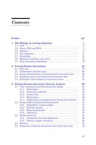
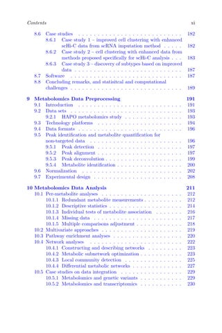

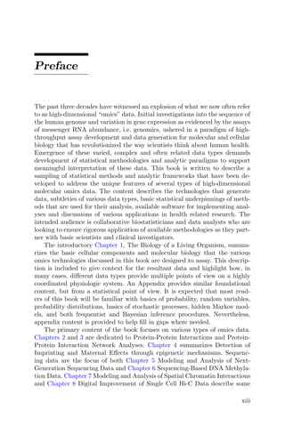
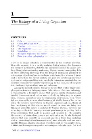
![26 Bioinformatics Methods: From Omics to Next Generation Sequencing
Repository for Interaction Datasets (BioGRID) (build 4.4.201) [322] and were
originally published by Gavin et al. (2002) [127]. A total of 1352 proteins and
3399 interactions are represented in Gavin2002. Data were originally reported
by Gavin et al. (2002) as a series of pairwise ‘interactions’ with designations of
bait or prey for each protein involved in the observed interaction. Several other
widely used TAP (or CoIP or AP-MS) data sets for Saccharomyces cerevisiae
are also publicly available [127,128,174,238,239].
The second data set, referred to here as “Ito2000,” is one of the earliest
published, comprehensive data sets that used yeast-two-hybrid (Y2H) tech-
nology to identify direct physical interactions among protein pairs in Saccha-
romyces cerevisiae. A total of 795 proteins and 839 interactions are represented
in Ito2000. Y2H screening is also known more simply as two-hybrid screening.
The Ito2001 data were also downloaded from BioGRID (build 4.4.201) [322]
and were originally published by Ito et al. (2001) [196].
While these two TAP and Y2H data sets in Saccharomyces cerevisiae
will be used for illustrative purposes, an ever-growing collection of interac-
tion data of various types in a variety of organisms are available in sev-
eral databases, including STRING [462], BioGRID [47, 412], IntACT [171],
MINT [46], APID [6] and HuRI [276]. There are also efforts to unite data
among these resources [102, 321]. Standardized schema for reporting experi-
mental interaction data are under development, for example the PSI-MI XML
and PSI-MI TAB molecular interaction data exchange formats supported by
the HUPO Proteomics Standards Initiative [170]. Table 2.1 lists several highly
referenced databases that contain protein interaction data for a wide variety
of organisms, along with their websites and references.
2.2 Technologies and data types
TAP or CoIP technology assays co-membership in protein complexes on be-
half of protein pairs. If two pairs are members of at least one protein complex
in a cell, TAP will detect an “interaction” for the two proteins. Direct physi-
cal binding is not requisite for a detected TAP interaction. A pair of proteins
that are directly bound to a third or additional proteins in a large multipro-
tein complex would still be identified as interacting pairs according to TAP
technology. For example, in the simple illustration in Figure 2.1, proteins A
and C are a part of the same protein complex. They do not bind directly to
each other, but are physically bound to other proteins in the same complex.
TAP technology would identify an interaction between proteins A and C due
to their membership in the same protein complex. There are several helpful re-
views describing specifics of TAP and CoIP technology in addition to original
publications of highly utilized data sets [127,128,174,238,239,247,349].
In contrast to TAP technology, Y2H technology assays direct physical bind-
ing among protein pairs. In Figure 2.1, proteins A and C would not be iden-
tified as interacting pairs. Even though they are part of the same protein](https://image.slidesharecdn.com/22039578-250603023538-f3f713eb/85/Bioinformatics-Methods-From-Omics-To-Next-Generation-Sequencing-Sujay-Datta-45-320.jpg)
![Protein-Protein Interactions 27
TABLE 2.1
Molecular interaction databases
Database Reference URL
APID Alonso-López et al. http://apid.dep.usal.es/
2019 [6]
BioGRID Chatr-Aryamontri et al. https://thebiogrid.org/
2017 [47]
DIP Salwinski et al. http://dip.doe-mbi.ucla.edu/dip/
2004 [372]
HPRD Prasad et al. http://www.hprd.org/
2009 [348]
HuRI Luck et al. http://www.interactime-atlas.org/
2020 [276]
IntACT Hermjakob et al. http://www.ebi.ac.uk/intact/
2004 [171]
MINT Chatraryamontri et al. http://mint.bio.uniroma2.it/
2007 [46]
PDB Rose et al., http://www.rcsb.org
2017 [368]
Reactome Fabregat et al. http://www.reactome.org/
2016 [101]
STRING Szklarczyk et al. 2015 http://string-db.org/
complex, they do not directly physically bind to each other. Y2H interactions
would be detected for proteins A and B, and for proteins B and C. Several
helpful reviews and publications of original Y2H data sets explain subtleties
of Y2H technology [38,113,163,196,197,438,446].
TAP and Y2H are both considered “bait-prey” technologies. In these set-
tings, for a designated “bait” protein, all proteins that interact with the bait
are identified as its “prey.” In concept, all cellular proteins are eligible for
A
B C
D
E
Example protein complexes
E
F
A -> B
A -> C
A -> D
A -> E
A -> B
A -> C
A -> D
A -> E
TAP Bait-Prey Interactions
A -> B
A -> C
A -> D
A -> E
C-> E
D -> C
E -> C
E -> F
F-> E
Y2H Bait-Prey Interactions
A -> B
B -> A
B -> C
C -> B
C-> D
B -> A
B -> C
B -> D
B -> E
C -> A
C -> B
C -> D
C -> E
D -> A
D -> B
D -> C
D -> E
E -> A
E -> B
E -> C
E -> D
E -> F
F -> E
FIGURE 2.1
Two simple, hypothetical protein complexes comprised of proteins labeled A
through F. Protein complex co-memberships are assayed by TAP technology
and direct physical interactions are assayed by Y2H technology. The lists of
directed bait-prey relationships that would be detectable by perfectly sensitive
and specific TAP and Y2H technologies are listed.](https://image.slidesharecdn.com/22039578-250603023538-f3f713eb/85/Bioinformatics-Methods-From-Omics-To-Next-Generation-Sequencing-Sujay-Datta-46-320.jpg)
![28 Bioinformatics Methods: From Omics to Next Generation Sequencing
● ●
●
●
●
●
●
●
●
●
●
●
●
●
●
●
●
●
●
●
●
●
●
●
●
●
●●
●
● ●
●
●
●
●
●
●
●
●
●
●
●
●
●
●
●
●
● ●
●
●
●
●
●
●
●
●
●
●
●
●
●
● ●
●
●
●
●
●
●
●
●
●
●
●
● ●
●
●
● ●
●
●
●
●
●
●
●
●
●
● ●
●
●
●
●●
●
●
● ●
●
●
●
●
●
●
●
●
●
●
●
●
●
●
●
●
●
●
●
●
●
●
● ●
●
●
●
●
●
●
●
●
●
●
●
●
●
●
●
●
● ●
●
●
●
●
●
●
●
●
●
●
●
●
●
●
●
●
●
●
●
●
●
●
●
●
●
●
● ●
● ●
●
●
●
●
●
●
●
●
●
●
●
●
●
●
●
●
●
●
●
●
●
●
●
●
●
●
●
●
●
●
●
●
●
● ●
●
●
●
●
●
●
●
●
●
●
●
●
●
●
●
●
●
●
●
●
●
●
●
●
●
●
●
●
●
●
●
●
●
●
●
●
●
●
●
●
●
●
●
●
●
●
●
●
●
●
●
●
●
●
●
● ●
●
●
●
●
●
●
●
●
●
● ●
●
●
●
●
●
●
●
●
●
●
●
●
●
●
●
●
●
●
●
●
●
●
●
● ●
●
●
●
●
●
●
●
Gavin2002 Ito2001
FIGURE 2.2
Plots of graphs for Gavin2002 TAP data and Ito2001 Y2H data for the set
of proteins in common for both experiments. Nodes without adjacent edges
have been removed. Bait proteins are blue and proteins that are only found
as prey and never used as baits are yellow. Gray directed edges extend from
baits to prey.
detection as interacting prey for a given bait, assuming they are present in
the cell under the given experimental conditions and are amenable to the tech-
nology. A set of baits is typically pre-specified at the outset of the experiment,
with the set ranging in size for a focused series of investigations [239] to all
viable proteins to the extent possible for comprehensive characterization of a
given system [128,238].
Figure 2.2 depicts Gavin2002 TAP and Ito2001 Y2H networks for the set
of proteins that were reported in both experiments. Nodes without adjacent
edges in this set of common nodes were removed before plotting. Bait pro-
teins are colored blue and proteins that were detected as prey but never used
as bait are colored yellow. Visual inspection suggests roughly similar topolo-
gies for the two networks, but with increased sparsity for the Ito2001 data.
This is to be expected since Y2H technology assays a direct subset of the edges
assayed by TAP technology. Data analytic implications according to bait-prey
designations will be discussed in later sections.
Protein complex co-memberships (such as those detected by TAP) and
direct physical binding interactions (such as those detected by Y2H) are fre-
quently used protein interaction data types. It should be noted, however, that
several other types of “interaction” data do exist, involving proteins as well
as other molecules. For example, synthetic lethal interactions are functional](https://image.slidesharecdn.com/22039578-250603023538-f3f713eb/85/Bioinformatics-Methods-From-Omics-To-Next-Generation-Sequencing-Sujay-Datta-47-320.jpg)
![Protein-Protein Interactions 29
interactions that are defined by functional disruption when both genes are
perturbed in an experiment but a normal functional phenotype when either
of the genes are mutated individually [214]. As another example, protein-DNA
interactions have also been reported, identifying proteins that bind directly
to a DNA sequence that may affect downstream gene expression [81]. Most
recently, these types of protein-DNA interactions are identifying using ChIP-
seq technology [208]. Compound-protein interaction data have also been cata-
logued in recent drug development efforts [265,273]. Understanding the nature
of the interactions being assayed and any subtleties among the measured re-
lationships is paramount to informative analysis. Analysis teams should work
closely with multidisciplinary collaborators to understand the subtleties of
interaction types and relevant interpretations of the data.
2.3 Graph representations of protein-protein interaction
data
Protein-protein interaction data are often modeled in node-and-edge graphs
in which nodes represent proteins and edges represent interactions. Two of the
earliest versions of this type of graph modeling for protein-protein interactions
were the “spoke” and “matrix” models for bait-prey data [17]. These models
designate protein interactions using undirected edges among protein pairs. The
spoke model is perhaps the most intuitive; all pairwise interactions that are
directly observed among baits and prey in an experiment are represented by
edges between these baits and prey. The matrix model extends spoke modeling
and attempts to capture edges that are not directly observed. In the matrix
model, all spoke model edges are included, and edges are also included among
all pairs of prey that were detected as interactors for at least one common bait.
The spoke model can be viewed as a direct representation of experimental
data, and the matrix model as an effort to impute additional edges among
pairs of prey that were not directly tested for interaction. Spoke and matrix
models are depicted in Figure 2.3 for a subset of the Gavin2002 TAP data.
When undirected edges are used for spoke and matrix modeling, reci-
procity in detecting interactions or lack thereof is not evident. For example, in
Figure 2.3, the protein Apl6 detects protein Apm3 as prey when used as a bait,
but when Apm3 is used as a bait, it does not detect Apl6 as prey. Strictly
speaking, bait-prey data are most accurately represented by directed graphs
in which directed edges extend from baits to their interacting prey. Includ-
ing direction summarizes the nature of the study design since edges will only
originate at proteins that are pre-specified as baits. A simple summary of
bait-prey data can be to list all “viable baits” and “viable prey.” Viable baits
can be defined as proteins for which an interaction with at least one prey
was observed in the experiment. Viable prey can be defined as proteins that](https://image.slidesharecdn.com/22039578-250603023538-f3f713eb/85/Bioinformatics-Methods-From-Omics-To-Next-Generation-Sequencing-Sujay-Datta-48-320.jpg)
![30 Bioinformatics Methods: From Omics to Next Generation Sequencing
Three TAP bait-prey sets
from Gavin2002
Bait: Detected Prey
Apl5: Apl6, Apm3, Aps1, Ckb1
Apl6: Apl5, Apm3, Eno2
Apm3: Apl6
Apm3
Apl6
Apl5
Aps1 Ckb1
Eno2
Apm3
Apl6
Apl5
Aps1 Ckb1
Eno2
Apm3
Apl6
Apl5
Aps1 Ckb1
Eno2
Spoke Model Matrix Model
FIGURE 2.3
Spoke and matrix models for three sets of TAP bait-prey edges for the
Gavin2002 data set. Spoke modeling includes all detected edges while ma-
trix modeling additionally imputes edges among all pairs of prey that share
at least one common bait.
were identified by at least one bait in an interaction. Summarizing viable baits
and prey can help describe experimental design and intent, and in some cases
identify the extent to which certain proteins were amenable to the specific
assay [53,54]. Recording bait-prey data in a directed graph is also crucial for
for understanding sampling subtleties in protein interaction data, identifying
systematic biases potentially due to technological factors and for estimating
measurement error. These concepts will be developed in detail in the next
sections.
Other types of node-and-edge graphs are often utilized for protein inter-
action data modeling as well. For example, bipartite graphs have been crucial
constructs in statistical algorithms that estimate protein complex member-
ship given observed TAP data. Bipartite graphs consist of two node classes
and edges among nodes represent affiliation of a node of one type with a node
of the other type. An example of a bipartite graph depicting membership of
proteins in the simple example protein complexes from Figure 2.1 is depicted
in Figure 2.4. A bipartite graph is also often represented as an affiliation ma-
trix in which rows correspond to one node type and columns to the other.
In Figure 2.4, for affiliation matrix A, the rows correspond to n proteins and
columns to m protein complexes. The co-affiliation matrix Y = [AAT
] − I
where [x] = 1 if x > 0 identifies protein pairs that are co-members in at least
one protein complex. Entries of 1 in Y represent complex co-membership. In
fact, Y represents the data that TAP and CoIP technologies are designed to
assay. Statistical approaches for estimating protein complexes recorded in A
given observations on Y will be described in the next chapter.
To accommodate the increasing use of node-and-edge graphs for
representing protein-protein interaction data, R statistical software](https://image.slidesharecdn.com/22039578-250603023538-f3f713eb/85/Bioinformatics-Methods-From-Omics-To-Next-Generation-Sequencing-Sujay-Datta-49-320.jpg)
![Protein-Protein Interactions 31
A
B
C
D
E
F
Complex 2
A
B
C
D
E
F
C
o
m
p
l
e
x
1
C
o
m
p
l
e
x
2
1
1
1
1
1
0
0
0
0
0
1
1
A
B
C
D
E
F
0
1
1
1
1
0
1
0
1
1
1
0
A B C D E F
1
1
0
1
1
0
1
1
1
0
1
0
1
1
1
1
0
1
0
0
0
0
1
0
A
A = Y =
Complex 1
FIGURE 2.4
Bipartite graph and related affiliation (A) and co-affiliation (Y = [AAT
]−I)
matrices for two simple example protein complexes.
(https:// cran.r-project.org/) packages including igraph [62] provide efficient
and informative data structures for storing and representing protein-protein
interaction data. As discussed in the next chapter, igraph also contains exten-
sive functionality for plotting graphs, and for characterizing both local and
global graph features.
2.4 Sampling issues in protein-protein interaction data
As explained previously, directed graph modeling of bait-prey data not only
records observed protein-protein interactions, but also summarizes the nature
of identified interactions from baits to their prey. Understanding the bait/prey
status of a protein is crucial for understanding which edges in a graph have
been tested and which have not. In concept, if all proteins in a cell are equally
amenable to the bait-prey technology, then given the full set of proteins, all
possible edges extending from baits to prey are tested in bait-prey experi-
ments. Edges in a graph model of bait-prey data are included only if the
interactions they represent are directly tested and observed according to the
technology being used. Importantly, edges that are not included in a graph
either represent potential interactions that were not observed when directly
assayed, or potential interactions that were never directly tested using the
bait-prey technology.
For any pair of proteins that are both used as baits in an experiment, the
possible edge between them is tested twice, one time for each bait. If technolo-
gies were perfectly sensitive and specific, identical results should be obtained](https://image.slidesharecdn.com/22039578-250603023538-f3f713eb/85/Bioinformatics-Methods-From-Omics-To-Next-Generation-Sequencing-Sujay-Datta-50-320.jpg)
![32 Bioinformatics Methods: From Omics to Next Generation Sequencing
Three TAP bait-prey
sets from Gavin2002
Bait: Detected Prey
Apl5: Apl6, Apm3, Aps1, Ckb1
Apl6: Apl5, Apm3, Eno2
Apm3: Apl6
Apm3
Apl6
Apl5
Aps1 Ckb1
Eno2
Apm3
Apl6
Apl5
Aps1 Ckb1
Eno2 tested 2x, observed 2x
tested 2x, observed 1x
tested 1x, observed 1x
tested 1x, not observed
untested
FIGURE 2.5
Directed graph model of TAP data from Gavin et al. (2002) [127]. Baits are
represented by blue nodes. Proteins not used as baits but detected as prey
are represented by yellow nodes. Edges among pairs of baits that are tested
twice are black with reciprocal arrows if observed twice and single arrows if
detected once. Edges among baits and prey-only nodes are gray and directed
from baits to prey if detected, and otherwise not included in the graph. All
edges among prey-only nodes are not tested by the TAP assay and therefore
edges are not included in the graph.
with both baits either detecting or failing to detect the pairwise interaction. In
reality, observed data for these doubly tested interactions are not always con-
sistent. An interaction may be observed when one protein is used as bait, but
not the other. This lack of reciprocity is generally attributed to measurement
error, either stochastic or systematic in nature [54]. For a pair of proteins in
which only one protein is used as bait but the other is not, the edge is only
tested once in the direction from the bait to the prey; reciprocal testing of the
edge from the prey to the bait is not performed. Importantly, edges among
pairs of proteins that are never used as baits are in fact never directly tested in
bait-prey technologies. While some graph modeling approaches, for example
the matrix model, induce edges among prey proteins that share a common
bait, in fact, there is no direct assay of the edge between two prey-only pro-
teins [382]. Similar to any statistical analysis, it is crucial to understand which
data are directly collected and which are not. Directed graph modeling and
bait-prey designations are key to distinguishing among edges in a graph that
have been tested and observed to exist, tested but no interaction was observed,
and never tested [7,379,383]. Figure 2.5 illustrates doubly tested edges among
pairs of bait proteins, singly tested edges from baits to prey-only proteins and
untested edges among pairs of prey-only proteins for a subset of the Gavin2002
TAP data. Note the simple but critical differences between the spoke model of
these same data in Figure 2.3. The directed edges and bait-prey distinctions
(denoted by color in Figure 2.5) more accurately represent the experimental
design and the frequency with which edges are tested and observed.](https://image.slidesharecdn.com/22039578-250603023538-f3f713eb/85/Bioinformatics-Methods-From-Omics-To-Next-Generation-Sequencing-Sujay-Datta-51-320.jpg)
![Protein-Protein Interactions 33
TABLE 2.2
Counts of VB, VP and VBP for several
TAP and Y2H data sets
Data set VB VP VBP
TAP data sets
Gavin2002 [127] 453 1169 270
Ho2002 [174] 492 1308 237
Krogan2004 [239] 28 39 24
Gavin2006 [128] 914 1361 845
Krogan2006 [238] 1605 2011 942
Y2H data sets
Cagney2001 [38] 19 39 10
Ito2001 [196] 465 506 195
Uetz2000 [446] 484 578 135
Tong2002 [438] 26 134 16
One simple way to describe the sampling scheme underlying bait-prey
experiments is to tabulate the numbers of proteins that serve as baits or
prey or both in a given experiment. According to Chiang et al. (2007) [54],
in bait-prey investigations, “viable baits” (VB) can be defined as proteins
for which at least one prey is detected as an interactor. “Viable prey” (VP)
can be defined as proteins which are detected as interactors by at least one
bait. “Viable bait and prey” (VBP) proteins satisfy both criteria and prove
amenable to assay as both baits and prey by the technology being used.
Table 2.2 reports the numbers of VB, VP and VBP for several TAP and Y2H
data sets in Saccharomyces cerevisiae. From these quantities, the numbers
of doubly tested, singly tested and untested edges among all pairs of proteins
can be directly calculated. These simple descriptive statistics demonstrate the
wide range of scope and comprehensiveness of bait-prey experiments.
Identifying tested and untested edges is similar in spirit to understanding
the sampling space of a graph. Tested edges are sampled for assay and untested
edges are not. The effects of sampling on global graph topology have been sum-
marized. Global summaries often treat untested edges in the exact same way
as edges that are tested but not observed. In many cases observed phenomena
can be explained by failure to identify the sampling structure when calcu-
lating summaries. For example, global topological graph characteristics that
identify graphs as “scale-free” can be replicated simply by partial sampling
of fuller networks with completely different topologies [155]. Other statistics
including overall node degree distributions, clustering coefficients, node be-
tweenness summary statistics and motif frequency can be greatly affected by
failure to account for sampling [71]. Specific statistical summaries describing
both global and local topological feature of graphs will be developed in detail
in the next chapter, but we note here that their typical calculation assumes](https://image.slidesharecdn.com/22039578-250603023538-f3f713eb/85/Bioinformatics-Methods-From-Omics-To-Next-Generation-Sequencing-Sujay-Datta-52-320.jpg)
![34 Bioinformatics Methods: From Omics to Next Generation Sequencing
that all edges in a graph have been tested to support direct observation of
edge existence or lack thereof.
Recognizing incomplete sampling in experimental settings, some attempts
have been made at compiling data from various sources to increase the qual-
ity of protein interaction reporting. Some strategies acknowledge variations in
direct assay of edges and incorporate this in their global rankings of hypoth-
esized interactions [444]. Other applications recognize the potential for bias
due to sampling and adjust for potential bias when identifying network char-
acteristics of proteins pertaining to disease, for example cancer [378]. There
are also several experimental design strategies that explicitly acknowledge the
sampling structure of bait-prey technologies and refine sampling according to
previous observations in a graph to ensure exploration of untested edges as
additional rounds of experimentation are conducted [64,243,383,387].
2.5 Systematic and stochastic measurement errors
Directed graph modeling of bait-prey interactions can also be key for identify-
ing systematic measurement error and for estimating stochastic false positive
and false negative probabilities for the set of observed edges. False positive
interactions are those detected by the technology that in truth do not exist.
False negative interactions are those that are tested but not detected by the
technology, when in fact they do exist. Here we define systematic measure-
ment error as errors that occur when edges are either observed or unobserved
due to differences in sensitivity and specificity for individual proteins for the
technology in use. Proteins maybe be “sticky” and identify more interact-
ing partners than are truly involved in cellular protein interactions. Proteins
may also be less amenable to the assay and either fail in large part to de-
tect interacting partners when used as baits or fail to be detected as prey by
most baits. While the cause for systematic error cannot always be diagnosed,
fairly straightforward statistical approaches can identify systematic bias in
observed data. Here, stochastic measurement errors are defined as false posi-
tive and false negative observations that occur at random and do not reflect
underlying amenability of the proteins or the experimental conditions to the
assay.
A simple test based on a binomial distribution can be used for diagnosing
bait proteins prone to systematic error by examining a subgraph consisting
only of nodes that are viable baits [54, 129]. Edges in this viable bait sub-
graph are all tested twice, once in each direction. If the bait-prey technology
was perfectly sensitive and perfectly specific, then reciprocal interactions or
lack thereof should be observed for all protein pairs. However, because of mea-
surement error, unreciprocated edges often do exist such that an interaction
is observed if one protein is used as bait, but not the other. If measurement](https://image.slidesharecdn.com/22039578-250603023538-f3f713eb/85/Bioinformatics-Methods-From-Omics-To-Next-Generation-Sequencing-Sujay-Datta-53-320.jpg)