
Downloaded 640 times
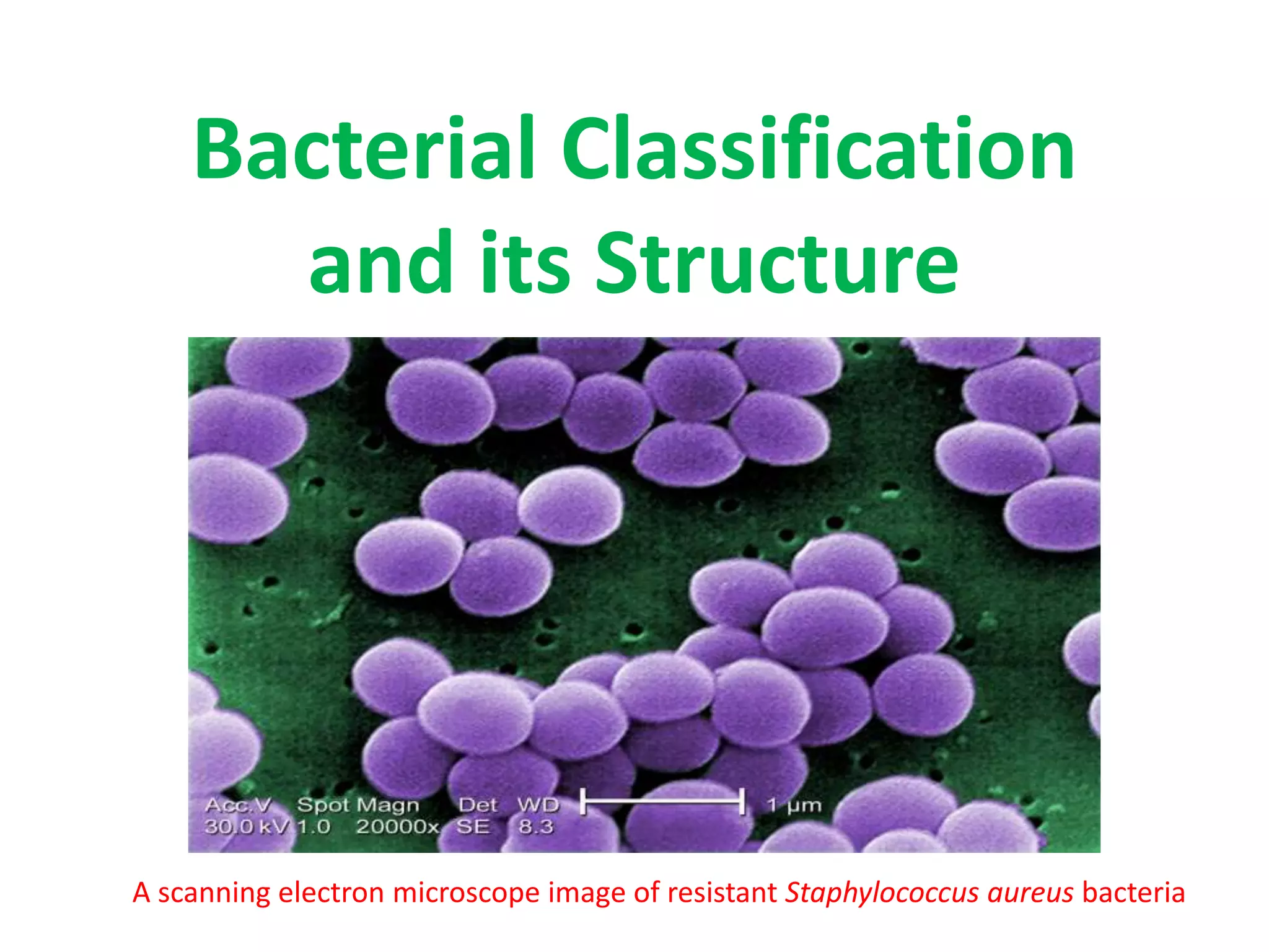
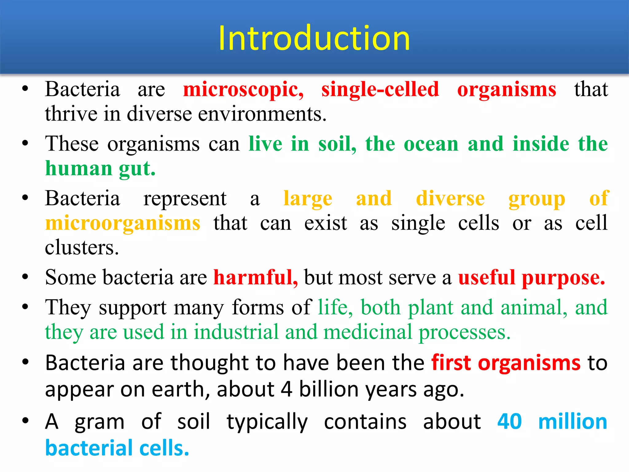
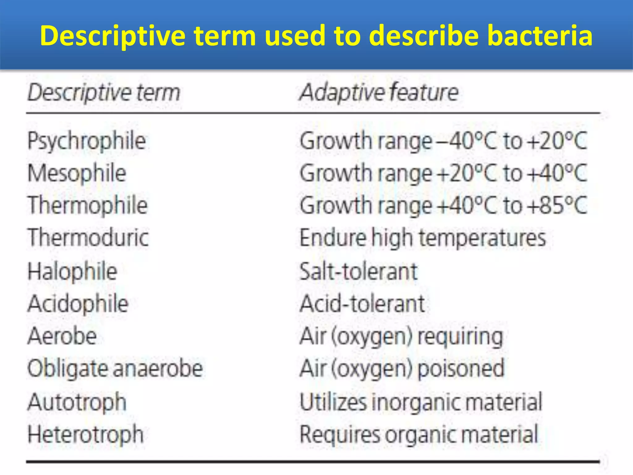
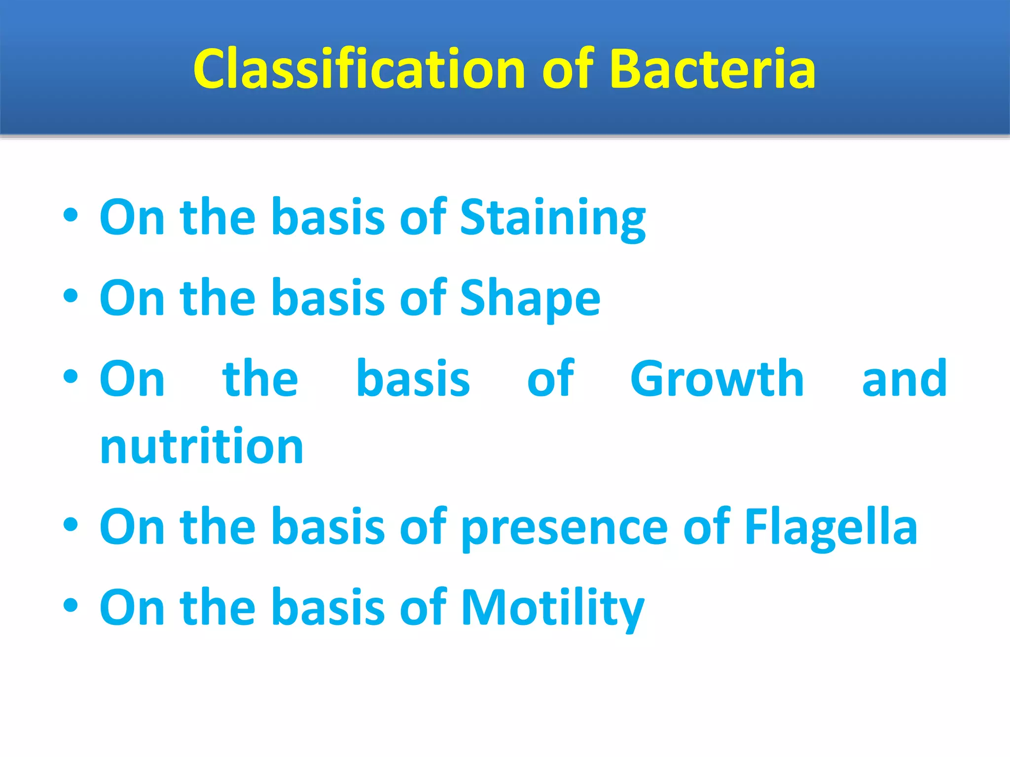
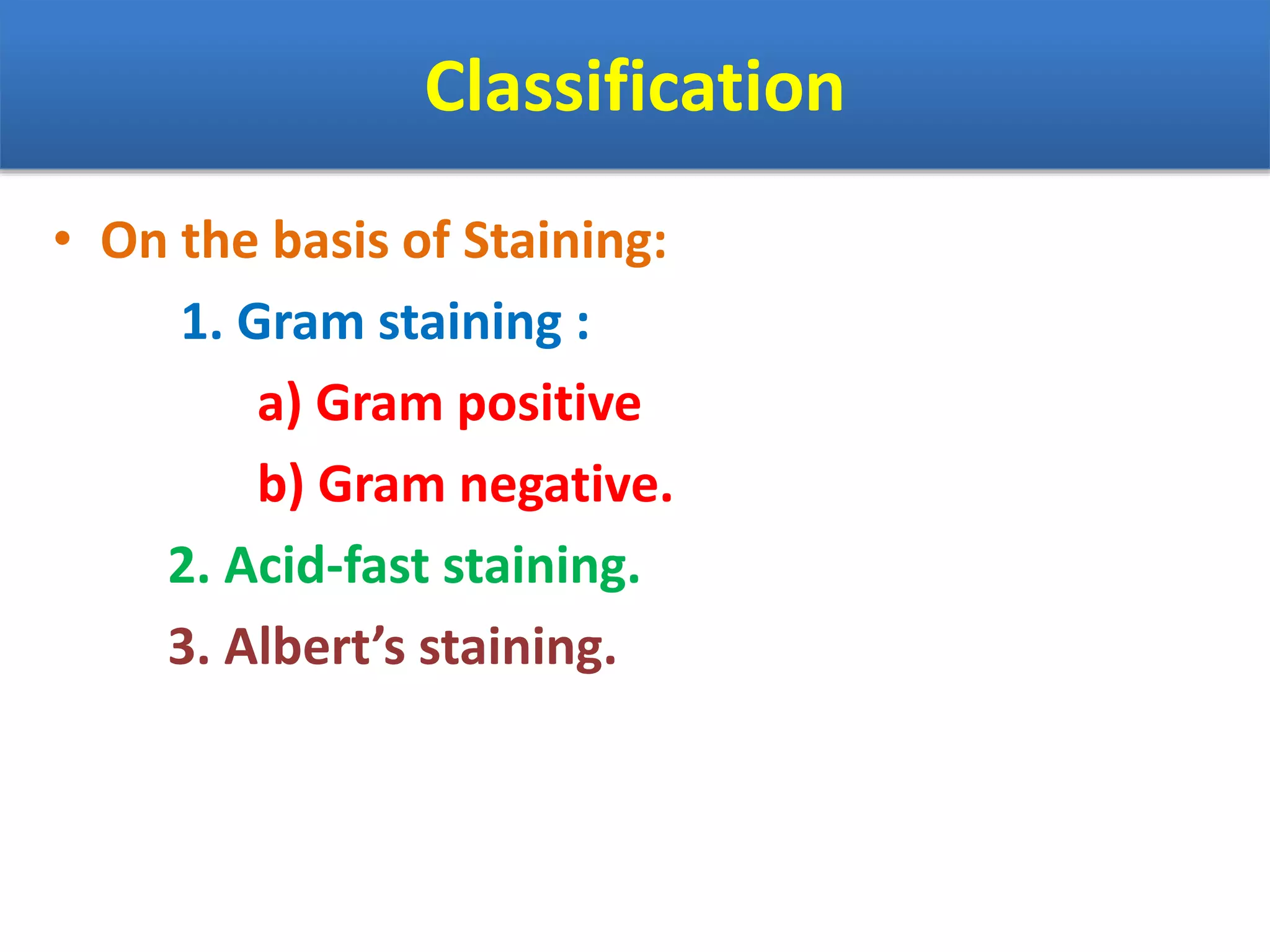

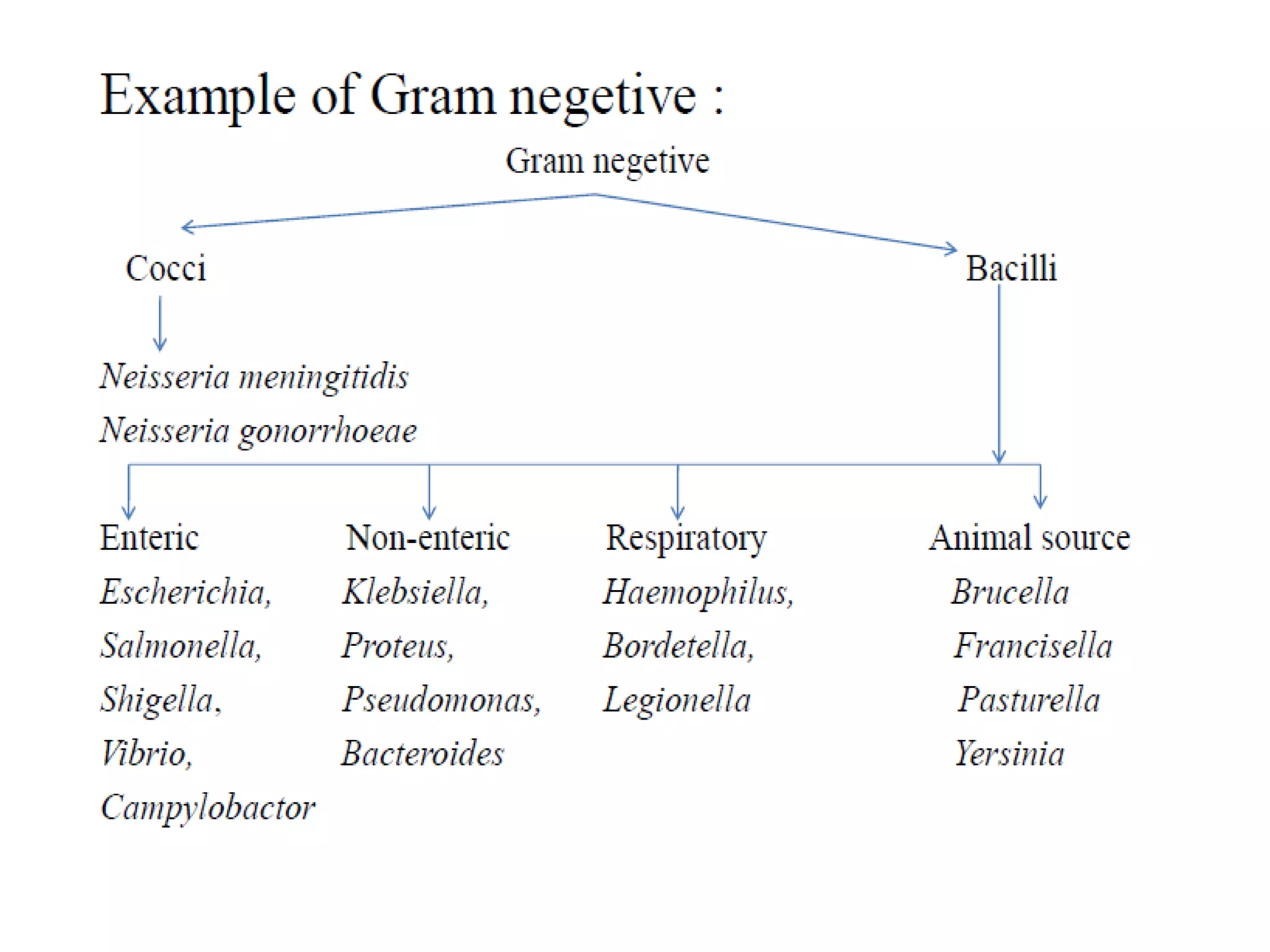
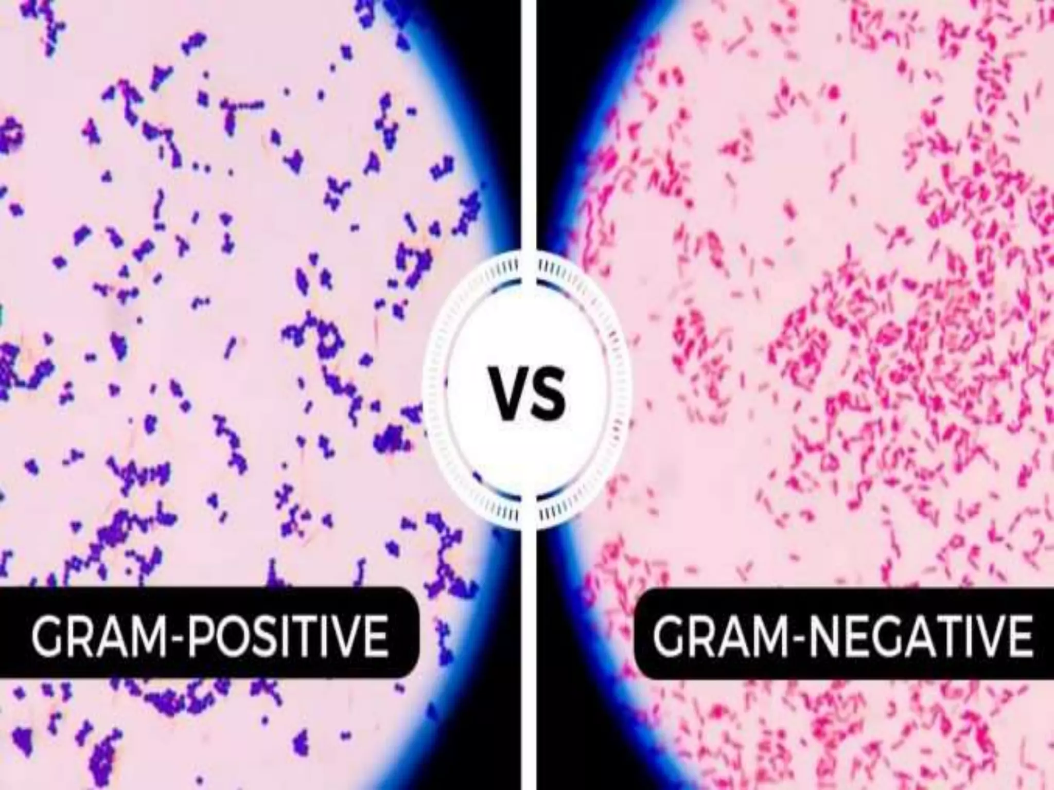
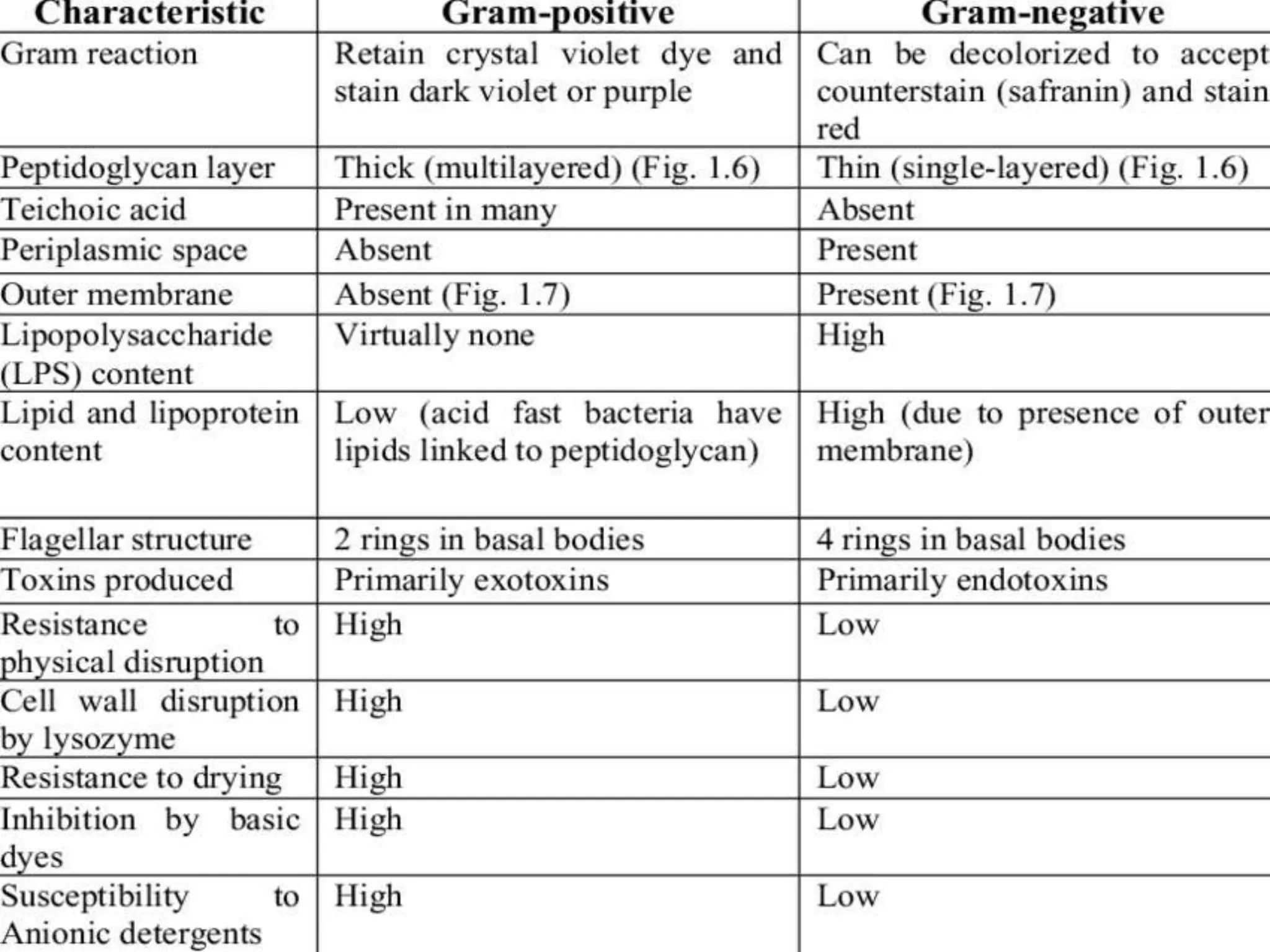


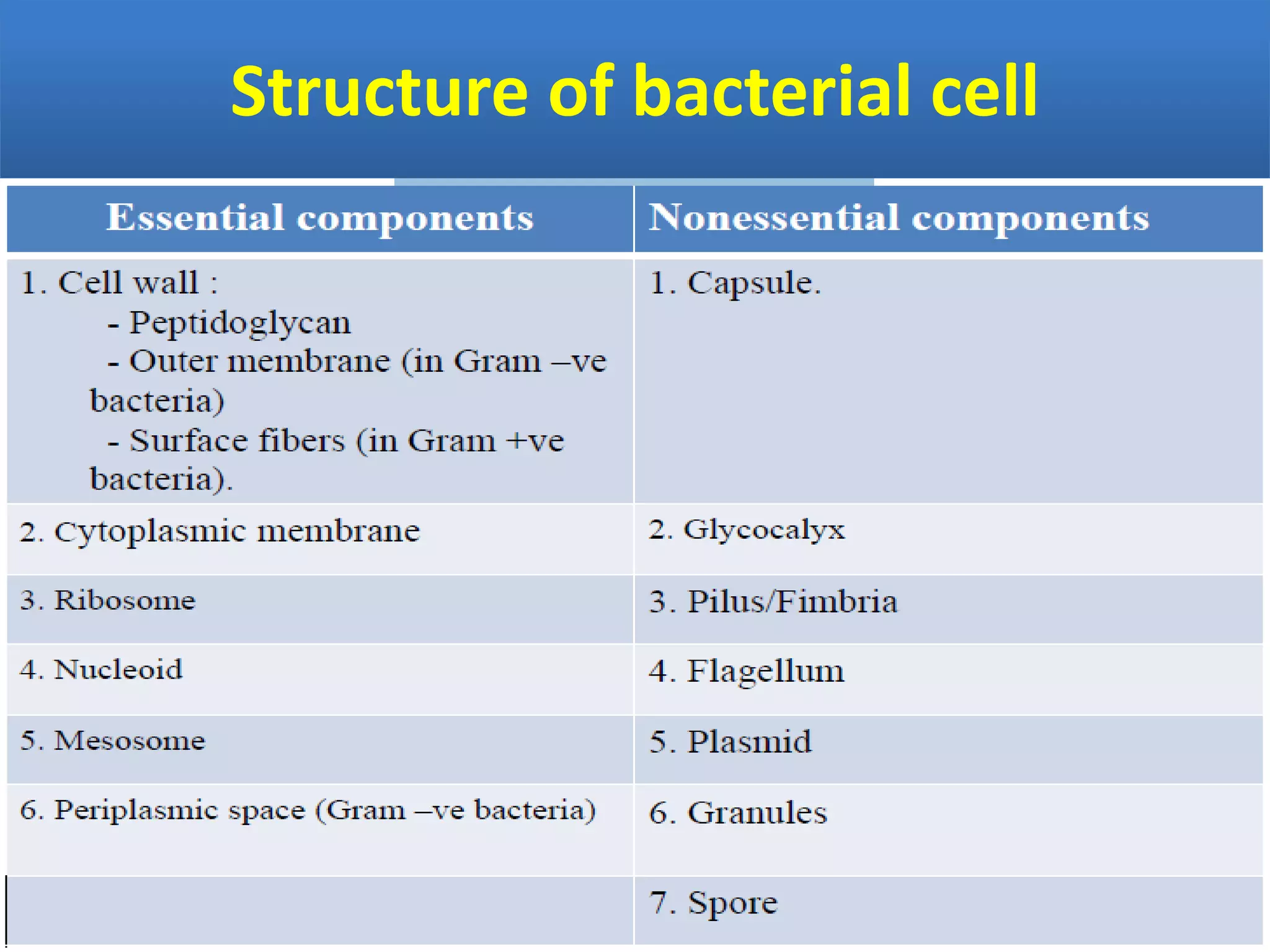
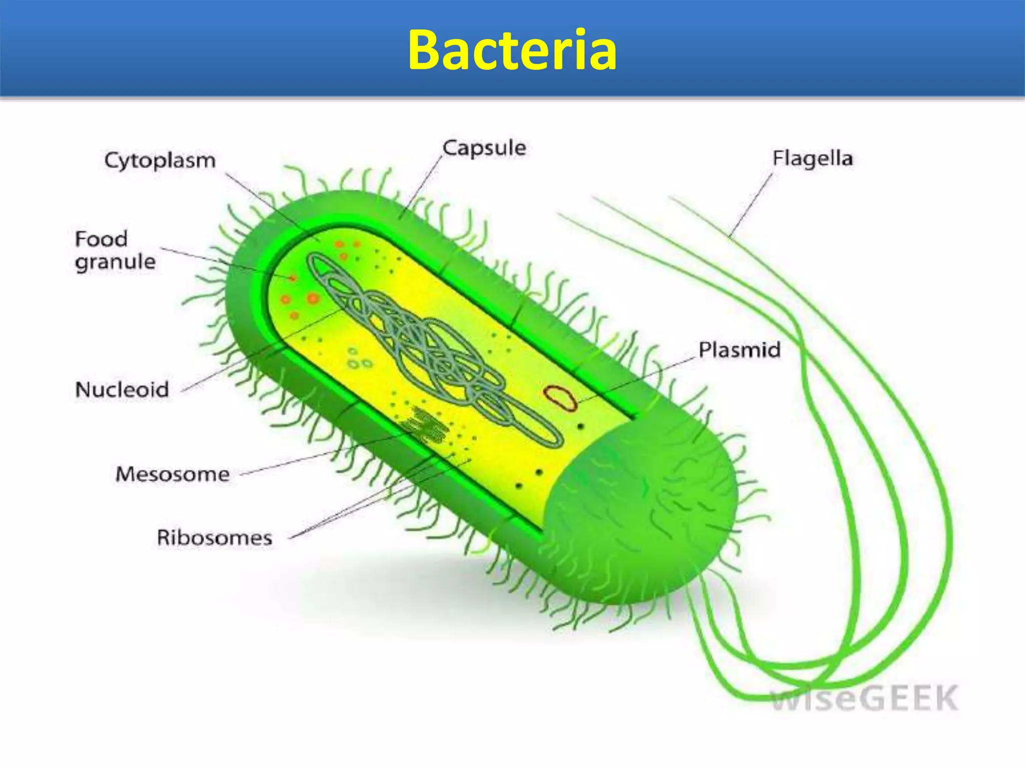
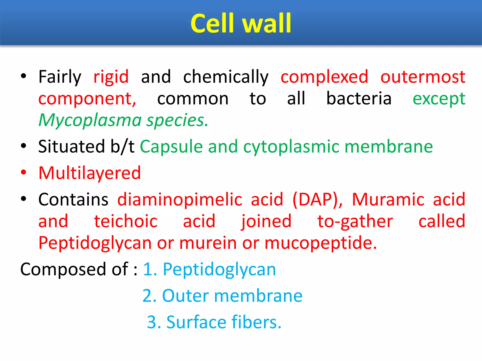
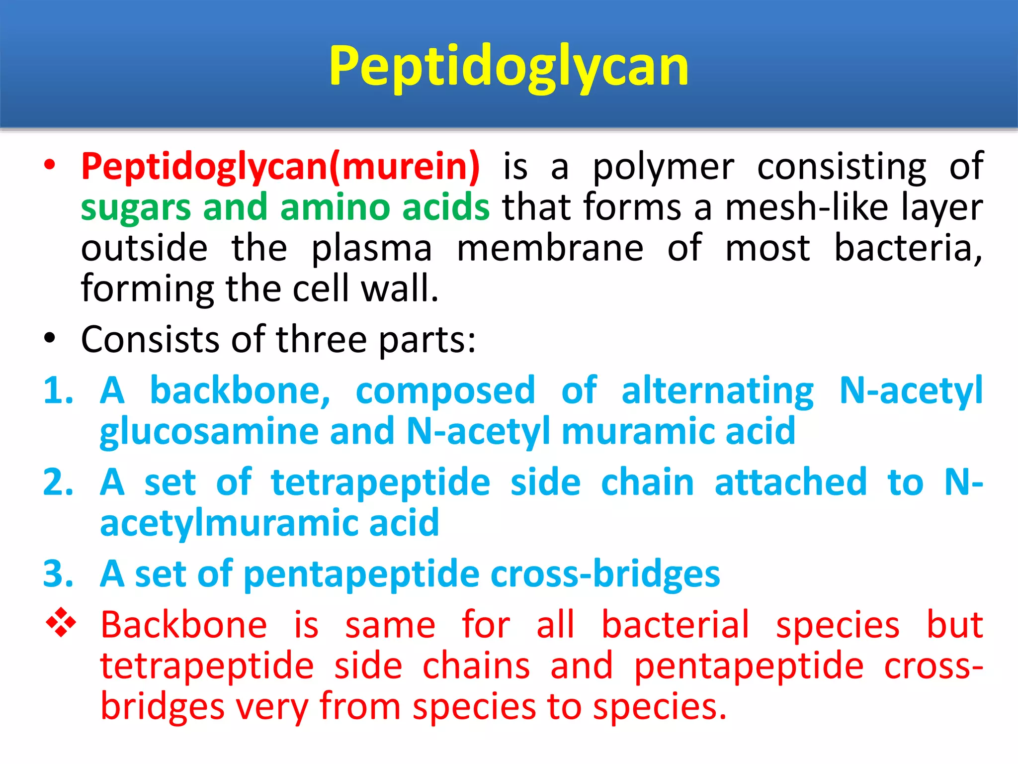
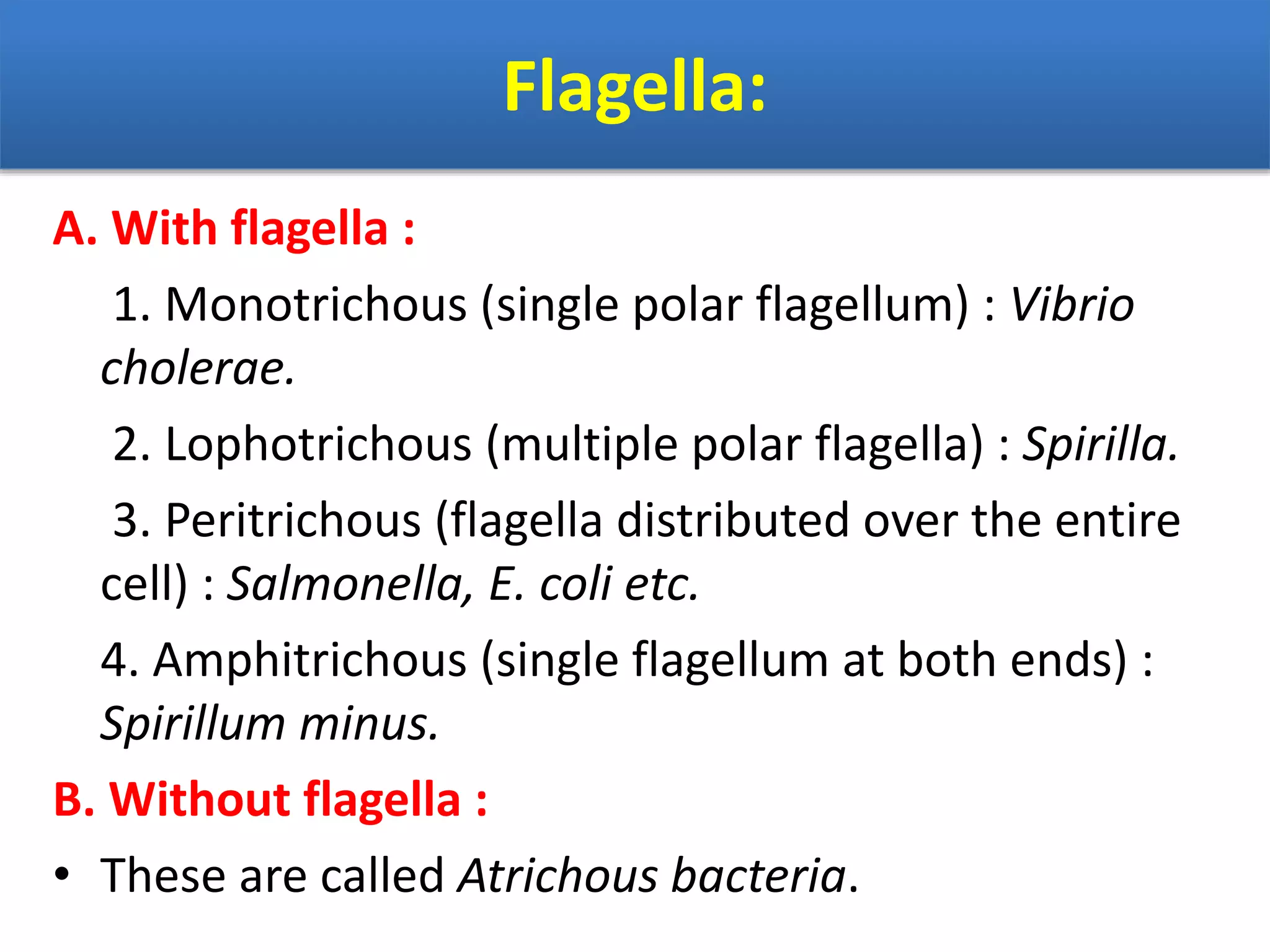

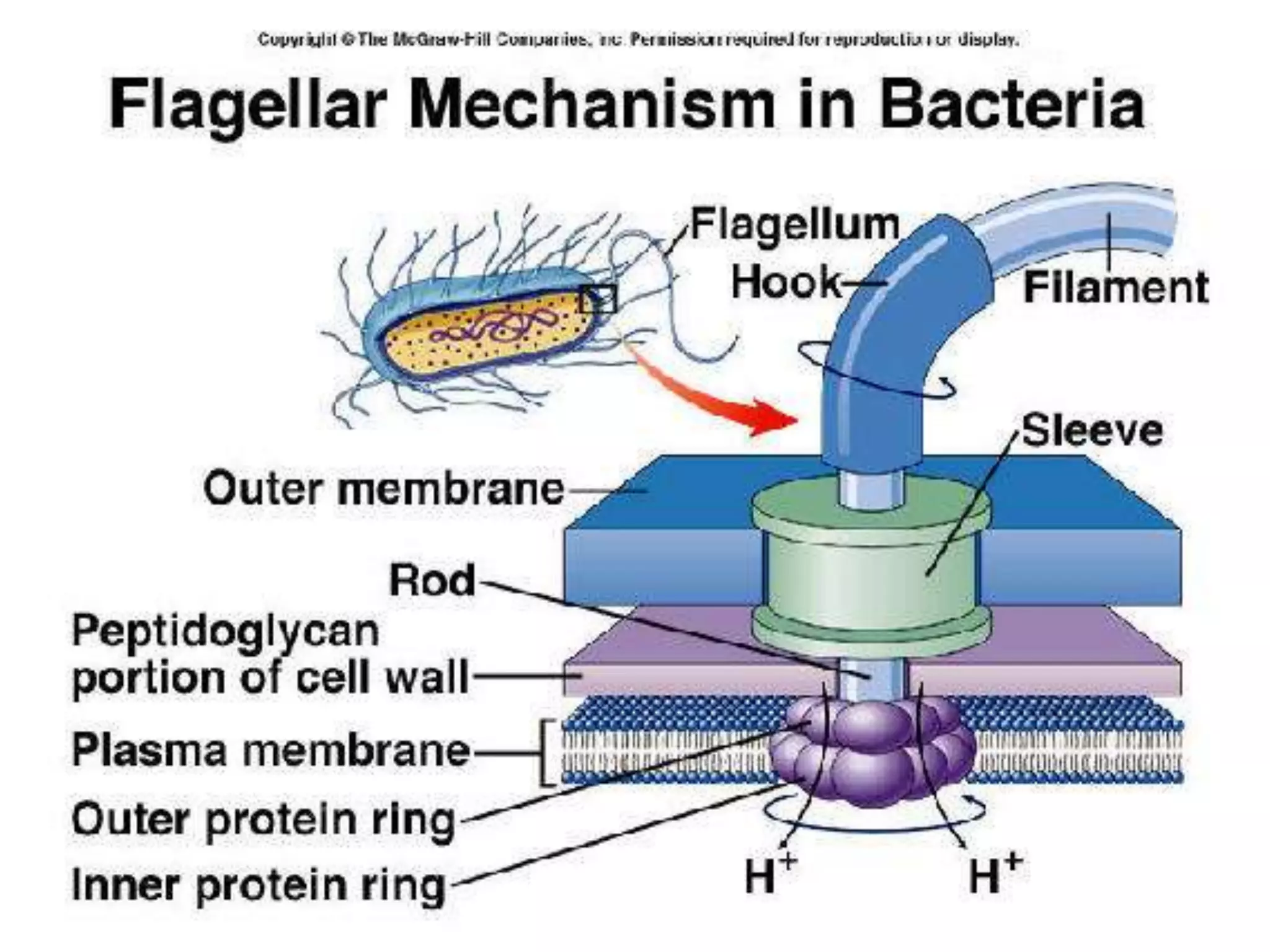
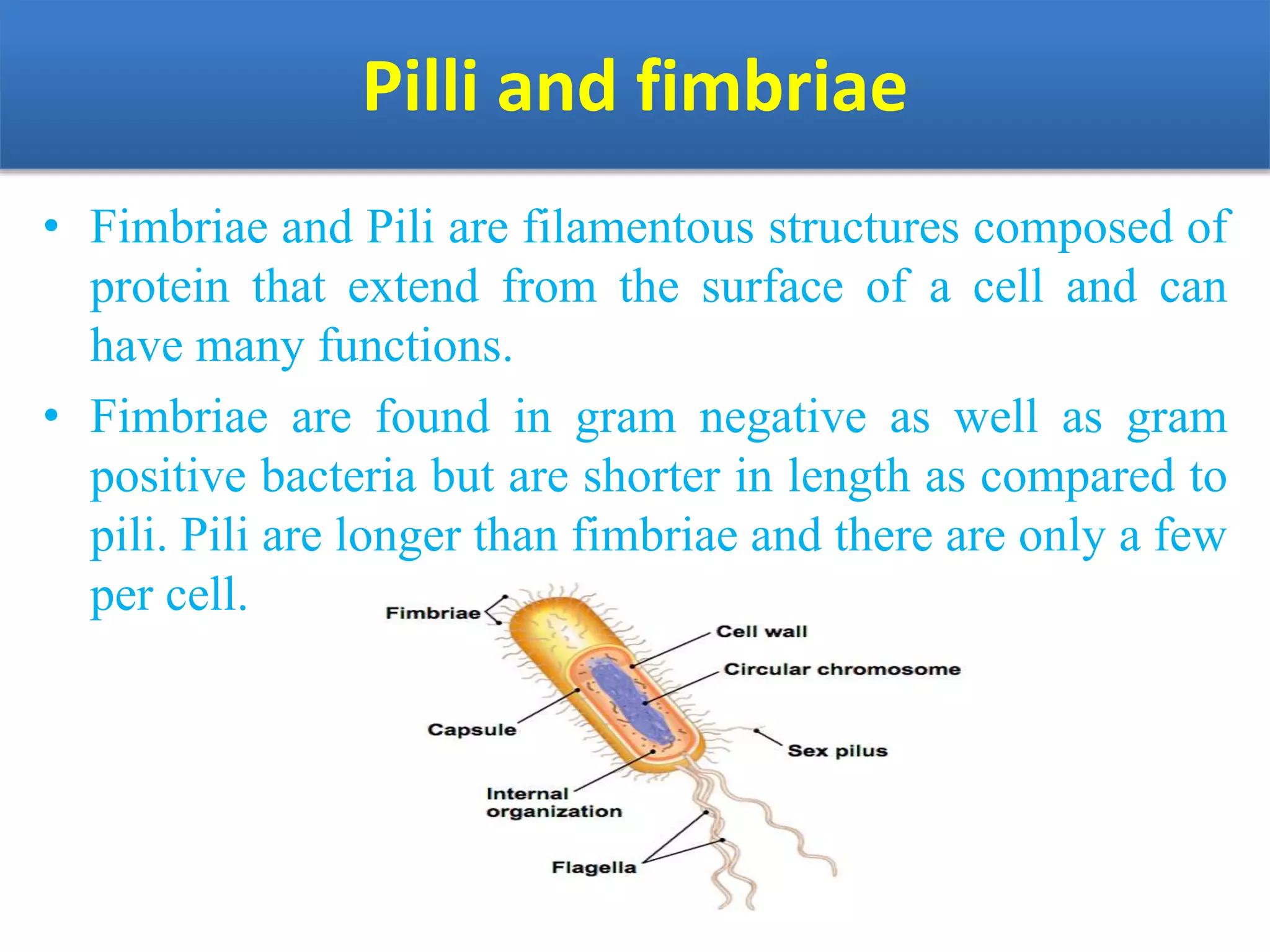
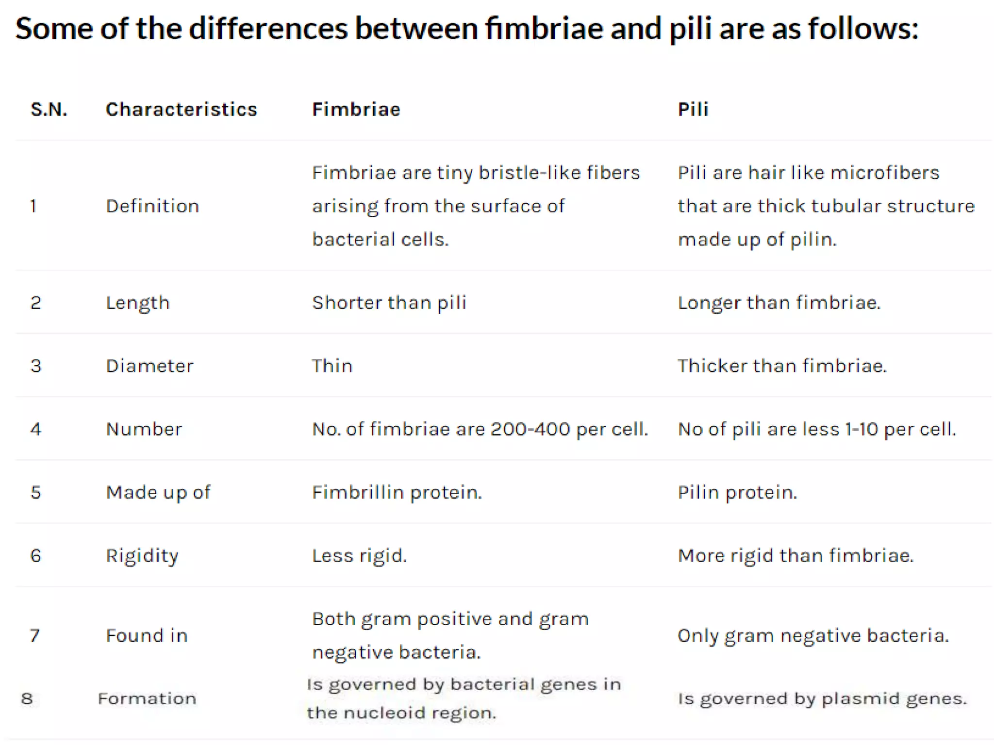
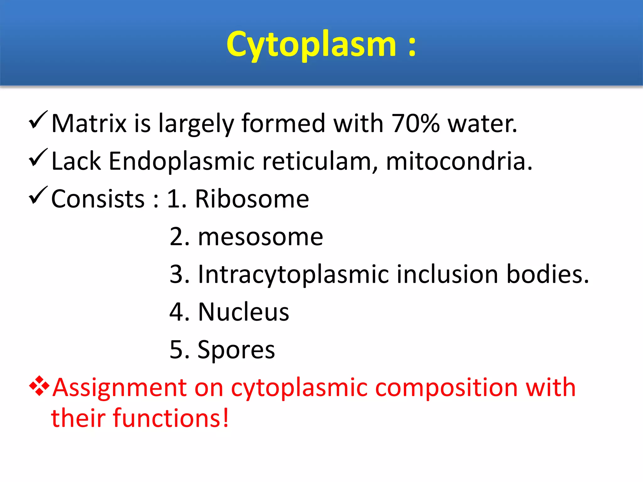


Bacteria are classified in several ways: 1. By staining (Gram positive/negative, acid-fast), shape (cocci, bacilli), motility, environment (aerobic/anaerobic). 2. The bacterial cell has a cell wall, cell membrane, flagella/fimbriae and cytoplasm. The cell wall provides structure and protection through its peptidoglycan layer. 3. Bacteria are further classified based on nutrition sources, temperature, pH and salt tolerance ranges they thrive in. Most bacteria serve important ecological roles while some can cause disease.


![06_Kingdom_Prokaryotae769[1].pptx](https://cdn.slidesharecdn.com/ss_thumbnails/06kingdomprokaryotae7691-240127144445-3a0d06ed-thumbnail.jpg?width=640&height=640&fit=bounds)


