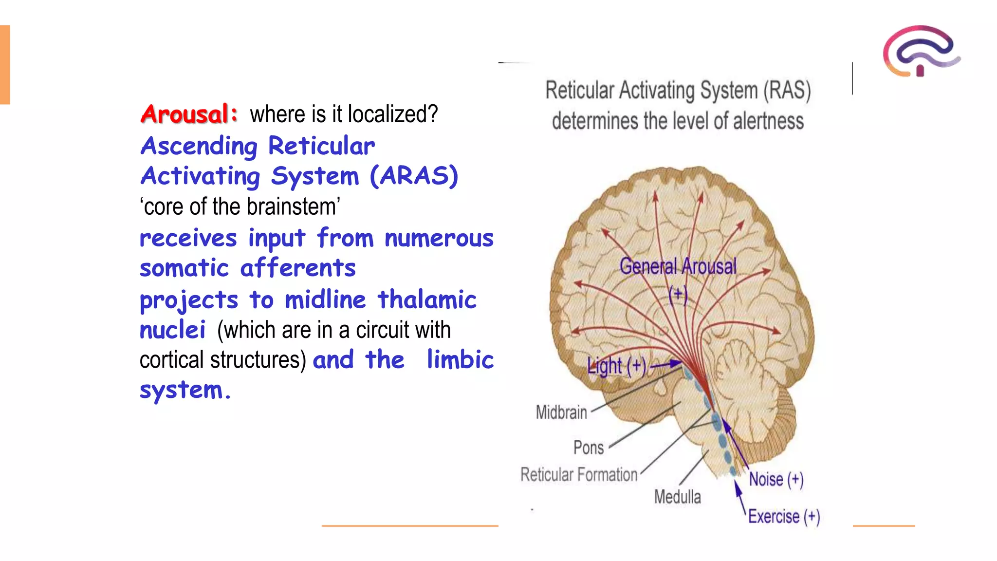
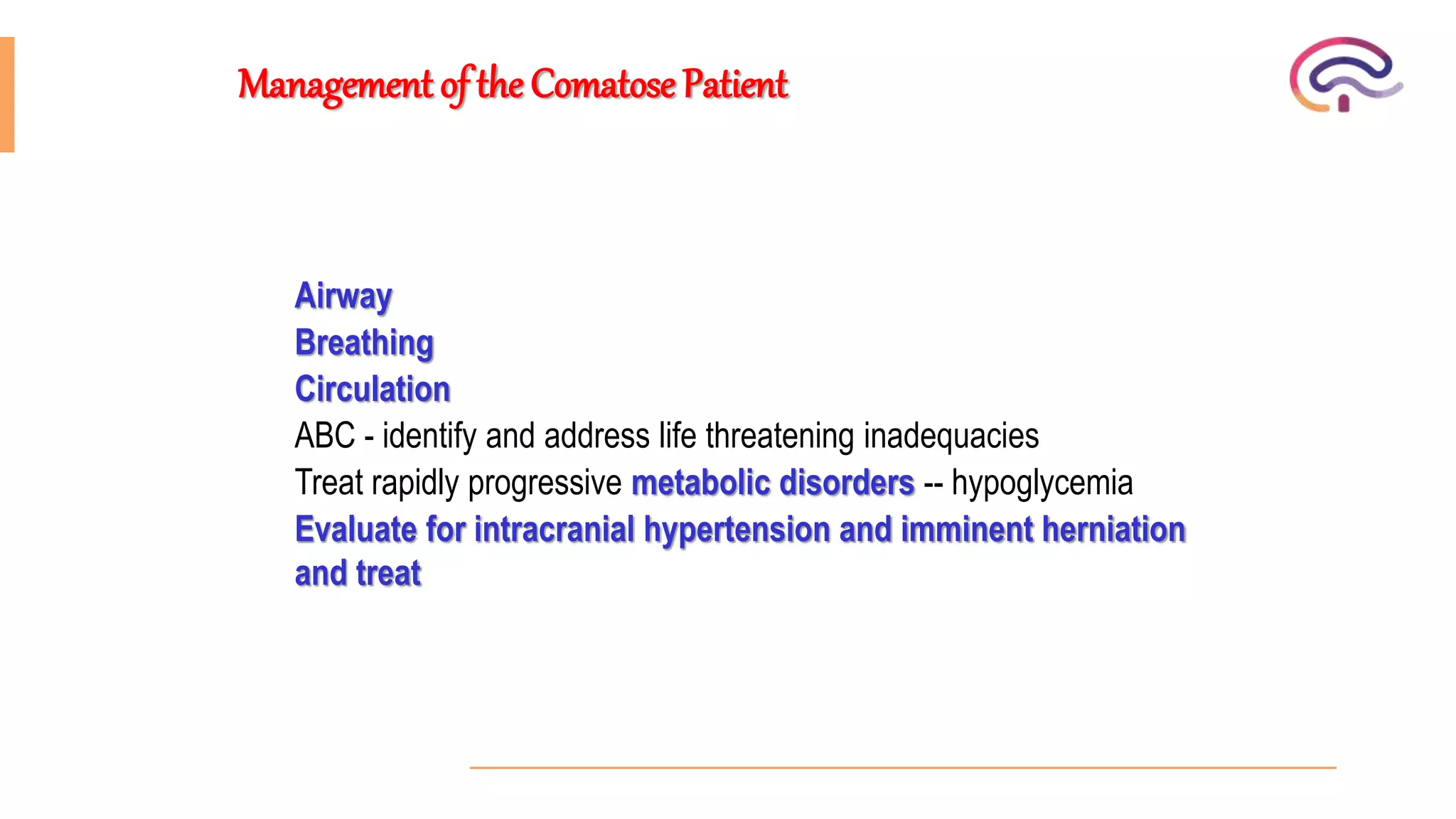
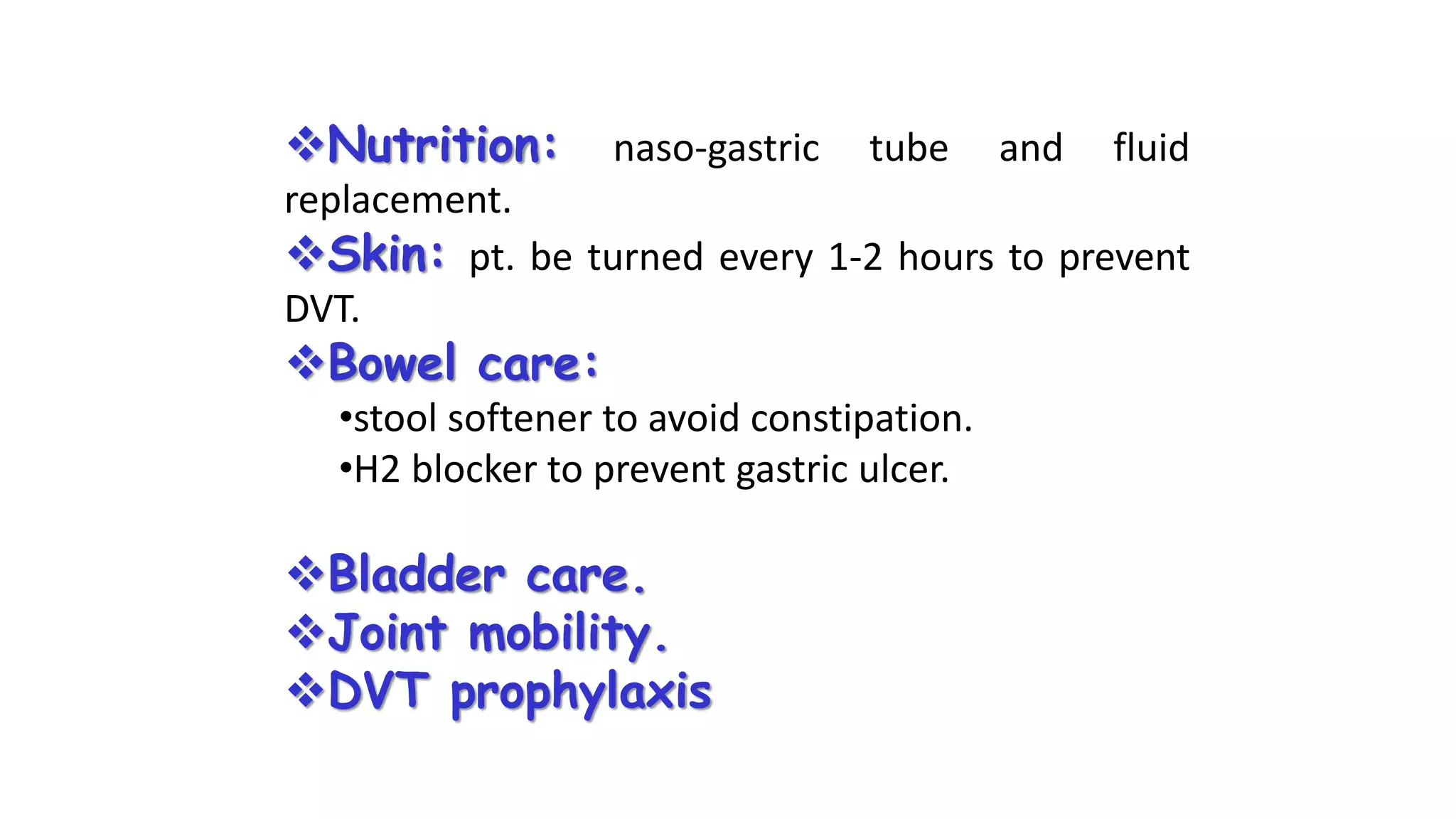
Approach to comatose patient
Mohamed rizk khodair
Lecturer of neurology
Mohamedrizk.med@o6u.edu.eg
October 6 university
Objectives
1. Consciousness.
2. Definition of coma
3. Causes of coma
4. Evaluation of a comatose patient.
5. How to manage a comatose patient.
Consciousness
Two components of conscious behavior
Content of consciousness:
the sum of cognitive and emotion( memory , attention , thinking )
State of consciousness (Arousal ):
Physiological state of being aware or reactive to stimuli.
Content depends on arousal, but normal arousal does not guarantee normal
content
• Awareness of self and environment
The content of consciousness depends upon the activities of the cerebral
cortex, the thalamus and their interrelationship.
Lesions of these structures will diminish the content of consciousness without as a
rule changing the state of consciousness.
Arousal: where is it localized?
Ascending Reticular
Activating System (ARAS)
‘core of the brainstem’
receives input from numerous
somatic afferents
projects to midline thalamic
nuclei (which are in a circuit with
cortical structures) and the limbic
system.
Inattentive persons are usually unable to do more than simplest
commands.
Speech is usually limited to a few words or phrases
Disoriented in time and place.
Confusion
Inability to sustain a wakeful state without the application of external stimuli.
The lids droop without closing completely; there may be snoring, the jaw and limb muscles are slack,
and the limbs are relaxed.
Drowsiness
 Patient can be roused only by vigorous and repeated stimuli.
 Response to spoken commands is either absent or slow and inadequate.
 When left unstimulated, patients quickly drift back into a sleep-like state.
 The eyes move outward and upward, a feature that is shared with sleep
 Tendon and plantar reflexes and breathing pattern may or may not be altered.
Stupor
 Unconscious and Incapable of being aroused by any stimuli however vigorous and painful.
 Eye closed.
 Absent sleep-wake cycle.
Coma
Evaluation
1-History
Recent events
When patient last seen?
How was the patient discovered?
Any preceding neurological complaints?
Any trauma?
Drug exposure?
Medical history.
Psychiatric history.
Medication.
Alcohol.
2-General Physical examination
Vital signs:
Severe HTN (hypertensive encephalopathy or structural lesion with increase ICP).
Skin: signs of trauma & needle mark, rash& jaundice.
Breath: alcohol & acetone ( diabetic ketoacidosis ) & fetor hepaticas ( hepatic coma ).
Head: fracture& hematomas& laceration.
Ear, nose and throat: CSF otorrhea or rhinohea &hemotympanum & tongue biting.
Neck (not manipulate if suspect of cervical spine fracture): stiffness.
3-Neurological examination
The goals of neurological examination are :
1) To localize the process leading to coma.
2) To determine the depth of coma.
1-General
appearance
2-Level of coma
3-pupil
4-Sign of meningeal
irritation
5-Ocular
movement
6-fundus
7-Corneal reflex
8-Gag reflex
9-respiration
10-Motor
response
1-General
appearance
2-Level of coma
3-pupil
4-Sign of meningeal
irritation
5-Ocular
movement
6-fundus
7-Corneal reflex
8-Gag reflex
9-respiration
10-Motor
response
Eye lid:
Closed lower Pons intact.
Blinking Psychogenic .
Puffiness renal &myxedema.
Open eye lids and slack jaw indicate
deep coma.
Head and gaze deviation suggest a
large ipsilateral hemispheric lesion.
Observe for:
myoclonus (metabolic cause).
Seizures.
Tetany.
1-General
appearance
2-Level of coma
3-pupil
4-Sign of meningeal
irritation
5-Ocular
movement
6-fundus
7-Corneal reflex
8-Gag reflex
9-respiration
10-Motor
response
1-General
appearance
2-Level of coma
3-pupil
4-Sign of meningeal
irritation
5-Ocular
movement
6-fundus
7-Corneal reflex
8-Gag reflex
9-respiration
10-Motor
response
Symmetry and normal reactive:
mean integrity of midbrain.
Midposition (2-5mm) fixed or
irregular: focal brain stem lesion.
Pinpoint reactive pupils:
Pontine damage
Opiates
cholinergic intoxication.
Unilateral dilated and fixed pupil:
uncal herniating.
Bilateral dilated fixed pupil:
 central herniation.
Global hypoxic ischemia.
Barbiturate and atropine
1-General
appearance
2-Level of coma
3-pupil
4-Sign of meningeal
irritation
5-Ocular
movement
6-fundus
7-Corneal reflex
8-Gag reflex
9-respiration
10-Motor
response
1-General
appearance
2-Level of coma
3-pupil
4-Sign of meningeal
irritation
5-Ocular
movement
6-fundus
7-Corneal reflex
8-Gag reflex
9-respiration
10-Motor
response
1) Position of eye at rest:
 Gaze deviation away from the
hemiparesis :
Hemispheric lesion contralateral
side of hemiparesis.
 Gaze deviated toward the
hemiparesis:
• Pontine lesion (fovill syndrome).
• ‘Wrong –way gaze’ thalamic
lesion contralateral to
hemiparesis.
1-General
appearance
2-Level of coma
3-pupil
4-Sign of meningeal
irritation
5-Ocular
movement
6-fundus
7-Corneal reflex
8-Gag reflex
9-respiration
10-Motor
response
Papilledema occurs
after prolonged (˃12h)
elevated of ICP.
1-General
appearance
2-Level of coma
3-pupil
4-Sign of meningeal
irritation
5-Ocular
movement
6-fundus
7-Corneal reflex
8-Gag reflex
9-respiration
10-Motor
response
Afferent CN 5
Efferent CN 7
1-General
appearance
2-Level of coma
3-pupil
4-Sign of meningeal
irritation
5-Ocular
movement
6-fundus
7-Corneal reflex
8-Gag reflex
9-respiration
10-Motor
response
In intubated
patients, this
reflex can be
tested by gently
manipulating the
endotracheal
tube.
1-General
appearance
2-Level of coma
3-pupil
4-Sign of meningeal
irritation
5-Ocular
movement
6-fundus
7-Corneal reflex
8-Gag reflex
9-respiration
10-Motor
response
Cheyne-stoke resp.
Central hyperventilation
Apneustic breathing
Cluster breathing
Ataxic breathing
1-General
appearance
2-Level of coma
3-pupil
4-Sign of meningeal
irritation
5-Ocular
movement
6-fundus
7-Corneal reflex
8-Gag reflex
9-respiration
10-Motor
response
Inspection:
Tone & reflexes
:symmetrical or not.
Response to painful
stimuli:
Flexor (decorticate)
posturing: damage of
corticospinal tracts at
deep hemisphere or
upper midbrain.
Extensor (decerebrate)
posturing: damage of
corticospinal tracts at
pons or upper medulla.
Management of the Comatose Patient
Airway
Breathing
Circulation
ABC - identify and address life threatening inadequacies
Treat rapidly progressive metabolic disorders -- hypoglycemia
Evaluate for intracranial hypertension and imminent herniation
and treat
Evaluate -- is airway patent. Can patient move air without
obstruction. Is there trauma or foreign body obstructing airway
Try chin lift to help open airway -- protect cervical spine
Place airway if indicated - nasal or oral airway, intubation, or
surgical airway
Airway
Intubate (protecting neck)
Any of the following are adequate criteria
GCS < 9
Airway not secure or open
Respiration not adequate
Any significant respiratory failure
Uncertainty regarding direction or rate of mental status changes, particularly if
constant observation not available (during CT scans, etc..)
Airway
Evaluate - is patient moving adequate air, is respiratory rate
appropriate, is gas exchange adequate, are breath sounds
adequate and symmetrical
Must assure oxygenation and ventilation
If intubated don’t forget to ventilate
Identify and immediately treat problems - pneumothorax,
airway obstruction, etc..
Breathing
Is patient in shock?
Check pulses, heart rate, blood pressure, perfusion
Remember hypotension is late sign of shock
Start treatment for shock
Do not restrict fluids in comatose patient with inadequate intravascular volume.
Cardiac output and cerebral perfusion are much more important than fluid restriction
Circulation
Use isotonic solutions and blood, as indicated.
Do not use hypotonic solutions to treat shock, particularly
patients with coma or possible cerebral edema
Identify life threatening hemorrhage and control it.
Circulation
During ABC’s and secondary survey:
Have someone start IV and obtain labs
ABG’s , Random blood sugar
Kidney function, LFT’s, ammonia, coagulation studies
Toxin screens
As soon as IV in and labs drawn, give
Glucose (D25, 2 - 4 cc per kilogram)
Consider thiamine
If scans normal, probably metabolic
Emergent causes of metabolic coma (even after ABC’s)
Hypoglycemia - give glucose
Infection - LP, consider antibiotics, acyclovir. If diagnostic studies delayed, treat
first
Certain toxins - antidepressants, salicylates, theophylline, alcohol (methanol
and ethylene glycol)
Subclinical status epilepticus
Nutrition: naso-gastric tube and fluid
replacement.
Skin: pt. be turned every 1-2 hours to prevent
DVT.
Bowel care:
•stool softener to avoid constipation.
•H2 blocker to prevent gastric ulcer.
Bladder care.
Joint mobility.
DVT prophylaxis
Thank you
Mohamedrizk.med@o6u.edu.eg

approach to comatose patient

  • 1.
    Approach to comatosepatient Mohamed rizk khodair Lecturer of neurology Mohamedrizk.med@o6u.edu.eg October 6 university
  • 2.
    Objectives 1. Consciousness. 2. Definitionof coma 3. Causes of coma 4. Evaluation of a comatose patient. 5. How to manage a comatose patient.
  • 3.
    Consciousness Two components ofconscious behavior Content of consciousness: the sum of cognitive and emotion( memory , attention , thinking ) State of consciousness (Arousal ): Physiological state of being aware or reactive to stimuli. Content depends on arousal, but normal arousal does not guarantee normal content • Awareness of self and environment
  • 4.
    The content ofconsciousness depends upon the activities of the cerebral cortex, the thalamus and their interrelationship. Lesions of these structures will diminish the content of consciousness without as a rule changing the state of consciousness.
  • 5.
    Arousal: where isit localized? Ascending Reticular Activating System (ARAS) ‘core of the brainstem’ receives input from numerous somatic afferents projects to midline thalamic nuclei (which are in a circuit with cortical structures) and the limbic system.
  • 6.
    Inattentive persons areusually unable to do more than simplest commands. Speech is usually limited to a few words or phrases Disoriented in time and place. Confusion
  • 7.
    Inability to sustaina wakeful state without the application of external stimuli. The lids droop without closing completely; there may be snoring, the jaw and limb muscles are slack, and the limbs are relaxed. Drowsiness
  • 8.
     Patient canbe roused only by vigorous and repeated stimuli.  Response to spoken commands is either absent or slow and inadequate.  When left unstimulated, patients quickly drift back into a sleep-like state.  The eyes move outward and upward, a feature that is shared with sleep  Tendon and plantar reflexes and breathing pattern may or may not be altered. Stupor
  • 9.
     Unconscious andIncapable of being aroused by any stimuli however vigorous and painful.  Eye closed.  Absent sleep-wake cycle. Coma
  • 11.
    Evaluation 1-History Recent events When patientlast seen? How was the patient discovered? Any preceding neurological complaints? Any trauma? Drug exposure? Medical history. Psychiatric history. Medication. Alcohol.
  • 12.
    2-General Physical examination Vitalsigns: Severe HTN (hypertensive encephalopathy or structural lesion with increase ICP). Skin: signs of trauma & needle mark, rash& jaundice. Breath: alcohol & acetone ( diabetic ketoacidosis ) & fetor hepaticas ( hepatic coma ). Head: fracture& hematomas& laceration. Ear, nose and throat: CSF otorrhea or rhinohea &hemotympanum & tongue biting. Neck (not manipulate if suspect of cervical spine fracture): stiffness.
  • 13.
    3-Neurological examination The goalsof neurological examination are : 1) To localize the process leading to coma. 2) To determine the depth of coma.
  • 14.
    1-General appearance 2-Level of coma 3-pupil 4-Signof meningeal irritation 5-Ocular movement 6-fundus 7-Corneal reflex 8-Gag reflex 9-respiration 10-Motor response
  • 15.
    1-General appearance 2-Level of coma 3-pupil 4-Signof meningeal irritation 5-Ocular movement 6-fundus 7-Corneal reflex 8-Gag reflex 9-respiration 10-Motor response Eye lid: Closed lower Pons intact. Blinking Psychogenic . Puffiness renal &myxedema. Open eye lids and slack jaw indicate deep coma. Head and gaze deviation suggest a large ipsilateral hemispheric lesion. Observe for: myoclonus (metabolic cause). Seizures. Tetany.
  • 16.
    1-General appearance 2-Level of coma 3-pupil 4-Signof meningeal irritation 5-Ocular movement 6-fundus 7-Corneal reflex 8-Gag reflex 9-respiration 10-Motor response
  • 17.
    1-General appearance 2-Level of coma 3-pupil 4-Signof meningeal irritation 5-Ocular movement 6-fundus 7-Corneal reflex 8-Gag reflex 9-respiration 10-Motor response Symmetry and normal reactive: mean integrity of midbrain. Midposition (2-5mm) fixed or irregular: focal brain stem lesion. Pinpoint reactive pupils: Pontine damage Opiates cholinergic intoxication. Unilateral dilated and fixed pupil: uncal herniating. Bilateral dilated fixed pupil:  central herniation. Global hypoxic ischemia. Barbiturate and atropine
  • 18.
    1-General appearance 2-Level of coma 3-pupil 4-Signof meningeal irritation 5-Ocular movement 6-fundus 7-Corneal reflex 8-Gag reflex 9-respiration 10-Motor response
  • 19.
    1-General appearance 2-Level of coma 3-pupil 4-Signof meningeal irritation 5-Ocular movement 6-fundus 7-Corneal reflex 8-Gag reflex 9-respiration 10-Motor response 1) Position of eye at rest:  Gaze deviation away from the hemiparesis : Hemispheric lesion contralateral side of hemiparesis.  Gaze deviated toward the hemiparesis: • Pontine lesion (fovill syndrome). • ‘Wrong –way gaze’ thalamic lesion contralateral to hemiparesis.
  • 20.
    1-General appearance 2-Level of coma 3-pupil 4-Signof meningeal irritation 5-Ocular movement 6-fundus 7-Corneal reflex 8-Gag reflex 9-respiration 10-Motor response Papilledema occurs after prolonged (˃12h) elevated of ICP.
  • 21.
    1-General appearance 2-Level of coma 3-pupil 4-Signof meningeal irritation 5-Ocular movement 6-fundus 7-Corneal reflex 8-Gag reflex 9-respiration 10-Motor response Afferent CN 5 Efferent CN 7
  • 22.
    1-General appearance 2-Level of coma 3-pupil 4-Signof meningeal irritation 5-Ocular movement 6-fundus 7-Corneal reflex 8-Gag reflex 9-respiration 10-Motor response In intubated patients, this reflex can be tested by gently manipulating the endotracheal tube.
  • 23.
    1-General appearance 2-Level of coma 3-pupil 4-Signof meningeal irritation 5-Ocular movement 6-fundus 7-Corneal reflex 8-Gag reflex 9-respiration 10-Motor response Cheyne-stoke resp. Central hyperventilation Apneustic breathing Cluster breathing Ataxic breathing
  • 24.
    1-General appearance 2-Level of coma 3-pupil 4-Signof meningeal irritation 5-Ocular movement 6-fundus 7-Corneal reflex 8-Gag reflex 9-respiration 10-Motor response Inspection: Tone & reflexes :symmetrical or not. Response to painful stimuli: Flexor (decorticate) posturing: damage of corticospinal tracts at deep hemisphere or upper midbrain. Extensor (decerebrate) posturing: damage of corticospinal tracts at pons or upper medulla.
  • 26.
    Management of theComatose Patient Airway Breathing Circulation ABC - identify and address life threatening inadequacies Treat rapidly progressive metabolic disorders -- hypoglycemia Evaluate for intracranial hypertension and imminent herniation and treat
  • 27.
    Evaluate -- isairway patent. Can patient move air without obstruction. Is there trauma or foreign body obstructing airway Try chin lift to help open airway -- protect cervical spine Place airway if indicated - nasal or oral airway, intubation, or surgical airway Airway
  • 28.
    Intubate (protecting neck) Anyof the following are adequate criteria GCS < 9 Airway not secure or open Respiration not adequate Any significant respiratory failure Uncertainty regarding direction or rate of mental status changes, particularly if constant observation not available (during CT scans, etc..) Airway
  • 29.
    Evaluate - ispatient moving adequate air, is respiratory rate appropriate, is gas exchange adequate, are breath sounds adequate and symmetrical Must assure oxygenation and ventilation If intubated don’t forget to ventilate Identify and immediately treat problems - pneumothorax, airway obstruction, etc.. Breathing
  • 30.
    Is patient inshock? Check pulses, heart rate, blood pressure, perfusion Remember hypotension is late sign of shock Start treatment for shock Do not restrict fluids in comatose patient with inadequate intravascular volume. Cardiac output and cerebral perfusion are much more important than fluid restriction Circulation
  • 31.
    Use isotonic solutionsand blood, as indicated. Do not use hypotonic solutions to treat shock, particularly patients with coma or possible cerebral edema Identify life threatening hemorrhage and control it. Circulation
  • 32.
    During ABC’s andsecondary survey: Have someone start IV and obtain labs ABG’s , Random blood sugar Kidney function, LFT’s, ammonia, coagulation studies Toxin screens As soon as IV in and labs drawn, give Glucose (D25, 2 - 4 cc per kilogram) Consider thiamine
  • 33.
    If scans normal,probably metabolic Emergent causes of metabolic coma (even after ABC’s) Hypoglycemia - give glucose Infection - LP, consider antibiotics, acyclovir. If diagnostic studies delayed, treat first Certain toxins - antidepressants, salicylates, theophylline, alcohol (methanol and ethylene glycol) Subclinical status epilepticus
  • 34.
    Nutrition: naso-gastric tubeand fluid replacement. Skin: pt. be turned every 1-2 hours to prevent DVT. Bowel care: •stool softener to avoid constipation. •H2 blocker to prevent gastric ulcer. Bladder care. Joint mobility. DVT prophylaxis
  • 35.