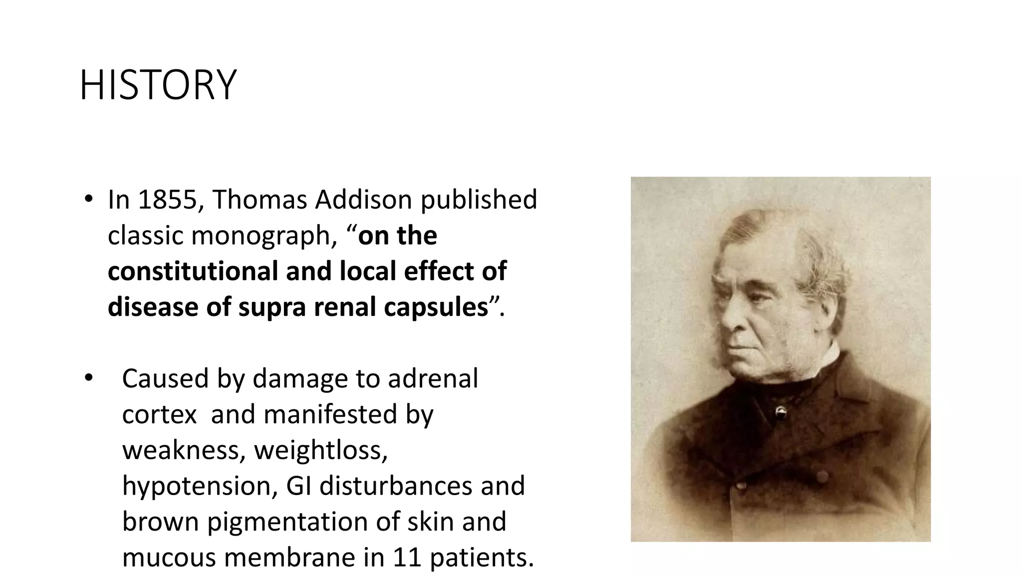
3. HISTORY
• In 1855, Thomas Addison published
classic monograph, “on the
constitutional and local effect of
disease of supra renal capsules”.
• Caused by damage to adrenal
cortex and manifested by
weakness, weightloss,
hypotension, GI disturbances and
brown pigmentation of skin and
mucous membrane in 11 patients.
4. Anatomy and physiology of adrenal gland
• Adrenal gland located at upper pole of each kidney.
• It comprises of two distinct endocrine organ.
• Adrenal cortex derived from intermediate mesoderm
(coelomic epithelium) and medulla from neural crest cells.
• Adrenal cortex is controlled by HPA axis and RAAS system and
medulla is controlled by sympathetic nervous system.
10. EPIDEMIOLOGY
• Prevalence of permanent AI 5 in 10,000 in gen population.
• Out of this 3 hypothalamic origin and 2 primary AI.
• Worldwide infection is m/c cause of PAI but in western word
autoimmune causes are m/c(75%)
• Exact prevalence of exogenous adrenal suppression is not known but
it is much more common(0.5-2% of population in developed
countries).
11. ETIOLOGY
• Causes of primary adrenal insufficiency:
• Autoimmune: Isolated autoimmune adrenalitis accounts
for 30–40%, whereas 60–70% develop adrenal insufficiency as part
of autoimmune polyglandular syndromes (APSs)
• Infection: TB is the m/c
• Genetic disorders:CAH, Adrenoleucodystrophy, Familial glucocorticoid
deficiency
• Infiltration: amyloid, hemochromatosis,metastasis
• Drug induced:mitoten, ketoconazole,rifampicin
15. CAUSES OF SECONDARY ADRENAL
INSUFFICIENCY
• It implies pituitary etiology
• HPA Axis suppression by exogenous GC : m/c/c
• considerable interindividual variability in response to GC
• no absolute cutoff values for the type of steroid taken, dose, route of
administration, duration of treatment,or time since steroid withdrawal
that predict adrenal suppression.
• adrenal atrophy and subsequent deficiency should be anticipated in
any subject who has taken more than the equivalent of 30 mg
hydrocortisone per day orally(>7.5 mg/day prednisolone or >0.75
mg/day dexamethasone) for longer than 3 weeks.
• In addition to the magnitude of the dose of glucocorticoid, the timing
of administration of may affect the degree of adrenal suppression
18. • PAI characterized by the loss of both glucocorticoid and
mineralocorticoid secretion
• In SAI only glucocorticoid deficiency is present.
• Hypothalamo pituitary disease may have additional features(d/t
involvement of thyroid, gonad, GH and prolactin)
• Signs and Symptomes due to GC deficiency:fatigue, lack of
energy, weight loss, anorexia, myalgia.
• Normochromic anaemia, eosinophilia, increase TSH,
hypoglycemia, low BP, hyponatremia.
19. • Signs and symptoms by MC Def: abd pain, nausea, vomiting, salt
craving, low BP, hyponatremia, hyperkalemia.
• Signs and symptoms due to adrenal androgen deficiency: Lac of
energy, loss of libido(women), loss of axillary and pubic hair(women)
• Others: hyperpigmentation, alabaster like paleness
20.
21. PAI SAI
Etiology Autoimmune, TB Pituitary disease
GC deficiency ++++ ++++
MC deficiency +++ ---
Androgen deficiency ++ ++
Salt craving and postural
hypotension
++++ --
Hyperpigmentation ++++ ----
ACTH High Low
S Potassium High Low
S Sodium Low Low
22. INVESTIGATIONS
1.Serum cortisol: in time of clinical urgency most important test in
diagnosing AI.
circadian and ultradian pulsatile cortisol secretion makes random
cortisol testing unreliable.
If morning cortisol(8 AM)- less than 3mcg/dl likely AI.
More than 18mcg/dl likely normal adrenal function
2. Cosyntropin stimulation test:plasma cortisol at 30 &/or60min
following synthetic ACTH in a dose of 250 mcg I/M or I/V at any time of
day.
23. • If serum cortisol less than 18mcg/dl s/o AI.(assay specific)
• It is cheapest, reliable, rapid and insensitive to interference from diet
and medication.
3. overnight metyrapone test: single dose 30mg/kg BW metyrapone
administered at midnight and 11 deoxycortisol and cortisol obtained at
morning.
Metyrapone blocks conversion of 11 deoxycortisol to cortisol by 11
beta hydroxylase.
Failure of rising 11 deoxycortisol after overnights administration of
metyrapone indicates AI.
24. • 3. Insulin intolerance test : For investigation of SAI remains the
gold standard test of the integrity of the HPA axis.
• C/I ischemic heart disease, epilepsy, or severe hypopituitarism
(i.e., 9 am plasma cortisol [<6.5 μg/dL])
IV regular insulin in a dose of 0.1 to 0.15 U/kg body weight, with
measurement of plasma cortisol at 0, 30, 45, 60, 90, and 120
minutes.
• Adequate hypoglycemia (blood glucose <45 mg/dl with signs of
neuroglycopenia—sweating and tachycardia) is required for a
fail result
• In normal subjects, the peak plasma cortisol concentration
exceeds 500 nmol/L (18 μg/dL).z
25. • 4.Adrenal antibody test :21 hydroxylase ab present 90% recent onset
autoimmune adrenalitis pt .
• 5.S .17 OH progesterone : cornerstone in diagnosing CAH .done in
suspected infant and selected child ,adults.
• 6.VLCFA: males with isolated AI ,without evidence of autoimmunity
Should be tested to exclude ALD or AMN .
7.Adrenal imaging : adrenal CT done in suspecting hge, infection
,infiltration ,neoplastic disease .
26. TREATMENT
• Treatment of AI - replacement of missing steroid hormone .
• cortisol in SAI and cortisol & aldosterone in PAI .
• Replacement of GC : hydrocortisone – oral 15-25mg/day in 2/3
divided doses
.long acting GC like- prednisolone(3-5mg daily), dexamethasone(0.3-
0.5mg daily) not preferred.
In pregnany dose may increased to 50% in last trimester.
In stressful condition dose may doubled.
Monitoring based on clinical assessment.
27. • MC replacement: fludricortisone 100-150mcg should be initiated.
dose adjustment involves control of symptoms of aldrosterone
deficiency and normalization of serum potassium and renin.
.Adrenal androgen replacement: replacement of DHEA not routinely
required ,may be considered in selected patients.
29. • Patient on steroid for more than 2 years- early recovery never occurs.
• So give physiologic dose of hydrocortisone and do SST after 6 months.
• If failed response- continue H.CORT for 4-5 months and do SST.
• If value close to 12mcg/dl- better chance of recovery and retest after
3 months.
30. APROACH
Clinical suspicion of adrenal insufficiency
(weight loss,fatigue,postural
hypotension,hyperpigmentation,hyponatremia)
Screening or confirmation of
diagnosis
(cosyntropin stimulation test
Cbc,na,k,TSH,urea,cr )
31. Differential diagnosis
(ACTH,Renin ,Aldosterone)
Primary adrenal
insufficiency
(High ACTH ,high Renin
,low aldosterone)
secondary adrenal
insufficiency (low normal
ACTH,normal renin ,normal
aldosterone)
MC and GC
replacement
GC
replacement
32.
33. ADRENAL CRISIS
• Life threatening emergency.
• Triggered by anything that increase the normal sress level.
• May lead to shock and vascular collapse
35. Management of adrenal crisis
• administration of intravenous hydrocortisone, 100 mg IV,
followed by 200 mg/24 hours as a continuous infusion or as IV (or IM)
hydrocortisone 50 mg every 6 hours, with subsequent does tailored to
clinical respons.
Replacement of fluid with normal saline is needed, according to
standard resuscitation guidelines; typically volumes of 2 to 3 liters are
required
36. Prevention of adrenal crisis
• • Prevention is possible using a combination of
• patient education,
• patient driven stress dosing of glucocorticoid,
• home parenteral hydrocortisone, and
• use of MedicAlert jewelry/patient information cards to advise
clinicians to promptly administer hydrocortisone in patients unable to
do so themselves.