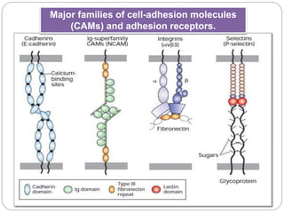
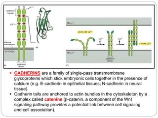
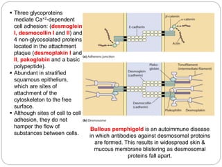
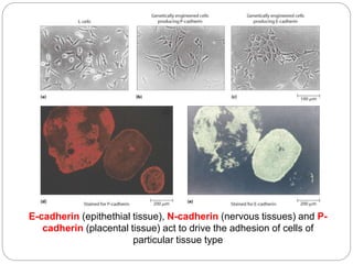
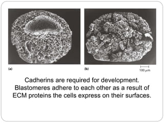
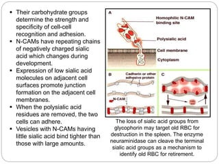
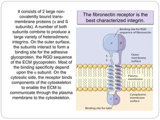
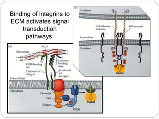
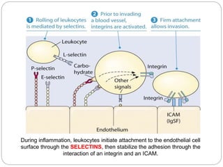
CELL ADHESION
MOLECULES
Department of Natural Sciences
University of St. La Salle
Bacolod City
Major families of cell-adhesion molecules
(CAMs) and adhesion receptors.
 Dimeric E-cadherins most commonly form homophilic
(self) cross-bridges with E-cadherins on adjacent cells.
 Members of the Immunoglobulin (Ig) superfamily of
CAMs can form both homophilic linkages and
heterophilic (nonself) linkages.
 Heterodimeric integrins function as CAMs or as
adhesion receptors that bind to very large, multiadhesive
matrix proteins such as fibronectin.
 Selectin dimers contain a carbohydrate-binding lectin
domain that recognizes specialized sugar structures on
glycoproteins and glycolipids on adjacent cells.
 Note that CAMs often form higher-order oligomers
within the plane of the plasma membrane.
 Many adhesive molecules contain multiple distinct
domains, some of which are found in more than one
kind of CAM.
 The cytoplasmic domains of these proteins are
often associated with adapter proteins that link them
to the cytoskeleton or to signaling pathways.
 The evolution of CAMs, adhesion receptors, and
ECM molecules with specialized structures and
functions permits cells to assemble into diverse
classes of tissues with varying functions.
 CADHERINS are a family of single-pass transmembrane
glycoproteins which stick embryonic cells together in the presence of
calcium (e.g. E-cadherin in epithelial tissues; N-cadherin in neural
tissue).
 Cadherin tails are anchored to actin bundles in the cytoskeleton by a
complex called catenins (-catenin, a component of the Wnt
signaling pathway provides a potential link between cell signaling
and cell association).
 Three glycoproteins
mediate Ca+2-dependent
cell adhesion: (desmoglein
I, desmocollin I and II) and
4 non-glycosolated proteins
located in the attachment
plaque (desmoplakin I and
II, pakoglobin and a basic
polypeptide).
 Abundant in stratified
squamous epithelium,
which are sites of
attachment of the
cytoskeleton to the free
surface.
 Although sites of cell to cell
adhesion, they do not
hamper the flow of
substances between cells.
Bullous pemphigold is an autoimmune disease
in which antibodies against desmosomal proteins
are formed. This results in widespread skin &
mucous membrane blistering as desmosomal
proteins fall apart.
E-cadherin (epithethial tissue), N-cadherin (nervous tissues) and P-
cadherin (placental tissue) act to drive the adhesion of cells of
particular tissue type
Cadherins are required for development.
Blastomeres adhere to each other as a result of
ECM proteins the cells express on their surfaces.
 CAMs (Cell Adhesion
Molecules) are single-pass
transmembrane glycoproteins
which do not require calcium to
bind to other cells.
 Neural cell adhesion molecules
(N-CAMs) are a large family of
proteins formed by alternative
splicing.
 When embryonic tissue is
exposed to antibodies that
interact with N-CAMs, the cells
do not bind to each other and
neural tissue is not formed.
 N-CAMs and cadherins mediate
cell-cell recognition and cell-cell
adhesion.
 Their carbohydrate groups
determine the strength and
specificity of cell-cell
recognition and adhesion.
 N-CAMs have repeating chains
of negatively charged sialic
acid which changes during
development.
 Expression of low sialic acid
molecules on adjacent cell
surfaces promote junction
formation on the adjacent cell
membranes.
 When the polysialic acid
residues are removed, the two
cells can adhere.
 Vesicles with N-CAMs having
little sialic acid bind tighter than
those with large amounts.
The loss of sialic acid groups from
glycophorin may target old RBC for
destruction in the spleen. The enzyme
neuraminidase can cleave the terminal
sialic acid groups as a mechanism to
identify old RBC for retirement.
 INTEGRINS (I-CAMs) are
cell surface receptors that
bind the ECM.
 They require (Ca+2 or
Mg+2), to interact with
ECM components
(fibronectin, laminin and
collagens).
 Important in epithelial cell
cohesion and attachment
to substrate and cell
migration during tissue
repair.
 Bound integrins prevent
transcription of genes that
specify apoptosis.
It consists of 2 large non-
covalently bound trans-
membrane proteins (α and ß
subunits). A number of both
subunits combine to produce a
large variety of heterodimeric
integrins. On the outer surface,
the subunits interact to form a
binding site for the adhesive
glycoprotein, the RGD sequence
of the ECM glycoprotein. Most of
the binding specificity depend
upon the α subunit. On the
cytosolic side, the receptor binds
components of the cytoskeleton
to enable the ECM to
communicate through the plasma
membrane to the cytoskeleton.
The fibronectin receptor is the
best characterized integrin.
Binding of integrins to
ECM activates signal
transduction
pathways.
During inflammation, leukocytes initiate attachment to the endothelial cell
surface through the SELECTINS, then stabilize the adhesion through the
interaction of an integrin and an ICAM.
Adhesionmolecules 111109065715-phpapp01

Adhesionmolecules 111109065715-phpapp01

  • 1.
    CELL ADHESION MOLECULES Department ofNatural Sciences University of St. La Salle Bacolod City
  • 2.
    Major families ofcell-adhesion molecules (CAMs) and adhesion receptors.
  • 3.
     Dimeric E-cadherinsmost commonly form homophilic (self) cross-bridges with E-cadherins on adjacent cells.  Members of the Immunoglobulin (Ig) superfamily of CAMs can form both homophilic linkages and heterophilic (nonself) linkages.  Heterodimeric integrins function as CAMs or as adhesion receptors that bind to very large, multiadhesive matrix proteins such as fibronectin.  Selectin dimers contain a carbohydrate-binding lectin domain that recognizes specialized sugar structures on glycoproteins and glycolipids on adjacent cells.
  • 4.
     Note thatCAMs often form higher-order oligomers within the plane of the plasma membrane.  Many adhesive molecules contain multiple distinct domains, some of which are found in more than one kind of CAM.  The cytoplasmic domains of these proteins are often associated with adapter proteins that link them to the cytoskeleton or to signaling pathways.  The evolution of CAMs, adhesion receptors, and ECM molecules with specialized structures and functions permits cells to assemble into diverse classes of tissues with varying functions.
  • 5.
     CADHERINS area family of single-pass transmembrane glycoproteins which stick embryonic cells together in the presence of calcium (e.g. E-cadherin in epithelial tissues; N-cadherin in neural tissue).  Cadherin tails are anchored to actin bundles in the cytoskeleton by a complex called catenins (-catenin, a component of the Wnt signaling pathway provides a potential link between cell signaling and cell association).
  • 6.
     Three glycoproteins mediateCa+2-dependent cell adhesion: (desmoglein I, desmocollin I and II) and 4 non-glycosolated proteins located in the attachment plaque (desmoplakin I and II, pakoglobin and a basic polypeptide).  Abundant in stratified squamous epithelium, which are sites of attachment of the cytoskeleton to the free surface.  Although sites of cell to cell adhesion, they do not hamper the flow of substances between cells. Bullous pemphigold is an autoimmune disease in which antibodies against desmosomal proteins are formed. This results in widespread skin & mucous membrane blistering as desmosomal proteins fall apart.
  • 7.
    E-cadherin (epithethial tissue),N-cadherin (nervous tissues) and P- cadherin (placental tissue) act to drive the adhesion of cells of particular tissue type
  • 8.
    Cadherins are requiredfor development. Blastomeres adhere to each other as a result of ECM proteins the cells express on their surfaces.
  • 9.
     CAMs (CellAdhesion Molecules) are single-pass transmembrane glycoproteins which do not require calcium to bind to other cells.  Neural cell adhesion molecules (N-CAMs) are a large family of proteins formed by alternative splicing.  When embryonic tissue is exposed to antibodies that interact with N-CAMs, the cells do not bind to each other and neural tissue is not formed.  N-CAMs and cadherins mediate cell-cell recognition and cell-cell adhesion.
  • 10.
     Their carbohydrategroups determine the strength and specificity of cell-cell recognition and adhesion.  N-CAMs have repeating chains of negatively charged sialic acid which changes during development.  Expression of low sialic acid molecules on adjacent cell surfaces promote junction formation on the adjacent cell membranes.  When the polysialic acid residues are removed, the two cells can adhere.  Vesicles with N-CAMs having little sialic acid bind tighter than those with large amounts. The loss of sialic acid groups from glycophorin may target old RBC for destruction in the spleen. The enzyme neuraminidase can cleave the terminal sialic acid groups as a mechanism to identify old RBC for retirement.
  • 11.
     INTEGRINS (I-CAMs)are cell surface receptors that bind the ECM.  They require (Ca+2 or Mg+2), to interact with ECM components (fibronectin, laminin and collagens).  Important in epithelial cell cohesion and attachment to substrate and cell migration during tissue repair.  Bound integrins prevent transcription of genes that specify apoptosis.
  • 12.
    It consists of2 large non- covalently bound trans- membrane proteins (α and ß subunits). A number of both subunits combine to produce a large variety of heterodimeric integrins. On the outer surface, the subunits interact to form a binding site for the adhesive glycoprotein, the RGD sequence of the ECM glycoprotein. Most of the binding specificity depend upon the α subunit. On the cytosolic side, the receptor binds components of the cytoskeleton to enable the ECM to communicate through the plasma membrane to the cytoskeleton. The fibronectin receptor is the best characterized integrin.
  • 13.
    Binding of integrinsto ECM activates signal transduction pathways.
  • 14.
    During inflammation, leukocytesinitiate attachment to the endothelial cell surface through the SELECTINS, then stabilize the adhesion through the interaction of an integrin and an ICAM.