6 Spontaneous milk fistula from an accessory breast an extremely rare case. Breast J. 2015.pdf
- 1. chemotherapeutic agents, the incidence of Stewart–
Treves syndrome has significantly decreased. This
case should not be considered as metastatic or
recurrent angiosarcoma, but as angiosarcoma sec-
ondary to irradiation, or secondary angiosarcoma. A
Dutch population-based study found that the abso-
lute risk of developing angiosarcoma after breast-
conserving therapy for breast cancer was 0.16% at
5 years. Reporting this case, we want to remember
that the angiosarcoma should be considered in any
postradiation patient with persistent or worsening
skin thickening and the occurrence of skin nodules.
Early diagnosis is crucial in fact it allows the
patient to quickly obtain aggressive surgical treat-
ment, which is the only chance for definitive treat-
ment and survival.
Spontaneous Milk Fistula from an Accessory Breast:
An Extremely Rare Case
Deniz Firat, MD,* Oguz Idiz, MD,* Arda Isik, MD,* Kemal Peker, MD,*
Nilgun Atar, MD,†
and Eylem Gul, MD‡
*General Surgery, Erzincan Unıversity, Erzincan, Turkey; †
Radiology, Erzincan Unıversity, Erzincan,
Turkey; ‡
Dermatology, Erzincan Uunıversity, Erzincan, Turkey
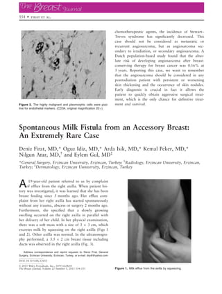
A19-year-old patient referred to us by complaint
of efflux from the right axilla. When patient his-
tory was investigated, it was learned that she has been
breast feeding since 5 months ago. Her efflux com-
plaint from her right axilla has started spontaneously
without any trauma, abscess or surgery 2 months ago.
Furthermore, she specified that a slowly growing
swelling occurred on the right axilla in parallel with
her delivery of her child. In her physical examination,
there was a soft mass with a size of 3 9 3 cm, which
excretes milk by squeezing on the right axilla (Figs 1
and 2). Other axilla was normal. In the ultrasonogra-
phy performed, a 3.5 9 2 cm breast tissue including
ducts was observed in the right axilla (Fig. 3).
Figure 1. Milk efflux from the axilla by squeezing.
Figure 3. The highly malignant and pleomorphic cells were posi-
tive for endothelial markers. (CD34, original magnification 209).
Address correspondence and reprint requests to: Deniz Firat, General
Surgery, Erzincan University, Erzincan, Turkey, or e-mail: drydf@yahoo.com
DOI: 10.1111/tbj.12452
© 2015 Wiley Periodicals, Inc., 1075-122X/15
The Breast Journal, Volume 21 Number 5, 2015 554–555
554 • firat et al.
- 2. The milk efflux from the right axilla completely
stopped within 1.5 month after termination of breast
feeding. Excision of the axillary breast tissue is
planned to prevent such problem for her further
pregnancies.
Giant Intracystic (Encysted) Papillary Carcinoma of the
Breast
Vincenzo Vigorita, MD,* Marco Bertucci Zoccali, MD,†
Marta Martinez
Miguez, MD,* Maria J. Ave Seijas, MD,‡
Rocio Fernandez Martin, MD,§
Enrique J. Casal Nu~
nez, MD,* and Gonzalo De Castro Parga, MD¶
*Department of General and Digestive Surgery, University of Vigo – Meixoeiro Hospital, Vigo, Spain;
†
General Surgery Unit, Department of Surgery, Catholic University Med. School—”A.Gemelli” Gen.
Hospital, Rome, Italy; ‡
Department of Radiology, University of Vigo – Meixoeiro Hospital, Vigo,
Spain; §
Department of Pathology, University of Vigo – Meixoeiro Hospital, Vigo, Spain; ¶
Breast Unit,
University of Vigo – Meixoeiro Hospital, Vigo, Spain
A79-year-old British woman came to our attention
due to acute gallstone pancreatitis (leukocytes
15.41 9 109
/L, n.v. 3.7–10.8; serum amylase
2,319.0 UI/l, n.v. 20.00–115.0; total bilirubin 1.7 mg/
dL, n.v. 0.0–1; AST 162 UI/L, n.v. 1.0–40). On physi-
cal exam, a greater than 20 cm, well-defined, multilo-
bulated cystic mass was palpated in her right breast,
with inverted nipple. The skin overlying the lesion
was slightly darker and there was no tenderness. No
axillary lymph nodes were palpable. The other breast
appeared normal (Fig. 1). The patient reported that
the lesion has been present for at least 8 years. Her
family history was remarkable for breast cancer in her
mother.
Ducts
Right Axilla
Figure 3. USG _
Image of the axilla and the ducts.
Figure 2. Spontaneous milk flowfrom the axilla.
Address correspondence and reprint requests to: Vincenzo Vigorita, MD,
Department of General and Digestive Surgery, University of Vigo – Meixoe-
iro Hospital, Meixoeiro s/n, 36200 Vigo, Spain, or e-mail: v.vigorita@gmail.
com
DOI: 10.1111/tbj.12454
© 2015 Wiley Periodicals, Inc., 1075-122X/15
The Breast Journal, Volume 21 Number 5, 2015 555–557
Intracystic Papillary Carcinoma • 555