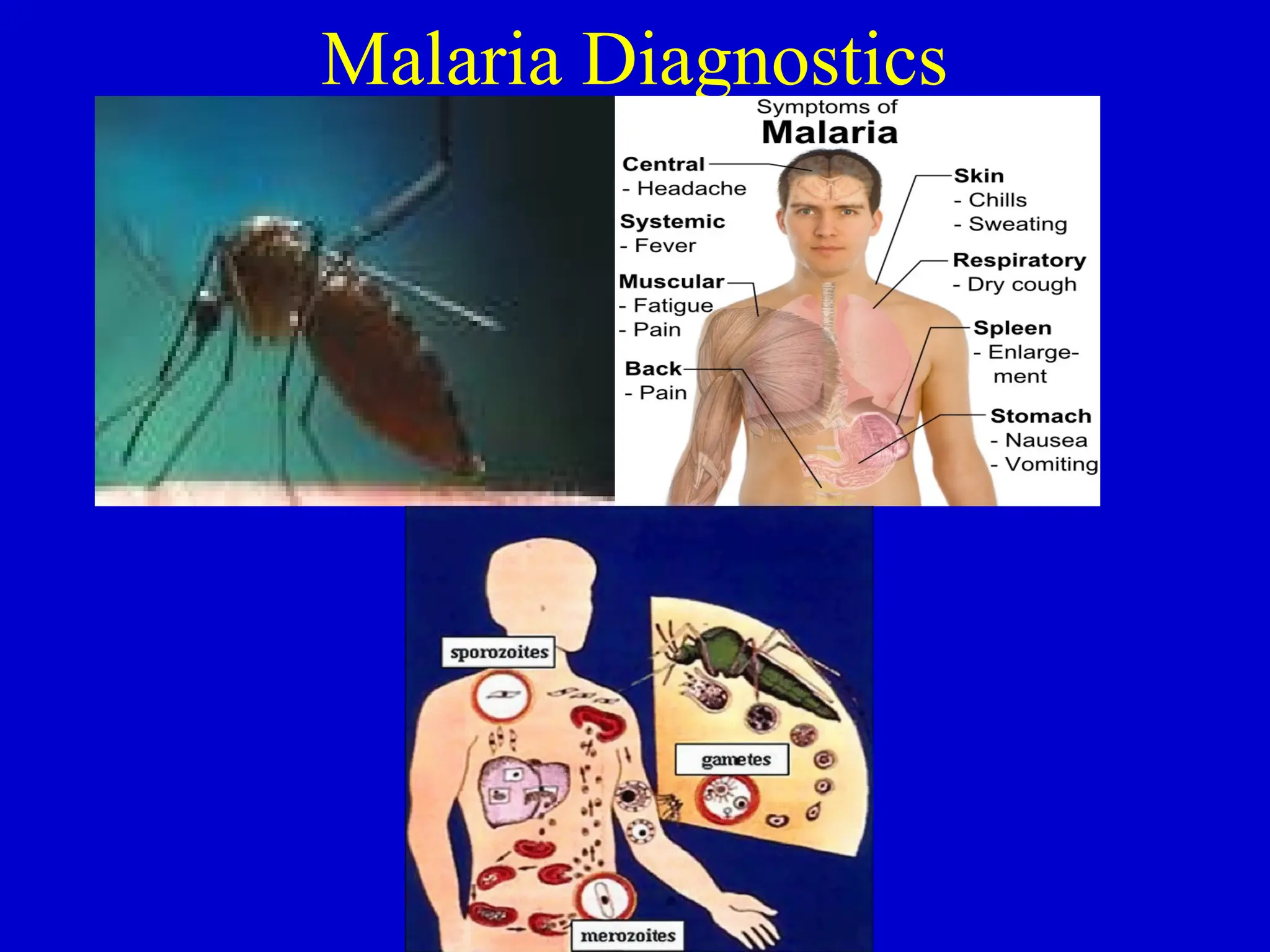
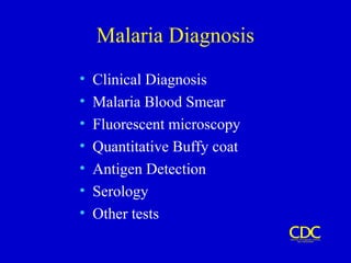
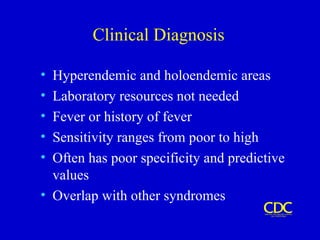
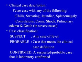
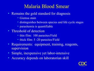
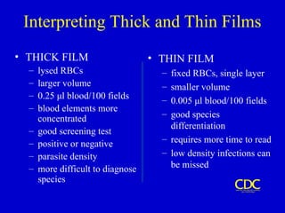
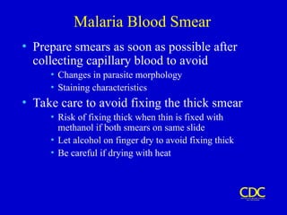
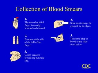
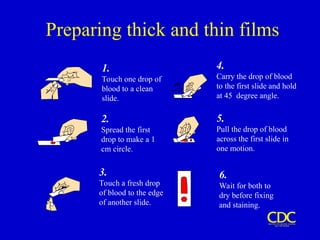
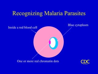
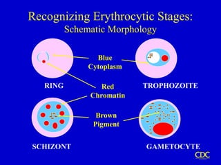
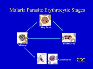
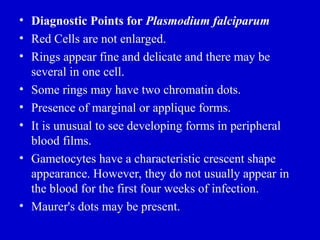
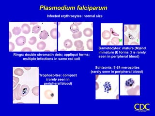
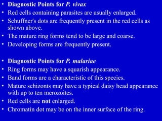
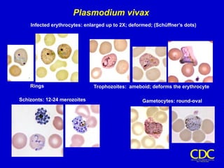
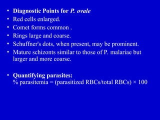
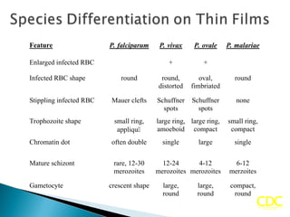
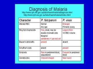
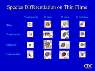
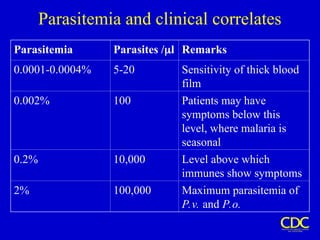
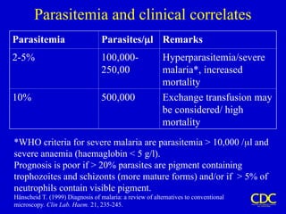
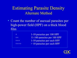
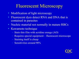
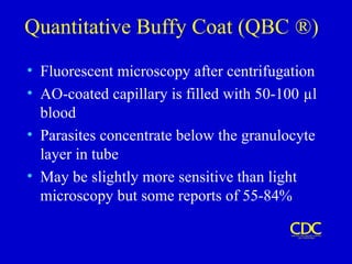
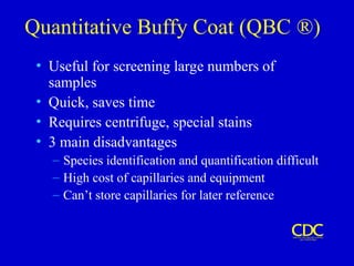
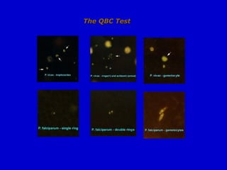
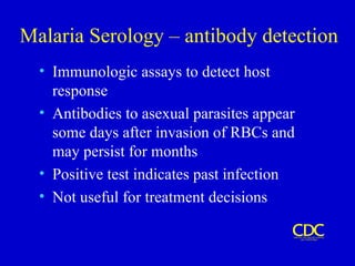
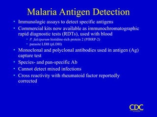
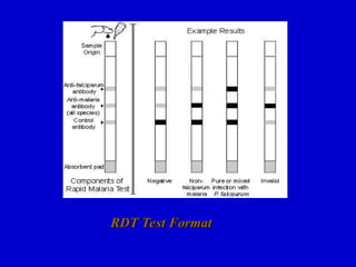
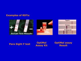
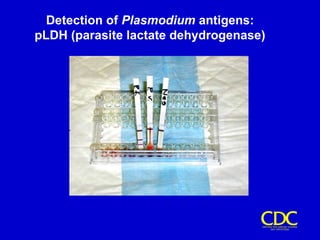
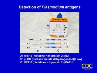
Malaria Diagnostics
Malaria Diagnosis
• Clinical Diagnosis
• Malaria Blood Smear
• Fluorescent microscopy
• Quantitative Buffy coat
• Antigen Detection
• Serology
• Other tests
Clinical Diagnosis
• Hyperendemic and holoendemic areas
• Laboratory resources not needed
• Fever or history of fever
• Sensitivity ranges from poor to high
• Often has poor specificity and predictive
values
• Overlap with other syndromes
• Clinical case description:
Fever case with any of the following:
Chills, Sweating, Jaundice, Splenomegaly
Convulsions, Coma, Shock, Pulmonary
edema & Death (in severe cases)
• Case classification:
SUSPECT : Any case of fever
PROBABLE : Case that meets the clinical
case definition
CONFIRMED: A suspected/probable case
that is laboratory confirmed
Malaria Blood Smear
• Remains the gold standard for diagnosis
• Giemsa stain
• distinguishes between species and life cycle stages
• parasitemia is quantifiable
• Threshold of detection
• thin film: 100 parasites/Field
• thick film: 5 -20 parasites/Field
• Requirements: equipment, training, reagents,
supervision
• Simple, inexpensive yet labor-intensive
• Accuracy depends on laboratorian skill
• Jaswant Singh Battacharya (JSB) Stain for
Jaswant Singh Battacharya (JSB) Stain for
thick and thin films:
thick and thin films: This is the standard method used by
the laboratories under the National Malaria Eradication Programme in
India.
• Preparation of the stain
Preparation of the stain:
:
• JSB I stain:
JSB I stain: Medicinal methylene blue (0.5 g) is dissolved in 500
ml of distilled water and 3 ml of 1% sulphuric acid (H2
SO4
) is
gradually added, followed by 0.5 g of potassium dichromate (K2
Cr2
O7
)
when a purple precipitate forms. 3.5 g of disodium hydrogen
phosphate dihydrate (Na2
HPO4
.2H2
O) is next added and when the
precipitate has dissolved, the solution is boiled in a flask with a reflex
condenser for 1 hour. The stain is ready for immediate use.
• JSB II stain:
JSB II stain: 1 g Eosin is dissolved in 500 ml tap water.
• Buffered water:
Buffered water: 0.22 g of disodium hydrogen phosphate
dihydrate (Na2
HPO4
.2H2
O) and 0.74 g of potassium acid
phosphate (KH2
PO4
) are added to 1000 ml of distilled water or
filtered tap water.
• Staining:
Staining:
• After dehemoglobinisation, dip the thick smear in JSB II stain
two to three times. Wash it by dipping in buffer water two to
three times. Then keep the thick film dipped in JSB I stain for
40-60 seconds. Wash it with buffer water. Drain, dry and
examine.
Interpreting Thick and Thin Films
• THICK FILM
– lysed RBCs
– larger volume
– 0.25 μl blood/100 fields
– blood elements more
concentrated
– good screening test
– positive or negative
– parasite density
– more difficult to diagnose
species
• THIN FILM
– fixed RBCs, single layer
– smaller volume
– 0.005 μl blood/100 fields
– good species
differentiation
– requires more time to read
– low density infections can
be missed
Malaria Blood Smear
• Prepare smears as soon as possible after
collecting capillary blood to avoid
• Changes in parasite morphology
• Staining characteristics
• Take care to avoid fixing the thick smear
• Risk of fixing thick when thin is fixed with
methanol if both smears on same slide
• Let alcohol on finger dry to avoid fixing thick
• Be careful if drying with heat
Collection of Blood Smears
5.
Touch the drop of
blood to the slide
from below.
4.
Slide must always be
grasped by its edges.
2.
Puncture at the side
of the ball of the
finger.
3.
Gently squeeze
toward the puncture
site.
1.
The second or third
finger is usually
selected and cleaned.
Preparing thick and thin films
1.
Touch one drop of
blood to a clean
slide.
2.
Spread the first
drop to make a 1
cm circle.
3.
Touch a fresh drop
of blood to the edge
of another slide.
6.
Wait for both to
dry before fixing
and staining.
5.
Pull the drop of blood
across the first slide in
one motion.
4.
Carry the drop of blood
to the first slide and hold
at 45degree angle.
Recognizing Malaria Parasites
Inside a red blood cell
One or more red chromatin dots
Blue cytoplasm
RING TROPHOZOITE
SCHIZONT GAMETOCYTE
Blue
Cytoplasm
Red
Chromatin
Brown
Pigment
Recognizing Erythrocytic Stages:
Schematic Morphology
Malaria Parasite Erythrocytic Stages
Ring form
Trophozoite
Schizont
Gametocytes
• Diagnostic Points for Plasmodium falciparum
• Red Cells are not enlarged.
• Rings appear fine and delicate and there may be
several in one cell.
• Some rings may have two chromatin dots.
• Presence of marginal or applique forms.
• It is unusual to see developing forms in peripheral
blood films.
• Gametocytes have a characteristic crescent shape
appearance. However, they do not usually appear in
the blood for the first four weeks of infection.
• Maurer's dots may be present.
Plasmodium falciparum
Rings: double chromatin dots; appliqué forms;
multiple infections in same red cell
Gametocytes: mature (M)and
immature (I) forms (I is rarely
seen in peripheral blood)
Trophozoites: compact
(rarely seen in
peripheral blood)
Schizonts: 8-24 merozoites
(rarely seen in peripheral blood)
Infected erythrocytes: normal size
M I
• Diagnostic Points for P. vivax
• Red cells containing parasites are usually enlarged.
• Schuffner's dots are frequently present in the red cells as
shown above.
• The mature ring forms tend to be large and coarse.
• Developing forms are frequently present.
• Diagnostic Points for P. malariae
• Ring forms may have a squarish appearance.
• Band forms are a characteristic of this species.
• Mature schizonts may have a typical daisy head appearance
with up to ten merozoites.
• Red cells are not enlarged.
• Chromatin dot may be on the inner surface of the ring.
Plasmodium vivax
Trophozoites: ameboid; deforms the erythrocyte
Gametocytes: round-oval
Schizonts: 12-24 merozoites
Rings
Infected erythrocytes: enlarged up to 2X; deformed; (Schüffner’s dots)
• Diagnostic Points for P. ovale
• Red cells enlarged.
• Comet forms common .
• Rings large and coarse.
• Schuffner's dots, when present, may be prominent.
• Mature schizonts similar to those of P. malariae but
larger and more coarse.
• Quantifying parasites:
% parasitemia = (parasitized RBCs/total RBCs) × 100
Feature P. falciparum P. vivax P. ovale P. malariae
Enlarged infected RBC + +
Infected RBC shape round round,
distorted
oval,
fimbriated
round
Stippling infected RBC Mauer clefts Schuffner
spots
Schuffner
spots
none
Trophozoite shape small ring,
appliqu
large ring,
amoeboid
large ring,
compact
small ring,
compact
Chromatin dot often double single large single
Mature schizont rare, 12-30
merozoites
12-24
merozoites
4-12
merozoites
6-12
merzoites
Gametocyte crescent shape large,
round
large,
round
compact,
round
Species Differentiation on Thin Films
P. falciparum P. vivax P. ovale P. malariae
Rings
Trophozoites
Schizonts
Gametocytes
Parasitemia and clinical correlates
Parasitemia Parasites /l Remarks
0.0001-0.0004% 5-20 Sensitivity of thick blood
film
0.002% 100 Patients may have
symptoms below this
level, where malaria is
seasonal
0.2% 10,000 Level above which
immunes show symptoms
2% 100,000 Maximum parasitemia of
P.v. and P.o.
Parasitemia and clinical correlates
Parasitemia Parasites/l Remarks
2-5% 100,000-
250,00
Hyperparasitemia/severe
malaria*, increased
mortality
10% 500,000 Exchange transfusion may
be considered/ high
mortality
*WHO criteria for severe malaria are parasitemia > 10,000 /l and
severe anaemia (haemaglobin < 5 g/l).
Prognosis is poor if > 20% parasites are pigment containing
trophozoites and schizonts (more mature forms) and/or if > 5% of
neutrophils contain visible pigment.
Hänscheid T. (1999) Diagnosis of malaria: a review of alternatives to conventional
microscopy. Clin Lab. Haem. 21, 235-245.
Estimating Parasite Density
Alternate Method
• Count the number of asexual parasites per
high-power field (HPF) on a thick blood
film
+ 1-10 parasites per 100 HPF
++ 11-100 parasites per 100 HPF
+++ 1-10 parasites per each HPF
++++ > 10 parasites per each HPF
Fluorescent Microscopy
• Modification of light microscopy
• Fluorescent dyes detect RNA and DNA that is
contained in parasites
• Nucleic material not normally in mature RBCs
• Kawamoto technique
– Stain thin film with acridine orange (AO)
– Requires special equipment – fluorescent microscope
– Staining itself is cheap
– Sensitivities around 90%
Quantitative Buffy Coat (QBC ®)
• Fluorescent microscopy after centrifugation
• AO-coated capillary is filled with 50-100 µl
blood
• Parasites concentrate below the granulocyte
layer in tube
• May be slightly more sensitive than light
microscopy but some reports of 55-84%
Quantitative Buffy Coat (QBC ®)
• Useful for screening large numbers of
samples
• Quick, saves time
• Requires centrifuge, special stains
• 3 main disadvantages
– Species identification and quantification difficult
– High cost of capillaries and equipment
– Can’t store capillaries for later reference
The QBC Test
The QBC Test
Malaria Serology – antibody detection
• Immunologic assays to detect host
response
• Antibodies to asexual parasites appear
some days after invasion of RBCs and
may persist for months
• Positive test indicates past infection
• Not useful for treatment decisions
Malaria Serology – antibody detection
• Valuable epidemiologic tool in some settings
• Useful for
– Identifying infective donor in transfusion-transmitted
malaria
– Investigating congenital malaria, esp. if mom’s smear
is negative
– Diagnosing, or ruling out, tropical splenomegaly
syndrome
– Retrospective confirmation of empirically-treated
non-immunes
Malaria Antigen Detection
• Immunologic assays to detect specific antigens
• Commercial kits now available as immunochromatographic
rapid diagnostic tests (RDTs), used with blood
• P. falciparum histidine-rich protein 2 (PfHRP-2)
• parasite LDH (pLDH)
• Monoclonal and polyclonal antibodies used in antigen (Ag)
capture test
• Species- and pan-specific Ab
• Cannot detect mixed infections
• Cross reactivity with rheumatoid factor reportedly
corrected
RDT Test Format
RDT Test Format
Examples of RDTs:
Para Sight F test
OptiMal
Assay Kit
OptiMal assay
Result
Detection of Plasmodium antigens:
pLDH (parasite lactate dehydrogenase)
Detection of Plasmodium antigens
A: HRP-2 (histidine-rich protein 2) (ICT)
B: pLDH (parasite lactate dehydrogenase)(Flow)
C: HRP-2 (histidine-rich protein 2) (PATH)
OTHER TESTS
• Polymerase chain reaction (PCR)
• Detection of Anti-malarial antibodies
• Intraleucocytic malaria pigment
• Flowcytometry
• Mass spectrometry
T
T
H
H
A
A
N
N
K
K
S
S

4malariadiagnosis-091202111915-phpapp02 (2).ppt

  • 1.
  • 2.
    Malaria Diagnosis • ClinicalDiagnosis • Malaria Blood Smear • Fluorescent microscopy • Quantitative Buffy coat • Antigen Detection • Serology • Other tests
  • 3.
    Clinical Diagnosis • Hyperendemicand holoendemic areas • Laboratory resources not needed • Fever or history of fever • Sensitivity ranges from poor to high • Often has poor specificity and predictive values • Overlap with other syndromes
  • 4.
    • Clinical casedescription: Fever case with any of the following: Chills, Sweating, Jaundice, Splenomegaly Convulsions, Coma, Shock, Pulmonary edema & Death (in severe cases) • Case classification: SUSPECT : Any case of fever PROBABLE : Case that meets the clinical case definition CONFIRMED: A suspected/probable case that is laboratory confirmed
  • 5.
    Malaria Blood Smear •Remains the gold standard for diagnosis • Giemsa stain • distinguishes between species and life cycle stages • parasitemia is quantifiable • Threshold of detection • thin film: 100 parasites/Field • thick film: 5 -20 parasites/Field • Requirements: equipment, training, reagents, supervision • Simple, inexpensive yet labor-intensive • Accuracy depends on laboratorian skill
  • 6.
    • Jaswant SinghBattacharya (JSB) Stain for Jaswant Singh Battacharya (JSB) Stain for thick and thin films: thick and thin films: This is the standard method used by the laboratories under the National Malaria Eradication Programme in India. • Preparation of the stain Preparation of the stain: : • JSB I stain: JSB I stain: Medicinal methylene blue (0.5 g) is dissolved in 500 ml of distilled water and 3 ml of 1% sulphuric acid (H2 SO4 ) is gradually added, followed by 0.5 g of potassium dichromate (K2 Cr2 O7 ) when a purple precipitate forms. 3.5 g of disodium hydrogen phosphate dihydrate (Na2 HPO4 .2H2 O) is next added and when the precipitate has dissolved, the solution is boiled in a flask with a reflex condenser for 1 hour. The stain is ready for immediate use. • JSB II stain: JSB II stain: 1 g Eosin is dissolved in 500 ml tap water. • Buffered water: Buffered water: 0.22 g of disodium hydrogen phosphate dihydrate (Na2 HPO4 .2H2 O) and 0.74 g of potassium acid phosphate (KH2 PO4 ) are added to 1000 ml of distilled water or filtered tap water. • Staining: Staining: • After dehemoglobinisation, dip the thick smear in JSB II stain two to three times. Wash it by dipping in buffer water two to three times. Then keep the thick film dipped in JSB I stain for 40-60 seconds. Wash it with buffer water. Drain, dry and examine.
  • 7.
    Interpreting Thick andThin Films • THICK FILM – lysed RBCs – larger volume – 0.25 μl blood/100 fields – blood elements more concentrated – good screening test – positive or negative – parasite density – more difficult to diagnose species • THIN FILM – fixed RBCs, single layer – smaller volume – 0.005 μl blood/100 fields – good species differentiation – requires more time to read – low density infections can be missed
  • 8.
    Malaria Blood Smear •Prepare smears as soon as possible after collecting capillary blood to avoid • Changes in parasite morphology • Staining characteristics • Take care to avoid fixing the thick smear • Risk of fixing thick when thin is fixed with methanol if both smears on same slide • Let alcohol on finger dry to avoid fixing thick • Be careful if drying with heat
  • 9.
    Collection of BloodSmears 5. Touch the drop of blood to the slide from below. 4. Slide must always be grasped by its edges. 2. Puncture at the side of the ball of the finger. 3. Gently squeeze toward the puncture site. 1. The second or third finger is usually selected and cleaned.
  • 10.
    Preparing thick andthin films 1. Touch one drop of blood to a clean slide. 2. Spread the first drop to make a 1 cm circle. 3. Touch a fresh drop of blood to the edge of another slide. 6. Wait for both to dry before fixing and staining. 5. Pull the drop of blood across the first slide in one motion. 4. Carry the drop of blood to the first slide and hold at 45degree angle.
  • 11.
    Recognizing Malaria Parasites Insidea red blood cell One or more red chromatin dots Blue cytoplasm
  • 12.
  • 13.
    Malaria Parasite ErythrocyticStages Ring form Trophozoite Schizont Gametocytes
  • 14.
    • Diagnostic Pointsfor Plasmodium falciparum • Red Cells are not enlarged. • Rings appear fine and delicate and there may be several in one cell. • Some rings may have two chromatin dots. • Presence of marginal or applique forms. • It is unusual to see developing forms in peripheral blood films. • Gametocytes have a characteristic crescent shape appearance. However, they do not usually appear in the blood for the first four weeks of infection. • Maurer's dots may be present.
  • 15.
    Plasmodium falciparum Rings: doublechromatin dots; appliqué forms; multiple infections in same red cell Gametocytes: mature (M)and immature (I) forms (I is rarely seen in peripheral blood) Trophozoites: compact (rarely seen in peripheral blood) Schizonts: 8-24 merozoites (rarely seen in peripheral blood) Infected erythrocytes: normal size M I
  • 16.
    • Diagnostic Pointsfor P. vivax • Red cells containing parasites are usually enlarged. • Schuffner's dots are frequently present in the red cells as shown above. • The mature ring forms tend to be large and coarse. • Developing forms are frequently present. • Diagnostic Points for P. malariae • Ring forms may have a squarish appearance. • Band forms are a characteristic of this species. • Mature schizonts may have a typical daisy head appearance with up to ten merozoites. • Red cells are not enlarged. • Chromatin dot may be on the inner surface of the ring.
  • 17.
    Plasmodium vivax Trophozoites: ameboid;deforms the erythrocyte Gametocytes: round-oval Schizonts: 12-24 merozoites Rings Infected erythrocytes: enlarged up to 2X; deformed; (Schüffner’s dots)
  • 18.
    • Diagnostic Pointsfor P. ovale • Red cells enlarged. • Comet forms common . • Rings large and coarse. • Schuffner's dots, when present, may be prominent. • Mature schizonts similar to those of P. malariae but larger and more coarse. • Quantifying parasites: % parasitemia = (parasitized RBCs/total RBCs) × 100
  • 19.
    Feature P. falciparumP. vivax P. ovale P. malariae Enlarged infected RBC + + Infected RBC shape round round, distorted oval, fimbriated round Stippling infected RBC Mauer clefts Schuffner spots Schuffner spots none Trophozoite shape small ring, appliqu large ring, amoeboid large ring, compact small ring, compact Chromatin dot often double single large single Mature schizont rare, 12-30 merozoites 12-24 merozoites 4-12 merozoites 6-12 merzoites Gametocyte crescent shape large, round large, round compact, round
  • 21.
    Species Differentiation onThin Films P. falciparum P. vivax P. ovale P. malariae Rings Trophozoites Schizonts Gametocytes
  • 22.
    Parasitemia and clinicalcorrelates Parasitemia Parasites /l Remarks 0.0001-0.0004% 5-20 Sensitivity of thick blood film 0.002% 100 Patients may have symptoms below this level, where malaria is seasonal 0.2% 10,000 Level above which immunes show symptoms 2% 100,000 Maximum parasitemia of P.v. and P.o.
  • 23.
    Parasitemia and clinicalcorrelates Parasitemia Parasites/l Remarks 2-5% 100,000- 250,00 Hyperparasitemia/severe malaria*, increased mortality 10% 500,000 Exchange transfusion may be considered/ high mortality *WHO criteria for severe malaria are parasitemia > 10,000 /l and severe anaemia (haemaglobin < 5 g/l). Prognosis is poor if > 20% parasites are pigment containing trophozoites and schizonts (more mature forms) and/or if > 5% of neutrophils contain visible pigment. Hänscheid T. (1999) Diagnosis of malaria: a review of alternatives to conventional microscopy. Clin Lab. Haem. 21, 235-245.
  • 24.
    Estimating Parasite Density AlternateMethod • Count the number of asexual parasites per high-power field (HPF) on a thick blood film + 1-10 parasites per 100 HPF ++ 11-100 parasites per 100 HPF +++ 1-10 parasites per each HPF ++++ > 10 parasites per each HPF
  • 25.
    Fluorescent Microscopy • Modificationof light microscopy • Fluorescent dyes detect RNA and DNA that is contained in parasites • Nucleic material not normally in mature RBCs • Kawamoto technique – Stain thin film with acridine orange (AO) – Requires special equipment – fluorescent microscope – Staining itself is cheap – Sensitivities around 90%
  • 26.
    Quantitative Buffy Coat(QBC ®) • Fluorescent microscopy after centrifugation • AO-coated capillary is filled with 50-100 µl blood • Parasites concentrate below the granulocyte layer in tube • May be slightly more sensitive than light microscopy but some reports of 55-84%
  • 27.
    Quantitative Buffy Coat(QBC ®) • Useful for screening large numbers of samples • Quick, saves time • Requires centrifuge, special stains • 3 main disadvantages – Species identification and quantification difficult – High cost of capillaries and equipment – Can’t store capillaries for later reference
  • 29.
  • 30.
    Malaria Serology –antibody detection • Immunologic assays to detect host response • Antibodies to asexual parasites appear some days after invasion of RBCs and may persist for months • Positive test indicates past infection • Not useful for treatment decisions
  • 31.
    Malaria Serology –antibody detection • Valuable epidemiologic tool in some settings • Useful for – Identifying infective donor in transfusion-transmitted malaria – Investigating congenital malaria, esp. if mom’s smear is negative – Diagnosing, or ruling out, tropical splenomegaly syndrome – Retrospective confirmation of empirically-treated non-immunes
  • 32.
    Malaria Antigen Detection •Immunologic assays to detect specific antigens • Commercial kits now available as immunochromatographic rapid diagnostic tests (RDTs), used with blood • P. falciparum histidine-rich protein 2 (PfHRP-2) • parasite LDH (pLDH) • Monoclonal and polyclonal antibodies used in antigen (Ag) capture test • Species- and pan-specific Ab • Cannot detect mixed infections • Cross reactivity with rheumatoid factor reportedly corrected
  • 33.
  • 34.
    Examples of RDTs: ParaSight F test OptiMal Assay Kit OptiMal assay Result
  • 35.
    Detection of Plasmodiumantigens: pLDH (parasite lactate dehydrogenase)
  • 36.
    Detection of Plasmodiumantigens A: HRP-2 (histidine-rich protein 2) (ICT) B: pLDH (parasite lactate dehydrogenase)(Flow) C: HRP-2 (histidine-rich protein 2) (PATH)
  • 37.
    OTHER TESTS • Polymerasechain reaction (PCR) • Detection of Anti-malarial antibodies • Intraleucocytic malaria pigment • Flowcytometry • Mass spectrometry
  • 38.