
Recommended
More Related Content
What's hot
What's hot (20)
Similar to Retention of open bite
Similar to Retention of open bite (20)
More from Maher Fouda
More from Maher Fouda (20)
Recently uploaded
Recently uploaded (20)
Retention of open bite
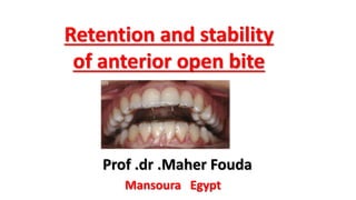
- 1. Retention and stability of anterior open bite Prof .dr .Maher Fouda Mansoura Egypt
- 2. What are retention considerations in open bite cases? An open bite malocclusion may be dental or skeletal in nature. A dental open bite may be caused by depression of the incisors because of a habit such as thumb- or finger-sucking or poor tongue posture.
- 3. A good cephalometric value to differentiate between a dental and skeletal open bite is incision-stomion; dental open bites have intruded maxillary incisors whereas skeletal open bites have a normal incisor position.
- 4. NCISION - STOMION DISTANCE: A 3 to 4 mm incision - stomion distance is esthetically pleasing.
- 5. The open bite must be accurately diagnosed and treated if relapse is to be prevented. Treatment and retention of relapsed anterior openbite with low tongue posture and tongue-tie: A 10-year follow-up [Korean J Orthod 2014;44(4):203-216]
- 6. The open bite must be accuratel y diagnose d and treated if relapse is to be prevented Facial and intraoral photographs of relapse after 3-year post-treatment Treatment and retention of relapsed anterior openbite with low tongue posture and tongue-tie: A 10-year follow-up [Korean J Orthod 2014;44(4):203-216]
- 7. In skeletal open bite, incisors are in a normal position, but the posterior teeth have elongated.
- 8. Controlling the eruption of the maxillary molars with high-pull headgears and a transpalatal bar with a midline acrylic palatal button 4 mm off of the palate is useful to control extrusion a Patient with Clark Twinblock with flying tubes to insert a Headgear. b Patient wearing Clark Twinblock with high-pull headgear
- 9. Camouflage of a high-angle skeletal Class II open-bite malocclusion in an adult after mini- implant failure during treatment American Journal of Orthodontics and Dentofacial Orthopedics March 2017 " Vol 151 " Issue 3
- 10. Retention of closed anterior open bite is a major problem. After surgery Pretreatment open bite Presurgical orthodontics 1year after appliance removal
- 11. One reason is that vertical growth and eruption of posterior teeth may continue until the late teenage years or early twenties, with vertical growth of the maxilla being the last stage of maturation.
- 12. Recurrence of the open bite malocclusion is possible and that’s why a long retention phase appears to be very important, particularly in following instances : Nonsurgical approach to Class I open-bite malocclusion with extrusion mechanics: A 3- year retention case report American Journal of Orthodontics and Dentofacial Orthopedics April 2015 Vol 147 Issue 4
- 13. Nonsurgical approach to Class I open-bite malocclusion with extrusion mechanics: A 3- year retention case report American Journal of Orthodontics and Dentofacial Orthopedics April 2015 Vol 147 Issue 4 Three-year retention facial and intraoral photographs A fixed retainer was attached to the lingual surface of the mandibular anterior teeth. Overlayed Hawley retainers were fabricated and delivered to secure the stability of both arches. A tongue crib was incorporated in the maxillary Hawley retainer to prevent relapse of the tongue- thrust habit. Total treatment time for this patient was 24 months.
- 14. Appearance pre-op (top row), post-q (middle row) and 36 months post-op (hottom row). 1-Macroglossia. 2-Dolichofacial growth after the completion of the treatment. RELAPSE FOLLOWING SURGICAL TREATMENT OF ANTERIOR OPEN BITE British Journal of Oral and Maxillofacial Surgery (1986) 24, 391-404 long retention phase appears to be very important in
- 15. (A) Example of a high angle case with mandibular retrusion and anterior open bite treated demonstrating relapse of the orthognathic surgical correction of a severe anterior open bite. (B) Extra and intraoral photographs before orthognathic surgical treatment after orthognathic surgical treatment (C) and 2 years after orthognathic surgical treatment
- 16. Orthodontic treatment of severe anterior open bite and alveolar bone defect complicated by an ankylosed maxillary central incisor: a case report Lin et al. Head & Face Medicine 2014, 10:47 Stages in the orthodontic tooth alignment. (A– F) Orthodontic traction and alignment of the tooth; (G) A modified Hawley retainer bonded with resin to the central incisors to provide retention for the tooth in the dental arch. Pre-treatment photographs showing a severe anterior open bite from the left maxillary canine to the right lateral incisor. The maxillary left central incisor was severely infra-occluded and the right central incisor had a fractured crown.
- 17. Orthodontic treatment of severe anterior open bite and alveolar bone defect complicated by an ankylosed maxillary central incisor: a case report Lin et al. Head & Face Medicine 2014, 10:47 intraoral photographs of patient at 2- year follow-up Post- treatment intraoral photographs
- 18. 3- Restart of dysfunctional habits. 4-Tongue function was not corrected well. long retention phase appears to be very important in
- 19. Relapse of AOB has been attributed to: Inappropriate orthodontic tooth movement, such as extrusion of incisors where their eruption had not been previously impeded , Appearance pre-op (top row), post- op (middle row). and 36 months post-op (bottom
- 20. Relapse of AOB has been attributed to: Surgery that has increased the posterior face height – as would occur if the aob is closed using a mandibular procedure only. , Appearance pre-op (top row). post- op (middle row) and 24 months post-op (bottom row).
- 21. Surgical Orthodontic Treatment of Severe Class Iii and Anterior Open Bite World Journal of Oral and Maxillofacial Surgery 2018 | Volume 1 | Issue 1 | Article 1004 Pre-surgical photographs. Initial photographs.
- 22. Surgical Orthodontic Treatment of Severe Class Iii and Anterior Open Bite World Journal of Oral and Maxillofacial Surgery 2018 | Volume 1 | Issue 1 | Article 1004 The surgery was performed under general anesthesia, with nasal intubation, and the sagittal surgical technique for the mandibular setback with counterclockwis e rotation was used. photographs of 1 month after surgery.
- 23. Surgical Orthodontic Treatment of Severe Class Iii and Anterior Open Bite World Journal of Oral and Maxillofacial Surgery 2018 | Volume 1 | Issue 1 | Article 1004 Intermaxillary elastics for posterior and anterior intercuspation were used, such as post- surgical blockage, for 10 days. photographs of 1 month after surgery.
- 24. Surgical Orthodontic Treatment of Severe Class Iii and Anterior Open Bite World Journal of Oral and Maxillofacial Surgery 2018 | Volume 1 | Issue 1 | Article 1004 After this time, the elastics continued to be used at night, for maintenance and stabilization of the occlusion obtained after surgery. photographs of 1.5 years follow-up.
- 25. Relapse of open bite can occur because of tongue size or posture, digit- sucking habits, respiratory problems,and condylar resorption. Vertical relapse after orthodontic and orthognathic surgical treatment in a patient with myotonic dystrophy European Journal of Paediatric Dentistry vol. 20/1-2019 Intra-oral photographs before treatment. Intra-oral photographs after active treatment. 13.5 years post-treatment: intra-oral photographs .
- 26. Anterior open bite due to idiopathic condylar resorption during orthodontic retention of a Class II Division 1 malocclusion American Journal of Orthodontics and Dentofacial Orthopedics October 2019 Vol 156 Issue 4 Pretreatment photographs and radiographs.
- 27. Anterior open bite due to idiopathic condylar resorption during orthodontic retention of a Class II Division 1 malocclusion American Journal of Orthodontics and Dentofacial Orthopedics October 2019 Vol 156 Issue 4 Posttreatment photographs and radiographs
- 28. Anterior open bite due to idiopathic condylar resorption during orthodontic retention of a Class II Division 1 malocclusion American Journal of Orthodontics and Dentofacial Orthopedics October 2019 Vol 156 Issue 4 Radiographic imaging (CBCT) of the patient's condylar resorption. A, 10 months posttreatment;
- 29. Anterior open bite due to idiopathic condylar resorption during orthodontic retention of a Class II Division 1 malocclusion American Journal of Orthodontics and Dentofacial Orthopedics October 2019 Vol 156 Issue 4 Radiographic imaging (CBCT) of the patient's condylar resorption. A, 10 months posttreatment;
- 30. Anterior open bite due to idiopathic condylar resorption during orthodontic retention of a Class II Division 1 malocclusion American Journal of Orthodontics and Dentofacial Orthopedics October 2019 Vol 156 Issue 4 Radiographic imaging (CBCT) of the patient's condylar resorption. B, 15 months posttreatment.
- 31. Anterior open bite due to idiopathic condylar resorption during orthodontic retention of a Class II Division 1 malocclusion American Journal of Orthodontics and Dentofacial Orthopedics October 2019 Vol 156 Issue 4 Radiographic imaging (CBCT) of the patient's condylar resorption. B, 15 months posttreatment.
- 32. Anterior open bite due to idiopathic condylar resorption during orthodontic retention of a Class II Division 1 malocclusion American Journal of Orthodontics and Dentofacial Orthopedics October 2019 Vol 156 Issue 4 Lateral cephalograms and panoramic radiographs showing the progress of ICR with aggravated open bite, condylar shape change, and shortening of the mandibular length. A, pretreatment;B, posttreatment;
- 33. Anterior open bite due to idiopathic condylar resorption during orthodontic retention of a Class II Division 1 malocclusion American Journal of Orthodontics and Dentofacial Orthopedics October 2019 Vol 156 Issue 4 Lateral cephalograms and panoramic radiographs showing the progress of ICR with aggravated open bite, condylar shape change, and shortening of the mandibular length. C, 10 months posttreatment; D, 15 months posttreatment.
- 34. Anterior open bite due to idiopathic condylar resorption during orthodontic retention of a Class II Division 1 malocclusion American Journal of Orthodontics and Dentofacial Orthopedics October 2019 Vol 156 Issue 4 Progress of anterior open bite over time shown in intraoral and profile photos. A, posttreatment (14.1 years old); B, 10 months posttreatment (14.11 years old); C, 15 months posttreatment (15.4 years old); D, 23 months posttreatment (16.0 years old).
- 35. Anterior open bite due to idiopathic condylar resorption during orthodontic retention of a Class II Division 1 malocclusion American Journal of Orthodontics and Dentofacial Orthopedics October 2019 Vol 156 Issue 4 . Phases of condylar degeneration from normal TMJ to destructive, repair, and finally stable stage. A, normal TMJ; B, beginning of active/destructive phase; C, “cup” shaped defect in superior surface of condyle (B and C, stages vulnerable to biomechanical force); D, beginning of repair stage; E, advanced repair phase; F, stable stage.
- 36. Preoperative Occlusion. Demonstration of Preoperativ e Frontal Repose. Preoperative Cephalometric Radiograph Preoperati ve Frontal Smile. 6-month Postoperative Occlusion 12-month Postoperative Occlusion Stability and Relapse in Orthognathic Surgery Neeraj Panchal, DDS, MD, MA Christine Ellis, DDS, MSD Paul Tiwana, DDS, MD, MS, FACS Relapse Due To Condylar Resorption One Year After Bilateral Sagittal Split Osteotomy To Correct Apertognat hia and Mandibular Deficiency.
- 37. Tongue habits, particularly tongue–thrust swallowing, are often blamed for relapse into open bite, but the evidence to support this contention is not convincing Patient with an anterior open bite which was believed to be due to an endogenous tongue thrust. Both upper and lower incisors were proclined. The patient did not have a digit-sucking habit.
- 38. Open-bile cases present a great challenge to the orthodontist. It is important lo evaluate tongue position and tongue habits in the finishing stages of treatment.
- 39. Hopefully, this problem was observed prior to this stage, and myofunclional therapy initiated if the habit was not corrected.
- 40. Active habits (of which thumb sucking is the best example) can produce intrusive forces on the incisors, while at the same time leading to an altered posture of the jaw that allows posterior teeth to erupt. The occlusal effects of a persistent digit-sucking habit. Note the anterior open bite and the unilateral posterior crossbite.
- 42. Relapse of AOB has been attributed to: Unfavourable growth (a posterior mandibular growth rotation) soft-tissue factors such as an unfavourable tongue posture Resumption of a digit-sucking habit; ,
- 43. - Bite elevation after eruption of the second molars. - After an active intrusion of molars or extrusion of the front.
- 44. Severe open bite due to traumatic condylar fractures treated nonsurgically with implanted miniscrew anchorage female patient was 36 years 0 months of age at the first examination. She had chief complaint of chewing difficulties associated with the anterior teeth.
- 45. Severe open bite due to traumatic condylar fractures treated nonsurgically with implanted miniscrew anchorage She had injured her head and face in a traffic accident 6 months before her visit. Initially, her frontal facial appearance showed left deviation of the mandible, and her lateral profile exhibited posterior mandibular positioning.
- 46. Severe open bite due to traumatic condylar fractures treated nonsurgically with implanted miniscrew anchorage Tongue thrusting was observed during conversation. The dental midline of her mandible was deviated 2 mm to the left relative to the facial midline. Her molars showed occlusal interferences, and her anteroposterior relationship was Angle Class II, with an overjet of 5.2 mm and an overbite of 4.0 mm .
- 47. Severe open bite due to traumatic condylar fractures treated nonsurgically with implanted miniscrew anchorage On the pretreatment 3- dimensional computed tomography images, we found that both condyles were deformed; however, the fracture lines were not clear . Pretreatment 3-dimensional computed tomography scans.
- 48. The results of an examination with a 3-dimensional, 6 degree of freedom jaw movement measurement apparatus (Gnathohexagraph system, version 1.31; Ono Sokki, Kanagawa, Japan) showed that the movements of the bilateral condyles during lateral sliding movements and anterior movements were irregular, with the incisal path unstable. With an occlusal force recording system (Dental Prescale and Occluzer; Fuji Film, Tokyo, Japan), her maximum occlusal force was found to be relatively weak (201 N). No impacted teeth were seen on the panoramic radiograph . The cephalometric analysis showed that she had a skeletal Class II anteroposterior jaw relationship with an ANB angle of 8.3
- 49. Pretreatment cephalogram and panoramic radiograph.
- 50. Oral photographs obtained: A, at the start of treatment;
- 51. Oral photographs obtained: C, 16 months later; D, 24 months later; E, 32 months later.
- 52. Posttreatment photographs. The retention involved the use of maxillomandibular circumferential retainers and the attachment of a lingual bonded retainer to the mandibular incisors.
- 53. Posttreatment oral photographs: A, 6 months posttreatment; B, 18 months posttreatment.
- 54. The fallacy of tongue thrust and non-surgical treatment of a severe anterior open bite Journal of Dental Health Oral Disorders & Therapy Volume 4 Issue 4 - 2016 The patient is a 33-year old female dentist who presents with a chief complaint of an open bite and poor posterior occlusion. As a 12-year old child growing up in Serbia, the patient accompanied by her parents first presented to the private family dentist for evaluation and treatment. She was diagnosed with a skeletal open bite secondary to a “tongue thrust problem” which her dentist described as continuous suckling.
- 55. She was given a series of removable habit correcting appliances which she used as instructed but tapered herself off in about a year because treatment was ineffective. Several years passed before the patient returned to a public dentist for treatment at the age of 19 where she was given removable orthodontic/orthopedic appliances followed by application of brackets prior to surgical orthodontic treatment.
- 56. Once again her anterior open bite was attributed to tongue thrust. Because of the uncertain outcome and difficulty associated with the surgical orthodontic procedure as described by her dentist and surgeon she decided not to pursue treatment and brackets were removed. Shortly thereafter she started dental school where she was seen by a professor in the department of orthodontics.
- 57. She was told that surgical orthodontics was the only viable treatment option but was once again cautioned of the difficulty and uncertain outcome of the procedure. She once again decided to forgo surgery and all orthodontic treatment for several years.
- 58. Treatment took 15 months with appointments scheduled approximately on a monthly basis. Brackets were initially placed on the four maxillary incisors for patient comfort for one month. At the second appointment brackets were placed on all remaining maxillary teeth including the properly occluding second molars.
- 59. This set up provided appropriate force and adequate torque for both the maxillary first molars and all premolar roots to upright and aligns the maxillary arch by inducing alveolar bone growth in order to provide proper occlusion with opposing mandibular teeth.
- 60. At the third visit and three months into treatment, brackets were placed on the mandibular teeth with elastics to close the anterior bite. The treating co- author notes that treatment time could have been substantially less had the patient diligently complied with the use of elastics.
- 61. Two-year post-treatment follow- up, intra-oral frontal view. Post-treatment photographs. non-surgical, non-extraction orthodontic treatment
- 62. A non-surgical correction of an open bite was proven to have a high stability rate as well as the surgical correction.
- 63. Most of the relapses happen during the first year of the retention. Many studies showed that extraction treatment had greater stability than a nonextraction treatment. Still non- extraction therapy provided a reliable stability of results.
- 64. Studies of long-term outcomes following orthodontic treatment for open bite, and following surgically treated cases indicated that the relapse rate is about 40%.
- 65. If correction of severe open bite is not started in the mixed dentition, it will most likely require orthognathic surgery in late adolescence or adulthood. The skeletal open bite phenotype is easily diagnosed in the early mixed dentition.
- 66. Controlling the eruption of posterior teeth during late vertical growth is the key to preventing open bite relapse..
- 67. Essix-type retainers with full-thickness plastic posterior coverage are the best retainers for open bites because they maintain intrusive force on the posterior teeth, which in turn maintains good anterior bite depth.
- 68. Clear and Fixed Retainer's Outcomes After Orthodontic Ally Treated Open Bite Cases : Clinical Study Medical Journal of Babylon Vol. 13- No. 2: 271 - 276 , 2016
- 69. There are another two major approaches to accomplishing this: a maxillary retainer with bite blocks (or a functional appliance) to impede eruption, or high-pull headgear.
- 70. Retention has been directed towards intrusion, or at least prevention of eruption, of maxillary posterior teeth, using headgear attached to an upper removable retainer. A high-pull headgear device attached to a removable maxillary retainer to control the post-orthopedic phase of growth and retain the dentoalveolar correction The removable retainer incorporates headgear tubes attached to the Adam’s clasps on the upper molars to facilitate insertion of the facebow
- 71. However, this should ideally be continued until the patient ceases growing, although compliance is obviously an issue.
- 72. Although highpull headgear can be quite effective in a cooperative patient, a removable appliance with bite blocks is a better choice for most patients for two reasons: it controls eruption of both the upper and lower molars, and usually it is better accepted because it is easier to wear.
- 73. The removable appliance with bite blocks stretches the patient’s soft tissues to provide a force opposing eruption.
- 74. High-pull headgear to the upper molars, in conjunction with a standard removable retainer to maintain tooth position, also can be effective, but the intraoral appliance is better tolerated and controls eruption of lower as well as upper posterior teeth. A high-pull headgear device attached to a removable maxillary retainer to control the post-orthopedic phase of growth and retain the dentoalveolar correction. he removable retainer incorporates head- gear tubes attached to the Adam’s clasps on the upper molars to facilitate insertion of the facebow.
- 75. following orthodontic treatment, more than one third of patients demonstrated a return of their AOB . Lopez- Gavito et al. reported that,
- 76. Neither the extent of the pretreatment open bite or mandibular plane angle nor any other single parameter of dentofacial form was a reliable predictor of post- treatment stability. Lopez- Gavito et al. also reported that,
- 77. Relapse into anterior open bite can occur by any combination of depression of the incisors and elongation of the molars.
- 78. There is general agreement among orthodontists that patients with AOBs are challenging to treat, and relapse is common after treatment with orthodontics alone or combined with orthognathic surgery.
- 80. Thus, clinicians should pay more attention during retention phase and long-term studies on post- treatment changes and stability should be encouraged.
- 81. Overcorrecting the anterior bite as much as possible is recommended, owing to the high incidence of relapse.
- 82. The problem of retaining open bite corrections is the same irrespective of the treatment that closed the open bite. A new skeletal retention system for retaining anterior open bites APOS Trends in Orthodontics| March 2013 |Vol 3 | Issue 2
- 83. It is expected that LeFort surgical open bite closure procedures will be relatively stable, and these treatments actually reduce the space for the tongue.
- 84. The extrusion arches probably have no more nor less ability to be stable than any other treatment procedure.
- 85. When considering the patient's investment of time, discomfort, and money, the issue of stability becomes even more important.
- 86. Conventional wraparound retainer with a tongue grid Skeletal class III and anterior open bite treatment with different retention protocols: a report of three cases Journal of Orthodontics, Vol. 39, 2012, 212–223 It is recommended that patients wear this retainer during the night while sleeping. The retainer consists of a conventional removable appliance made with a 0.9- mm stainless steel labial bow placed at the middle third of the crown and with a tongue grid fixed on the acrylic portion to discourage abnormal tongue posture, which is hopefully corrected by the muscle exercises during the day.
- 87. modified retainer adapted at the cementoenamel junction of the anterior teeth Skeletal class III and anterior open bite treatment with different retention protocols: a report of three cases Journal of Orthodontics, Vol. 39, 2012, 212–223 Daytime wraparound retention with modified contour It is recommended that patients wear this retainer during the day. It has a 0.8-mm stainless steel wire that contours around the gingival margin of the anterior teeth. The aim is to reduce relapse due to intrusion or protrusion of the anterior teeth
- 88. Skeletal class III and anterior open bite treatment with different retention protocols: a report of three cases Journal of Orthodontics, Vol. 39, 2012, 212–223 Case 1 — post- treatment intra-oral photographs (a) frontal, (b) right side, (c) left side, (d) upper occlusal, (e) lower occlusal, (f) daytime wraparound retention and (g) night-time wraparound retention
- 89. Skeletal class III and anterior open bite treatment with different retention protocols: a report of three cases Journal of Orthodontics, Vol. 39, 2012, 212–223 Case 1 — 2 years post- retention intra-oral photographs : (a) frontal, (b) right side and (c) left side
- 90. Skeletal class III and anterior open bite treatment with different retention protocols: a report of three cases Journal of Orthodontics, Vol. 39, 2012, 212–223 Case 2 — pre- treatment intra-oral photographs: (a) frontal, (b) right side, (c) left side, (d) upper occlusal and (e) lower
- 91. Skeletal class III and anterior open bite treatment with different retention protocols: a report of three cases Journal of Orthodontics, Vol. 39, 2012, 212–223 Case 2 — post- treatment intra-oral photographs: (a) frontal, (b) right side, (c) left side, (d) upper occulsal, (e) lower occlusal, (f) and (g) daytime retention
- 92. Skeletal class III and anterior open bite treatment with different retention protocols: a report of three cases Journal of Orthodontics, Vol. 39, 2012, 212–223 Case 2 — 6-year post-retention intra-oral photographs: (a) frontal, (b) right side and (c) left side
- 93. Skeletal class III and anterior open bite treatment with different retention protocols: a report of three cases Journal of Orthodontics, Vol. 39, 2012, 212–223 Case 3 — pre- treatment intra-oral photographs: (a) frontal, (b) right side, (c) left side, (d) upper occlusal and (e) lower occlusal
- 94. Skeletal class III and anterior open bite treatment with different retention protocols: a report of three cases Journal of Orthodontics, Vol. 39, 2012, 212–223 Case 3 — post- treatment intra- oral photographs P: (a) frontal, (b) right side, (c) left side, (d) upper occlusal, (e) lower occlusal and (f) daytime wraparound retention
- 95. Skeletal class III and anterior open bite treatment with different retention protocols: a report of three cases Journal of Orthodontics, Vol. 39, 2012, 212–223 0 Case 3 — 5 years post-retention intra-oral photographs: (a) frontal, (b) right side and (c) left side These three cases demonstrate adequate stability with this retention protocol, which we believe is suitable for open bite patients treated with incisor extrusion.
- 96. Drawings showing the differences between the action of the two retainers — conventional (a) and modified (b)
- 97. American Journal of Orthodontics and Dentofacial Orthopedics July 2010 Stability of anterior open-bite treatment with occlusal adjustment The sample consisted of 17 patients (7 male, 10 female). All patients originally had an anterior open-bite malocclusion, had undergone orthodontic treatment with fixed appliances, had anterior open-bite relapse after a mean posttreatment period of 4.15 years (range, 1-6 years), and were retreated with the occlusal adjustment procedure.
- 98. If the intra-arch alignment of the teeth is acceptable, and the position of her anterior teeth in the face is reasonable the first option generally consider for an anterior open bite is occlusal equilibration; altering the posterior occlusion by reshaping the teeth, effectively closing the vertical dimension and bringing the anterior teeth into contact OCCLUSION & WEAR A Guide to Anterior Open Bites By Frank Spear on February 18, 2018 Intraoral photographs before occlusal adjustment show the anterior open bite. Intraoral photographs after occlusal adjustment show the corrected anterior open bite
- 99. The risk of equilibration is that it may require excessive tooth reduction, exposing dentin in the process, and it may be inadequate to achieve the necessary closure to gain anterior contact. For these reasons, a trial equilibration is typically performed on mounted models to see if the desired occlusion can be obtained with just equilibration - and to evaluate the amount of alterations necessary to the posterior teeth OCCLUSION & WEAR A Guide to Anterior Open Bites By Frank Spear on February 18, 2018
- 100. American Journal of Orthodontics and Dentofacial Orthopedics July 2010 Stability of anterior open-bite treatment with occlusal adjustment Intraoral photographs taken before the occlusal adjustment show the anterior open bite Intraoral photographs taken after the occlusal adjustment show correction of the anterior open bite, with a positive overbite. Intraoral photographs taken in the long term after the occlusal adjustment show clinical stability of the open-bite correction with the occlusal adjustment. However, a slight reduction in the overbite is obvious.
- 101. American Journal of Orthodontics and Dentofacial Orthopedics July 2010 Stability of anterior open-bite treatment with occlusal adjustment CONCLUSIONS 1. There was a statistically significant relapse of anterior open bite in the whole sample; growth seemed to have contributed to a significant amount of the relapse. 2. The primary factor that contributed to the relapse was the increase in posterior molar height, consequent to compensatory posterior tooth eruption. 3. There was clinically significant stability in 66.7% of the patients. 4. Dentinal sensitivity remained within the normal range in the long term.
- 102. Relapse tendency can be minimized if a habit-breaking appliance or a tongue crib is used for at least 6 months prior to any incisor extrusion mechanics.
- 103. The selection of retention device should be based on etiology and the mechanics employed to close the bite. Bluegrass Maxillary Hawley retainer
- 104. In this way, some patients learn to modify their tongue position or activity, by holding the tip of the tongue in the roof of the palate during swallowing and other activities.
- 105. However, in some cases, a tongue will reassert itself, despite the best efforts of the patient and the orthodontist. The patient should be informed of this possibility before treatment.
- 106. These cases will often benefit from the use of positioners to help bite closure. If a conventional upper retainer is to be used, a small hole can be placed in the palatal surface of the acrylic, for tongue positioning.
- 107. In patients who do not place some object between the front teeth, return of open bite is almost always the result of elongation of the posterior teeth, particularly the upper molars, without any evidence of intrusion of incisors Facial and intraoral photographs of relapse after 3-year post- treatment
- 108. Controlling eruption of the upper molars therefore is the key to retention in open bite patients Facial and intraoral photographs of relapse after 3-year post- treatment
- 109. . Four years after removal of the orthodontic appliances, this 17-year-old has an anterior open bite, 5 mm of overjet with an end-on molar relationship, and severe crowding of the mandibular incisors..
- 110. Relapse of this type is associated with little or no mandibular growth and a downward and backward mandibular rotation as the maxilla grows downward and upper posterior teeth erupt, as shown in the cephalometric superimposition from the end of treatment to 4-year recall.
- 111. . The incisor crowding is due to uprighting and lingual repositioning of the incisors as the mandibular rotation thrusts them into the lower lip.
- 112. The question of retainers is difficult because the typical Hawley type retainer has little impact on open bites. Bluegrass Maxillary Hawley retainer
- 113. Improving Retention of Anterior Open-Bite Cases To improve the retention of an anterior open-bite case or recover an open-bite relapse, small composite bumps can be bonded to the labial surfaces of the incisors. Before light-curing,the paste is shaped into smooth round balls using a brush saturated with primer solution
- 114. Improving Retention of Anterior Open-Bite Cases The labial wire of a Hawley-type retainer is slightly activated incisally and placed against the gingival edges of the composite bumps.
- 115. Improving Retention of Anterior Open-Bite Cases For more positive retention, the gingival edges can be shaped into horizontal steps with a fluted bur . These steps are not required when using positioners or clear plastic retainers.
- 116. A removable retainer can keep teeth from tipping to the facial, which, if occurring, will reduce overbite. Removable retainers cannot prevent actual translation of the center of resistance in an intrusive direction.
- 117. A) and (B): Retention plate with anterior tongue crib to avoid tongue thrust in the anterior teeth, anterior tongue posture, and a posterior bite block to restrict vertical development of the posterior teeth.
- 118. (C) and (D): Retention plate with an orifice in the region of incisive papillae to condition correct tongue position.
- 119. Positioners can be considered during retention, because of their bite- closing effect. POSITIONER
- 120. A silicone positioner is often helpful if used for 4 hours during the day and during sleep for at least 6 months.
- 121. After the positioner use, a wraparound retainer for the upper arch is better than a conventional Hawley, as the objective should be to prevent wires between the occluding surfaces
- 122. Using lingual upper and lower fixed retainers is suggested Resin fiberglass band retainer bonded to all anterior teeth from first premolar to first premolar
- 123. The removal of acrylic from the incisor area of the upper retainer is recommended, along with the placement of a small hole in the anterior region as a reminder for the tongue. Upper Hawley style removable retainer. removal of acrylic from the incisor area
- 124. Results achieved with extrusion arches have been generally positive. The preferred method to control relapse toward anterior open bite is an appliance with bite blocks between the posterior teeth that creates several millimeters of jaw separation (an open bite activator or bionator. Severe anterior open bites (front teeth that do not touch)
- 125. In a patient with the long-face growth pattern, either must be continued as a nighttime retainer through the late teens.
- 126. Excessive vertical growth and eruption of the posterior teeth often continue until late in the teens or early twenties, so retention also must continue well beyond the typical completion of active treatment.
- 127. Cephalometric superimpositions showing the chin position as a result of growth changes in a patient between 7 and 14 years of age. It was observed that the chin moved downward and forward till the age of 11 years; thereafter, it was displaced downward and backward as a result of less condylar growth and more vertical growth in the molar area
- 128. A patient with a severe open bite problem is particularly likely to benefit from having conventional maxillary and mandibular retainers for daytime wear and an open bite bionator as a nighttime retainer from the beginning of the retention period.
- 129. The etiology of the anterior open bite should be determine d before treatment. Facial and intraoral photographs of relapse after 3-year post-treatment Treatment and retention of relapsed anterior openbite with low tongue posture and tongue-tie: A 10-year follow-up
- 130. Understanding Post-orthodontic Relapse and Retention by Eli Halabi, DMD the Journal: summer 2015 Feature Article Higher incidence of relapse has been noted in cases when the incisors are extruded to close an open bite that is skeletal in nature.
- 131. A more appropriate approach for such patients would be orthognathic surgery, or the use of temporary anchorage devices (TADs) to intrude the posterior teeth so as to close the anterior open bite.
- 132. Further, newfound success has been shown in using Clear Aligner Therapy to intrude the posterior teeth to aid in closing the anterior open bite.
- 133. Accordingly, Essix-type retainers with full-thickness plastic posterior coverage are the best retainers for open bites because they maintain intrusive force on the posterior teeth, which in turn maintains good anterior bite depth.
- 134. The postulated clinical theory purports that the thermoplastic between the posterior teeth causes a “bite- block effect,” keeping the posterior teeth from extruding and therefore preventing open-bite relapse
- 135. Fabrication of the spur-implanted Essix Retainers . Before the fabrication of Essix plate on the upper cast, a small amount of dental stone was added near the incisive papilla region in order to create space for insertion and securing of the wire. C type Essix plates (0.40) were fabricated over the casts and trimmed. A 0.9mm stainless steel laboratory wire is bent in “U” shape with a small helix at the base for retention. a) Upper cast. b) Bump on the lingual side of incisors. c) U shaped wire. d) Essix plate for the upper cast. e) Insertion of the wire. f) Addition of self-curing acrylic. g) Retainer after polishing. progress in orthodontics 1 1 (2010) 45–52 A new type of modified Essix Retainer for anterior open bite retention
- 136. The wire is heated and inserted to the space created on the lingual side of incisors. The hot wire easily punctures, and when cooled, sticks to the Essix plate. A small amount of self- curing acrylic is applied to fill the rest of the space in order to keep the wire firmly in place. The spurs are sharpened and polished accordingly after the curing of the acrylic (Fig. 9).
- 137. Retainers used after active phase of orthodontic treatment. (a) Two retainers with posterior bite blocks were constructed; (b) Daytime retainer with an orifice close to the incisive papillae and; (c) Night-time retainer with a palatal crib Wrap-around retainers were provided for the maxillary arch and a 0.028-inch stainless-steel arch wire segment was bonded to the mandibular anterior teeth. Nonsurgical treatment and stability of an adult with a severe anterior open-bite malocclusion 2018 Journal of Orthodontic Science
- 138. Retainers used after active phase of orthodontic treatment. (a) Two retainers with posterior bite blocks were constructed; (b) Daytime retainer with an orifice close to the incisive papillae and; (c) Night-time retainer with a palatal crib Two maxillary appliances with posterior bite blocks to retain the posterior intrusion were constructed for day and night-time use
- 139. Retainers used after active phase of orthodontic treatment. (a) Two retainers with posterior bite blocks were constructed; (b) Daytime retainer with an orifice close to the incisive papillae and; (c) Night-time retainer with a palatal crib The daytime retainer incorporated an orifice close to the incisive papillae which guided correct tongue position
- 140. Retainers used after active phase of orthodontic treatment. (a) Two retainers with posterior bite blocks were constructed; (b) Daytime retainer with an orifice close to the incisive papillae and; (c) Night-time retainer with a palatal crib .The night-time retainer contained a palatal crib to prevent lingual pressure on the anterior teeth .
- 141. A loop was made in the mandibular retention wire to allow subsequent restoration of the left central incisor
- 142. Relapse of the non- extraction groups mainly happened in the first year after finishing treatment. Many studies showed that the post- treatment phase of non- extraction therapy was longer in comparison to extraction treatment, therefore producing a higher tendency for relapse.
- 143. In some cases the wrap-around retainer (in the maxilla) is used 24 h/day in the first 8 months, half a day (at night) for an additional 3 months and every other night in the last month of use. A lower retainer in the six anterior teeth (3-3) are set for undetermined ending time . The patient is urged to maintain her orofacial myofunctional therapy with the speech therapist for additional 12 months. Case report
- 144. Treatment and retention of relapsed anterior openbite with low tongue posture and tongue-tie: A 10-year follow-up pISSN 2234-7518 • eISSN 2005-372X http://dx.doi.org/10.4041/kjod.2014.44.4.203 Design of the tongue elevator: The acrylic base occupies the entire mouth floor except for the region that can disturb the movement of the lingual frenum.
- 145. Treatment and retention of relapsed anterior openbite with low tongue posture and tongue-tie: A 10-year follow-up pISSN 2234-7518 • eISSN 2005-372X http://dx.doi.org/10.4041/kjod.2014.44.4.203 The occlusal rests are placed on the lingual occlusal grooves of the posterior teeth. In a modified tongue elevator , the volume and height of the resin part are reduced for tongue-tie.
- 146. If the open bite is due to a persistent low tongue posture, a tongue elevator is applied as an active retreatment alternative, and is used thereafter as a retainer
- 147. A tongue elevator is a removable appliance comprising an acrylic base, occlusal rests, and several retentive elements
- 148. The acrylic base occupies the entire sublingual space, except for the lingual frenum, and the occlusal rests are placed in the lingual occlusal grooves of the posterior teeth. For retention, a labial bow and other retentive clasps can be added.
- 149. The function of the tongue elevator is to keep the tongue in a higher than usual position, which is accomplished by the sheer volume of the acrylic base. The positional change induced by the tongue elevator brings about three dentoalveolar effects
- 150. First, the tongue tends to go back to its original position when elevated, which generates a downward force. This force is then transmitted to the occlusal rests of the appliance, which results in intrusion of the lower posterior teeth. Second, when elevated, the tongue occupies the space under the palatal vault, and contacts the upper dentition
- 151. In this position, the tongue exerts an outward force that results in upper arch expansion. Finally, the pushing force exerted by the elevated tongue can be used in conjunction with a transpalatal arch, or upper removable retainer with occlusal rests, to intrude the upper posterior teeth.
- 152. Thus, the anterior open-bite can be corrected by inhibition of posterior alveolar growth
- 153. Journal of Orthodontic Science | 2018 Nonsurgical treatment and stability of an adult with a severe anterior open-bite malocclusion
- 154. Wrap-around retainers were provided for the maxillary arch and a 0.028-inch stainless-steel arch wire segment was bonded to the mandibular anterior teeth. Two maxillary appliances with posterior bite blocks to retain the posterior intrusion were constructed for day and night-time use. Retainers used after active phase of orthodontic treatment. (a) Two retainers with posterior bite blocks were constructed; (b) Daytime retainer with an orifice close to the incisive papillae and; (c) Night-time retainer with a palatal crib
- 155. The daytime retainer incorporated an orifice close to the incisive papillae which guided correct tongue position.The night-time retainer contained a palatal crib to prevent lingual pressure on the anterior Figure 4: Miniscrews placed and biomechanics employed for molar intrusion teeth. Retainers used after active phase of orthodontic treatment. (a) Two retainers with posterior bite blocks were constructed; (b) Daytime retainer with an orifice close to the incisive papillae and; (c) Night-time retainer with a palatal crib
- 156. A loop was made in the mandibular retention wire to allow subsequent restoration of the left central incisor .