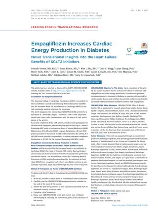
1. LEADING EDGE IN TRANSLATIONAL RESEARCH
Empagliflozin Increases Cardiac
Energy Production in Diabetes
Novel Translational Insights Into the Heart Failure
Benefits of SGLT2 Inhibitors
Subodh Verma, MD, PHD,a,
* Sonia Rawat, BSC,b,
* Kim L. Ho, BSC,b,
* Cory S. Wagg,b
Liyan Zhang, PHD,b
Hwee Teoh, PHD,a,c
John E. Dyck,b
Golam M. Uddin, PHD,b
Gavin Y. Oudit, MD, PHD,b
Eric Mayoux, PHD,d
Michael Lehrke, MD,e
Nikolaus Marx, MD,e
Gary D. Lopaschuk, PHDb
JACC: BASIC TO TRANSLATIONAL SCIENCE CME/MOC/ECME
This article has been selected as this month’s JACBTS CME/MOC/ECME
activity, available online at http://www.acc.org/jacc-journals-cme by
selecting the JACC Journals CME/MOC/ECME tab.
Accreditation and Designation Statement
The American College of Cardiology Foundation (ACCF) is accredited by
the Accreditation Council for Continuing Medical Education (ACCME)
and the European Board for Accreditation in Cardiology (EBAC) to pro-
vide continuing medical education for physicians.
The ACCF designates this Journal-based CME/MOC/ECME activity for a
maximum of 1 AMAPRA Category 1 Credit or 1 EBAC Credit. Physicians
should only claim credit commensurate with the extent of their partici-
pation in the activity.
Successful completion of this CME activity, which includes participation in
the evaluation component, enables the participant to earn up to 1 Medical
Knowledge MOC point in the American Board of Internal Medicine’s (ABIM)
Maintenance of Certification (MOC) program. Participants will earn MOC
points equivalent to the amount of CME credits claimed for the activity. It is
the CME activity provider’s responsibility to submit participant completion
information to ACCME for the purpose of granting ABIM MOC credit.
Empagliflozin Increases Cardiac Energy Production in Diabetes:
Novel Translational Insights Into the Heart Failure Benefits of SGLT2
Inhibitors will be accredited by the European Board for Accreditation in
Cardiology (EBAC) for 1 hour of External CME credits. Each participant
should claim only those hours of credit that have actually been spent in the
educational activity. The Accreditation Council for Continuing Medical
Education (ACCME) and the European Board for Accreditation in Cardi-
ology (EBAC) have recognized each other’s accreditation systems as sub-
stantially equivalent. Apply for credit through the post-course evaluation.
Method of Participation and Receipt of CME/MOC/ECME Certificate
To obtain credit for JACC: Basicto Translational Science CME/MOC/ECME, you
must:
1. Be an ACC member or JACC: Basic to Translational Science subscriber.
2. Carefully read the CME/MOC/ECME-designated article available on-
line and in this issue of the journal.
3. Answer the post-test questions. At least 2 questions provided must be
answered correctly to obtain credit.
4. Complete a brief evaluation.
5. Claim your CME/MOC/ECME credit and receive your certificate
electronically by following the instructions given at the conclusion of
the activity.
CME/MOC/ECME Objective for This Article: Upon completion of this activ-
ity, the learner should be able to: 1) discuss the effects of treatment with
empagliflozin on cardiac energy production; 2) examine the guideline
recommendations for treatment of diabetes in patients with or at risk for
cardiovascular disease; and 3) define the potential cardiovascular benefits
associated with the treatment of diabetes mellitus with empaglifozin
CME/MOC/ECME Editor Disclosure: CME/MOC/ECME Editor L. Kristin
Newby, MD, is supported by research grants from Amylin, Bristol-Myers
Squibb Company, GlaxoSmithKline, Sanofi, Verily Life Sciences (formerly
Google Life Sciences), the MURDOCK Study, NIH, and PCORI; receives
consultant fees/honoraria from BioKier, DemeRx, MedScape/The-
Heart.org, Metanomics, Philips Healthcare, Roche Diagnostics, CMAC
Health Education & Research Institute; serves as an Officer, Director,
Trustee, or other fiduciary role for the AstraZeneca HealthCare Founda-
tion and the Society of Chest Pain Centers (now part of ACC); and serves
in another role for the American Heart Association and is the Deputy
Editor of JACC: Basic to Translational Science.
Author Disclosures: This work was supported through an unrestricted
grant from Boehringer Ingelheim, and operating grants from the Canadian
Institutes of Health Research to Drs. Verma and Lopaschuk. Dr. Verma
holds a Tier 1 Canada Research Chair in Cardiovascular Surgery; and has
received speaker honoraria from Abbott, Amgen, AstraZeneca, Bayer,
Boehringer Ingelheim, Eli Lilly, Janssen, Merck, NovoNordisk, and Sanofi;
and received research support from Amgen, AstraZeneca, Boehringer
Ingelheim, and Eli Lilly. Dr. Oudit has received speaker honoraria from
Sanofi-Genzyme, Novartis, and Amgen. Dr. Lopaschuk is a shareholder in
Metabolic Modulators Research Ltd; and has received grant support from
Servier, Boehringer Ingelheim, Sanofi, and REMED Biopharmaceuticals.
Dr. Marx has received support for clinical trial leadership from Boehringer
Ingelheim; has served as a consultant to Amgen, Bayer, Boehringer Ingel-
heim, Sanofi, Merck Sharp & Dohme, Bristol-Myers Squibb, AstraZeneca,
NovoNordisk; has received grant support from Boehringer Ingelheim and
Merck Sharp & Dohme; and has served as speaker for Amgen, Bayer,
Boehringer Ingelheim, Sanofi, Merck Sharp & Dohme, Bristol-Myers
Squibb, AstraZeneca, Lilly, NovoNordisk. All other authors have reported
that they have no relationships relevant to the contents of this paper to
disclose.
Medium of Participation: Online (article and quiz).
CME/MOC/ECME Term of Approval
Issue Date: October 2018
Expiration Date: September 30, 2019
ISSN 2452-302X https://doi.org/10.1016/j.jacbts.2018.07.006
J A C C : B A S I C T O T R A N S L A T I O N A L S C I E N C E V O L . 3 , N O . 5 , 2 0 1 8
ª 2 0 1 8 T H E A U T H O R S . P U B L I S H E D B Y E L S E V I E R O N B E H A L F O F T H E A M E R I C A N
C O L L E G E O F C A R D I O L O G Y F O U N D A T I O N . T H I S I S A N O P E N A C C E S S A R T I C L E U N D E R
T H E C C B Y - N C - N D L I C E N S E ( h t t p : / / c r e a t i v e c o m m o n s . o r g / l i c e n s e s / b y - n c - n d / 4 . 0 / ) .
2. Empagliflozin Increases Cardiac
Energy Production in Diabetes
Novel Translational Insights Into the Heart Failure
Benefits of SGLT2 Inhibitors
Subodh Verma, MD, PHD,a,*
Sonia Rawat, BSC,b,*
Kim L. Ho, BSC,b,*
Cory S. Wagg,b
Liyan Zhang, PHD,b
Hwee Teoh, PHD,a,c
John E. Dyck,b
Golam M. Uddin, PHD,b
Gavin Y. Oudit, MD, PHD,b
Eric Mayoux, PHD,d
Michael Lehrke, MD,e
Nikolaus Marx, MD,e
Gary D. Lopaschuk, PHDb
VISUAL ABSTRACT
Verma, S. et al. J Am Coll Cardiol Basic Trans Science. 2018;3(5):575–87.
HIGHLIGHTS
This study evaluated cardiac energy
production and bioenergetics in an
experimental model of diabetes treated
with the SGLT2 inhibitor empagliflozin.
Rates of glucose oxidation, fatty acid
oxidation, ketone oxidation, glycolysis,
and cardiac function were measured in
diabetic (db/db) mice treated with or
without empagliflozin.
Rates of glucose and ketone oxidation in
the hearts of untreated db/db mice were
markedly decreased, whereas fatty acid
oxidation was increased with a significant
overall reduction in cardiac ATP
production compared to nondiabetic mice.
Empagliflozin treatment increased overall
cardiac ATP production by w30% and
prevented cardiac failure; this effect was
due to an increase in the rate of glucose
and fatty acid oxidation, but with no a
change in the rate of ketone oxidation.
The authors conclude that the SGLT2
inhibitor empagliflozin enhances the
cardiac energy pool by increasing cardiac
energy production from glucose and fatty
acid oxidation, but not ketone oxidation.
From the a
Division of Cardiac Surgery, Keenan Research Centre for Biomedical Science and Li Ka Shing Knowledge Institute of St.
Michael’s Hospital, Toronto, Ontario, Canada; b
Cardiovascular Research Centre, University of Alberta, Edmonton, Alberta, Can-
ada; c
Division of Endocrinology and Metabolism, Keenan Research Centre for Biomedical Science and Li Ka Shing Knowledge
Institute of St. Michael’s Hospital, Toronto, Ontario, Canada; d
Boehringer Ingelheim Pharma GmbH Co. KG, Biberach, Germany;
and the e
Department of Internal Medicine I–Cardiology, University Hospital Aachen, Aachen, Germany. This work was supported
through an unrestricted grant from Boehringer Ingelheim, and operating grants from the Canadian Institutes of Health Research
to Drs. Verma and Lopaschuk. Dr. Verma holds a Tier 1 Canada Research Chair in Cardiovascular Surgery; and has received
speaker honoraria from Abbott, Amgen, AstraZeneca, Bayer, Boehringer Ingelheim, Eli Lilly, Janssen, Merck, NovoNordisk, and
Sanofi; and received research support from Amgen, AstraZeneca, Boehringer Ingelheim, and Eli Lilly. Dr. Oudit has received
speaker honoraria from Sanofi, Novartis, and Amgen. Dr. Lopaschuk is a shareholder in Metabolic Modulators Research Ltd; and
has received grant support from Servier, Boehringer Ingelheim, Sanofi, and REMED Biopharmaceuticals. Dr. Marx has received
support for clinical trial leadership from Boehringer Ingelheim; has served as a consultant to Amgen, Bayer, Boehringer Ingel-
heim, Sanofi, Merck Sharp Dohme, Bristol-Myers Squibb, AstraZeneca, NovoNordisk; has received grant support from
Verma et al. J A C C : B A S I C T O T R A N S L A T I O N A L S C I E N C E V O L . 3 , N O . 5 , 2 0 1 8
SGLT2 Inhibition and Cardiac Bioenergetics in Diabetes O C T O B E R 2 0 1 8 : 5 7 5 – 8 7
576
4. aberrant changes in metabolic flexibility and sub-
strate use play an early and permissive role in the
development of heart failure and are believed to
promote adverse remodeling and progression to se-
vere heart failure in both experimental and clinical
settings (12,13,20).
It has been hypothesized that SGLT2 inhibitors
may prevent heart failure through improving ATP
generation from ketone body oxidation, thereby
enhancing cardiac efficiency (cardiac work/O2
consumed) (21,22). However, this hypothesis remains
controversial (23), and to date, no cogent evidence to
support this proposed beneficial mechanism has been
provided. Because SGLT2 inhibitors are known to in-
crease the production of ketone bodies (such as
b-hydroxybutyrate [ßOHB]), it has been suggested
that an increase in myocardial ketone body oxidation
may serve to improve cardiac ATP production as a
preferred substrate to fatty acids or glucose in the
diabetic heart. Whereas ketone bodies have been
shown to be a source of energy supply in the hyper-
trophied failing heart (24), the relationship that exists
between ketone bodies, glucose, and fatty acid
oxidation in the diabetic heart remains unclear,
particularly as it relates to SGLT2 inhibition.
We sought to shed light on the potential effects of
SGLT2 inhibitors on myocardial energetics and energy
substrate use. Mouse surrogates for diabetes (db/db
mice) were treated with 10 mg/kg/day empagliflozin
or the vehicle for 4 weeks before the rates of glycol-
ysis, fatty acid, glucose, and ketone oxidation were
evaluated in isolated perfused working hearts har-
vested from these mice.
METHODS
EXPERIMENTAL ANIMALS. The experimental proced-
ures described herein were approved by the Univer-
sity of Alberta Institutional Animal Care and Use
Committee and conform to the guidelines of the
Canadian Council of Animal Care. At 18 weeks of
age, male db/db (Leprdb
/J, The Jackson Laboratory,
Bar Harbor, Maine) mice were treated with either
empagliflozin (10 mg/kg/day via their food) or the
vehicle for 4 weeks. Concomitantly, 20-week-old
male C57BL/6J mice were treated for 4 weeks with the
vehicle via their food.
A separate group of C57BL/6J and db/db mice was
treated with a single 10 mg/kg dose of empagliflozin
via oral gavage before being subjected to a 24-h fast.
Lastly, 23-week-old naive male db/db mice were
used to study the short-term effects of ketones on the
db/db mouse heart.
TRANSTHORACIC ECHOCARDIOGRAPHY. The cardiac
function of anesthetized (3% isoflurane) mice was
assessed with a Vevo 3100 high-resolution imaging
system that was coupled to a 30-MHz transducer
(RMV-707B; VisualSonics, Toronto, Canada). Systolic
and diastolic parameters were assessed as previously
described (25).
BLOOD METABOLITE LEVELS. For both fed and
fasted protocols, total plasma ketones levels were
measured using a 2-part kit from Wako Diagnostics
(Cat: 415-73301 and 411-73491, Wako Diagnostics,
Mountain View, California), blood glucose levels
were measured using a glucometer, and plasma fatty
acid levels were measured using a kit from Roche
(Cat: 11383175001, Roche, Basel, Switzerland).
ISOLATED WORKING HEART PERFUSION. Male 22-week
to 23-week-old db/db mice and age-matched control
C57BL/6J mice were sacrificed with sodium pento-
barbital after their hearts were excised and blood
collected for plasma isolation. The hearts were
perfused for 60 min in the isolated working mode as
previously described (26) with Krebs-Henseleit solu-
tion containing 2.5 mmol/l Ca2þ
, 5 mmol/l [5-3
H/
U-14
C]glucose, 0.8 mmol/l palmitate (pre-bound to 3%
albumin), and 500 mmol/l bOHB in the presence or
absence of 500 mU/ml insulin. A second set of hearts
were perfused under identical conditions, but with 5
mmol/l glucose, 0.8 mmol/l [9,10-3
H]palmitate (pre-
bound to 3% albumin), and 500 mmol/l [3-14
C]bOHB.
To study the short-term effect of ketones on the
hearts from db/db mice, these hearts were acutely
perfused in the absence or presence of 600 mmol/l b-
OHB. At the end of the perfusion protocol, the left
ventricles were snap-frozen with liquid nitrogen and
stored at À80
C.
IMMUNOBLOTTING. Frozen left ventricular (LV)
tissue samples (30 mg) were homogenized, resolved
by SDS-PAGE, and transferred onto nitrocellulose.
The membranes were blocked in 5% fat-free milk
for 1 h before being probed with primary antibodies
that included: anti–acetyl-lysine (Millipore, AB3879,
MilliporeSigma, St. Louis, Missouri), anti–b-hydrox-
yacyl-CoA-dehydrogenase (bHAD) (ab37673, Abcam,
Cambridge, United Kingdom), anti–long-chain-acyl-
CoA-dehydrogenase (LCAD) (ab129711, Abcam), anti–
pyruvate-dehydrogenase (PDH) (2784, Cell Signaling
Technology, Danvers, Massachusetts), and anti–
phospho-PDH (PDH ε1a [Ser293]) (Calbiochem,
AP1062, MilliporeSigma). The membranes were then
incubated with the appropriate secondary antibodies
SEE PAGE 588
Verma et al. J A C C : B A S I C T O T R A N S L A T I O N A L S C I E N C E V O L . 3 , N O . 5 , 2 0 1 8
SGLT2 Inhibition and Cardiac Bioenergetics in Diabetes O C T O B E R 2 0 1 8 : 5 7 5 – 8 7
578
5. (goat anti-rabbit, catalog sc-2054; goat anti-mouse,
catalog sc-2055; and goat anti-chicken, catalog
sc-2901; Santa Cruz Biotechnology, Dallas, Texas).
Protein bands were visualized with enhanced chem-
iluminescence and semiquantified via densitometric
analysis using the Image J 1.50i software (NIH,
Bethesda, Maryland). Tubulin (catalog T6074; Milli-
poreSigma) acted as the loading control to normalize
any variation in protein loading.
IMMUNOPRECIPITATION. Lysates (300 mg) were
pre-cleared with 20 ml of protein A/G agarose beads,
incubated overnight at 4
C with anti–acetyl lysine
(2 ml/300 mg lysate, EMD Millipore, catalog AB3879;
MilliporeSigma) before the acetylated proteins were
pulled down with A/G agarose beads (15). The heavy
chain of IgG was used as the loading control.
MYOCARDIAL MALONYL-CoA LEVELS. An assay
to determine malonyl-CoA levels from frozen
myocardial tissue was performed based on a previ-
ously described modified ultra high–pressure liquid
chromatography procedure (27).
STATISTICAL ANALYSIS. The data are presented as
mean Æ SEM and were either analyzed by one-way
analysis of variance (ANOVA) and the least significant
difference post hoc test or repeated one-way ANOVA
and the least significant difference post hoc test.
A p value of 0.05 was considered significant.
RESULTS
LONG-TERM EMPAGLIFLOZIN TREATMENT IMPROVES
CARDIAC FUNCTION IN DB/DB MICE. C57BL/6J mice
FIGURE 1 Body Weight, LV Mass, and Cardiac Function in Empagliflozin-Treated db/db Mice
A B
C D
Body weight (n ¼ 16 to 19/group) (A) and ventricular mass measured after sacrifice (n ¼ 12 to 13/group) (B) are shown. Cardiac work pre-
sented in J ∙ minÀ1
∙ gram dry weightÀ1
(C) and cardiac output in ml ∙ minÀ1
(D) (n ¼ 19 to 20/group) determined by isolated working heart
perfusion are shown. Data are presented as mean Æ SEM. Data were analyzed by 1-way analysis of variance (ANOVA) followed by least
significant difference (LSD) post hoc test. Cardiac work and cardiac output were analyzed by repeated 1-way ANOVA followed by LSD post
hoc test. *p 0.05 was considered as a significantly different comparison with C57BL/6J þ Vehicle. †p 0.05 was considered as a
significantly different comparison with db/db þ Vehicle.
J A C C : B A S I C T O T R A N S L A T I O N A L S C I E N C E V O L . 3 , N O . 5 , 2 0 1 8 Verma et al.
O C T O B E R 2 0 1 8 : 5 7 5 – 8 7 SGLT2 Inhibition and Cardiac Bioenergetics in Diabetes
579
6. were significantly lighter than the db/db mice
(Figure 1A). Empagliflozin treatment did not appre-
ciably affect the weights of db/db mice. Ventricular
mass, a surrogate measurement of hypertrophy, was
higher for vehicle-treated db/db mice relative to that
for C57BL/6J mice. As shown in Figure 1B, mean
ventricular mass for empagliflozin-treated db/db mice
was lower than that for vehicle-treated db/db mice
and comparable to that for C57BL/6J mice. Empagli-
flozin significantly lowered fasting blood glucose
levels (Table 1). Although plasma ketone levels in the
vehicle-treated fed and fasted db/db mice were not
different from those of the C57BL/6J mice, signifi-
cantly higher levels of fed and fasted ketone levels
were detected in the empagliflozin-treated db/db
mice (Table 1). Free fatty acid levels, generally higher
in db/db mice relative to C57BL/6J mice, were unaf-
fected by empagliflozin treatment (Table 1).
Cardiac function, as measured in the isolated
working heart model, was significantly compromised
in the hearts of db/db mice when compared with that
of hearts from C57BL/6J mice (Figures 1C and 1D). This
agrees with previous reports (28,29). Of note, the
cardiac function of hearts from empagliflozin-treated
db/db mice was significantly improved compared
with db/db vehicle-treated mice. The differences in
cardiac function between the empagliflozin- and
vehicle-treated db/db mice were not as dramatic
when assessed by in vivo echocardiography and
did not have a significant impact on individual LV
dimensions (Table 1).
EMPAGLIFLOZIN IMPROVES CARDIAC ATP
PRODUCTION. Fatty acid oxidation rates in the
hearts of vehicle-treated db/db mice were signifi-
cantly higher than those measured in the hearts of
C57BL/6J mice (Figure 2A). Mean palmitate oxidation
rate, in the presence of insulin, in the hearts of
empagliflozin-treated db/db mice was 133% that
of hearts from C57BL/6J mice (Figure 2A). Conversely,
glucose oxidation rates in the hearts of vehicle-
treated db/db mice, in the presence and absence of
insulin, were significantly lower compared with those
of hearts from C57BL/6J mice (Figure 2B). Although
the presence of insulin increased glucose oxidation in
the hearts of C57BL/6J mice, the insulin-associated
effect was absent in hearts from db/db mice, which
is consistent with the presence of cardiac insulin
resistance (28,29). Glycolysis rates measured in the
presence of insulin were lower in the empagliflozin-
treated db/db mouse hearts compared with C57BL/6J
hearts (Figure 2C). As shown in Figure 2D, the mean
cardiac ketone oxidation rate in the hearts of db/db
mice was approximately 43% lower than that for
hearts from C57BL/6J mice (Figure 2D). Notably,
this outcome was apparent regardless of whether in-
sulin was present or not. Of further interest is the
observation that empagliflozin had no influence on
the ketone oxidation rates of hearts from db/db mice
(Figure 2D).
Overall cardiac ATP production rates in the hearts
of vehicle-treated db/db mice were 36% lower than
those measured in the C57BL/6J mice, with fatty
acid oxidation providing 42%, glucose oxidation 26%,
ketone oxidation 10%, and glycolysis 22% of energy
production in the presence of insulin (Figure 2E). In
empagliflozin-treated db/db mice, cardiac ATP pro-
duction rates increased by 31%, such that they were
restored to levels similar to those measured for the
hearts from C57BL/6J mice (Figure 2E).
Cardiac efficiency (cardiac work/O2 consumed)
decreased by 28% in the hearts of vehicle-db/db mice
compared with that of the hearts from vehicle-treated
C57BL/6J mice (Figure 2F). Under our experimental
TABLE 1 Physiologic and Echocardiographic Parameters
C57BL/6J þ Vehicle db/db þ Vehicle db/db þ Empagliflozin
Blood metabolites, mM
Fed group glucose 9.0 Æ 0.2 11.3 Æ 1.7 7.6 Æ 0.3†
Fed group ketones 0.10 Æ 0.02 0.11 Æ 0.01 0.22 Æ 0.03†
Fed group FFA 0.38 Æ 0.06 0.87 Æ 0.11* 1.00 Æ 0.19
Fasted group glucose 7.2 Æ 0.6 21.4 Æ 1.8* 11.8 Æ 1.3†
Fasted group ketones 0.59 Æ 0.07 0.47 Æ 0.14 1.20 Æ 0.19†
Fasted group FFA 0.51 Æ 0.04 1.00 Æ 0.09* 0.94 Æ 0.04
Echocardiography data
Fractional shortening, % 31.1 Æ 1.0 38.8 Æ 1.2* 35.9 Æ 1.3*
Ejection fraction, % 59.4 Æ 1.5 69.5 Æ 1.5* 65.7 Æ 1.8*
MV E, mm/s 586 Æ 21 616 Æ 35 507 Æ 38†
MV A, mm/s 482 Æ 25 498 Æ 33 423 Æ 35
MV E/E’ 25.6 Æ 1.0 27.1 Æ 2.2 26.3 Æ 2.4
IVS;d, mm 0.95 Æ 0.04 0.97 Æ 0.02 0.92 Æ 0.03
IVS;s, mm 1.31 Æ 0.05 1.50 Æ 0.04* 1.42 Æ 0.04
LVID;d, mm 3.77 Æ 0.06 3.83 Æ 0.08 3.81 Æ 0.07
LVID;s, mm 2.65 Æ 0.07 2.38 Æ 0.08* 2.47 Æ 0.09
LVPW;d, mm 0.88 Æ 0.05 0.91 Æ 0.02 0.91 Æ 0.03
LVPW;s, mm 1.29 Æ 0.06 1.32 Æ 0.02 1.29 Æ 0.04
Values are mean Æ SD. Mice in the fed group were treated for 4 weeks with empagliflozin (10 mg/kg/day) or
vehicle. For the fasting measurements, an independent set of mice were given a single 10 mg/kg dose of
empagliflozin or vehicle and then subjected to a 24-h fast. Blood metabolite levels are shown in the fed group
for glucose (n ¼ 16 per group), ketones (n ¼ 14 to16 per group), and FFA (n ¼ 8 to 15 per group), and in the fasted
group (n ¼ 8 to 10 per group). Fractional shortening and ejection fraction of the left ventricle (n ¼ 14 to 19 per
group) and mitral valve (MV) E, MV A, and MV E/E’ (n ¼ 14 to19 per group) are shown. Ventricular measurements,
including interventricular septum at end diastole/systole (IVS;d, IVS;s), left ventricular internal diameter at end
diastole/systole (LVID;d, LVID;s) and left ventricular posterior wall at end diastole/systole (LVPW;d, LVPW;s) are
included. Data are presented as mean Æ SEM and analyzed by One-Way ANOVA followed by LSD post hoc test.
*p0.05 was considered as significantly different compared to C57BL/6J þ Vehicle. †p0.05 was considered as
significantly different compared to db/db þ Vehicle.
ANOVA ¼ analysis of variance; FFA ¼ free fatty acids; IVS ¼ interventricular septum; LSD ¼ least significant
difference; LVID ¼ left ventricular internal diameter; LVPW ¼ left ventricular posterior wall; MV ¼ mitral valve.
Verma et al. J A C C : B A S I C T O T R A N S L A T I O N A L S C I E N C E V O L . 3 , N O . 5 , 2 0 1 8
SGLT2 Inhibition and Cardiac Bioenergetics in Diabetes O C T O B E R 2 0 1 8 : 5 7 5 – 8 7
580
7. FIGURE 2 Absolute Metabolic Rates and Cardiac Efficiency in Ex Vivo Isolated Working Hearts From Empagliflozin-Treated db/db Mice
Metabolic rates are shown for palmitate oxidation (n ¼ 8 to 10/group) (A), glucose oxidation (n ¼ 8 to 10/group) (B), glycolysis (8 to 9/group) (C), b-hydroxybutyrate
(bOHB) oxidation (n ¼ 8 to 9/group) (D), and total adenosine triphosphate (ATP) production (8 to 10/group) (E). Cardiac efficiency calculated as cardiac work/O2
consumed (n ¼ 9 to 12/group) (F) is also shown. Data are presented as mean Æ SEM. Data were analyzed by 1-way ANOVA to determine differences within the same
insulin status and between groups in the presence or absence of insulin followed by LSD post hoc test. Four separate 1-way ANOVAs were performed to determine
each substrate’s contribution to ATP production. Cardiac efficiency was analyzed by repeated 1-way ANOVA followed by LSD post hoc test. *p 0.05 was considered
as a significantly different comparison with C57BL/6J þ Vehicle. For E, ‡p 0.05 was considered as a significantly different in comparison with C57BL/6J þ Vehicle
within the same insulin status. Abbreviations as in Figure 1.
J A C C : B A S I C T O T R A N S L A T I O N A L S C I E N C E V O L . 3 , N O . 5 , 2 0 1 8 Verma et al.
O C T O B E R 2 0 1 8 : 5 7 5 – 8 7 SGLT2 Inhibition and Cardiac Bioenergetics in Diabetes
581
8. FIGURE 3 Cardiac Metabolic Rates, Energy Production, and Cardiac Efficiency in db/db Mouse Hearts Perfused With or Without 600 mmol/l bOHB
Working heart perfusion derived palmitate (fatty acid) oxidation (n ¼ 7 for db/db, n ¼ 6 for db/db þ 600 mM bOHB) (A), glucose oxidation (n ¼ 5 for db/db, n ¼ 8 for
db/db þ 600 mM bOHB) (B), and glycolysis (n ¼ 4 to 5 for db/db, n ¼ 7 to 8 for db/db þ 600 mM bOHB) (C) levels. bOHB (ketone body) oxidation levels (n ¼ 8 for
db/db þ 600 mM bOHB) (D) are also shown. Cardiac ATP production and comparison between contribution from glycolysis and the oxidation of glucose, palmitate,
and bOHB (n ¼ 5 to 8 for all groups) (E). Cardiac efficiency of the ex vivo heart as determined by normalizing cardiac work to oxygen consumption (n ¼ 10 for db/db,
n ¼ 15 for db/db þ 600 mM bOHB) (F). Data are presented as mean Æ SEM. Data were analyzed by 1-way ANOVA followed by LSD post hoc test. Three separate 1-way
ANOVAs were performed to determine each substrate’s contribution to ATP production. *p 0.05 was considered as a significantly different comparison to the
insulin-absent levels of the same group. For E, ‡p 0.05 was considered as significantly different in comparison with the same group in the absence of insulin.
Abbreviations as in Figures 1 and 2.
Verma et al. J A C C : B A S I C T O T R A N S L A T I O N A L S C I E N C E V O L . 3 , N O . 5 , 2 0 1 8
SGLT2 Inhibition and Cardiac Bioenergetics in Diabetes O C T O B E R 2 0 1 8 : 5 7 5 – 8 7
582
9. condition, isolated hearts from empagliflozin-treated
db/db mice did not exhibit an increase in cardiac
efficiency compared with db/db mice treated with
vehicle.
EX VIVO ADDITION OF KETONES ADDS AN
ADDITIONAL SOURCE OF ATP PRODUCTION, BUT
DOES NOT IMPROVE CARDIAC EFFICIENCY IN THE
DIABETIC MOUSE HEART. Given that empagliflozin
has been shown to increase circulating blood ketone
levels and that the “thrifty” fuel hypothesis suggests
that ketones may be responsible for the cardiovas-
cular benefits observed with empagliflozin (21,22),
we conducted studies to determine whether the ex
vivo addition of ketones to db/db mice hearts would
improve cardiac energetic status and efficiency.
In db/db mice treated with 600 mmol/l bOHB, there
was an insulin-dependent increase in glucose oxida-
tion, accompanied with a drop in palmitate oxidation
(Figures 3A and 3B). The addition of 600 mmol/l bOHB
to isolated perfused hearts from db/db mice did
not affect palmitate oxidation, glucose oxidation,
or glycolysis (Figures 3A and 3C). However, addition
of bOHB resulted in increased ketone oxidation
(Figure 3D), which was independent of insulin.
Interestingly, the addition of bOHB increased total
energy (ATP) production (Figure 3E) without impair-
ing glucose oxidation or exhibiting any sort
of significant substrate competition. However, the
additional presence of ketones, representing only a
few percent of total calories from available fuels, did
not result in an increase in cardiac efficiency and
cardiac work in the hearts of db/db mice (Figure 3F,
Supplemental Figure 1).
CARDIAC OXIDATIVE ENZYME EXPRESSION
AND ACETYLATION WERE MAINTAINED WITH
EMPAGLIFLOZIN TREATMENT. PDH is the rate-
limiting component of glucose oxidation. Protein
levels of PDH were significantly higher in the hearts
of vehicle-treated db/db mice relative to those in the
hearts of vehicle-treated C57BL/6 mice. Notably, PDH
levels in the hearts of db/db mice were unaffected by
empagliflozin treatment (Figure 4A). The phosphory-
lation and deactivation of PDH on serine residue 293
and the expression of pyruvate dehydrogenase kinase
4 (PDK4) were similar across the 3 groups of mice
studied (Figures 4B and 4C). Cardiac protein expres-
sion of LCAD and b-HAD were significantly increased
in db/db mice treated with vehicle, whereas empa-
gliflozin treatment in db/db mice did not result in
altered expression (Figures 4D and 4E). We also
looked at cardiac acetylation because an increase in
acetylation is positively associated with increased
fatty acid oxidation (15,30). Total acetylation signifi-
cantly increased in db/db mice, although this was not
significantly different in empagliflozin-treated db/db
mice (Figures 4F and 4G). Lysine acetylation of PDH,
LCAD, and b-HAD were also not significantly different
between the groups (Figures 4H and 4J). Malonyl CoA,
a known inhibitor of fatty acid oxidation (31),
was significantly higher in db/db mice treated with
vehicle compared with C57BL/6J, although this was
not altered with empagliflozin treatment (12.3 Æ
0.8 nmol/g dry weight in C57BL/6J, 17.9 Æ 1.6 nmol/g
dry weight in the hearts of vehicle-treated db/db
mice, and 16.8 Æ 1.6 nmol/g dry weight in the hearts
from empagliflozin-treated db/db mice).
DISCUSSION
The current study was undertaken to characterize the
effects of empagliflozin on myocardial energetics and
substrate use in diabetes with the specific aim of
determining if the impact of SGLT2 inhibitors on the
prevention of heart failure are related to
improvements in cardiac energy production. Several
important and translational insights have been
gained in this regard. First and foremost, as has been
shown previously, we observed that the decline in
cardiac function in db/db mice was associated with a
reduction in cardiac ATP production. This was due to
a combined decrease in glucose oxidation and fatty
acid oxidation rates (17). We also provide the novel
observation that ketone oxidation is dramatically
inhibited in the db/db mouse heart, contributing to
the decrease in ATP production in these hearts. It
is well known that in diabetes, decreased insulin
stimulation of glucose oxidation in the failing heart
contributes to both an impairment of heart efficiency
and the development of cardiac failure (12,17–19). In
support of this concept, we have demonstrated that
the low glucose oxidation rates in the db/db mouse
hearts was accompanied by a decrease in cardiac
efficiency. We also observed that compared with the
hearts of C57BL/6J mice, db/db mouse hearts had an
w36% reduction in cardiac ATP production, with fatty
acid oxidation providing 42%, glucose oxidation 26%,
ketone oxidation 10%, and glycolysis 22% of total
ATP production. By contrast, in empagliflozin-treated
db/db mice, cardiac ATP production rates increased
by 31% compared with vehicle-treated db/db mice.
This was not accompanied by a significant change in
overall cardiac efficiency, suggesting that the cardiac
benefits associated with empagliflozin were associ-
ated with an increase in cardiac ATP production.
Furthermore, when db/db mouse hearts were
perfused with higher ketone levels (which was seen
in vivo with empagliflozin treatment), overall cardiac
ATP production was increased, supporting the
J A C C : B A S I C T O T R A N S L A T I O N A L S C I E N C E V O L . 3 , N O . 5 , 2 0 1 8 Verma et al.
O C T O B E R 2 0 1 8 : 5 7 5 – 8 7 SGLT2 Inhibition and Cardiac Bioenergetics in Diabetes
583
10. concept that the beneficial effects of empagliflozin
are associated with an increased ATP production in
the heart.
Of note, rates of ketone oxidation were markedly
depressed in db/db mouse hearts. Intriguingly, this
phenomenon did not seem to be responsible for the
observed decrease in cardiac efficiency because
infusion of db/db hearts with bOHB did not change
the rates of either glucose or fatty acid oxidation.
Importantly, exogenous bOHB infusion was associ-
ated with an overall increase in ATP production in
db/db mouse hearts, not through changes in glucose
or fatty acid oxidation, but rather via an independent
increase in ATP production through ketone oxidation.
Finally, empagliflozin treatment did not affect the
rates of ketone oxidation in db/db hearts. Taken
together, these data suggest that empagliflozin
treatment is associated with an increase in overall
FIGURE 4 Cardiac Protein Expression and Regulation of Glucose and Fatty Acid Oxidation Enzymes and Its Regulation by Acetylation in
Empagliflozin-Treated db/db Mice
Cardiac protein expression of glucose oxidation enzyme pyruvate dehydrogenase (PDH) (n ¼ 6–7/group) (A), phosphorylation of PDH at the
serine 293 residue (n ¼ 6 to 7/group) (B), protein expression of pyruvate dehydrogenase kinase 4 (PDK4) (n ¼ 6 to 7/group) (C), fatty acid
b-oxidation enzymes long chain acyl CoA dehydrogenase (LCAD) (D), and b-hydroxyacyl CoA dehydrogenase (b-HAD) (E) (n ¼ 6 to 7/group)
are shown. Total cardiac lysine acetylation blot (F) with quantification (G) (n ¼ 7/group) along with lysine acetylation levels of PDH, LCAD,
and b-HAD normalized to the anti-acetyllysine immunoglobulin heavy chain (n ¼ 5 to 6/group) (H–J) are shown. Data are presented as mean
Æ SEM. Data were analyzed by 1-way ANOVA followed by LSD post hoc test. *p 0.05 was considered as a significantly different comparison
with C57BL/6J þ Vehicle. Abbreviations as Figures 1 and 2.
Continued on the next page
Verma et al. J A C C : B A S I C T O T R A N S L A T I O N A L S C I E N C E V O L . 3 , N O . 5 , 2 0 1 8
SGLT2 Inhibition and Cardiac Bioenergetics in Diabetes O C T O B E R 2 0 1 8 : 5 7 5 – 8 7
584
11. cardiac ATP production, largely through a mechanism
of increased glucose oxidation and fatty acid
oxidation versus changes in glycolysis, or rates of
ketone oxidation. Whereas empagliflozin does not
alter rates of myocardial ketone oxidation per se,
it is plausible that an increase in serum ketone bodies
that occurs with empagliflozin may serve as an
additional source of cardiac ATP production without
changing/inhibiting rates of glucose or fatty acid
oxidation.
A decrease in the glucose oxidation contribution to
ATP production was evident in the db/db mouse
hearts. The decrease in glucose oxidation did not
appear to be due to an increase phosphorylation and
inhibition of PDH, the rate-limiting enzyme for
glucose oxidation (Figure 4B). The decreased glucose
oxidation was also not the result of an increased
acetylation of PDH (Figure 4H) (which inhibits PDH
[32] and glucose oxidation). Rather, it appears the
decrease in glucose oxidation observed in db/db
mouse hearts was due to an increased expression of
fatty acid oxidative enzymes (Figures 4D and 4E),
which would decrease glucose oxidation secondary to
an increase in fatty acid oxidation (i.e., the Randle
Cycle [33]). Interestingly, the permissive effect
empagliflozin exerts on ATP production from glucose
oxidation did not appear to result from altered fatty
acid oxidation enzyme expression in db/db mouse
hearts.
In addition to the changes in myocardial ener-
getics noted in this study, various other mechanisms
by which SGLT2 inhibition confers cardiovascular
benefits have been suggested (7,8,34). These include
indirect effects to improve filling conditions, through
a reduction in preload and afterload, effects on
attenuating the expression of cardiac sodium-
hydrogen exchanger (NHE) (7,8,34,35), changes in
adipokines, inflammatory biomarkers, natriuretic
peptides, and epicardial adipose tissue volume (7).
Importantly, the benefits empagliflozin exert on
cardiac function appear to be independent of hyper-
glycemia. In animal models of heart failure that do
not factor in diabetes (induced by transverse aortic
constriction), empagliflozin has been reported to
prevent the decline in ejection fraction (11). In this
study, the benefits of empagliflozin were observed in
addition to extrinsic factors that regulate cardiac
function, such as hemodynamics and ketone oxida-
tion, because these benefits were observed in the
setting of matching preload and afterload, and in the
presence of similar concentrations of insulin, fatty
acids, glucose, or ketones. These data suggest that
outcomes mediated by empagliflozin may be over
and above indirect hemodynamic effects, a
FIGURE 4 Continued
J A C C : B A S I C T O T R A N S L A T I O N A L S C I E N C E V O L . 3 , N O . 5 , 2 0 1 8 Verma et al.
O C T O B E R 2 0 1 8 : 5 7 5 – 8 7 SGLT2 Inhibition and Cardiac Bioenergetics in Diabetes
585
12. hypothesis that is supported by our current study
demonstrating an effect to improve overall cardiac
ATP generation through increases in rates of glucose
oxidation. Recent data suggest that empagliflozin
may inhibit myocardial NHE activity (36), in part
through binding with the extracellular domain of
NHE. In addition to reducing intracellular sodium
and calcium, inhibition of NHE has been suggested to
exhibit a protective effect on mitochondrial function,
attenuating mitochondrial permeability transition
pore opening and improving cardiac respiratory
function (37).
STUDY LIMITATIONS. First, we did not include an
empagliflozin-treated C57BL/6J group because our
primary focus was on the effects of empagliflozin in a
diabetic setting. In future studies, the addition of an
empagliflozin-treated C57BL/6J group would help
assess whether empagliflozin increases cardiac
glucose oxidation and fatty acid oxidation in a model
in which glucose oxidation is not suppressed as in the
db/db mouse heart. Second, we also did not include a
C57BL/6J group in the ex vivo ketone study.
Accordingly, it would be important in future in-
vestigations to investigate the short-term effect of
ketones in a healthy heart. Third, we used 5 mmol/l
of glucose in the diabetic mouse heart perfusions.
This is subphysiological in a diabetic setting. How-
ever, 5 mmol/l glucose is within the normal glucose
levels for both the healthy model and an
empagliflozin-treated model, and was maintained
throughout all groups for consistency. Finally, the
concentration of insulin added to the ex vivo heart
buffer 30 min after the initiation of perfusion was
lower in the ex vivo ketone study versus the chronic
empagliflozin feeding study, which should be
considered when making comparisons between these
2 studies.
CONCLUSIONS
The present study provides important translational
clues on the effects of empagliflozin on cardiac
energetics in experimental diabetes. We conclude
that the salutatory effects of SGLT2 inhibitors on
cardiac failure may be, in part, due to an increase in
cardiac ATP production via an increase in rates of
cardiac oxidation of glucose and fatty acids, and
increased supply and oxidation of ketones by the
heart. We observed that overall rates of ketone
oxidation were dramatically depressed in experi-
mental diabetes, and remained unchanged with
empagliflozin treatment, but could be increased by
increasing ketone supply to the heart. This suggests
that the ability of empagliflozin to increase
circulating ketone levels may provide the heart with
an additional source of energy to sustain contractile
function.
ADDRESS FOR CORRESPONDENCE: Dr. Gary D.
Lopaschuk, 423 HMRC, Cardiovascular Research
Centre, University of Alberta, Edmonton, Alberta T6G
2S2, Canada. E-mail: gary.lopaschuk@ualberta.ca.
PERSPECTIVES
COMPETENCY IN MEDICAL KNOWLEDGE:
Although SGLT2 inhibitors have been shown to reduce
the rates of heart failure hospitalizations in individuals
with type 2 diabetes, the underlying mechanisms by
which these benefits occur remain unclear. Cardiac
failure is characterized by changes in myocardial fuel
metabolism and bioenergetics, and changes in sub-
strate use have been demonstrated to play a causal
and permissive role in the development and natural
history of heart failure. There has been a growing in-
terest in the hypothesis that SGLT2 inhibitors improve
cardiac function through an effect on cardiac energy
production, in part through increasing ketone body
production/oxidation. To this aim, the present study
provides a characterization of the cardiac energetics
and fuel metabolic flux in an experimental model of
diabetes treated with and without the SGLT2 inhibitor,
empagliflozin.
TRANSLATIONAL OUTLOOK: We observed that
experimental diabetes led to a decrease in cardiac
function, coincident with a reduction in overall cardiac
ATP production. This was due to a reduction in rates of
glucose and ketone oxidation, with a concomitant in-
crease in fatty acid oxidation. Empagliflozin treatment
prevented the decrease in cardiac function and
increased cardiac ATP production without changing
overall cardiac efficiency. This increase in cardiac en-
ergy production was ascribed to a combined increase
in glucose oxidation and fatty acid oxidation rates,
without changes in the rates of glycolysis or ketone
oxidation. However, increasing ketone supply to the
heart may also contribute to the beneficial effects of
empagliflozin on increasing cardiac ATP production.
These data provide translational clues as to how
SGLT2 inhibitors may prevent cardiac failure, through
augmenting glucose and fatty acid oxidation. Contrary
to prior hypotheses, increased rates of cardiac ATP
production, as opposed to increased cardiac effi-
ciency, may explain the beneficial effects of SGLT2
inhibitors on improving cardiac function in diabetes.
Verma et al. J A C C : B A S I C T O T R A N S L A T I O N A L S C I E N C E V O L . 3 , N O . 5 , 2 0 1 8
SGLT2 Inhibition and Cardiac Bioenergetics in Diabetes O C T O B E R 2 0 1 8 : 5 7 5 – 8 7
586
13. R E F E R E N C E S
1. Zinman B, Wanner C, Lachin JM, et al. Empa-
gliflozin, cardiovascular outcomes, and mortality
in type 2 diabetes. N Engl J Med 2015;373:
2117–28.
2. Neal B, Perkovic V, Mahaffey KW, et al. CANVAS
Program Collaborative Group, Canagliflozin and
cardiovascular and renal events in type 2 diabetes.
N Engl J Med 2017;377:644–57.
3. Farkouh ME, Verma S. Prevention of heart fail-
ure with SGLT-2 inhibition: insights from CVD-
REAL. J Am Coll Cardiol 2018;71:2507–10.
4. Cavender MA, Norhammar A, Birkeland KI,
et al., CVD-REAL Investigators and Study Group.
SGLT-2 inhibitors and cardiovascular risk: an
analysis of CVD-REAL. J Am Coll Cardiol 2018;71:
2497–506.
5. Verma S, Mazer CD, Al-Omran M, et al. Car-
diovascular outcomes and safety of empagli-
flozin in patients with type 2 diabetes mellitus
and peripheral artery disease: a subanalysis of
EMPA-REG OUTCOME. Circulation 2018;137:
405–7.
6. Verma S, Mazer CD, Fitchett D, et al.
Empagliflozin reduces cardiovascular events,
mortality and renal events in participants with
type 2 diabetes after coronary artery bypass
graft surgery: subanalysis of the EMPA-REG
OUTCOME(R) randomised trial. Diabetologia
2018;61:1712–23.
7. Verma S, McMurray JJV. SGLT2 inhibitors and
mechanisms of cardiovascular benefit: a state-of-
the-art review. Diabetologia 2018. In press.
8. Verma S, McMurray JJV, Cherney DZI. The
metabolodiuretic promise of sodium-dependent
glucose cotransporter 2 inhibition: the search for
the sweet spot in heart failure. JAMA Cardiol 2017;
2:939–40.
9. Shi X, Verma S, Yun J, et al. Effect of empa-
gliflozin on cardiac biomarkers in a zebrafish
model of heart failure: clues to the EMPA-REG
OUTCOME trial? Mol Cell Biochem 2017;433:
97–102.
10. Verma S, Garg A, Yan AT, et al. Effect of
empagliflozin on left ventricular mass and dia-
stolic function in individuals with diabetes: an
important clue to the EMPA-REG OUTCOME trial?
Diabetes Care 2016;39:e212–3.
11. Byrne NJ, Parajuli N, Levasseur JL, et al.
Empagliflozin prevents worsening of cardiac
function in an experimental model of pressure
overload-induced heart failure. J Am Coll Cardiol
Basic Trans Science 2017;1:347–54.
12. Lopaschuk GD, Ussher JR, Folmes CD,
Jaswal JS, Stanley WC. Myocardial fatty acid
metabolism in health and disease. Physiol Rev
2010;90:207–58.
13. Wende AR, Brahma MK, McGinnis GR,
Young ME. Metabolic origins of heart failure. J Am
Coll Cardiol Basic Trans Science 2017;2:297–310.
14. Singh KK, Shukla PC, Yanagawa B, et al.
Regulating cardiac energy metabolism and bio-
energetics by targeting the DNA damage repair
protein BRCA1. J Thorac Cardiovasc Surg 2013;
146:702–9.
15. Fukushima A, Zhang L, Huqi A, et al. Acetyla-
tion contributes to hypertrophy-caused matura-
tional delay of cardiac energy metabolism. JCI
Insight 2018;3:99239.
16. Karwi QG, Uddin GM, Ho KL, Lopaschuk GD.
Loss of metabolic flexibility in the failing heart.
Front Cardiovasc Med 2018;5:68.
17. Mori J, Patel VB, Abo Alrob O, et al. Angio-
tensin 1-7 ameliorates diabetic cardiomyopathy
and diastolic dysfunction in db/db mice by
reducing lipotoxicity and inflammation. Circ Heart
Fail 2014;7:327–39.
18. Zhabyeyev P, Gandhi M, Mori J, et al.
Pressure-overload-induced heart failure induces
a selective reduction in glucose oxidation at
physiological afterload. Cardiovasc Res 2013;97:
676–85.
19. Mori J, Alrob OA, Wagg CS, Harris RA,
Lopaschuk GD, Oudit GY. ANG II causes insulin
resistance and induces cardiac metabolic switch
and inefficiency: a critical role of PDK4. Am J
Physiol Heart Circ Physiol 2013;304:H1103–13.
20. Huss JM, Kelly DP. Mitochondrial energy
metabolism in heart failure: a question of balance.
J Clin Invest 2005;115:547–55.
21. Ferrannini E, Mark M, Mayoux E. CV protection
in the EMPA-REG OUTCOME trial: a “thrifty sub-
strate” hypothesis. Diabetes Care 2016;39:
1108–14.
22. Mudaliar S, Alloju S, Henry RR. Can a shift in fuel
energetics explain the beneficial cardiorenal out-
comes in the EMPA-REG OUTCOME study? a uni-
fying hypothesis. Diabetes Care 2016;39:1115–22.
23. Lopaschuk GD, Verma S. Empagliflozin’s fuel
hypothesis: not so soon. Cell Metab 2016;24:
200–2.
24. Aubert G, Martin OJ, Horton JL, et al. The
failing heart relies on ketone bodies as a fuel.
Circulation 2016;133:698–705.
25. Basu R, Oudit GY, Wang X, et al. Type 1 dia-
betic cardiomyopathy in the Akita (Ins2WT/C96Y)
mouse model is characterized by lipotoxicity and
diastolic dysfunction with preserved systolic
function. Am J Physiol Heart Circ Physiol 2009;
297:H2096–108.
26. Larsen TS, Belke DD, Sas R, et al. The isolated
working mouse heart: methodological consider-
ations. Pflugers Arch 1999;437:979–85.
27. Lopaschuk GD, Witters LA, Itoi T, Barr R, Barr A.
Acetyl-CoA carboxylase involvement in the rapid
maturation of fatty acid oxidation in the newborn
rabbit heart. J Biol Chem 1994;269:25871–8.
28. Buchanan J, Mazumder PK, Hu P, et al.
Reduced cardiac efficiency and altered substrate
metabolism precedes the onset of hyperglycemia
and contractile dysfunction in two mouse models
of insulin resistance and obesity. Endocrinology
2005;146:5341–9.
29. Mazumder PK, O’Neill BT, Roberts MW, et al.
Impaired cardiac efficiency and increased fatty
acid oxidation in insulin-resistant ob/ob mouse
hearts. Diabetes 2004;53:2366–74.
30. Alrob OA, Sankaralingam S, Ma C, et al.
Obesity-induced lysine acetylation increases car-
diac fatty acid oxidation and impairs insulin sig-
nalling. Cardiovasc Res 2014;103:485–97.
31. Dyck JR, Barr AJ, Barr RL, Kolattukudy PE,
Lopaschuk GD. Characterization of cardiac
malonyl-CoA decarboxylase and its putative role
in regulating fatty acid oxidation. Am J Physiol
1998;275:H2122–9.
32. Ozden O, Park SH, Wagner BA, et al. SIRT3
deacetylates and increases pyruvate dehydroge-
nase activity in cancer cells. Free Radic Biol Med
2014;76:163–72.
33. Randle PJ, Garland PB, Hales CN,
Newsholme EA. The glucose fatty-acid cycle its role
in insulin sensitivity and the metabolic disturbances
of diabetes mellitus. Lancet 1963;281:785–9.
34. Sherman SE, Bell GO, Teoh H, et al. Canagli-
flozin improves the recovery of blood flow in an
experimental model of severe limb ischemia. J Am
Coll Cardiol Basic Trans Science 2017;3:327–9.
35. Packer M, Anker SD, Butler J, Filippatos G,
Zannad F. Effects of sodium-glucose cotransporter
2 inhibitors for the treatment of patients with
heart failure: proposal of a novel mechanism of
action. JAMA Cardiol 2017;2:1025–9.
36. Uthman L, Baartscheer A, Bleijlevens B, et al.
Class effects of SGLT2 inhibitors in mouse car-
diomyocytes and hearts: inhibition of Na(þ)/H(þ)
exchanger, lowering of cytosolic Na(þ) and vaso-
dilation. Diabetologia 2018;61:722–6.
37. Javadov S, Huang C, Kirshenbaum L,
Karmazyn M. NHE-1 inhibition improves impaired
mitochondrial permeability transition and respira-
tory function during postinfarction remodelling in
the rat. J Mol Cell Cardiol 2005;38:135–43.
KEY WORDS cardiac efficiency, db/db,
glucose oxidation, ketone oxidation
APPENDIX For a supplemental figure, please
see the online version of this paper.
Go to http://www.acc.org/
jacc-journals-cme to take
the CME/MOC/ECME quiz
for this article.
J A C C : B A S I C T O T R A N S L A T I O N A L S C I E N C E V O L . 3 , N O . 5 , 2 0 1 8 Verma et al.
O C T O B E R 2 0 1 8 : 5 7 5 – 8 7 SGLT2 Inhibition and Cardiac Bioenergetics in Diabetes
587