SlideShare a Scribd company logo
1 of 9
Download to read offline
REVIEW 
Review 
Preimplantation genetic diagnosis (PGD) was introduced at the beginning of the 1990s as an alternative to prenatal 
diagnosis, to prevent termination of pregnancy in couples with a high risk for offspring affected by a sex-linked genetic 
disease. At that time, embryos obtained in vitro were tested to ascertain their sex, and only female embryos were 
transferred. Since then, techniques for genetic analysis at the single-cell level, involving assessment of first and 
second polar bodies from oocytes or blastomeres from cleavage-stage embryos, have evolved. Fluorescence in-situ 
hybridisation (FISH) has been introduced for the analysis of chromosomes and PCR for the analysis of genes in cases 
of monogenic diseases. In-vitro culture of embryos has also improved through the use of sequential media. Here, we 
provide an overview of indications for, and techniques used in, PGD, and discuss results obtained with the technique 
and outcomes of pregnancies. A brief review of new technologies is also included. 
Preimplantation genetic diagnosis (PGD) is an early form of 
prenatal diagnosis, in which embryos created in vitro are 
analysed for well-defined genetic defects; only those free of 
the defects are replaced into the womb.1 The technique is 
used mainly in two broad indication groups. The first group 
are individuals at high risk of having a child with a genetic 
disease—eg, carriers of a monogenic disease or of 
chromosomal structural aberrations, such as transloca-tions— 
who have repeatedly opted to terminate their 
pregnancies on the basis of results of prenatal tests, have 
concurrent infertility (as in congenital bilateral absence of 
the vas deferens), have had recurrent miscarriages (as is 
often the case in translocation carriers), or have religious or 
moral objections to abortion. The second group are those 
being treated with in-vitro fertilisation (IVF), who might 
have a low genetic risk but whose embryos are screened for 
chromosome aneuploidies to enhance their chance of an 
ongoing pregnancy. PGD for aneuploidy screening (PGD-AS) 
is mainly applied when a low IVF success rate might be 
attributable to chromosomal aneuploidies in the embryos, 
as is sometimes the case in women older than age 
37–40 years.2 
PGD was first described in a clinical setting in a ground-breaking 
report published in 1990,3 which had a great effect 
on UK legislation with respect to embryo research. 
Preliminary experiments had, however, been described 
several years earlier.4,5 The first application for PGD was in 
patients who were carriers of an X-linked disease and had 
thus one chance in four of having an affected child.3 
Sequences on the Y-chromosomes were amplified by PCR 
to discriminate male from female embryos, and only female 
embryos were transferred. Since then, PCR has been used 
and refined for diagnosis of several of the more common 
monogenic diseases.6 In the early 1990s, a method that 
allowed single-cell analysis at the chromosomal level was 
Lancet 2004; 363: 1633–41 
Centre for Medical Genetics (Prof K Sermon MD, Prof I Liebaers MD) 
and Centre for Reproductive Medicine (Prof A Van Steirteghem MD), 
University Hospital and Medical School, Dutch-speaking Brussels 
Free University, Brussels, Belgium 
Correspondence to: Prof Karen Sermon, Centre for Medical 
Genetics, AZ-VUB, Laarbeeklaan 101, 1090 Brussels, Belgium 
(e-mail: Karen.sermon@az.vub.ac.be) 
described; fluorescence in-situ hybridisation (FISH) has 
since replaced PCR as a reliable method for the sexing of 
embryos,7,8 and has been widely used for PGD-AS and for 
detection of imbalanced forms of chromosomal 
aberrations.2,9,10 
Here, we discuss methods used for diagnosis of genetic 
diseases, indications for PGD and PGD-AS, results 
obtained with the techniques, and subsequent outcomes 
of pregnancies. We then look ahead at some of the new 
methods being introduced. 
In-vitro diagnostic procedures 
Procedures used to obtain oocytes through ovarian 
hyperstimulation, IVF techniques such as intracyto-plasmic 
sperm injection, and embryo culture conditions 
have been widely reviewed elsewhere11,12 and are 
considered outside the scope of this review. 
Biopsy of polar bodies and embryos 
Polar-body biopsy 
Polar bodies can be biopsied without affecting an egg’s 
rate of fertilisation or eventual cleavage of the embryo, 
and can be used to deduce the genotype of the oocyte. To 
obtain polar bodies, a slit is made in the zona pellucida by 
mechanical means (with sharp needles) or with laser 
technology. The polar body is then gently drawn out of 
the egg with a biopsy pipette. 
Verlinsky and colleagues13 pioneered the approach, to 
begin with on the first polar body only and later on the 
second (extruded after fertilisation and completion of the 
second meiotic division) to increase accuracy of 
diagnosis.14 In a large series of first and second polar-body 
analyses for single-gene disorders, Verlinsky and 
co-workers15 correctly identified a genetic disorder in 98% 
(157 of 160) of oocytes tested. 
Preimplantation genetic diagnosis 
Karen Sermon, André Van Steirteghem, Inge Liebaers 
Search strategy and selection criteria 
We did a PubMed search with the phrase “preimplantation 
genetic diagnosis” for articles published between 1989 and 
2003. Other key phrases used were single-cell PCR, 
comparative genomic hybridisation, and any monogenic 
disease. Also included were a selection of the most 
important or trend-setting articles on the subject. We limited 
our search to articles published in English. 
THE LANCET • Vol 363 • May 15, 2004 • www.thelancet.com 1633 
For personal use. Only reproduce with permission from The Lancet.
Figure 1: Cleavage-stage biopsy 
A: hole in zona pellucida made with laser. B: removal of first blastomere 
from embryo. C: deposition of blastomere in medium. D: removal of 
second blastomere. 
Cleavage-stage biopsy 
Cleavage-stage biopsy is a widely used technique,16 for 
which embryos are grown in vitro until they reach their 
third division (when they have six or more cells), which 
occurs on the third day after insemination. The zona 
pellucida is then breached, using an acid solution 
(Tyrode’s solution) or laser technology. A biopsy pipette 
is introduced through the hole into the embryo and one or 
two blastomeres are gently aspirated (figure 1). 
Alternatives to aspiration are extrusion, which involves 
making a hole in the embryo and squeezing the cells out, 
and fine-needle aspiration, though these techniques are 
less widely used.16,17 
Results of a retrospective study18 indicate no difference 
in implantation potential between embryos from which 
two cells are taken and non-biopsied embryos obtained 
after intracytoplasmic sperm injection. However, doubt 
remains about whether the biopsy of one cell would not be 
less detrimental to the embryo than biopsy of two cells. 
Randomised prospective studies to address this question 
are underway. 
Blastocyst biopsy 
The blastocyst is the latest stage at which an embryo can 
be biopsied. The advantage of biopsy at 
this stage is that more cells can be 
obtained, but the disadvantages are that 
only about 36% of embryos mature this 
far and that little time is left for diagnosis, 
since embryos should be transferred 
before day 5 or 6.19,20 Hence, blastocyst 
biopsy has been attempted by only one 
centre to date.21 
Analysis of cells obtained 
PCR 
PCR is used to amplify sufficient DNA 
from cells obtained from an oocyte or 
embryo to diagnose monogenic diseases.1 
In brief, a polar body or a blastomere is 
placed in a solution that lyses the cell and 
releases the DNA. A PCR reaction mix is 
then added and PCR begins. Because of 
its high sensitivity, contamination of the 
study sample with extraneous DNA is a 
danger and has led to the adoption of 
rigorous laboratory procedures and 
standards, such as the use of intracytoplasmic sperm 
injection.15 Moreover, amplification of only one, rather 
than both, of the genes present in a cell can result in 
misdiagnosis of disease and the transfer and implantation 
of affected embryos.22 To overcome this potential 
difficulty, dubbed allele-drop out, various techniques have 
evolved for the analysis of PCR fragments; fluorescent 
PCR and fragment analysis on automated sequencers were 
introduced first and, later, multiplex PCR was developed.1 
Since then, the introduction of automated sequencing,23 
minisequencing, and real-time PCR has further refined our 
diagnostic capabilities.24–28 
FISH 
FISH is the most frequently used method for analysis of 
the chromosomal complement of blastomeres.2,7–10 
Collected cells are spread on a glass microscope slide, 
after which DNA probes labelled with fluorochromes 
specific for the chromosomes of interest are applied to 
their nuclei (figure 2). The type and number of FISH 
probes used depends on the indication: for sexing, only 
two probes are needed (for the X and Y chromosomes), 
though probes for autosomal chromosomes are often 
added to check for aneuploidy; for identification of 
translocations, two or three probes are used as explained 
later; for aneuploidy screening, the number of 
chromosomes that can be analysed simultaneously is 
limited by the number of fluorochromes available—kits 
that contain probes for chromosomes X, Y, 13, 18, and 21 
(figure 3), or for chromosomes X, Y, 13, 16, 18, 21, and 
22 are available, but separate probes can only usually be 
obtained with a red or a green label. Mixing of two colours 
and the application of two, or even three, rounds of FISH 
can enhance the number of chromosomes analysed 
simultaneously to nine with acceptable efficiency. In large 
series, the approximate FISH error level is about 
10%.2,29–31 
Comparative genomic hybridisation 
Another way to identify chromosomal aberrations is to use 
comparative genomic hybridisation, a new technique 
introduced by two groups.32–35 Here, pre-amplified DNA 
from a single test cell is labelled with one fluorochrome— 
eg, red—and then mixed with pre-amplified DNA from a 
control sample labelled with a different fluorochrome— 
eg, green—with which it is compared. The mixture is 
REVIEW 
Chromosome on 
microscope slide Denature 
* * * 
T A G A T C C T 
G G 
A T 
A A 
* 
C C 
* 
T 
T 
T 
C 
* 
* 
Hybridise probe 
to denatured 
chromosome 
DNA double 
helix 
T A G A T C C T 
A T C T A G G 
A 
Denature 
* * * 
T A G A T C C T 
A T C T A G G 
A 
* * 
* 
* * 
* 
* 
** 
* * 
* * 
* 
* 
Complementary 
probe labelled wih 
fluorescent marker 
A A 
T 
T A G A T C C T 
A T C T A A 
G 
* 
G G A T C T A G G A 
* * 
* * * 
T A G A T C C T 
A T C T A G G 
A 
Figure 2: Principle of FISH 
1634 THE LANCET • Vol 363 • May 15, 2004 • www.thelancet.com 
For personal use. Only reproduce with permission from The Lancet.
Figure 3: Result of five-colour FISH applied to nuclei of single blastomere 
Different chromosomes are identified with their number. A: male nucleus (X and Y chromosome present). B: female nucleus (two X chromosomes present). 
applied to a normal metaphase spread, and the colour 
ratio measured. Areas with more red indicate that the test 
sample contained more of this genetic material, whereas 
areas showing more green indicate that the test sample 
contained too little of this genetic material. 
The advantage of comparative genomic hybridisation 
over FISH is that the whole chromosome complement is 
analysed, though polyploidy and balanced translocations 
cannot be detected. The disadvantage is that the whole 
procedure takes about 72 h, which is why the method is 
either applied to polar bodies, leaving 5 days for analysis, 
or to cells obtained at the cleavage stage of development, 
after which embryos are cryopreserved and transferred in 
a later cycle if identified as healthy.34,35 The time taken to 
complete the procedure could be shortened by replacing 
the metaphase spread with microarrays of carefully chosen 
sequences from all 24 chromosomes, so-called microarray 
comparative genomic hybridisation.36 This technique is 
described under future developments. 
Cryopreservation of biopsied embryos 
After PGD, few embryos usually remain for 
cryopreservation.6 Although most centres do cryopreserve 
surplus embryos, the survival rate of the embryos is 
extremely low, and only one pregnancy has been 
described after standard procedures were followed.37–39 
Jericho and colleagues40 have, however, adapted these 
standard procedures to make the freezing and thawing 
processes more gentle, and have successfully applied their 
new technique to embryos analysed with comparative 
genomic hybridisation. The group40 noted a survival rate 
of 75% (by comparison with 43% with the standard 
protocol) and an implantation rate of 12%; 50 embryos 
were transferred in 36 transfer procedures, leading to 
six ongoing pregnancies. 
Indications for PGD and PGD-AS 
Monogenic diseases 
PCR was the first technique developed for the analysis of 
DNA from single cells. That monogenic diseases were the 
first genetic abnormalities to be diagnosed in embryos is, 
therefore, not surprising. PGD in general and single-cell 
PCR in particular are demanding labour-intensive 
techniques however, so only the most common 
monogenic diseases have been studied extensively. 
REVIEW 
One such disease is cystic fibrosis, which has a 
prevalence of one in 2500 births in white people and is 
caused, in most instances, by the same mutation—namely 
the F508 mutation. A multitude of reports describe 
protocols for preimplantation diagnosis of the 
disease,13,14,41–48 which was the first to be identified by 
PGD.41 Several methods of PGD have also been described 
for a group of autosomal recessive diseases with a high 
prevalence in certain populations—ie, defects in the 
 globin gene, such as sickle-cell anaemia (in African 
populations) and  thalassaemia (in Mediterranean 
populations).49–53 Spinal muscular atrophy has also 
received a lot of attention.54–56 
Autosomal dominant diseases—eg, myotonic 
dystrophy,57,58 Huntington’s disease,59,60 and Marfan’s 
syndrome61–63—are less prevalent than autosomal recessive 
diseases. However, because the risk to their offspring is 
50%, rather than 25% for recessive diseases, individuals 
with an autosomal dominant disease will more readily opt 
for PGD. 
X-linked diseases, such as Duchenne muscular 
dystrophy and hypoxanthine-guanine phosphoribosyl-transferase 
(HPRT) deficiency, were among the first for 
which PGD was undertaken.3 With a simple technique, 
the sex of the embryos is ascertained and all male embryos 
disposed of. A drawback of this approach is that half of 
the discarded male embryos are healthy and half of the 
female embryos transferred are carriers of the condition. 
For more and more X-linked diseases, the specific genetic 
defect has now been identified, however, allowing a 
specific DNA diagnosis. Again, most reports are for 
PGD methods for the more frequent X-linked diseases, 
such as Duchenne muscular dystrophy and fragile X 
syndrome64–67 (which should not be diagnosed through 
sexing because girls can also be affected). Ethically, for 
X-linked recessive diseases, most would agree that to 
transfer non-carrier girls and healthy boys is preferable 
than to transfer carrier girls.68,69 
The third report of the European Society for Human 
Reproduction and Embryology (ESHRE) PGD 
consortium,6 which presents data from 26 different PGD 
centres, lists 33 monogenic diseases for which PGD had 
been applied. The list is comparable to the list of diseases 
for which prenatal diagnosis is undertaken, though it is 
shorter. We do not believe that the list will increase in 
THE LANCET • Vol 363 • May 15, 2004 • www.thelancet.com 1635 
For personal use. Only reproduce with permission from The Lancet.
Indication Method Number of Number of 
Reference 
Ao et al, 199870 Familial adenomatous polyposis coli Primer extension pre-amplification+PCR for mutation 1 0 
Strom et al, 199871 Autosomal dominant retinitis pigmentosa First and second polar body, nested multiplex 1 0 
Strom et al, 199814 Cystic fibrosis First and second polar body, nested multiplex 3 3 
Kuliev et al, 199951  thalassaemia First and second polar body, nested multiplex 17 7 
Van de Velde et al, 199972 21-hydroxylase deficiency Fluorescent PCR of mutation 2 1 
Kanavakis et al, 199953  thalassaemia Denaturing gradient gel electrophoresis for 11 6 
Sermon et al, 199963 Marfan’s syndrome Fluorescent PCR of marker 3 0 
Ray et al, 200073 Ornithine transcarbamylase deficiency Nested PCR (mutation+marker) 2 1 
Dreesen et al, 200044 Cystic fibrosis Fluorescent marker multiplex NA NA 
Sermon et al, 200074 Medium-chain acyl-CoA dehydrogenase deficiency Fluorescent PCR of mutation 1 1 
De Rycke et al, 200150  thalassaemia, sickle-cell anaemia Fluorescent PCR of mutation 12 3 
Ray et al, 200165 Duchenne muscular dystrophy Nested multiplex NA NA 
Georgiou et al, 200175 Spinal and bulbar muscular atrophy Fluorescent PCR of mutation 1 1 
Sermon et al, 200158 Myotonic dystrophy, Huntington’s disease, Nested and fluorescent PCR of mutation 92 19 
fragile X syndrome 
Piyamongkol et al, 200157 Myotonic dystrophy Fluorescent multiplex (mutation+marker) 3 2 
Moutou et al, 200247 Cystic fibrosis Fluorescent multiplex (mutation+marker) 3 0 
Harper et al, 200276 Myotonic dystrophy, cystic fibrosis, Fluorescent multiplex 13 4 
fragile X syndrome and cystic fibrosis, 
neurofibromatosis 2, Crouzon 
Sermon et al, 200277 Huntington’s disease exclusion Fluorescent multiplex 7 1 
Hussey et al, 200223  thalassaemia Fluorescent sequencing 8 2 
Rechitsky et al, 200278 Familial adenomatous polyposis coli, First and second polar body, nested multiplex 20 5 
von Hippel-Lindau disease, retinoblastoma, 
Li-Fraumeni syndrome, neurofibromatosis 
1 and 2, familial posterios fossa brain tumour 
De Vos et al, 200379 Charcot-Marie-Tooth disease type 1A Fluorescent multiplex 13 3 
Fiorentino et al, 200325 Cystic fibrosis,  thalassaemia, sickle-cell Fluorescent multiplex and minisequencing 55 15 
anaemia, haemophilia A, retinoblastoma, 
spinal muscular atrophy 
Girardet et al, 200380 Retinoblastoma Fluorescent multiplex 1 0 
Moutou et al, 200381 Achondroplasia Fluorescent PCR of mutation 4 0 
Rechitsky et al, 200382 Familial dysautonomia First and second polar body, nested multiplex 1 1 
Goossens et al, 200383 Cystic fibrosis Fluorescent multiplex (mutation+marker) 22 4 
NA=not applied clinically. 
Monogenic diseases for which PGD protocols have been assessed 
length at a rapid rate, because only large PGD centres have 
the time and resources needed to research and develop the 
needed tests. The table gives an overview of monogenic 
diseases for which PGD has been assessed in individuals 
with up-to-date technologies.14,23,25,44,47,50,51,53,57,58,63,65,70–83 
Ethically difficult indications 
Since PGD can be viewed as an early form of prenatal 
diagnosis, many believed that the indications for both types 
of diagnosis would be similar. However, for some diseases, 
PGD but not prenatal diagnosis can be defended from an 
ethical point of view. 
In non-disclosure PGD, which has been described for 
Huntington’s disease but could also be applied to other 
late-onset diseases, patients do not wish to know their 
carrier status but want to have disease-free offspring. Their 
embryos are, therefore, tested without revealing any of the 
details of the cycle or diagnosis.60 Because non-disclosure 
testing puts practitioners in a difficult ethical position—eg, 
having to undertake PGD cycles even when the results of 
previous cycles preclude the individual as a carrier, or 
having to do mock-transfers if no embryos are available— 
the ESHRE ethics task force discourages non-disclosure 
testing, encouraging instead exclusion testing,68,84 which 
involves assessment of embryos with linked markers for the 
presence of a chromosome from the affected grandparent; 
the mutation itself is not analysed. 
Another new indication for PGD involves the selection of 
embryos, according to their HLA type, so that a child born 
out of a PGD cycle can be a stem-cell donor for a sick 
treatment clinical 
cycles pregnancies 
(gestation 
20 weeks) 
+PCR for marker 
12 different mutations 
sibling. The first such case was described by Verlinsky and 
colleagues85 and concerned a family with a child who had 
Fanconi anaemia. In this instance, PGD was undertaken to 
select embryos not affected with Fanconi anaemia, but 
which had the same HLA type as the affected sibling. This 
application of PGD is controversial. 
The main ethical arguments against are that a child is 
being used as a treatment, hence breaching the Kantian 
imperative that a person should never be used as a means. 
Furthermore, how will the child feel when they are told that 
they were made for the purpose of saving a sibling? 
Pennings and colleagues,86 however, argue that the Kantian 
imperative is not violated because the future donor child is 
not solely made for the purpose of donating stem cells, but 
will also be loved and cared for by the family as an 
individual. Careful counselling by a psychologist should be 
arranged before PGD is used for this purpose to ascertain 
the real motivations of the prospective parents. Pennings 
and co-workers also note that a child who is told that they 
were made to save a sibling will not experience feelings of 
low self-esteem but will, on the contrary, be proud to have 
contributed in such an important way to the well-being of a 
sibling. Furthermore, giving cord blood at birth could be 
considered something one family member would do 
naturally for another. Finally, they argued that parents 
would not hesitate to volunteer an existing child as a bone-marrow 
donor for a sick sibling, and that it is ethically 
acceptable to make a child for a reason if it is acceptable to 
use an existing child for the same reason. Several other 
authors have reached the same conclusion.68,69,87 
REVIEW 
1636 THE LANCET • Vol 363 • May 15, 2004 • www.thelancet.com 
For personal use. Only reproduce with permission from The Lancet.
The use of PGD to diagnose risk of late-onset diseases 
(such as Huntington’s disease and Alzheimer’s disease) 
and to search for genes that predispose for cancer 
(BRCA1, BRCA2, Li-Fraumeni, neurofibromatosis 1 
and 2) is also ethically debatable.59,60,76,78,88 These two 
groups are distinct in pathology: late-onset genetic 
diseases have full penetrance, cancer predisposition genes 
do not; late-onset diseases are often not curable, whereas 
cancer predisposition syndromes can be treated, although 
the value of treatment needs to be demonstrated. 
However, the ethical arguments with respect to PGD for 
these diseases are quite similar. Many believe PGD is 
ethically acceptable for these indications because of the 
heavy burden imposed on patients who are carriers of one 
of these diseases, be it because of the burden imposed by 
the eventual fate in late-onset diseases, or because of the 
burden on the quality of life imposed by far-reaching 
preventive measures in cancer predisposition 
syndromes.68,69,87 
Finally, several reports have been published on the use 
of sexing for social reasons and have provoked mixed 
reactions.6,68,89–91 Opponents argue that sex selection for 
non-medical purposes is sexist and tantamount to 
discrimination against women. Those in favour argue that 
sex selection at the embryonic stage of development is 
preferable to sex selection after prenatal diagnosis, or even 
after birth. Moreover, they claim that overall sex 
distribution will not be skewed because only a few people 
will use PGD to choose their child’s sex.89 A distinction 
should be made, however, between sex selection for 
convenience and for so-called family balancing. In the 
latter instance, the family should have at least one or two 
children of one sex before they can ask for a child of the 
opposite sex.90 In family balancing, the issues of sex 
discrimination and skewed sex ratios are avoided, giving 
the family an increased autonomy without conflicts with 
other ethical principles.84,87,90 
Sex-linked diseases 
Women who are carriers of a recessive X-linked disease 
have an a-priori risk of 25% of having an affected boy, but 
are themselves, like their carrier daughters, healthy. 
Hence, sexing of embryos with FISH has been used 
extensively to avoid the birth of boys affected by X-linked 
disease.8,92 As noted previously, however, half of discarded 
male embryos are healthy and carrier girls cannot be 
distinguished from healthy girls with this technique. If 
available, individuals therefore prefer specific DNA 
diagnosis. Nonetheless, sexing remains a useful method 
when the gene responsible for a disease is unknown— 
eg, non-fragile X, X-linked mental retardation—when the 
search for a specific mutation is judged too difficult— 
eg, X-linked retinitis pigmentosa—or when individuals do 
not want to wait for the specific test to be developed— 
eg, when the woman is already aged older than 37 years. 
Chromosomal abnormalities 
Reciprocal and Robertsonian translocations 
Reciprocal translocations are characterised by the 
exchange of fragments of DNA between chromosomes, 
whereas in Robertsonian translocations a whole chromo-some 
is translocated to another through centromeric 
fusion. Healthy carriers of such translocations are at risk of 
having children with congenital anomalies and mental 
retardation due to chromosomal imbalances, or are likely 
to have recurrent miscarriages or to be infertile (especially 
if the man is a carrier). 
To offer PGD to female carriers of balanced 
translocations, Munné and colleagues9 analysed the first 
REVIEW 
polar body of their eggs; if biopsied within a couple of 
hours after oocyte retrieval, first polar bodies are still in 
metaphase and can be assessed by chromosome 
painting—ie, a type of FISH in which chromosomes are 
identified over their entire length. A major drawback of 
this method is that only translocations carried by the 
mother can be analysed. The current most widely used 
method hence involves analysis of cleavage-stage embryos. 
Here, two FISH probes are used for Robertsonian 
translocations, one locus-specific probe centromeric to the 
breakpoint of one of the chromosomes involved, and one 
on the telomere of the other chromosome involved, and 
three FISH probes are used for assessment of reciprocal 
translocations—two on the centromeric side of the 
breakpoints of the chromosomes involved, and a third on 
one of the telomeres on the other side of the breakpoint. 
The two or three probes used have to carry a different 
fluorochrome, and it must be possible to use them 
together in one assay.10,93,94 Identification of the probes 
needed for a particular couple can be difficult, since 
commercially available probes usually come in only two 
different fluorochromes (red and green). Once identified, 
the combination of probes centromeric and telomeric to 
the breakpoints allows embryos that carry an unbalanced 
chromosome complement to be distinguished from 
healthy ones, but cannot help to distinguish between 
embryos without the translocation or carrying the 
balanced translocation. Since reciprocal translocations are 
often different for each couple, a new probe mixture needs 
to be tested in each instance. Moreover, several centres 
have shown that a large proportion (over 80%) of embryos 
are chromosomally abnormal and cannot be 
transferred,6,93–97 leading to a low pregnancy rate per cycle 
(but not per transfer) in individuals with reciprocal 
translocations. However, because of the occurrence of 
multiple miscarriages or infertility in these patients, PGD 
is often the only way to achieve an ongoing healthy 
pregnancy. 
Aneuploidy screening 
Results of investigations98,99 with classic cytogenetic 
methods indicate that between 23% and 80% of embryos 
are aneuploid, and use of FISH is providing some 
information about the types of chromosomal abnormal-ities 
and why they arise. For example, Delhanty and 
colleagues100 undertook FISH with probes for the X and Y 
chromosomes to assess embryos discarded after PGD, 
and noted a range of anomalies, such as monosomy X 
and mosaics for trisomy/triploidy, and Munné and 
co-workers101 noted, after analysis of normally as well as 
abnormally developing embryos with five-colour FISH for 
chromosomes X, Y, 13, 18, and 21, that 70% of the 
abnormally developing embryos were abnormal for at 
least one of the chromosomes studied. Furthermore, 
analysing the chromosomal abnormalities in 1255 non-viable 
cleavage-stage embryos, Marquez and colleagues102 
showed that aneuploidy increases with maternal age (from 
1·4% in patients aged 20–34 years to 52·4% in patients 
aged 40–47 years), whereas polyploidy and mosaicism are 
related to poor embryo morphology. Considering these 
data, together with the well documented correlation 
between advanced maternal age and fetal aneuploidy and 
the recognition that 50–60% of all spontaneous abortions 
from clinically recognised pregnancies carry a grossly 
abnormal and lethal chromosome complement,103 
researchers soon realised that embryo selection based on 
chromosome complement would improve IVF results in 
individuals with poor outcome and reduce the number of 
babies born with chromosomal defects. 
THE LANCET • Vol 363 • May 15, 2004 • www.thelancet.com 1637 
For personal use. Only reproduce with permission from The Lancet.
PGD-AS has been used to identify problems in 
individuals of advanced maternal age,29,104 in those in whom 
embryos have repeatedly not implanted, and in women 
who have recurrent miscarriages not due to 
translocations.2,100,105 PGD-AS has, in fact, become the most 
widely used application of PGD, because of the relative 
ease of the technique compared with others and because of 
the large potential group of patients.6 The misdiagnosis rate 
after biopsy of one blastomere was estimated to be 7% in a 
large series;29 nearly 6% of blastomeres are misdiagnosed 
due to mosaicism. Findings of large retrospective series106,107 
have indicated a higher implantation rate after polar-body 
biopsy than without selection through biopsy, and a non-significant 
increase in implantation rate with a significant 
decrease in spontaneous abortion rate after single 
blastomere biopsy. More recent data108 indicate that the use 
of eight or nine different probes (X, Y, 13, 14, 15, 16, 18, 
21, and 22) for diagnosis greatly increases implantation 
rates. However, prospective randomised studies are needed 
before the real efficiency of PGD-AS is known.2 
Outcome 
Misdiagnosis 
Reports of misdiagnoses are anecdotal, making an 
approximation of rates of misdiagnosis difficult. In the 
early days of PGD, one misdiagnosis for sexing, using 
PCR, and one misdiagnosis for cystic fibrosis in a 
compound heterozygote embryo were reported. Both 
misdiagnoses were due to the low efficiency of single-cell 
PCR, and technical developments—eg, FISH for sexing 
and multiplex fluorescent PCR—have ruled out the 
reoccurrence of such errors.22 Munné and colleagues107 
reported one misdiagnosis (trisomy 21 after aneuploidy 
screening) in a series of 57 pregnancies. Pickering and co-workers109 
reported one misdiagnosis, for spinal muscular 
atrophy, in a series of 18 pregnancies. 
The third report of the ESHRE PGD consortium6 
details the occurrence of eight misdiagnoses in 451 (2%) 
pregnancies (or of eight in 265 [3%] pregnancies if only 
those fetal sacs for which a control had been undertaken 
are counted): five of 145 (3%) pregnancies after PCR 
(two for sexing [one for Duchenne muscular dystrophy 
and one for retinitis pigmentosa], one each for 
 thalassaemia, cystic fibrosis, and myotonic dystrophy) 
and three of 305 (1%) after FISH (one each for sexing for 
social reasons, translocation [11;22], and trisomy 21 
[after PGD-AS]). We believe the reporting of these 
misdiagnoses was an important step in the identification 
of technical flaws and shortcomings, which have led to the 
adoption of stringent diagnostic criteria. 
Pregnancy outcome 
Because of the newness and technical complexity of PGD, 
large series on the practice and outcome of the various 
techniques involved are rare. Within ESHRE, therefore, 
the ESHRE PGD consortium was formed to obtain data 
on PGD cycles and their outcome.6 
Of all the treatment cycles in couples whose embryos 
were tested by PGD in the 26 centres which are members 
and contribute their data, clinical pregnancies resulted in: 
62 of 368 (17%) after testing for structural chromosomal 
abnormalities, including translocations, 41 of 254 (16%) 
after sexing, and 119 of 575 (21%) after testing for 
monogenic diseases. These numbers are lower than would 
be expected in a regular IVF cycle (20–25%), though it is 
noteworthy that many embryos are diagnosed as affected 
or abnormal and cannot therefore be transferred. 
These findings contrast with the results for PGD-AS, in 
which instance patients are not at high risk of having 
genetically abnormal offspring, but which involves the 
selection of embryos most likely to implant in the womb. 
Here, of the 799 cycles reported,6 199 or 25% led to a 
clinical pregnancy in individuals with a previously poor 
prognosis because of advanced maternal age, repetitive 
IVF failure, and recurrent miscarriages. Results of further 
analyses of these figures indicate that women of advanced 
age who have had recurrent miscarriages are particularly 
likely to become pregnant (28%) after PGD-AS, whereas 
patients who have little success after IVF are less likely to 
become pregnant (8%). Although the PGD-AS data are 
not compared with a suitable control group, most believe 
that PGD-AS increases the chances of pregnancy for 
patients with a poor prognosis. Wilton noted that most of 
the large studies published on PGD-AS and its outcome 
did not include an appropriate control group,2,29,102–104 but 
large, multicentre, randomised studies are underway that 
will, hopefully, provide information on the efficiency of 
PGD-AS and the groups of individuals most likely to 
benefit from it. 
The ESHRE PGD consortium is also interested in the 
outcomes of pregnancies after PGD. The most important 
cause of morbidity and mortality is multiple pregnancy. 
For this aspect and many others, such as birthweight and 
rate of congenital malformations, statistics for children 
born after PGD are comparable to those for children born 
after intracytoplasmic sperm injection. 
In their report,110 the International Working Group on 
Preimplantation Genetics noted that more than 3000 PGD 
cycles had been undertaken by mid-2001 since the early 
1990s, resulting in a pregnancy rate of about 24%. Nearly 
700 children were born as a result of the pregnancies and 
5% of the babies had some kind of abnormality. 
Furthermore, when data for this period from the four 
most active centres worldwide were combined, a total of 
2774 PGD cycles are shown to have resulted in 
2265 transfers of embryos to the womb and 652 subsequent 
clinical pregnancies (29%).110 In the report,110 it is stressed 
that outcome of pregnancy was comparable to that for IVF 
populations. The authors of a study on another large cohort 
of 109 children,111 not included in the ESHRE PGD 
consortium report, also conclude that children born after 
PGD are comparable to those born after intracytoplasmic 
sperm injection with respect to pregnancy and birth 
parameters, and that PGD is a safe method of avoiding the 
birth of children with genetic defects. 
Future developments 
New methods for diagnosis of monogenic diseases are 
being developed at a rapid rate, some of which are very 
suitable for use with single-cell PCR. 
Real-time PCR 
In real-time PCR, the accumulation of PCR product is 
measured during cycling, when PCR is in its exponential 
phase, instead of after the process is complete. To 
differentiate between different PCR products—eg, a gene 
with a mutation and a wild-type gene—molecular beacons 
are added to the reaction mixture. These beacons consist 
of short oligonucleotides complementary to the region of 
interest within the PCR fragment. The oligonucleotides 
have complementary ends, one of which carries a 
fluorochrome and the other a quencher, so that if they do 
not anneal to the DNA and remain in solution the 
beacons are folded on themselves and do not fluoresce.27,28 
Other methods are also commercially available—eg, 
fluorescence resonance energy transfer (FRET). In real-time 
PCR, short PCR fragments are generated, thus 
reducing the likelihood of allele drop-out. Moreover, 
REVIEW 
1638 THE LANCET • Vol 363 • May 15, 2004 • www.thelancet.com 
For personal use. Only reproduce with permission from The Lancet.
continuous measurement of PCR product during the 
exponential phase of the process gives a more accurate idea 
of the homozygous or heterozygous state of a single cell. 
Real-time PCR has been used for PGD of sex, by analysing 
the presence of a Y chromosome, and for diagnosis of Tay- 
Sachs disease.27,28 However, its use has not been described 
in the diagnosis of mitochondrial diseases, despite the fact 
that real-time PCR allows for quantitative measurements. 
Minisequencing 
Minisequencing permits the quick and accurate detection 
of single nucleotide polymorphisms (SNPs). SNPs are 
widespread in the genome and can be either true 
polymorphisms, which do not affect functioning of genes, 
or mutations. SNPs can be used as linked polymorphisms 
to enhance the accuracy of the PGD, in the same way that 
microsatellites are used in duplex and multiplex PCR for 
PGD. The advantage of SNPs is that they are more 
common than microsatellites, thus giving a higher chance 
of finding a linked informative SNP, and that they can be 
detected with short PCR fragments. When starting from a 
single cell, the amplification of small stretches of DNA is 
much more efficient than for larger fragments; thus SNP 
analysis is ideally suited for single-cell PCR. In PGD, only a 
few loci need to be analysed simultaneously—eg, two 
mutations and two SNPs—so minisequencing on 
microcapillary systems are ideal.25 
Microarrays 
Mutation-specific primers can also be spotted on 
microarrays,112 which are another promising development. 
First described in the analysis of expression in cells, 
microarrays have moved from RNA to DNA technology. 
Small glass slides or other supports are covered with well-defined 
DNA fragments, to which fluorescently labelled 
sample DNA is then allowed to hybridise. The full array 
can be read by an automated system. 
Microarrays could be used in PGD in two ways. First, 
they could be used to diagnose common genetic diseases 
caused by more than one mutation—eg, cystic fibrosis 
(associated with more than 1000 mutations) or 
 thalassaemia.112 Because only small stretches of DNA 
need to be amplified, preliminary work-up should be simple 
and straightforward, and analysis of microarrays could be 
standardised for all patients. Second, microarrays could be 
used in comparative genomic hybridisation, where they 
would replace the metaphase spread to which the mixture 
of test and comparative DNA are hybridised. In this 
instance, the microarray would contain not sequences from 
one gene but sequences spread over the whole genome. 
The specificity of the sequences in the microarrays would 
lead to shorter hybridisation and computer analysis times 
than arise with comparative genomic hybridisation, 
obviating the need for polar-body analysis or 
cryopreservation. Other advantages of microarray 
comparative genomic hybridisation would be its greater 
resolution (100–200 kb instead of 2–10 Mb) compared 
with the use of metaphase spreads, while the important 
standardisation of the whole procedure by use of computers 
would allow more IVF centres to use microarray 
comparative genomic hybridisation routinely for 
aneuploidy screening.2,36 
Conflict of interest statement 
None declared. 
Acknowledgments 
We thank Catherine Staessen, Hubert Joris, and Elvire Van Assche for 
their advice about cryopreservation and aneuploidy issues, and for their 
critical reading of the manuscript. 
REVIEW 
K Sermon is supported by a postdoctoral fellowship from the Fund for 
Scientific Research, Flanders. The PGD team is supported by grants from 
the Fund for Scientific Research Flanders, the Research Council of the 
Dutch-speaking Brussels Free University, the Forton Fund, and the 
Concerted Action of the Brussels Free University. None of the sponsors 
had a role in the research or writing of this Review. 
References 
1 Sermon K. Current concepts in preimplantation genetic diagnosis 
(PGD): a molecular biologist’s view. Hum Reprod Update 2002, 8: 1–10. 
2 Wilton L. Preimplantation genetic diagnosis for aneuploidy screening 
in early human embryos: a review. Prenat Diagn 2002; 22: 312–18. 
3 Handyside A, Kontogianni E, Hardy K, Winston R. Pregnancies from 
biopsied human preimplantation embryos sexed by Y-specific DNA 
amplification. Nature 1990; 344: 768–70. 
4 Coutelle C, Williams C, Handyside H, Hardy K, Winston R, 
Williamson R. Genetic analysis of DNA from single human oocytes: 
a model for preimplantation diagnosis of cystic fibrosis. BMJ 1989; 
299: 22–24. 
5 Holding C, Monk M. Diagnosis of beta-thalassaemia by DNA 
amplification in single blastomeres from mouse preimplantation 
embryos. Lancet 1989; 2: 532–35. 
6 ESHRE PGD Consortium Steering Committee. ESHRE 
Preimplantation Genetic Diagnosis Consortium: data collection III 
(May 2001). Hum Reprod 2002; 17: 233–46. 
7 Griffin D, Wilton L, Handyside A. Dual fluorescent in situ 
hybridisation for simultaneous detection of X and Y chromosome-specific 
probes for sexing of human preimplantation embryonic 
nuclei. Hum Genet 1992; 89: 18–22. 
8 Staessen C, Van Assche E, Joris H, et al. Clinical experience of sex 
determination by fluorescent in situ hybridisation for preimplantation 
genetic diagnosis. Mol Hum Reprod 1999; 5: 392–99. 
9 Munné S, Sandalinas M, Escudero T, Fung J, Gianaroli L, Cohen J. 
Outcome of preimplantation genetic diagnosis of translocations. 
Fertil Steril 2000; 73: 1209–18. 
10 Conn C, Harper J, Winston R, Delhanty J. Infertile couples with 
Robertsonian translocations: preimplantation genetic analysis of 
embryos reveals chaotic cleavage divisions. Hum Genet 1998; 102: 
117–23. 
11 Thornton K. Advances in assisted reproductive technologies. 
Obstet Gynecol Clin North Am 2000; 27: 517–27. 
12 Liebaers I, Sermon K, Staessen C, et al. Clinical experience with 
preimplantation genetic diagnosis and intracytoplasmic sperm 
injection. Hum Reprod 1998; 13 (suppl 1): 186–95. 
13 Verlinsky Y, Rechitsky S, Evsikov S, et al. Preconception and 
preimplantation diagnosis for cystic fibrosis. Prenat Diagn 1992; 12: 
103–10. 
14 Strom C, Ginsberg N, Rechitsky S, et al. Three births after 
preimplantation genetic diagnosis for cystic fibrosis with sequential 
first and second polar body analysis. Am J Obstet Gynecol 1998; 178: 
1298–306. 
15 Rechitsky S, Strom C, Verlinsky O, et al. Accuracy of preimplantation 
diagnosis of single-gene disorders by polar body analysis of oocytes. 
J Assist Reprod Genet 1999; 16: 192–98. 
16 De Vos A, Van Steirteghem A. Aspects of biopsy procedures prior to 
preimplantation genetic diagnosis. Prenat Diagn 2001; 21: 767–80. 
17 Inzunza J, Iwarsson E, Fridström M, et al. Application of single-needle 
blastomere biopsy in human preimplantation genetic diagnosis. 
Prenat Diagn 1998; 18: 1381–88. 
18 Van de Velde H, De Vos A, Sermon K, et al. Embryo implantation 
after biopsy of one or two cells from cleavage-stage embryos with a 
view to preimplantation genetic diagnosis. Prenat Diagn 2000; 20: 
1030–37. 
19 Verheyen G, Van Landuyt L, Joris H, Devroey P, Van Steirteghem A. 
Different embryo developmental patterns in different sequential 
media appear to result in similar pregnancy rates. Hum Reprod 2003; 
18 (suppl 1): xviii37. 
20 Veiga A, Sandalinas M, Benkhalifa M, et al. Laser blastocyst biopsy 
for preimplantation diagnosis in the human. Zygote 1997; 5: 351–54. 
21 Boada M, Carrera M, De la Iglesia C, Sandalinas M, Barri PN, 
Veiga A. Successful use of a laser for human embryo biopsy in 
preimplantation genetic diagnosis: report of two cases. 
J Assist Reprod Genet 1998; 15: 302–07. 
22 Lissens W, Sermon K. Preimplantation genetic diagnosis: current 
status and new developments. Hum Reprod 1997; 12: 1756–61. 
23 Hussey N, Davis T, Hall J, et al. Preimplantation genetic diagnosis for 
-thalassemia using sequencing of single cell PCR products to detect 
mutations and polymorphic loci. Mol Hum Reprod 2002; 8: 1136–43. 
24 Syvänen A-C. From gels to chips: “Minisequencing” primer extension 
for analysis of point mutations and single nucleotide polymorphisms. 
Hum Mutat 1999; 13: 1–10. 
THE LANCET • Vol 363 • May 15, 2004 • www.thelancet.com 1639 
For personal use. Only reproduce with permission from The Lancet.
25 Fiorentino F, Magli MC, Podini D, et al. The minisequencing method: 
an alternative strategy for preimplantation genetic diagnosis of single 
gene disorders. Mol Hum Reprod 2003; 9: 399–410. 
26 Szuhai K, Ouweland J, Dirks R, et al. Simultaneous A8344G 
heteroplasmy and mitochondrial DNA copy number quatification in 
myoclonus epilepsy and ragged-red fibers (MERFF) syndrome by a 
multiplex molecular beacon based real-time fluorescence PCR. 
Nucleic Acids Res 2001; 29: E13. 
27 Pierce K, Rice J, Aquiles Sanchez J, Brenner C, Wangh L. Real-time 
PCR using molecular beacons for accurate detection of the 
Y-chromosome in single human blastomeres. Mol Hum Reprod 2000, 6: 
1155–64. 
28 Rice J, Aquiles Sanchez J, Pierce K, Wangh L. Real-time PCR with 
molecular beacons provides a highly accurate assay for detection of 
Tay-Sachs alleles in single cells. Prenat Diagn 2002; 22: 1130–34. 
29 Munné S, Sandalinas M, Escudero T, Marquez C, Cohen J. 
Chromosome mosaicism in cleavage-stage human embryos: evidence of 
a maternal age effect. Repr BioMed Online 2003; 4: 223–32. 
30 Munné S, Magli C, Bahçe M, et al. Preimplantation diagnosis of the 
aneuploidies most commonly found in spontaneous abortions and live 
births: XY, 13, 14, 15, 16, 18, 21, 22. Prenat Diagn 1998; 18: 1459–66. 
31 Pehlivan T, Rubio C, Rodrigo L, et al. Impact of preimplantation 
genetic diagnosis on IVF outcome in implantation failure patients. 
Reprod BioMed Online 2002; 6: 232–37. 
32 Voullaire L, Slater H, Williamson R, Wilton L. Chromosome analysis 
of blastomeres from human embryos by using CGH. Hum Genet 2000; 
105: 210–17. 
33 Wells D, Delhanty J. Comprehensive chromosomal analysis of human 
preimplantation embryos using whole genome amplification and single 
cell comparative gnomic hybridisation. Mol Hum Reprod 2000; 6: 
1055–62. 
34 Wilton L, Williamson R, McBain J, Edgar D, Voullaire L. Birth of a 
healthy infant after preimlantation confirmation of euploidy by 
comparative genomic hybridisation. N Engl J Med 2001; 345: 1537–41. 
35 Wells D, Escudero T, Levy B, Hirschhorn K, Delhanty J, Munné S. 
First clinical application of comparative genomic hybridisation and 
polar body testing for preimplantation genetic diagnosis of aneuploidy. 
Fertil Steril 2002; 78: 543–49. 
36 Wells D, Brynn L. Cytogenetics in reproductive medicine: the 
contribution of comparative genomic hybridisation (CGH). BioEssays 
2003; 25: 289–300. 
37 Joris H, Van den Abbeel E, De Vos A, Van Steirteghem A. Reduced 
survival after human embryo biopsy and subsequent cryopreservation. 
Hum Reprod 1999; 14: 2833–37. 
38 Magli M, Gianaroli L, Fortini D, Ferrarreti A, Munné S. Impact of 
blastomere biopsy and cryopreservation techniques on human embryo 
viability. Hum Reprod 1999; 14: 770–73. 
39 Lee M, Munné S. Pregnancy after polar body biopsy and freezing and 
thawing of human embryos. Fertil Steril 2000; 73: 645–47. 
40 Jericho H, Wilton L, Gook D, Edgar D. A modified cryopreservation 
method increases the survival of human biopsied cleavage stage 
embryos. Hum Reprod 2003; 18: 568–71. 
41 Handyside A, Lesko J, Tarin J, Winston R, Hughes M. Birth of a 
normal girl after in vitro ferilization and preimplantation diagnostic 
testing for cystic fibrosis. N Engl J Med 1992; 327: 905–09. 
42 Liu J, Lissens W, Silber S, Devroey P, Liebaers I, Van Steirteghem A. 
Birth after preimplantation diagnosis of the cystic fibrosis F508 
mutation by polymerase chain reaction in human embryos resulting 
from intracytoplasmic sperm injection with epididymal sperm. JAMA 
1994; 272: 23. 
43 Moutou C, Viville S. Improvement of preimplantation genetic diagnosis 
(PGD) for cystic fibrosis mutation F508 by fluorescent polymerase 
chain reaction. Prenat Diagn 1999; 19: 1248–50. 
44 Dreesen J, Jacobs L, Bras M, et al. Multiplex PCR of polymorphic 
markers flanking the CFTR gene: a general approach for 
preimplantation genetic diagnosis of cystic fibrosis. Mol Hum Reprod 
2000; 6: 391–96. 
45 Goossens V, Sermon K, Lissens W, et al. Clinical application of 
preimplantation genetic diagnosis for cystic fibrosis. Prenat Diagn 2000; 
20: 571–81. 
46 Eftedal I, Schwartz M, Bendtsen H, Andersen A, Zieve S. Single 
intragenic microsatellite preimplantation genetic diagnosis for cystic 
fibrosis provides positive allele identification of all CFTR genotypes for 
informative couples. Mol Hum Reprod 2001; 7: 307–12. 
47 Moutou C, Gardes N, Viville S. Multiplex PCR combining delta 
F508 mutation and intragenic microsatellites of the CFTR gene for 
pre-implantation genetic diagnosis (PGD) of cystic fibrosis. 
Eur J Hum Genet 2002; 10: 231–38. 
48 Vrettou C, Tzetis M, Traeger-Synodinos J, Palmer G, Kanavakis E. 
Multiplex sequence variation detection throughout the CFTR gene 
appropriate for preimplantation genetic diagnosis in populations with 
heterogeneity of cystic fibrosis mutations. Mol Hum Reprod 2002; 8: 
880–86. 
49 Xu K, Shi Z, Veeck L, Hughes M, Rosenwaks Z. First unaffected 
pregnancy using preimplantation genetic diagnosis for sickle cell 
anemia. JAMA 1999; 281: 1701–06. 
50 De Rycke M, Van de Velde H, Sermon K, et al. Preimplantation 
genetic diagnosis for sickle-cell anemia and for -thalassemia. 
Prenat Diagn 2001; 21: 214–22. 
51 Kuliev A, Rechistky S, Verlinsky O, et al. Birth of healthy children 
after preimplantation diagnosis of thalassemias. J Assist Reprod Genet 
1999; 16: 207–11. 
52 Vrettou C, Palmer G, Kanavakis E, et al. A widely applicable strategy 
for single cell genotyping of -thalassemia mutations using DGGE 
analysis: application to preimplantation genetic diagnosis. 
Prenat Diagn 1999, 19: 1209–16. 
53 Kanavakis E, Vrettou C, Palmer G, Tretis M, Mastrominas M, 
Traeger-Synodinos J. Preimplantation genetic diagnosis in 10 couples 
at risk for transmitting -thalassemia major: clinical experience 
including the initiation of six singleton pregnancies. Prenat Diagn 
1999; 19: 1217–22. 
54 Dreesen J, Bras M, de Die-Smulders C, et al. Preimplantation genetic 
diagnosis of spinal muscular atrophy. Mol Hum Reprod 1998; 4: 
881–85. 
55 Fallon L, Harton GL, Sisson ME, et al. Preimplantation genetic 
diagnosis for spinal muscular atrophy type I. Neurology 1999; 53: 
1087–90. 
56 Moutou C, Gardes N, Rongières C, et al. Allele-specific amplification 
for preimplantation genetic diagnosis (PGD) of spinal muscular 
atrophy. Prenat Diagn 2001; 21: 498–503. 
57 Piyamongkol W, Harper J, Sherlock J, et al. A successful strategy for 
preimplantation genetic diagnosis of myotonic dystrophy using 
multiplex fluorescent PCR. Prenat Diagn 2001; 21: 223–32. 
58 Sermon K, Seneca S, De Rycke M, et al. PGD in the lab for triplet 
repeat diseases-myotonic dystrophy, Huntington’s disease and 
Fragile-X syndrome. Moll Cell Endocrinol 2001; 183 (suppl 1): 77–85. 
59 Sermon K, Goossens V, Seneca S, et al. Preimplantation diagnosis for 
Huntington’s disease (HD): clinical application and analysis of the 
HD expansion in affected embryos. Prenat Diagn 1998; 18: 1427–36. 
60 Stern H, Harton G, Sisson M, et al. Non-disclosing preimplantation 
genetic diagnosis for Huntington disease. Prenat Diagn 2002; 22: 
303–07. 
61 Harton G, Tsipouras P, Sisson M, et al. Preimplantation genetic 
testing for Marfan syndrome. Mol Hum Reprod 1996; 2: 713–15. 
62 Blaszczyk A, Tang Y, Dietz H, et al. Preimplantation genetic 
diagnosis of human embryos for Marfan’s syndrome. 
J Assist Reprod Genet 1998; 15: 281–84. 
63 Sermon K, Lissens W, Messiaen L, et al. Preimplantation genetic 
diagnosis of Marfan syndrome with the use of fluorescent polymerase 
chain reaction and the Automated Laser Fluorescence DNA 
Sequencer. Fertil Steril 1999; 71: 163–65. 
64 Hussey N, Donggui H, Froiland D, et al. Analysis of five Duchenne 
muscular dystrophy exons and gender determination using 
conventional duplex polymerase chain reaction on single cells. 
Mol Hum Reprod 1999; 5: 1089–94. 
65 Ray P, Vekemans M, Munnich A. Single cell multiplex PCR 
amplification of five dystrophin gene exons combined with gender 
determination. Mol Hum Reprod 2001; 7: 489–94. 
66 Sermon K, Seneca S, Vanderfaeillie A, et al. Preimplantation 
diagnosis for Fragile X syndrome based on the detection of the 
non-expanded paternal and maternal CGG. Prenat Diagn 1999; 
19: 1223–30. 
67 Apessos A, Abou-Sleiman P, Harper J, Delhanty J. Preimplantation 
genetic diagnosis of the fragile X syndrome by use of polymorphic 
linked markers. Prenat Diagn 2001; 21: 504–11. 
68 Shenfield F, Pennings G, Devroey P, Sureau C, Tarlatzis B, Cohen J. 
Taskforce 5: preimplantation genetic diagnosis. Hum Reprod 2003; 
18: 649–51. 
69 de Wert G. Ethics of assisted reproduction. In: Fauser B, Bouchard P, 
Hsueh A, Rutherford A, Simpson JL, Strauss J, Van Steirteghem A, 
eds. Reproductive medicine: molecular, cellular and genetic 
fundamentals. London: Parthenon Publishing, 2003: 645–65. 
70 Ao A, Wells D, Handyside A, Winston R, Delhanty J. 
Preimplantation genetic diagnosis of inherited cancer: familial 
adenomatous polyposis coli. J Assist Reprod Genet 1998; 15: 140–44. 
71 Strom C, Rechitsky S, Wolf G, Cieslak J, Kuliev A, Verlinsky Y. 
Preimplantation diagnosis of autosomal dominant retinitis 
pigmentosum using two simultaneous single cell assays for a point 
mutation in the rhodopsin gene. Mol Hum Reprod 1998; 4: 351–55. 
72 Van de Velde H, Sermon K, De Vos A, et al. Fluorescent PCR and 
automated fragment analysis in preimplantation genetic diagnosis for 
21-hydroxylase deficiency in congenital adrenal hyperplasia. 
Mol Hum Reprod 1999; 5: 691–96. 
73 Ray PF, Gigarel N, Bonnefont JP, et al. First specific preimplantation 
genetic diagnosis for ornithine transcarbamylase deficiency. 
Prenat Diagn 2000; 20: 1048–54. 
REVIEW 
1640 THE LANCET • Vol 363 • May 15, 2004 • www.thelancet.com 
For personal use. Only reproduce with permission from The Lancet.
74 Sermon K, Henderix P, Lissens W, et al. Preimplantation genetic 
diagnosis for medium-chain acyl-CoA dehydrogenase (MCAD) 
deficiency. Mol Hum Reprod 2000; 6: 1165–68. 
75 Georgiou I, Sermon K, Lissens W, et al. Preimplantation genetic 
diagnosis for spinal and bulbar muscular atrophy (SBMA). 
Hum Genet 2001; 108: 494–98. 
76 Harper J, Wells D, Piyamongkol W, et al. Preimplantation genetic 
diagnosis for single gene disorders: experience with five single gene 
disorders. Prenat Diagn 2002; 22: 525–33. 
77 Sermon K, De Rycke M, Lissens W, et al. Preimplantation genetic 
diagnosis for Huntington’s disease with exclusion testing. 
Eur J Hum Genet 2002; 10: 591–98. 
78 Rechitsky S, Verlinsky O, Chistokhina A, et al. Preimplantation 
genetic diagnosis for cancer predisposition. Reprod BioMed Online 
2002; 4: 148–55. 
79 De Vos A, Sermon K, De Rycke M, et al. Preimplantation genetic 
diagnosis for Charcot–Marie-Tooth disease type 1A. 
Molec Hum Reprod 2003; 9: 429–35. 
80 Girardet A, Hamamah S, Anahory T, et al. First preimplantation 
genetic diagnosis of hereditary retinoblastoma using informative 
microsatellite markers. Mol Hum Reprod 2003; 9: 111–16. 
81 Moutou C, Rongieres C, Bettahar-Lebugle K, Gardes N, Philippe C, 
Viville S. Preimplantation genetic diagnosis for achondroplasia: 
genetics and gynaecological limits and difficulties. Hum Reprod 2003; 
18: 509–14. 
82 Rechitsky S, Verlinsky O, Kuliev A, et al. Preimplantation genetic 
diagnosis for familial dysautonomia. Reprod BioMed Online 2003; 6: 
488–93. 
83 Goossens V, Sermon K, Lissens W, et al. Improving clinical 
preimplantation genetic diagnosis for cystic fibrosis by duplex-PCR 
using two polymorphic markers or one polymorphic marker in 
combination with the detection of the F508 mutation. 
Molec Hum Reprod 2003; 9: 559–67. 
84 Sermon K, De Rijcke M, Lissens W, et al. Preimplantation genetic 
diagnosis for Huntington’s disease with exclusion testing. 
Eur J Hum Genet 2002; 10: 591–98. 
85 Verlinsky Y, Rechitsky S, Schoolcraft W, Strom C, Kuliev A. 
Preimplantation diagnosis for Fanconi anemia combined with HLA 
matching. JAMA 2001; 285: 3130–33. 
86 Pennings G, Schots R, Liebaers I. Ethical considerations on 
preimplantation genetic diagnosis for HLA typing to match a future 
child as a donor of haematopoietic stem cells to a sibling. Hum Reprod 
2002; 17: 534–38. 
87 Robertson J. Extending preimplantation genetic diagnosis: the ethical 
debate—ethical issues in new uses of preimplantation genetic 
diagnosis. Hum Reprod 2003; 18: 465–71. 
88 Verlinsky Y, Rechitsky S, Verlinsky O, Masciangelo C, Lederer K, 
Kuliev A. Preimplantation diagnosis for early-onset Alzheimer disease 
caused by V717L mutation. JAMA 2002; 287: 1018–21. 
89 Malpani A. The use of preimplantation genetic diagnosis in sex 
selection for family balancing in India. Reprod BioMed Online 2002; 4: 
16–20. 
90 Pennings G. Personal desires of patients and social obligations of 
geneticists: applying preimlantation genetic diagnosis for non-medical 
sex selection. Prenat Diagn 2002; 22: 1123–29. 
91 Ray P, Munnich A, Nisand I, Frydman R, Vekemans M, Viville S. 
Sex selection by preimplantation genetic diagnosis: should it be 
carried out for social purposes?: Is preimplantation genetic diagnosis 
for “social sexing” desirable in today’s and tomorrow’s society? 
Hum Reprod 2003; 18: 463–64. 
92 Harper JC, Coonen E, Ramaekers FC, et al. Identification of the sex 
of human preimplantation embryos in two hours using an improved 
spreading method and fluorescent in-situ hybridization (FISH) using 
directly labelled probes. Hum Reprod 1994; 9: 721–24. 
REVIEW 
93 Coonen E, Martini E, Dumoulin J, et al. Preimplantation genetic 
diagnosis of a reciprocal translocation t(3;11)(q27.3;q24.3) in 
siblings. Mol Hum Reprod 2000; 6: 199–206. 
94 Van Assche E, Staessen C, Vegetti W, et al. Preimplantation genetic 
diagnosis and sperm analysis by fluorescence in situ hybridisation for 
the most common reciprocal translocation t(11;22). Mol Hum Reprod 
1999; 5: 682–90. 
95 Evsikov S, Cieslak J, Verlinsky Y. Effect of chromosomal 
translocations on the development of preimplantation human 
embryos in vitro. Fertil Steril 2000; 74: 672–77. 
96 Iwarsson E, Malmgren H, Inzunza J, et al. Highly abnormal cleavage 
divisions in preimplantation embryos from translocation carriers. 
Prenat Diagn 2000; 20: 1038–47. 
97 Gianaroli L, Magli MC, Ferraretti AP, et al. Possible 
interchromosomal effect in embryos generated by gametes from 
translocation carriers. Hum Reprod 2002; 17: 3201–07. 
98 Zenzes MT, Casper RF. Cytogenetics of human oocytes, zygotes, and 
embryos after in vitro fertilization. Hum Genet 1992; 88: 367–75. 
99 Pellestor F, Girardet A, Andréo B, Arnal F, Humeau C. Relationship 
between morphology and chromosomal constituttion in human 
preimplantation embryo. Mol Reprod Dev 1994; 39: 141–46. 
100 Delhanty J, Griffin D, Handyside A, et al. Detection of aneuploidy 
and chromosomal mosaicism in human embryos during 
preimplantation sex determination by fluorescent in situ 
hybridisation, (FISH). Hum Mol Genet 1993; 2: 1183–85. 
101 Munné S, Lee A, Rosenwaks Z, Grifo J, Cohen J. Diagnosis of major 
chromosome aneuploidies in human preimplantation embryos. 
Hum Reprod 1993; 8: 2185–91. 
102 Marquez C, Sandalinas M, Bahçe M, Alikani M, Munné S. 
Chromosome abnormalities in 1255 cleavage-stage human embryos. 
Reprod BioMed Online 2000; 1: 17–26. 
103 Boué A, Boué J, Gropp A. Cytogenetics of pregnancy wastage. 
Adv Hum Genet 1985; 14: 1–57. 
104 Verlinsky Y, Cieslak J, Ivakhnenko V, et al. Prevention of age-related 
aneupoidies by polar body testing of oocytes. J Assist Reprod Genet 
1999; 16: 165–69. 
105 Rubio C, Simon C, Vidal F, et al. Chromosomal abnormalities and 
embryo development in recurrent miscarriage couples. Hum Reprod 
2003; 18: 182–88. 
106 Kuliev A, Cieslak J, Ilkevitch Y, Verlinsky Y. Chromosomal 
abnormalities in a series of 6733 human oocytes in preimplantation 
diagnosis for age-related aneuploidies. Reprod BioMed Online 2002; 6: 
54–59. 
107 Munné S, Magli C, Cohan J, et al. Positive outcome after 
preimplantation diagnosis of aneuploidy in human embryos. 
Hum Reprod 1999; 14: 2191–99. 
108 Munné S, Cohen J, Sable D. Preimplantation genetic diagnosis for 
advanced maternal age and other indications. Fertil Steril 2002; 78: 
234–36. 
109 Pickering S, Polidoropoulos N, Caller J, et al. Strategies and 
outcomes of the first 100 cycles of preimplantation genetic 
diagnosis at the Guy’s and St Thomas’ Center. Fertil Steril 2003; 79: 
81–90. 
110 Anon. Report of the 11th annual meeting of International Working 
Group on Preimplantation Genetics: preimplantation genetic 
diagnosis—experience of 3000 clinical cycles. Reprod BioMed Online 
2001; 3: 49–53. 
111 Strom C, Levin R, Strom S, Masciangelo C, Kuliev A, Verlinsky Y. 
Neonatal outcome of preimplantation genetic diagnosis by polar body 
removal: the first 109 infants. Pediatrics 2000; 106: 650–53. 
112 Kurg A, Tonisson N, Georgiou I, Shumaker J, Tollett J, Metspalu A. 
Arrayed primer extension: solid-phase four-color DNA 
resequencing and mutation detection technology. Genet Test 2000; 4: 
1–7. 
THE LANCET • Vol 363 • May 15, 2004 • www.thelancet.com 1641 
For personal use. Only reproduce with permission from The Lancet.

More Related Content

What's hot

Pre genetic screen (1)pgs
Pre genetic screen (1)pgsPre genetic screen (1)pgs
Pre genetic screen (1)pgs
鋒博 蔡
 
Introduction: Preimplantation genetic screening is alive and very well. Meldr...
Introduction: Preimplantation genetic screening is alive and very well. Meldr...Introduction: Preimplantation genetic screening is alive and very well. Meldr...
Introduction: Preimplantation genetic screening is alive and very well. Meldr...
鋒博 蔡
 
1 s2.0-s0015028214020809-main
1 s2.0-s0015028214020809-main1 s2.0-s0015028214020809-main
1 s2.0-s0015028214020809-main
鋒博 蔡
 
Application of NGS technologies to Preimplantation Genetic Diagnosis (PGD)
Application of NGS technologies to Preimplantation Genetic Diagnosis (PGD)Application of NGS technologies to Preimplantation Genetic Diagnosis (PGD)
Application of NGS technologies to Preimplantation Genetic Diagnosis (PGD)
Andreu Paytuví
 
1 s2.0-s1472648313005798-main
1 s2.0-s1472648313005798-main1 s2.0-s1472648313005798-main
1 s2.0-s1472648313005798-main
鋒博 蔡
 
Development and validation of an accurate quantitative real time polymerase c...
Development and validation of an accurate quantitative real time polymerase c...Development and validation of an accurate quantitative real time polymerase c...
Development and validation of an accurate quantitative real time polymerase c...
t7260678
 

What's hot (16)

PGT Applications and Biopsy procedures - COOK Media workshop- Dubai 2014
PGT Applications and Biopsy procedures - COOK Media workshop- Dubai  2014PGT Applications and Biopsy procedures - COOK Media workshop- Dubai  2014
PGT Applications and Biopsy procedures - COOK Media workshop- Dubai 2014
 
Pre genetic screen (1)pgs
Pre genetic screen (1)pgsPre genetic screen (1)pgs
Pre genetic screen (1)pgs
 
Snp nipt pnd
Snp nipt pndSnp nipt pnd
Snp nipt pnd
 
Preimplantation genetic diagnosis (pgd) PGD
Preimplantation genetic diagnosis (pgd) PGDPreimplantation genetic diagnosis (pgd) PGD
Preimplantation genetic diagnosis (pgd) PGD
 
Introduction: Preimplantation genetic screening is alive and very well. Meldr...
Introduction: Preimplantation genetic screening is alive and very well. Meldr...Introduction: Preimplantation genetic screening is alive and very well. Meldr...
Introduction: Preimplantation genetic screening is alive and very well. Meldr...
 
1 s2.0-s0015028214020809-main
1 s2.0-s0015028214020809-main1 s2.0-s0015028214020809-main
1 s2.0-s0015028214020809-main
 
Preimplantation Genetic Diagnosis and Screening
Preimplantation Genetic Diagnosis and Screening�Preimplantation Genetic Diagnosis and Screening�
Preimplantation Genetic Diagnosis and Screening
 
Application of NGS technologies to Preimplantation Genetic Diagnosis (PGD)
Application of NGS technologies to Preimplantation Genetic Diagnosis (PGD)Application of NGS technologies to Preimplantation Genetic Diagnosis (PGD)
Application of NGS technologies to Preimplantation Genetic Diagnosis (PGD)
 
Preimplantation genetic screening & diagnosis brochures
Preimplantation genetic screening & diagnosis brochuresPreimplantation genetic screening & diagnosis brochures
Preimplantation genetic screening & diagnosis brochures
 
35170
3517035170
35170
 
1 s2.0-s1472648313005798-main
1 s2.0-s1472648313005798-main1 s2.0-s1472648313005798-main
1 s2.0-s1472648313005798-main
 
Preimplantation genetic diagnosis
Preimplantation genetic diagnosisPreimplantation genetic diagnosis
Preimplantation genetic diagnosis
 
Munne Array Cgh Update 2010 05
Munne Array Cgh Update 2010 05Munne Array Cgh Update 2010 05
Munne Array Cgh Update 2010 05
 
Uu
UuUu
Uu
 
PGD and PGS in India - the good, bad and ugly
PGD and PGS in India - the good, bad and uglyPGD and PGS in India - the good, bad and ugly
PGD and PGS in India - the good, bad and ugly
 
Development and validation of an accurate quantitative real time polymerase c...
Development and validation of an accurate quantitative real time polymerase c...Development and validation of an accurate quantitative real time polymerase c...
Development and validation of an accurate quantitative real time polymerase c...
 

Viewers also liked

Clinical manaement of in vitrofertilizatonwithpreimplantation geneticdiagnosis
Clinical manaement of in vitrofertilizatonwithpreimplantation geneticdiagnosisClinical manaement of in vitrofertilizatonwithpreimplantation geneticdiagnosis
Clinical manaement of in vitrofertilizatonwithpreimplantation geneticdiagnosis
鋒博 蔡
 
Ivf and pgd 23 2-11
Ivf and pgd 23 2-11Ivf and pgd 23 2-11
Ivf and pgd 23 2-11
鋒博 蔡
 
Clinical manaement of in vitrofertilizatonwithpreimplantation geneticdiagnosi...
Clinical manaement of in vitrofertilizatonwithpreimplantation geneticdiagnosi...Clinical manaement of in vitrofertilizatonwithpreimplantation geneticdiagnosi...
Clinical manaement of in vitrofertilizatonwithpreimplantation geneticdiagnosi...
鋒博 蔡
 
101 5000 bldsaf-ind-sd_agenda
101 5000 bldsaf-ind-sd_agenda101 5000 bldsaf-ind-sd_agenda
101 5000 bldsaf-ind-sd_agenda
鋒博 蔡
 
Pgd 121115232648-phpapp02 (2)
Pgd 121115232648-phpapp02 (2)Pgd 121115232648-phpapp02 (2)
Pgd 121115232648-phpapp02 (2)
鋒博 蔡
 
New developments in reproductive medicine
New developments in reproductive medicineNew developments in reproductive medicine
New developments in reproductive medicine
鋒博 蔡
 
J med genet 2014-wells-553-62
J med genet 2014-wells-553-62J med genet 2014-wells-553-62
J med genet 2014-wells-553-62
鋒博 蔡
 
Clinical manaement of in vitrofertilizatonwithpreimplantation geneticdiagnosi...
Clinical manaement of in vitrofertilizatonwithpreimplantation geneticdiagnosi...Clinical manaement of in vitrofertilizatonwithpreimplantation geneticdiagnosi...
Clinical manaement of in vitrofertilizatonwithpreimplantation geneticdiagnosi...
鋒博 蔡
 
Cordes sao paulo_29_11 (1)
Cordes sao paulo_29_11 (1)Cordes sao paulo_29_11 (1)
Cordes sao paulo_29_11 (1)
鋒博 蔡
 
Dr. irene souter pgd stickler foundation (1)
Dr. irene souter pgd stickler foundation (1)Dr. irene souter pgd stickler foundation (1)
Dr. irene souter pgd stickler foundation (1)
鋒博 蔡
 
Mol. hum. reprod. 2014-fragouli-117-26
Mol. hum. reprod. 2014-fragouli-117-26Mol. hum. reprod. 2014-fragouli-117-26
Mol. hum. reprod. 2014-fragouli-117-26
鋒博 蔡
 
Array cgh ftnw
Array cgh ftnwArray cgh ftnw
Array cgh ftnw
鋒博 蔡
 
Estevesevolutionofovarianstimulationforart towardsanindividualizedapproach-fi...
Estevesevolutionofovarianstimulationforart towardsanindividualizedapproach-fi...Estevesevolutionofovarianstimulationforart towardsanindividualizedapproach-fi...
Estevesevolutionofovarianstimulationforart towardsanindividualizedapproach-fi...
鋒博 蔡
 
17endometriosis infertility
17endometriosis infertility17endometriosis infertility
17endometriosis infertility
鋒博 蔡
 

Viewers also liked (18)

Otago039404
Otago039404Otago039404
Otago039404
 
Clinical manaement of in vitrofertilizatonwithpreimplantation geneticdiagnosis
Clinical manaement of in vitrofertilizatonwithpreimplantation geneticdiagnosisClinical manaement of in vitrofertilizatonwithpreimplantation geneticdiagnosis
Clinical manaement of in vitrofertilizatonwithpreimplantation geneticdiagnosis
 
Ivf and pgd 23 2-11
Ivf and pgd 23 2-11Ivf and pgd 23 2-11
Ivf and pgd 23 2-11
 
Clinical manaement of in vitrofertilizatonwithpreimplantation geneticdiagnosi...
Clinical manaement of in vitrofertilizatonwithpreimplantation geneticdiagnosi...Clinical manaement of in vitrofertilizatonwithpreimplantation geneticdiagnosi...
Clinical manaement of in vitrofertilizatonwithpreimplantation geneticdiagnosi...
 
101 5000 bldsaf-ind-sd_agenda
101 5000 bldsaf-ind-sd_agenda101 5000 bldsaf-ind-sd_agenda
101 5000 bldsaf-ind-sd_agenda
 
Pgd 121115232648-phpapp02 (2)
Pgd 121115232648-phpapp02 (2)Pgd 121115232648-phpapp02 (2)
Pgd 121115232648-phpapp02 (2)
 
New developments in reproductive medicine
New developments in reproductive medicineNew developments in reproductive medicine
New developments in reproductive medicine
 
Cont edmat
Cont edmatCont edmat
Cont edmat
 
J med genet 2014-wells-553-62
J med genet 2014-wells-553-62J med genet 2014-wells-553-62
J med genet 2014-wells-553-62
 
Clinical manaement of in vitrofertilizatonwithpreimplantation geneticdiagnosi...
Clinical manaement of in vitrofertilizatonwithpreimplantation geneticdiagnosi...Clinical manaement of in vitrofertilizatonwithpreimplantation geneticdiagnosi...
Clinical manaement of in vitrofertilizatonwithpreimplantation geneticdiagnosi...
 
Cordes sao paulo_29_11 (1)
Cordes sao paulo_29_11 (1)Cordes sao paulo_29_11 (1)
Cordes sao paulo_29_11 (1)
 
Dr. irene souter pgd stickler foundation (1)
Dr. irene souter pgd stickler foundation (1)Dr. irene souter pgd stickler foundation (1)
Dr. irene souter pgd stickler foundation (1)
 
L08 munne
L08 munneL08 munne
L08 munne
 
Mol. hum. reprod. 2014-fragouli-117-26
Mol. hum. reprod. 2014-fragouli-117-26Mol. hum. reprod. 2014-fragouli-117-26
Mol. hum. reprod. 2014-fragouli-117-26
 
Array cgh ftnw
Array cgh ftnwArray cgh ftnw
Array cgh ftnw
 
Estevesevolutionofovarianstimulationforart towardsanindividualizedapproach-fi...
Estevesevolutionofovarianstimulationforart towardsanindividualizedapproach-fi...Estevesevolutionofovarianstimulationforart towardsanindividualizedapproach-fi...
Estevesevolutionofovarianstimulationforart towardsanindividualizedapproach-fi...
 
Antagonist
AntagonistAntagonist
Antagonist
 
17endometriosis infertility
17endometriosis infertility17endometriosis infertility
17endometriosis infertility
 

Similar to Pg dreview

1 s2.0-s0015028213027441-main
1 s2.0-s0015028213027441-main1 s2.0-s0015028213027441-main
1 s2.0-s0015028213027441-main
鋒博 蔡
 
Snp microarray
Snp microarraySnp microarray
Snp microarray
t7260678
 
Preimplantation genetic screening (pgs) current ppt
Preimplantation genetic screening (pgs)  current     pptPreimplantation genetic screening (pgs)  current     ppt
Preimplantation genetic screening (pgs) current ppt
鋒博 蔡
 
Preimplantation genetic screening (pgs) current ppt
Preimplantation genetic screening (pgs)  current     pptPreimplantation genetic screening (pgs)  current     ppt
Preimplantation genetic screening (pgs) current ppt
鋒博 蔡
 
1 s2.0-s1472648313006366-main
1 s2.0-s1472648313006366-main1 s2.0-s1472648313006366-main
1 s2.0-s1472648313006366-main
鋒博 蔡
 
qpcr 1 s2.0-s0015028213005499-main (1)
qpcr           1 s2.0-s0015028213005499-main (1)qpcr           1 s2.0-s0015028213005499-main (1)
qpcr 1 s2.0-s0015028213005499-main (1)
鋒博 蔡
 
1 s2.0-s0015028213005499-main (1)
1 s2.0-s0015028213005499-main (1)1 s2.0-s0015028213005499-main (1)
1 s2.0-s0015028213005499-main (1)
鋒博 蔡
 
1 s2.0-s0929664613000958-main
1 s2.0-s0929664613000958-main1 s2.0-s0929664613000958-main
1 s2.0-s0929664613000958-main
鋒博 蔡
 
Snp microarray based 24 chromosome
Snp microarray based 24 chromosomeSnp microarray based 24 chromosome
Snp microarray based 24 chromosome
t7260678
 
複製 J med genet-2014-wells-553-62
複製  J med genet-2014-wells-553-62複製  J med genet-2014-wells-553-62
複製 J med genet-2014-wells-553-62
t7260678
 
ngs J med genet 2014-wells-553-62
ngs        J med genet 2014-wells-553-62ngs        J med genet 2014-wells-553-62
ngs J med genet 2014-wells-553-62
鋒博 蔡
 
J med genet 2014-wells-553-62
J med genet 2014-wells-553-62J med genet 2014-wells-553-62
J med genet 2014-wells-553-62
t7260678
 
Development and validation of an accurate quantitative real time polymerase c...
Development and validation of an accurate quantitative real time polymerase c...Development and validation of an accurate quantitative real time polymerase c...
Development and validation of an accurate quantitative real time polymerase c...
t7260678
 

Similar to Pg dreview (20)

1 s2.0-s0015028213027441-main
1 s2.0-s0015028213027441-main1 s2.0-s0015028213027441-main
1 s2.0-s0015028213027441-main
 
prenatal diagnosis
prenatal diagnosisprenatal diagnosis
prenatal diagnosis
 
P lo s on
P lo s onP lo s on
P lo s on
 
Snp microarray
Snp microarraySnp microarray
Snp microarray
 
acn-prenataldiagnosis-101013214352-phpapp02.pdf
acn-prenataldiagnosis-101013214352-phpapp02.pdfacn-prenataldiagnosis-101013214352-phpapp02.pdf
acn-prenataldiagnosis-101013214352-phpapp02.pdf
 
Preimplantation genetic screening (pgs) current ppt
Preimplantation genetic screening (pgs)  current     pptPreimplantation genetic screening (pgs)  current     ppt
Preimplantation genetic screening (pgs) current ppt
 
35170
3517035170
35170
 
35170
3517035170
35170
 
Preimplantation genetic screening (pgs) current ppt
Preimplantation genetic screening (pgs)  current     pptPreimplantation genetic screening (pgs)  current     ppt
Preimplantation genetic screening (pgs) current ppt
 
1 s2.0-s1472648313006366-main
1 s2.0-s1472648313006366-main1 s2.0-s1472648313006366-main
1 s2.0-s1472648313006366-main
 
qpcr 1 s2.0-s0015028213005499-main (1)
qpcr           1 s2.0-s0015028213005499-main (1)qpcr           1 s2.0-s0015028213005499-main (1)
qpcr 1 s2.0-s0015028213005499-main (1)
 
1 s2.0-s0015028213005499-main (1)
1 s2.0-s0015028213005499-main (1)1 s2.0-s0015028213005499-main (1)
1 s2.0-s0015028213005499-main (1)
 
1 s2.0-s0929664613000958-main
1 s2.0-s0929664613000958-main1 s2.0-s0929664613000958-main
1 s2.0-s0929664613000958-main
 
Snp microarray based 24 chromosome
Snp microarray based 24 chromosomeSnp microarray based 24 chromosome
Snp microarray based 24 chromosome
 
複製 J med genet-2014-wells-553-62
複製  J med genet-2014-wells-553-62複製  J med genet-2014-wells-553-62
複製 J med genet-2014-wells-553-62
 
ngs J med genet 2014-wells-553-62
ngs        J med genet 2014-wells-553-62ngs        J med genet 2014-wells-553-62
ngs J med genet 2014-wells-553-62
 
J med genet 2014-wells-553-62
J med genet 2014-wells-553-62J med genet 2014-wells-553-62
J med genet 2014-wells-553-62
 
Adjuncts in IVF laboratory: Current evidence
Adjuncts in IVF laboratory: Current evidenceAdjuncts in IVF laboratory: Current evidence
Adjuncts in IVF laboratory: Current evidence
 
Cgh
CghCgh
Cgh
 
Development and validation of an accurate quantitative real time polymerase c...
Development and validation of an accurate quantitative real time polymerase c...Development and validation of an accurate quantitative real time polymerase c...
Development and validation of an accurate quantitative real time polymerase c...
 

More from 鋒博 蔡

1 s2.0-s1472648313006354-main
1 s2.0-s1472648313006354-main1 s2.0-s1472648313006354-main
1 s2.0-s1472648313006354-main
鋒博 蔡
 
Fetal fraction nipt pnd
Fetal fraction nipt pndFetal fraction nipt pnd
Fetal fraction nipt pnd
鋒博 蔡
 
Fetal quant cuhk bioinformatics
Fetal quant cuhk bioinformaticsFetal quant cuhk bioinformatics
Fetal quant cuhk bioinformatics
鋒博 蔡
 
Gb 2011-12-9-228
Gb 2011-12-9-228Gb 2011-12-9-228
Gb 2011-12-9-228
鋒博 蔡
 
Mat weight ga fetal frac nipt pnd
Mat weight ga fetal frac nipt pndMat weight ga fetal frac nipt pnd
Mat weight ga fetal frac nipt pnd
鋒博 蔡
 
Nihms379831 stephen quake
Nihms379831 stephen quakeNihms379831 stephen quake
Nihms379831 stephen quake
鋒博 蔡
 
Twin zygosity nipt
Twin zygosity niptTwin zygosity nipt
Twin zygosity nipt
鋒博 蔡
 
Art%3 a10.1007%2fs10815 014-0355-4
Art%3 a10.1007%2fs10815 014-0355-4Art%3 a10.1007%2fs10815 014-0355-4
Art%3 a10.1007%2fs10815 014-0355-4
鋒博 蔡
 
Art%3 a10.1007%2fs10815 014-0355-4 (1)
Art%3 a10.1007%2fs10815 014-0355-4 (1)Art%3 a10.1007%2fs10815 014-0355-4 (1)
Art%3 a10.1007%2fs10815 014-0355-4 (1)
鋒博 蔡
 
Art%3 a10.1007%2fs10815 014-0230-3
Art%3 a10.1007%2fs10815 014-0230-3Art%3 a10.1007%2fs10815 014-0230-3
Art%3 a10.1007%2fs10815 014-0230-3
鋒博 蔡
 
1 s2.0-s0015028214018603-main
1 s2.0-s0015028214018603-main1 s2.0-s0015028214018603-main
1 s2.0-s0015028214018603-main
鋒博 蔡
 
Michele evans (1)
Michele evans (1)Michele evans (1)
Michele evans (1)
鋒博 蔡
 
New developments in reproductive medicine (1)
New developments in reproductive medicine (1)New developments in reproductive medicine (1)
New developments in reproductive medicine (1)
鋒博 蔡
 
Munneoverviewpgdchina200911withsound 12588814598727-phpapp02
Munneoverviewpgdchina200911withsound 12588814598727-phpapp02Munneoverviewpgdchina200911withsound 12588814598727-phpapp02
Munneoverviewpgdchina200911withsound 12588814598727-phpapp02
鋒博 蔡
 
Pgd 121115232648-phpapp02
Pgd 121115232648-phpapp02Pgd 121115232648-phpapp02
Pgd 121115232648-phpapp02
鋒博 蔡
 
Pgd 121115232648-phpapp02 (1)
Pgd 121115232648-phpapp02 (1)Pgd 121115232648-phpapp02 (1)
Pgd 121115232648-phpapp02 (1)
鋒博 蔡
 

More from 鋒博 蔡 (20)

Shapiro
ShapiroShapiro
Shapiro
 
Fresh or frozen embryos – which are better
Fresh or frozen embryos – which are betterFresh or frozen embryos – which are better
Fresh or frozen embryos – which are better
 
1 s2.0-s1472648313006354-main
1 s2.0-s1472648313006354-main1 s2.0-s1472648313006354-main
1 s2.0-s1472648313006354-main
 
Fetal fraction nipt pnd
Fetal fraction nipt pndFetal fraction nipt pnd
Fetal fraction nipt pnd
 
Fetal quant cuhk bioinformatics
Fetal quant cuhk bioinformaticsFetal quant cuhk bioinformatics
Fetal quant cuhk bioinformatics
 
Gb 2011-12-9-228
Gb 2011-12-9-228Gb 2011-12-9-228
Gb 2011-12-9-228
 
Pone.0034901
Pone.0034901Pone.0034901
Pone.0034901
 
Mat weight ga fetal frac nipt pnd
Mat weight ga fetal frac nipt pndMat weight ga fetal frac nipt pnd
Mat weight ga fetal frac nipt pnd
 
Nihms379831 stephen quake
Nihms379831 stephen quakeNihms379831 stephen quake
Nihms379831 stephen quake
 
Twin zygosity nipt
Twin zygosity niptTwin zygosity nipt
Twin zygosity nipt
 
Art%3 a10.1007%2fs10815 014-0355-4
Art%3 a10.1007%2fs10815 014-0355-4Art%3 a10.1007%2fs10815 014-0355-4
Art%3 a10.1007%2fs10815 014-0355-4
 
Art%3 a10.1007%2fs10815 014-0355-4 (1)
Art%3 a10.1007%2fs10815 014-0355-4 (1)Art%3 a10.1007%2fs10815 014-0355-4 (1)
Art%3 a10.1007%2fs10815 014-0355-4 (1)
 
Ecas1 1410
Ecas1 1410Ecas1 1410
Ecas1 1410
 
Art%3 a10.1007%2fs10815 014-0230-3
Art%3 a10.1007%2fs10815 014-0230-3Art%3 a10.1007%2fs10815 014-0230-3
Art%3 a10.1007%2fs10815 014-0230-3
 
1 s2.0-s0015028214018603-main
1 s2.0-s0015028214018603-main1 s2.0-s0015028214018603-main
1 s2.0-s0015028214018603-main
 
Michele evans (1)
Michele evans (1)Michele evans (1)
Michele evans (1)
 
New developments in reproductive medicine (1)
New developments in reproductive medicine (1)New developments in reproductive medicine (1)
New developments in reproductive medicine (1)
 
Munneoverviewpgdchina200911withsound 12588814598727-phpapp02
Munneoverviewpgdchina200911withsound 12588814598727-phpapp02Munneoverviewpgdchina200911withsound 12588814598727-phpapp02
Munneoverviewpgdchina200911withsound 12588814598727-phpapp02
 
Pgd 121115232648-phpapp02
Pgd 121115232648-phpapp02Pgd 121115232648-phpapp02
Pgd 121115232648-phpapp02
 
Pgd 121115232648-phpapp02 (1)
Pgd 121115232648-phpapp02 (1)Pgd 121115232648-phpapp02 (1)
Pgd 121115232648-phpapp02 (1)
 

Pg dreview

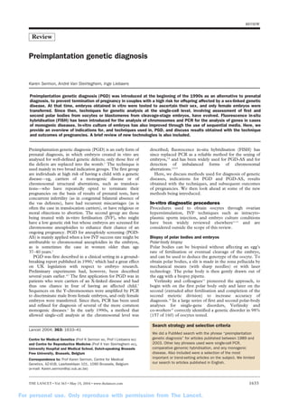
  • 1. REVIEW Review Preimplantation genetic diagnosis (PGD) was introduced at the beginning of the 1990s as an alternative to prenatal diagnosis, to prevent termination of pregnancy in couples with a high risk for offspring affected by a sex-linked genetic disease. At that time, embryos obtained in vitro were tested to ascertain their sex, and only female embryos were transferred. Since then, techniques for genetic analysis at the single-cell level, involving assessment of first and second polar bodies from oocytes or blastomeres from cleavage-stage embryos, have evolved. Fluorescence in-situ hybridisation (FISH) has been introduced for the analysis of chromosomes and PCR for the analysis of genes in cases of monogenic diseases. In-vitro culture of embryos has also improved through the use of sequential media. Here, we provide an overview of indications for, and techniques used in, PGD, and discuss results obtained with the technique and outcomes of pregnancies. A brief review of new technologies is also included. Preimplantation genetic diagnosis (PGD) is an early form of prenatal diagnosis, in which embryos created in vitro are analysed for well-defined genetic defects; only those free of the defects are replaced into the womb.1 The technique is used mainly in two broad indication groups. The first group are individuals at high risk of having a child with a genetic disease—eg, carriers of a monogenic disease or of chromosomal structural aberrations, such as transloca-tions— who have repeatedly opted to terminate their pregnancies on the basis of results of prenatal tests, have concurrent infertility (as in congenital bilateral absence of the vas deferens), have had recurrent miscarriages (as is often the case in translocation carriers), or have religious or moral objections to abortion. The second group are those being treated with in-vitro fertilisation (IVF), who might have a low genetic risk but whose embryos are screened for chromosome aneuploidies to enhance their chance of an ongoing pregnancy. PGD for aneuploidy screening (PGD-AS) is mainly applied when a low IVF success rate might be attributable to chromosomal aneuploidies in the embryos, as is sometimes the case in women older than age 37–40 years.2 PGD was first described in a clinical setting in a ground-breaking report published in 1990,3 which had a great effect on UK legislation with respect to embryo research. Preliminary experiments had, however, been described several years earlier.4,5 The first application for PGD was in patients who were carriers of an X-linked disease and had thus one chance in four of having an affected child.3 Sequences on the Y-chromosomes were amplified by PCR to discriminate male from female embryos, and only female embryos were transferred. Since then, PCR has been used and refined for diagnosis of several of the more common monogenic diseases.6 In the early 1990s, a method that allowed single-cell analysis at the chromosomal level was Lancet 2004; 363: 1633–41 Centre for Medical Genetics (Prof K Sermon MD, Prof I Liebaers MD) and Centre for Reproductive Medicine (Prof A Van Steirteghem MD), University Hospital and Medical School, Dutch-speaking Brussels Free University, Brussels, Belgium Correspondence to: Prof Karen Sermon, Centre for Medical Genetics, AZ-VUB, Laarbeeklaan 101, 1090 Brussels, Belgium (e-mail: Karen.sermon@az.vub.ac.be) described; fluorescence in-situ hybridisation (FISH) has since replaced PCR as a reliable method for the sexing of embryos,7,8 and has been widely used for PGD-AS and for detection of imbalanced forms of chromosomal aberrations.2,9,10 Here, we discuss methods used for diagnosis of genetic diseases, indications for PGD and PGD-AS, results obtained with the techniques, and subsequent outcomes of pregnancies. We then look ahead at some of the new methods being introduced. In-vitro diagnostic procedures Procedures used to obtain oocytes through ovarian hyperstimulation, IVF techniques such as intracyto-plasmic sperm injection, and embryo culture conditions have been widely reviewed elsewhere11,12 and are considered outside the scope of this review. Biopsy of polar bodies and embryos Polar-body biopsy Polar bodies can be biopsied without affecting an egg’s rate of fertilisation or eventual cleavage of the embryo, and can be used to deduce the genotype of the oocyte. To obtain polar bodies, a slit is made in the zona pellucida by mechanical means (with sharp needles) or with laser technology. The polar body is then gently drawn out of the egg with a biopsy pipette. Verlinsky and colleagues13 pioneered the approach, to begin with on the first polar body only and later on the second (extruded after fertilisation and completion of the second meiotic division) to increase accuracy of diagnosis.14 In a large series of first and second polar-body analyses for single-gene disorders, Verlinsky and co-workers15 correctly identified a genetic disorder in 98% (157 of 160) of oocytes tested. Preimplantation genetic diagnosis Karen Sermon, André Van Steirteghem, Inge Liebaers Search strategy and selection criteria We did a PubMed search with the phrase “preimplantation genetic diagnosis” for articles published between 1989 and 2003. Other key phrases used were single-cell PCR, comparative genomic hybridisation, and any monogenic disease. Also included were a selection of the most important or trend-setting articles on the subject. We limited our search to articles published in English. THE LANCET • Vol 363 • May 15, 2004 • www.thelancet.com 1633 For personal use. Only reproduce with permission from The Lancet.
  • 2. Figure 1: Cleavage-stage biopsy A: hole in zona pellucida made with laser. B: removal of first blastomere from embryo. C: deposition of blastomere in medium. D: removal of second blastomere. Cleavage-stage biopsy Cleavage-stage biopsy is a widely used technique,16 for which embryos are grown in vitro until they reach their third division (when they have six or more cells), which occurs on the third day after insemination. The zona pellucida is then breached, using an acid solution (Tyrode’s solution) or laser technology. A biopsy pipette is introduced through the hole into the embryo and one or two blastomeres are gently aspirated (figure 1). Alternatives to aspiration are extrusion, which involves making a hole in the embryo and squeezing the cells out, and fine-needle aspiration, though these techniques are less widely used.16,17 Results of a retrospective study18 indicate no difference in implantation potential between embryos from which two cells are taken and non-biopsied embryos obtained after intracytoplasmic sperm injection. However, doubt remains about whether the biopsy of one cell would not be less detrimental to the embryo than biopsy of two cells. Randomised prospective studies to address this question are underway. Blastocyst biopsy The blastocyst is the latest stage at which an embryo can be biopsied. The advantage of biopsy at this stage is that more cells can be obtained, but the disadvantages are that only about 36% of embryos mature this far and that little time is left for diagnosis, since embryos should be transferred before day 5 or 6.19,20 Hence, blastocyst biopsy has been attempted by only one centre to date.21 Analysis of cells obtained PCR PCR is used to amplify sufficient DNA from cells obtained from an oocyte or embryo to diagnose monogenic diseases.1 In brief, a polar body or a blastomere is placed in a solution that lyses the cell and releases the DNA. A PCR reaction mix is then added and PCR begins. Because of its high sensitivity, contamination of the study sample with extraneous DNA is a danger and has led to the adoption of rigorous laboratory procedures and standards, such as the use of intracytoplasmic sperm injection.15 Moreover, amplification of only one, rather than both, of the genes present in a cell can result in misdiagnosis of disease and the transfer and implantation of affected embryos.22 To overcome this potential difficulty, dubbed allele-drop out, various techniques have evolved for the analysis of PCR fragments; fluorescent PCR and fragment analysis on automated sequencers were introduced first and, later, multiplex PCR was developed.1 Since then, the introduction of automated sequencing,23 minisequencing, and real-time PCR has further refined our diagnostic capabilities.24–28 FISH FISH is the most frequently used method for analysis of the chromosomal complement of blastomeres.2,7–10 Collected cells are spread on a glass microscope slide, after which DNA probes labelled with fluorochromes specific for the chromosomes of interest are applied to their nuclei (figure 2). The type and number of FISH probes used depends on the indication: for sexing, only two probes are needed (for the X and Y chromosomes), though probes for autosomal chromosomes are often added to check for aneuploidy; for identification of translocations, two or three probes are used as explained later; for aneuploidy screening, the number of chromosomes that can be analysed simultaneously is limited by the number of fluorochromes available—kits that contain probes for chromosomes X, Y, 13, 18, and 21 (figure 3), or for chromosomes X, Y, 13, 16, 18, 21, and 22 are available, but separate probes can only usually be obtained with a red or a green label. Mixing of two colours and the application of two, or even three, rounds of FISH can enhance the number of chromosomes analysed simultaneously to nine with acceptable efficiency. In large series, the approximate FISH error level is about 10%.2,29–31 Comparative genomic hybridisation Another way to identify chromosomal aberrations is to use comparative genomic hybridisation, a new technique introduced by two groups.32–35 Here, pre-amplified DNA from a single test cell is labelled with one fluorochrome— eg, red—and then mixed with pre-amplified DNA from a control sample labelled with a different fluorochrome— eg, green—with which it is compared. The mixture is REVIEW Chromosome on microscope slide Denature * * * T A G A T C C T G G A T A A * C C * T T T C * * Hybridise probe to denatured chromosome DNA double helix T A G A T C C T A T C T A G G A Denature * * * T A G A T C C T A T C T A G G A * * * * * * * ** * * * * * * Complementary probe labelled wih fluorescent marker A A T T A G A T C C T A T C T A A G * G G A T C T A G G A * * * * * T A G A T C C T A T C T A G G A Figure 2: Principle of FISH 1634 THE LANCET • Vol 363 • May 15, 2004 • www.thelancet.com For personal use. Only reproduce with permission from The Lancet.
  • 3. Figure 3: Result of five-colour FISH applied to nuclei of single blastomere Different chromosomes are identified with their number. A: male nucleus (X and Y chromosome present). B: female nucleus (two X chromosomes present). applied to a normal metaphase spread, and the colour ratio measured. Areas with more red indicate that the test sample contained more of this genetic material, whereas areas showing more green indicate that the test sample contained too little of this genetic material. The advantage of comparative genomic hybridisation over FISH is that the whole chromosome complement is analysed, though polyploidy and balanced translocations cannot be detected. The disadvantage is that the whole procedure takes about 72 h, which is why the method is either applied to polar bodies, leaving 5 days for analysis, or to cells obtained at the cleavage stage of development, after which embryos are cryopreserved and transferred in a later cycle if identified as healthy.34,35 The time taken to complete the procedure could be shortened by replacing the metaphase spread with microarrays of carefully chosen sequences from all 24 chromosomes, so-called microarray comparative genomic hybridisation.36 This technique is described under future developments. Cryopreservation of biopsied embryos After PGD, few embryos usually remain for cryopreservation.6 Although most centres do cryopreserve surplus embryos, the survival rate of the embryos is extremely low, and only one pregnancy has been described after standard procedures were followed.37–39 Jericho and colleagues40 have, however, adapted these standard procedures to make the freezing and thawing processes more gentle, and have successfully applied their new technique to embryos analysed with comparative genomic hybridisation. The group40 noted a survival rate of 75% (by comparison with 43% with the standard protocol) and an implantation rate of 12%; 50 embryos were transferred in 36 transfer procedures, leading to six ongoing pregnancies. Indications for PGD and PGD-AS Monogenic diseases PCR was the first technique developed for the analysis of DNA from single cells. That monogenic diseases were the first genetic abnormalities to be diagnosed in embryos is, therefore, not surprising. PGD in general and single-cell PCR in particular are demanding labour-intensive techniques however, so only the most common monogenic diseases have been studied extensively. REVIEW One such disease is cystic fibrosis, which has a prevalence of one in 2500 births in white people and is caused, in most instances, by the same mutation—namely the F508 mutation. A multitude of reports describe protocols for preimplantation diagnosis of the disease,13,14,41–48 which was the first to be identified by PGD.41 Several methods of PGD have also been described for a group of autosomal recessive diseases with a high prevalence in certain populations—ie, defects in the globin gene, such as sickle-cell anaemia (in African populations) and thalassaemia (in Mediterranean populations).49–53 Spinal muscular atrophy has also received a lot of attention.54–56 Autosomal dominant diseases—eg, myotonic dystrophy,57,58 Huntington’s disease,59,60 and Marfan’s syndrome61–63—are less prevalent than autosomal recessive diseases. However, because the risk to their offspring is 50%, rather than 25% for recessive diseases, individuals with an autosomal dominant disease will more readily opt for PGD. X-linked diseases, such as Duchenne muscular dystrophy and hypoxanthine-guanine phosphoribosyl-transferase (HPRT) deficiency, were among the first for which PGD was undertaken.3 With a simple technique, the sex of the embryos is ascertained and all male embryos disposed of. A drawback of this approach is that half of the discarded male embryos are healthy and half of the female embryos transferred are carriers of the condition. For more and more X-linked diseases, the specific genetic defect has now been identified, however, allowing a specific DNA diagnosis. Again, most reports are for PGD methods for the more frequent X-linked diseases, such as Duchenne muscular dystrophy and fragile X syndrome64–67 (which should not be diagnosed through sexing because girls can also be affected). Ethically, for X-linked recessive diseases, most would agree that to transfer non-carrier girls and healthy boys is preferable than to transfer carrier girls.68,69 The third report of the European Society for Human Reproduction and Embryology (ESHRE) PGD consortium,6 which presents data from 26 different PGD centres, lists 33 monogenic diseases for which PGD had been applied. The list is comparable to the list of diseases for which prenatal diagnosis is undertaken, though it is shorter. We do not believe that the list will increase in THE LANCET • Vol 363 • May 15, 2004 • www.thelancet.com 1635 For personal use. Only reproduce with permission from The Lancet.
  • 4. Indication Method Number of Number of Reference Ao et al, 199870 Familial adenomatous polyposis coli Primer extension pre-amplification+PCR for mutation 1 0 Strom et al, 199871 Autosomal dominant retinitis pigmentosa First and second polar body, nested multiplex 1 0 Strom et al, 199814 Cystic fibrosis First and second polar body, nested multiplex 3 3 Kuliev et al, 199951 thalassaemia First and second polar body, nested multiplex 17 7 Van de Velde et al, 199972 21-hydroxylase deficiency Fluorescent PCR of mutation 2 1 Kanavakis et al, 199953 thalassaemia Denaturing gradient gel electrophoresis for 11 6 Sermon et al, 199963 Marfan’s syndrome Fluorescent PCR of marker 3 0 Ray et al, 200073 Ornithine transcarbamylase deficiency Nested PCR (mutation+marker) 2 1 Dreesen et al, 200044 Cystic fibrosis Fluorescent marker multiplex NA NA Sermon et al, 200074 Medium-chain acyl-CoA dehydrogenase deficiency Fluorescent PCR of mutation 1 1 De Rycke et al, 200150 thalassaemia, sickle-cell anaemia Fluorescent PCR of mutation 12 3 Ray et al, 200165 Duchenne muscular dystrophy Nested multiplex NA NA Georgiou et al, 200175 Spinal and bulbar muscular atrophy Fluorescent PCR of mutation 1 1 Sermon et al, 200158 Myotonic dystrophy, Huntington’s disease, Nested and fluorescent PCR of mutation 92 19 fragile X syndrome Piyamongkol et al, 200157 Myotonic dystrophy Fluorescent multiplex (mutation+marker) 3 2 Moutou et al, 200247 Cystic fibrosis Fluorescent multiplex (mutation+marker) 3 0 Harper et al, 200276 Myotonic dystrophy, cystic fibrosis, Fluorescent multiplex 13 4 fragile X syndrome and cystic fibrosis, neurofibromatosis 2, Crouzon Sermon et al, 200277 Huntington’s disease exclusion Fluorescent multiplex 7 1 Hussey et al, 200223 thalassaemia Fluorescent sequencing 8 2 Rechitsky et al, 200278 Familial adenomatous polyposis coli, First and second polar body, nested multiplex 20 5 von Hippel-Lindau disease, retinoblastoma, Li-Fraumeni syndrome, neurofibromatosis 1 and 2, familial posterios fossa brain tumour De Vos et al, 200379 Charcot-Marie-Tooth disease type 1A Fluorescent multiplex 13 3 Fiorentino et al, 200325 Cystic fibrosis, thalassaemia, sickle-cell Fluorescent multiplex and minisequencing 55 15 anaemia, haemophilia A, retinoblastoma, spinal muscular atrophy Girardet et al, 200380 Retinoblastoma Fluorescent multiplex 1 0 Moutou et al, 200381 Achondroplasia Fluorescent PCR of mutation 4 0 Rechitsky et al, 200382 Familial dysautonomia First and second polar body, nested multiplex 1 1 Goossens et al, 200383 Cystic fibrosis Fluorescent multiplex (mutation+marker) 22 4 NA=not applied clinically. Monogenic diseases for which PGD protocols have been assessed length at a rapid rate, because only large PGD centres have the time and resources needed to research and develop the needed tests. The table gives an overview of monogenic diseases for which PGD has been assessed in individuals with up-to-date technologies.14,23,25,44,47,50,51,53,57,58,63,65,70–83 Ethically difficult indications Since PGD can be viewed as an early form of prenatal diagnosis, many believed that the indications for both types of diagnosis would be similar. However, for some diseases, PGD but not prenatal diagnosis can be defended from an ethical point of view. In non-disclosure PGD, which has been described for Huntington’s disease but could also be applied to other late-onset diseases, patients do not wish to know their carrier status but want to have disease-free offspring. Their embryos are, therefore, tested without revealing any of the details of the cycle or diagnosis.60 Because non-disclosure testing puts practitioners in a difficult ethical position—eg, having to undertake PGD cycles even when the results of previous cycles preclude the individual as a carrier, or having to do mock-transfers if no embryos are available— the ESHRE ethics task force discourages non-disclosure testing, encouraging instead exclusion testing,68,84 which involves assessment of embryos with linked markers for the presence of a chromosome from the affected grandparent; the mutation itself is not analysed. Another new indication for PGD involves the selection of embryos, according to their HLA type, so that a child born out of a PGD cycle can be a stem-cell donor for a sick treatment clinical cycles pregnancies (gestation 20 weeks) +PCR for marker 12 different mutations sibling. The first such case was described by Verlinsky and colleagues85 and concerned a family with a child who had Fanconi anaemia. In this instance, PGD was undertaken to select embryos not affected with Fanconi anaemia, but which had the same HLA type as the affected sibling. This application of PGD is controversial. The main ethical arguments against are that a child is being used as a treatment, hence breaching the Kantian imperative that a person should never be used as a means. Furthermore, how will the child feel when they are told that they were made for the purpose of saving a sibling? Pennings and colleagues,86 however, argue that the Kantian imperative is not violated because the future donor child is not solely made for the purpose of donating stem cells, but will also be loved and cared for by the family as an individual. Careful counselling by a psychologist should be arranged before PGD is used for this purpose to ascertain the real motivations of the prospective parents. Pennings and co-workers also note that a child who is told that they were made to save a sibling will not experience feelings of low self-esteem but will, on the contrary, be proud to have contributed in such an important way to the well-being of a sibling. Furthermore, giving cord blood at birth could be considered something one family member would do naturally for another. Finally, they argued that parents would not hesitate to volunteer an existing child as a bone-marrow donor for a sick sibling, and that it is ethically acceptable to make a child for a reason if it is acceptable to use an existing child for the same reason. Several other authors have reached the same conclusion.68,69,87 REVIEW 1636 THE LANCET • Vol 363 • May 15, 2004 • www.thelancet.com For personal use. Only reproduce with permission from The Lancet.
  • 5. The use of PGD to diagnose risk of late-onset diseases (such as Huntington’s disease and Alzheimer’s disease) and to search for genes that predispose for cancer (BRCA1, BRCA2, Li-Fraumeni, neurofibromatosis 1 and 2) is also ethically debatable.59,60,76,78,88 These two groups are distinct in pathology: late-onset genetic diseases have full penetrance, cancer predisposition genes do not; late-onset diseases are often not curable, whereas cancer predisposition syndromes can be treated, although the value of treatment needs to be demonstrated. However, the ethical arguments with respect to PGD for these diseases are quite similar. Many believe PGD is ethically acceptable for these indications because of the heavy burden imposed on patients who are carriers of one of these diseases, be it because of the burden imposed by the eventual fate in late-onset diseases, or because of the burden on the quality of life imposed by far-reaching preventive measures in cancer predisposition syndromes.68,69,87 Finally, several reports have been published on the use of sexing for social reasons and have provoked mixed reactions.6,68,89–91 Opponents argue that sex selection for non-medical purposes is sexist and tantamount to discrimination against women. Those in favour argue that sex selection at the embryonic stage of development is preferable to sex selection after prenatal diagnosis, or even after birth. Moreover, they claim that overall sex distribution will not be skewed because only a few people will use PGD to choose their child’s sex.89 A distinction should be made, however, between sex selection for convenience and for so-called family balancing. In the latter instance, the family should have at least one or two children of one sex before they can ask for a child of the opposite sex.90 In family balancing, the issues of sex discrimination and skewed sex ratios are avoided, giving the family an increased autonomy without conflicts with other ethical principles.84,87,90 Sex-linked diseases Women who are carriers of a recessive X-linked disease have an a-priori risk of 25% of having an affected boy, but are themselves, like their carrier daughters, healthy. Hence, sexing of embryos with FISH has been used extensively to avoid the birth of boys affected by X-linked disease.8,92 As noted previously, however, half of discarded male embryos are healthy and carrier girls cannot be distinguished from healthy girls with this technique. If available, individuals therefore prefer specific DNA diagnosis. Nonetheless, sexing remains a useful method when the gene responsible for a disease is unknown— eg, non-fragile X, X-linked mental retardation—when the search for a specific mutation is judged too difficult— eg, X-linked retinitis pigmentosa—or when individuals do not want to wait for the specific test to be developed— eg, when the woman is already aged older than 37 years. Chromosomal abnormalities Reciprocal and Robertsonian translocations Reciprocal translocations are characterised by the exchange of fragments of DNA between chromosomes, whereas in Robertsonian translocations a whole chromo-some is translocated to another through centromeric fusion. Healthy carriers of such translocations are at risk of having children with congenital anomalies and mental retardation due to chromosomal imbalances, or are likely to have recurrent miscarriages or to be infertile (especially if the man is a carrier). To offer PGD to female carriers of balanced translocations, Munné and colleagues9 analysed the first REVIEW polar body of their eggs; if biopsied within a couple of hours after oocyte retrieval, first polar bodies are still in metaphase and can be assessed by chromosome painting—ie, a type of FISH in which chromosomes are identified over their entire length. A major drawback of this method is that only translocations carried by the mother can be analysed. The current most widely used method hence involves analysis of cleavage-stage embryos. Here, two FISH probes are used for Robertsonian translocations, one locus-specific probe centromeric to the breakpoint of one of the chromosomes involved, and one on the telomere of the other chromosome involved, and three FISH probes are used for assessment of reciprocal translocations—two on the centromeric side of the breakpoints of the chromosomes involved, and a third on one of the telomeres on the other side of the breakpoint. The two or three probes used have to carry a different fluorochrome, and it must be possible to use them together in one assay.10,93,94 Identification of the probes needed for a particular couple can be difficult, since commercially available probes usually come in only two different fluorochromes (red and green). Once identified, the combination of probes centromeric and telomeric to the breakpoints allows embryos that carry an unbalanced chromosome complement to be distinguished from healthy ones, but cannot help to distinguish between embryos without the translocation or carrying the balanced translocation. Since reciprocal translocations are often different for each couple, a new probe mixture needs to be tested in each instance. Moreover, several centres have shown that a large proportion (over 80%) of embryos are chromosomally abnormal and cannot be transferred,6,93–97 leading to a low pregnancy rate per cycle (but not per transfer) in individuals with reciprocal translocations. However, because of the occurrence of multiple miscarriages or infertility in these patients, PGD is often the only way to achieve an ongoing healthy pregnancy. Aneuploidy screening Results of investigations98,99 with classic cytogenetic methods indicate that between 23% and 80% of embryos are aneuploid, and use of FISH is providing some information about the types of chromosomal abnormal-ities and why they arise. For example, Delhanty and colleagues100 undertook FISH with probes for the X and Y chromosomes to assess embryos discarded after PGD, and noted a range of anomalies, such as monosomy X and mosaics for trisomy/triploidy, and Munné and co-workers101 noted, after analysis of normally as well as abnormally developing embryos with five-colour FISH for chromosomes X, Y, 13, 18, and 21, that 70% of the abnormally developing embryos were abnormal for at least one of the chromosomes studied. Furthermore, analysing the chromosomal abnormalities in 1255 non-viable cleavage-stage embryos, Marquez and colleagues102 showed that aneuploidy increases with maternal age (from 1·4% in patients aged 20–34 years to 52·4% in patients aged 40–47 years), whereas polyploidy and mosaicism are related to poor embryo morphology. Considering these data, together with the well documented correlation between advanced maternal age and fetal aneuploidy and the recognition that 50–60% of all spontaneous abortions from clinically recognised pregnancies carry a grossly abnormal and lethal chromosome complement,103 researchers soon realised that embryo selection based on chromosome complement would improve IVF results in individuals with poor outcome and reduce the number of babies born with chromosomal defects. THE LANCET • Vol 363 • May 15, 2004 • www.thelancet.com 1637 For personal use. Only reproduce with permission from The Lancet.
  • 6. PGD-AS has been used to identify problems in individuals of advanced maternal age,29,104 in those in whom embryos have repeatedly not implanted, and in women who have recurrent miscarriages not due to translocations.2,100,105 PGD-AS has, in fact, become the most widely used application of PGD, because of the relative ease of the technique compared with others and because of the large potential group of patients.6 The misdiagnosis rate after biopsy of one blastomere was estimated to be 7% in a large series;29 nearly 6% of blastomeres are misdiagnosed due to mosaicism. Findings of large retrospective series106,107 have indicated a higher implantation rate after polar-body biopsy than without selection through biopsy, and a non-significant increase in implantation rate with a significant decrease in spontaneous abortion rate after single blastomere biopsy. More recent data108 indicate that the use of eight or nine different probes (X, Y, 13, 14, 15, 16, 18, 21, and 22) for diagnosis greatly increases implantation rates. However, prospective randomised studies are needed before the real efficiency of PGD-AS is known.2 Outcome Misdiagnosis Reports of misdiagnoses are anecdotal, making an approximation of rates of misdiagnosis difficult. In the early days of PGD, one misdiagnosis for sexing, using PCR, and one misdiagnosis for cystic fibrosis in a compound heterozygote embryo were reported. Both misdiagnoses were due to the low efficiency of single-cell PCR, and technical developments—eg, FISH for sexing and multiplex fluorescent PCR—have ruled out the reoccurrence of such errors.22 Munné and colleagues107 reported one misdiagnosis (trisomy 21 after aneuploidy screening) in a series of 57 pregnancies. Pickering and co-workers109 reported one misdiagnosis, for spinal muscular atrophy, in a series of 18 pregnancies. The third report of the ESHRE PGD consortium6 details the occurrence of eight misdiagnoses in 451 (2%) pregnancies (or of eight in 265 [3%] pregnancies if only those fetal sacs for which a control had been undertaken are counted): five of 145 (3%) pregnancies after PCR (two for sexing [one for Duchenne muscular dystrophy and one for retinitis pigmentosa], one each for thalassaemia, cystic fibrosis, and myotonic dystrophy) and three of 305 (1%) after FISH (one each for sexing for social reasons, translocation [11;22], and trisomy 21 [after PGD-AS]). We believe the reporting of these misdiagnoses was an important step in the identification of technical flaws and shortcomings, which have led to the adoption of stringent diagnostic criteria. Pregnancy outcome Because of the newness and technical complexity of PGD, large series on the practice and outcome of the various techniques involved are rare. Within ESHRE, therefore, the ESHRE PGD consortium was formed to obtain data on PGD cycles and their outcome.6 Of all the treatment cycles in couples whose embryos were tested by PGD in the 26 centres which are members and contribute their data, clinical pregnancies resulted in: 62 of 368 (17%) after testing for structural chromosomal abnormalities, including translocations, 41 of 254 (16%) after sexing, and 119 of 575 (21%) after testing for monogenic diseases. These numbers are lower than would be expected in a regular IVF cycle (20–25%), though it is noteworthy that many embryos are diagnosed as affected or abnormal and cannot therefore be transferred. These findings contrast with the results for PGD-AS, in which instance patients are not at high risk of having genetically abnormal offspring, but which involves the selection of embryos most likely to implant in the womb. Here, of the 799 cycles reported,6 199 or 25% led to a clinical pregnancy in individuals with a previously poor prognosis because of advanced maternal age, repetitive IVF failure, and recurrent miscarriages. Results of further analyses of these figures indicate that women of advanced age who have had recurrent miscarriages are particularly likely to become pregnant (28%) after PGD-AS, whereas patients who have little success after IVF are less likely to become pregnant (8%). Although the PGD-AS data are not compared with a suitable control group, most believe that PGD-AS increases the chances of pregnancy for patients with a poor prognosis. Wilton noted that most of the large studies published on PGD-AS and its outcome did not include an appropriate control group,2,29,102–104 but large, multicentre, randomised studies are underway that will, hopefully, provide information on the efficiency of PGD-AS and the groups of individuals most likely to benefit from it. The ESHRE PGD consortium is also interested in the outcomes of pregnancies after PGD. The most important cause of morbidity and mortality is multiple pregnancy. For this aspect and many others, such as birthweight and rate of congenital malformations, statistics for children born after PGD are comparable to those for children born after intracytoplasmic sperm injection. In their report,110 the International Working Group on Preimplantation Genetics noted that more than 3000 PGD cycles had been undertaken by mid-2001 since the early 1990s, resulting in a pregnancy rate of about 24%. Nearly 700 children were born as a result of the pregnancies and 5% of the babies had some kind of abnormality. Furthermore, when data for this period from the four most active centres worldwide were combined, a total of 2774 PGD cycles are shown to have resulted in 2265 transfers of embryos to the womb and 652 subsequent clinical pregnancies (29%).110 In the report,110 it is stressed that outcome of pregnancy was comparable to that for IVF populations. The authors of a study on another large cohort of 109 children,111 not included in the ESHRE PGD consortium report, also conclude that children born after PGD are comparable to those born after intracytoplasmic sperm injection with respect to pregnancy and birth parameters, and that PGD is a safe method of avoiding the birth of children with genetic defects. Future developments New methods for diagnosis of monogenic diseases are being developed at a rapid rate, some of which are very suitable for use with single-cell PCR. Real-time PCR In real-time PCR, the accumulation of PCR product is measured during cycling, when PCR is in its exponential phase, instead of after the process is complete. To differentiate between different PCR products—eg, a gene with a mutation and a wild-type gene—molecular beacons are added to the reaction mixture. These beacons consist of short oligonucleotides complementary to the region of interest within the PCR fragment. The oligonucleotides have complementary ends, one of which carries a fluorochrome and the other a quencher, so that if they do not anneal to the DNA and remain in solution the beacons are folded on themselves and do not fluoresce.27,28 Other methods are also commercially available—eg, fluorescence resonance energy transfer (FRET). In real-time PCR, short PCR fragments are generated, thus reducing the likelihood of allele drop-out. Moreover, REVIEW 1638 THE LANCET • Vol 363 • May 15, 2004 • www.thelancet.com For personal use. Only reproduce with permission from The Lancet.
  • 7. continuous measurement of PCR product during the exponential phase of the process gives a more accurate idea of the homozygous or heterozygous state of a single cell. Real-time PCR has been used for PGD of sex, by analysing the presence of a Y chromosome, and for diagnosis of Tay- Sachs disease.27,28 However, its use has not been described in the diagnosis of mitochondrial diseases, despite the fact that real-time PCR allows for quantitative measurements. Minisequencing Minisequencing permits the quick and accurate detection of single nucleotide polymorphisms (SNPs). SNPs are widespread in the genome and can be either true polymorphisms, which do not affect functioning of genes, or mutations. SNPs can be used as linked polymorphisms to enhance the accuracy of the PGD, in the same way that microsatellites are used in duplex and multiplex PCR for PGD. The advantage of SNPs is that they are more common than microsatellites, thus giving a higher chance of finding a linked informative SNP, and that they can be detected with short PCR fragments. When starting from a single cell, the amplification of small stretches of DNA is much more efficient than for larger fragments; thus SNP analysis is ideally suited for single-cell PCR. In PGD, only a few loci need to be analysed simultaneously—eg, two mutations and two SNPs—so minisequencing on microcapillary systems are ideal.25 Microarrays Mutation-specific primers can also be spotted on microarrays,112 which are another promising development. First described in the analysis of expression in cells, microarrays have moved from RNA to DNA technology. Small glass slides or other supports are covered with well-defined DNA fragments, to which fluorescently labelled sample DNA is then allowed to hybridise. The full array can be read by an automated system. Microarrays could be used in PGD in two ways. First, they could be used to diagnose common genetic diseases caused by more than one mutation—eg, cystic fibrosis (associated with more than 1000 mutations) or thalassaemia.112 Because only small stretches of DNA need to be amplified, preliminary work-up should be simple and straightforward, and analysis of microarrays could be standardised for all patients. Second, microarrays could be used in comparative genomic hybridisation, where they would replace the metaphase spread to which the mixture of test and comparative DNA are hybridised. In this instance, the microarray would contain not sequences from one gene but sequences spread over the whole genome. The specificity of the sequences in the microarrays would lead to shorter hybridisation and computer analysis times than arise with comparative genomic hybridisation, obviating the need for polar-body analysis or cryopreservation. Other advantages of microarray comparative genomic hybridisation would be its greater resolution (100–200 kb instead of 2–10 Mb) compared with the use of metaphase spreads, while the important standardisation of the whole procedure by use of computers would allow more IVF centres to use microarray comparative genomic hybridisation routinely for aneuploidy screening.2,36 Conflict of interest statement None declared. Acknowledgments We thank Catherine Staessen, Hubert Joris, and Elvire Van Assche for their advice about cryopreservation and aneuploidy issues, and for their critical reading of the manuscript. REVIEW K Sermon is supported by a postdoctoral fellowship from the Fund for Scientific Research, Flanders. The PGD team is supported by grants from the Fund for Scientific Research Flanders, the Research Council of the Dutch-speaking Brussels Free University, the Forton Fund, and the Concerted Action of the Brussels Free University. None of the sponsors had a role in the research or writing of this Review. References 1 Sermon K. Current concepts in preimplantation genetic diagnosis (PGD): a molecular biologist’s view. Hum Reprod Update 2002, 8: 1–10. 2 Wilton L. Preimplantation genetic diagnosis for aneuploidy screening in early human embryos: a review. Prenat Diagn 2002; 22: 312–18. 3 Handyside A, Kontogianni E, Hardy K, Winston R. Pregnancies from biopsied human preimplantation embryos sexed by Y-specific DNA amplification. Nature 1990; 344: 768–70. 4 Coutelle C, Williams C, Handyside H, Hardy K, Winston R, Williamson R. Genetic analysis of DNA from single human oocytes: a model for preimplantation diagnosis of cystic fibrosis. BMJ 1989; 299: 22–24. 5 Holding C, Monk M. Diagnosis of beta-thalassaemia by DNA amplification in single blastomeres from mouse preimplantation embryos. Lancet 1989; 2: 532–35. 6 ESHRE PGD Consortium Steering Committee. ESHRE Preimplantation Genetic Diagnosis Consortium: data collection III (May 2001). Hum Reprod 2002; 17: 233–46. 7 Griffin D, Wilton L, Handyside A. Dual fluorescent in situ hybridisation for simultaneous detection of X and Y chromosome-specific probes for sexing of human preimplantation embryonic nuclei. Hum Genet 1992; 89: 18–22. 8 Staessen C, Van Assche E, Joris H, et al. Clinical experience of sex determination by fluorescent in situ hybridisation for preimplantation genetic diagnosis. Mol Hum Reprod 1999; 5: 392–99. 9 Munné S, Sandalinas M, Escudero T, Fung J, Gianaroli L, Cohen J. Outcome of preimplantation genetic diagnosis of translocations. Fertil Steril 2000; 73: 1209–18. 10 Conn C, Harper J, Winston R, Delhanty J. Infertile couples with Robertsonian translocations: preimplantation genetic analysis of embryos reveals chaotic cleavage divisions. Hum Genet 1998; 102: 117–23. 11 Thornton K. Advances in assisted reproductive technologies. Obstet Gynecol Clin North Am 2000; 27: 517–27. 12 Liebaers I, Sermon K, Staessen C, et al. Clinical experience with preimplantation genetic diagnosis and intracytoplasmic sperm injection. Hum Reprod 1998; 13 (suppl 1): 186–95. 13 Verlinsky Y, Rechitsky S, Evsikov S, et al. Preconception and preimplantation diagnosis for cystic fibrosis. Prenat Diagn 1992; 12: 103–10. 14 Strom C, Ginsberg N, Rechitsky S, et al. Three births after preimplantation genetic diagnosis for cystic fibrosis with sequential first and second polar body analysis. Am J Obstet Gynecol 1998; 178: 1298–306. 15 Rechitsky S, Strom C, Verlinsky O, et al. Accuracy of preimplantation diagnosis of single-gene disorders by polar body analysis of oocytes. J Assist Reprod Genet 1999; 16: 192–98. 16 De Vos A, Van Steirteghem A. Aspects of biopsy procedures prior to preimplantation genetic diagnosis. Prenat Diagn 2001; 21: 767–80. 17 Inzunza J, Iwarsson E, Fridström M, et al. Application of single-needle blastomere biopsy in human preimplantation genetic diagnosis. Prenat Diagn 1998; 18: 1381–88. 18 Van de Velde H, De Vos A, Sermon K, et al. Embryo implantation after biopsy of one or two cells from cleavage-stage embryos with a view to preimplantation genetic diagnosis. Prenat Diagn 2000; 20: 1030–37. 19 Verheyen G, Van Landuyt L, Joris H, Devroey P, Van Steirteghem A. Different embryo developmental patterns in different sequential media appear to result in similar pregnancy rates. Hum Reprod 2003; 18 (suppl 1): xviii37. 20 Veiga A, Sandalinas M, Benkhalifa M, et al. Laser blastocyst biopsy for preimplantation diagnosis in the human. Zygote 1997; 5: 351–54. 21 Boada M, Carrera M, De la Iglesia C, Sandalinas M, Barri PN, Veiga A. Successful use of a laser for human embryo biopsy in preimplantation genetic diagnosis: report of two cases. J Assist Reprod Genet 1998; 15: 302–07. 22 Lissens W, Sermon K. Preimplantation genetic diagnosis: current status and new developments. Hum Reprod 1997; 12: 1756–61. 23 Hussey N, Davis T, Hall J, et al. Preimplantation genetic diagnosis for -thalassemia using sequencing of single cell PCR products to detect mutations and polymorphic loci. Mol Hum Reprod 2002; 8: 1136–43. 24 Syvänen A-C. From gels to chips: “Minisequencing” primer extension for analysis of point mutations and single nucleotide polymorphisms. Hum Mutat 1999; 13: 1–10. THE LANCET • Vol 363 • May 15, 2004 • www.thelancet.com 1639 For personal use. Only reproduce with permission from The Lancet.
  • 8. 25 Fiorentino F, Magli MC, Podini D, et al. The minisequencing method: an alternative strategy for preimplantation genetic diagnosis of single gene disorders. Mol Hum Reprod 2003; 9: 399–410. 26 Szuhai K, Ouweland J, Dirks R, et al. Simultaneous A8344G heteroplasmy and mitochondrial DNA copy number quatification in myoclonus epilepsy and ragged-red fibers (MERFF) syndrome by a multiplex molecular beacon based real-time fluorescence PCR. Nucleic Acids Res 2001; 29: E13. 27 Pierce K, Rice J, Aquiles Sanchez J, Brenner C, Wangh L. Real-time PCR using molecular beacons for accurate detection of the Y-chromosome in single human blastomeres. Mol Hum Reprod 2000, 6: 1155–64. 28 Rice J, Aquiles Sanchez J, Pierce K, Wangh L. Real-time PCR with molecular beacons provides a highly accurate assay for detection of Tay-Sachs alleles in single cells. Prenat Diagn 2002; 22: 1130–34. 29 Munné S, Sandalinas M, Escudero T, Marquez C, Cohen J. Chromosome mosaicism in cleavage-stage human embryos: evidence of a maternal age effect. Repr BioMed Online 2003; 4: 223–32. 30 Munné S, Magli C, Bahçe M, et al. Preimplantation diagnosis of the aneuploidies most commonly found in spontaneous abortions and live births: XY, 13, 14, 15, 16, 18, 21, 22. Prenat Diagn 1998; 18: 1459–66. 31 Pehlivan T, Rubio C, Rodrigo L, et al. Impact of preimplantation genetic diagnosis on IVF outcome in implantation failure patients. Reprod BioMed Online 2002; 6: 232–37. 32 Voullaire L, Slater H, Williamson R, Wilton L. Chromosome analysis of blastomeres from human embryos by using CGH. Hum Genet 2000; 105: 210–17. 33 Wells D, Delhanty J. Comprehensive chromosomal analysis of human preimplantation embryos using whole genome amplification and single cell comparative gnomic hybridisation. Mol Hum Reprod 2000; 6: 1055–62. 34 Wilton L, Williamson R, McBain J, Edgar D, Voullaire L. Birth of a healthy infant after preimlantation confirmation of euploidy by comparative genomic hybridisation. N Engl J Med 2001; 345: 1537–41. 35 Wells D, Escudero T, Levy B, Hirschhorn K, Delhanty J, Munné S. First clinical application of comparative genomic hybridisation and polar body testing for preimplantation genetic diagnosis of aneuploidy. Fertil Steril 2002; 78: 543–49. 36 Wells D, Brynn L. Cytogenetics in reproductive medicine: the contribution of comparative genomic hybridisation (CGH). BioEssays 2003; 25: 289–300. 37 Joris H, Van den Abbeel E, De Vos A, Van Steirteghem A. Reduced survival after human embryo biopsy and subsequent cryopreservation. Hum Reprod 1999; 14: 2833–37. 38 Magli M, Gianaroli L, Fortini D, Ferrarreti A, Munné S. Impact of blastomere biopsy and cryopreservation techniques on human embryo viability. Hum Reprod 1999; 14: 770–73. 39 Lee M, Munné S. Pregnancy after polar body biopsy and freezing and thawing of human embryos. Fertil Steril 2000; 73: 645–47. 40 Jericho H, Wilton L, Gook D, Edgar D. A modified cryopreservation method increases the survival of human biopsied cleavage stage embryos. Hum Reprod 2003; 18: 568–71. 41 Handyside A, Lesko J, Tarin J, Winston R, Hughes M. Birth of a normal girl after in vitro ferilization and preimplantation diagnostic testing for cystic fibrosis. N Engl J Med 1992; 327: 905–09. 42 Liu J, Lissens W, Silber S, Devroey P, Liebaers I, Van Steirteghem A. Birth after preimplantation diagnosis of the cystic fibrosis F508 mutation by polymerase chain reaction in human embryos resulting from intracytoplasmic sperm injection with epididymal sperm. JAMA 1994; 272: 23. 43 Moutou C, Viville S. Improvement of preimplantation genetic diagnosis (PGD) for cystic fibrosis mutation F508 by fluorescent polymerase chain reaction. Prenat Diagn 1999; 19: 1248–50. 44 Dreesen J, Jacobs L, Bras M, et al. Multiplex PCR of polymorphic markers flanking the CFTR gene: a general approach for preimplantation genetic diagnosis of cystic fibrosis. Mol Hum Reprod 2000; 6: 391–96. 45 Goossens V, Sermon K, Lissens W, et al. Clinical application of preimplantation genetic diagnosis for cystic fibrosis. Prenat Diagn 2000; 20: 571–81. 46 Eftedal I, Schwartz M, Bendtsen H, Andersen A, Zieve S. Single intragenic microsatellite preimplantation genetic diagnosis for cystic fibrosis provides positive allele identification of all CFTR genotypes for informative couples. Mol Hum Reprod 2001; 7: 307–12. 47 Moutou C, Gardes N, Viville S. Multiplex PCR combining delta F508 mutation and intragenic microsatellites of the CFTR gene for pre-implantation genetic diagnosis (PGD) of cystic fibrosis. Eur J Hum Genet 2002; 10: 231–38. 48 Vrettou C, Tzetis M, Traeger-Synodinos J, Palmer G, Kanavakis E. Multiplex sequence variation detection throughout the CFTR gene appropriate for preimplantation genetic diagnosis in populations with heterogeneity of cystic fibrosis mutations. Mol Hum Reprod 2002; 8: 880–86. 49 Xu K, Shi Z, Veeck L, Hughes M, Rosenwaks Z. First unaffected pregnancy using preimplantation genetic diagnosis for sickle cell anemia. JAMA 1999; 281: 1701–06. 50 De Rycke M, Van de Velde H, Sermon K, et al. Preimplantation genetic diagnosis for sickle-cell anemia and for -thalassemia. Prenat Diagn 2001; 21: 214–22. 51 Kuliev A, Rechistky S, Verlinsky O, et al. Birth of healthy children after preimplantation diagnosis of thalassemias. J Assist Reprod Genet 1999; 16: 207–11. 52 Vrettou C, Palmer G, Kanavakis E, et al. A widely applicable strategy for single cell genotyping of -thalassemia mutations using DGGE analysis: application to preimplantation genetic diagnosis. Prenat Diagn 1999, 19: 1209–16. 53 Kanavakis E, Vrettou C, Palmer G, Tretis M, Mastrominas M, Traeger-Synodinos J. Preimplantation genetic diagnosis in 10 couples at risk for transmitting -thalassemia major: clinical experience including the initiation of six singleton pregnancies. Prenat Diagn 1999; 19: 1217–22. 54 Dreesen J, Bras M, de Die-Smulders C, et al. Preimplantation genetic diagnosis of spinal muscular atrophy. Mol Hum Reprod 1998; 4: 881–85. 55 Fallon L, Harton GL, Sisson ME, et al. Preimplantation genetic diagnosis for spinal muscular atrophy type I. Neurology 1999; 53: 1087–90. 56 Moutou C, Gardes N, Rongières C, et al. Allele-specific amplification for preimplantation genetic diagnosis (PGD) of spinal muscular atrophy. Prenat Diagn 2001; 21: 498–503. 57 Piyamongkol W, Harper J, Sherlock J, et al. A successful strategy for preimplantation genetic diagnosis of myotonic dystrophy using multiplex fluorescent PCR. Prenat Diagn 2001; 21: 223–32. 58 Sermon K, Seneca S, De Rycke M, et al. PGD in the lab for triplet repeat diseases-myotonic dystrophy, Huntington’s disease and Fragile-X syndrome. Moll Cell Endocrinol 2001; 183 (suppl 1): 77–85. 59 Sermon K, Goossens V, Seneca S, et al. Preimplantation diagnosis for Huntington’s disease (HD): clinical application and analysis of the HD expansion in affected embryos. Prenat Diagn 1998; 18: 1427–36. 60 Stern H, Harton G, Sisson M, et al. Non-disclosing preimplantation genetic diagnosis for Huntington disease. Prenat Diagn 2002; 22: 303–07. 61 Harton G, Tsipouras P, Sisson M, et al. Preimplantation genetic testing for Marfan syndrome. Mol Hum Reprod 1996; 2: 713–15. 62 Blaszczyk A, Tang Y, Dietz H, et al. Preimplantation genetic diagnosis of human embryos for Marfan’s syndrome. J Assist Reprod Genet 1998; 15: 281–84. 63 Sermon K, Lissens W, Messiaen L, et al. Preimplantation genetic diagnosis of Marfan syndrome with the use of fluorescent polymerase chain reaction and the Automated Laser Fluorescence DNA Sequencer. Fertil Steril 1999; 71: 163–65. 64 Hussey N, Donggui H, Froiland D, et al. Analysis of five Duchenne muscular dystrophy exons and gender determination using conventional duplex polymerase chain reaction on single cells. Mol Hum Reprod 1999; 5: 1089–94. 65 Ray P, Vekemans M, Munnich A. Single cell multiplex PCR amplification of five dystrophin gene exons combined with gender determination. Mol Hum Reprod 2001; 7: 489–94. 66 Sermon K, Seneca S, Vanderfaeillie A, et al. Preimplantation diagnosis for Fragile X syndrome based on the detection of the non-expanded paternal and maternal CGG. Prenat Diagn 1999; 19: 1223–30. 67 Apessos A, Abou-Sleiman P, Harper J, Delhanty J. Preimplantation genetic diagnosis of the fragile X syndrome by use of polymorphic linked markers. Prenat Diagn 2001; 21: 504–11. 68 Shenfield F, Pennings G, Devroey P, Sureau C, Tarlatzis B, Cohen J. Taskforce 5: preimplantation genetic diagnosis. Hum Reprod 2003; 18: 649–51. 69 de Wert G. Ethics of assisted reproduction. In: Fauser B, Bouchard P, Hsueh A, Rutherford A, Simpson JL, Strauss J, Van Steirteghem A, eds. Reproductive medicine: molecular, cellular and genetic fundamentals. London: Parthenon Publishing, 2003: 645–65. 70 Ao A, Wells D, Handyside A, Winston R, Delhanty J. Preimplantation genetic diagnosis of inherited cancer: familial adenomatous polyposis coli. J Assist Reprod Genet 1998; 15: 140–44. 71 Strom C, Rechitsky S, Wolf G, Cieslak J, Kuliev A, Verlinsky Y. Preimplantation diagnosis of autosomal dominant retinitis pigmentosum using two simultaneous single cell assays for a point mutation in the rhodopsin gene. Mol Hum Reprod 1998; 4: 351–55. 72 Van de Velde H, Sermon K, De Vos A, et al. Fluorescent PCR and automated fragment analysis in preimplantation genetic diagnosis for 21-hydroxylase deficiency in congenital adrenal hyperplasia. Mol Hum Reprod 1999; 5: 691–96. 73 Ray PF, Gigarel N, Bonnefont JP, et al. First specific preimplantation genetic diagnosis for ornithine transcarbamylase deficiency. Prenat Diagn 2000; 20: 1048–54. REVIEW 1640 THE LANCET • Vol 363 • May 15, 2004 • www.thelancet.com For personal use. Only reproduce with permission from The Lancet.
  • 9. 74 Sermon K, Henderix P, Lissens W, et al. Preimplantation genetic diagnosis for medium-chain acyl-CoA dehydrogenase (MCAD) deficiency. Mol Hum Reprod 2000; 6: 1165–68. 75 Georgiou I, Sermon K, Lissens W, et al. Preimplantation genetic diagnosis for spinal and bulbar muscular atrophy (SBMA). Hum Genet 2001; 108: 494–98. 76 Harper J, Wells D, Piyamongkol W, et al. Preimplantation genetic diagnosis for single gene disorders: experience with five single gene disorders. Prenat Diagn 2002; 22: 525–33. 77 Sermon K, De Rycke M, Lissens W, et al. Preimplantation genetic diagnosis for Huntington’s disease with exclusion testing. Eur J Hum Genet 2002; 10: 591–98. 78 Rechitsky S, Verlinsky O, Chistokhina A, et al. Preimplantation genetic diagnosis for cancer predisposition. Reprod BioMed Online 2002; 4: 148–55. 79 De Vos A, Sermon K, De Rycke M, et al. Preimplantation genetic diagnosis for Charcot–Marie-Tooth disease type 1A. Molec Hum Reprod 2003; 9: 429–35. 80 Girardet A, Hamamah S, Anahory T, et al. First preimplantation genetic diagnosis of hereditary retinoblastoma using informative microsatellite markers. Mol Hum Reprod 2003; 9: 111–16. 81 Moutou C, Rongieres C, Bettahar-Lebugle K, Gardes N, Philippe C, Viville S. Preimplantation genetic diagnosis for achondroplasia: genetics and gynaecological limits and difficulties. Hum Reprod 2003; 18: 509–14. 82 Rechitsky S, Verlinsky O, Kuliev A, et al. Preimplantation genetic diagnosis for familial dysautonomia. Reprod BioMed Online 2003; 6: 488–93. 83 Goossens V, Sermon K, Lissens W, et al. Improving clinical preimplantation genetic diagnosis for cystic fibrosis by duplex-PCR using two polymorphic markers or one polymorphic marker in combination with the detection of the F508 mutation. Molec Hum Reprod 2003; 9: 559–67. 84 Sermon K, De Rijcke M, Lissens W, et al. Preimplantation genetic diagnosis for Huntington’s disease with exclusion testing. Eur J Hum Genet 2002; 10: 591–98. 85 Verlinsky Y, Rechitsky S, Schoolcraft W, Strom C, Kuliev A. Preimplantation diagnosis for Fanconi anemia combined with HLA matching. JAMA 2001; 285: 3130–33. 86 Pennings G, Schots R, Liebaers I. Ethical considerations on preimplantation genetic diagnosis for HLA typing to match a future child as a donor of haematopoietic stem cells to a sibling. Hum Reprod 2002; 17: 534–38. 87 Robertson J. Extending preimplantation genetic diagnosis: the ethical debate—ethical issues in new uses of preimplantation genetic diagnosis. Hum Reprod 2003; 18: 465–71. 88 Verlinsky Y, Rechitsky S, Verlinsky O, Masciangelo C, Lederer K, Kuliev A. Preimplantation diagnosis for early-onset Alzheimer disease caused by V717L mutation. JAMA 2002; 287: 1018–21. 89 Malpani A. The use of preimplantation genetic diagnosis in sex selection for family balancing in India. Reprod BioMed Online 2002; 4: 16–20. 90 Pennings G. Personal desires of patients and social obligations of geneticists: applying preimlantation genetic diagnosis for non-medical sex selection. Prenat Diagn 2002; 22: 1123–29. 91 Ray P, Munnich A, Nisand I, Frydman R, Vekemans M, Viville S. Sex selection by preimplantation genetic diagnosis: should it be carried out for social purposes?: Is preimplantation genetic diagnosis for “social sexing” desirable in today’s and tomorrow’s society? Hum Reprod 2003; 18: 463–64. 92 Harper JC, Coonen E, Ramaekers FC, et al. Identification of the sex of human preimplantation embryos in two hours using an improved spreading method and fluorescent in-situ hybridization (FISH) using directly labelled probes. Hum Reprod 1994; 9: 721–24. REVIEW 93 Coonen E, Martini E, Dumoulin J, et al. Preimplantation genetic diagnosis of a reciprocal translocation t(3;11)(q27.3;q24.3) in siblings. Mol Hum Reprod 2000; 6: 199–206. 94 Van Assche E, Staessen C, Vegetti W, et al. Preimplantation genetic diagnosis and sperm analysis by fluorescence in situ hybridisation for the most common reciprocal translocation t(11;22). Mol Hum Reprod 1999; 5: 682–90. 95 Evsikov S, Cieslak J, Verlinsky Y. Effect of chromosomal translocations on the development of preimplantation human embryos in vitro. Fertil Steril 2000; 74: 672–77. 96 Iwarsson E, Malmgren H, Inzunza J, et al. Highly abnormal cleavage divisions in preimplantation embryos from translocation carriers. Prenat Diagn 2000; 20: 1038–47. 97 Gianaroli L, Magli MC, Ferraretti AP, et al. Possible interchromosomal effect in embryos generated by gametes from translocation carriers. Hum Reprod 2002; 17: 3201–07. 98 Zenzes MT, Casper RF. Cytogenetics of human oocytes, zygotes, and embryos after in vitro fertilization. Hum Genet 1992; 88: 367–75. 99 Pellestor F, Girardet A, Andréo B, Arnal F, Humeau C. Relationship between morphology and chromosomal constituttion in human preimplantation embryo. Mol Reprod Dev 1994; 39: 141–46. 100 Delhanty J, Griffin D, Handyside A, et al. Detection of aneuploidy and chromosomal mosaicism in human embryos during preimplantation sex determination by fluorescent in situ hybridisation, (FISH). Hum Mol Genet 1993; 2: 1183–85. 101 Munné S, Lee A, Rosenwaks Z, Grifo J, Cohen J. Diagnosis of major chromosome aneuploidies in human preimplantation embryos. Hum Reprod 1993; 8: 2185–91. 102 Marquez C, Sandalinas M, Bahçe M, Alikani M, Munné S. Chromosome abnormalities in 1255 cleavage-stage human embryos. Reprod BioMed Online 2000; 1: 17–26. 103 Boué A, Boué J, Gropp A. Cytogenetics of pregnancy wastage. Adv Hum Genet 1985; 14: 1–57. 104 Verlinsky Y, Cieslak J, Ivakhnenko V, et al. Prevention of age-related aneupoidies by polar body testing of oocytes. J Assist Reprod Genet 1999; 16: 165–69. 105 Rubio C, Simon C, Vidal F, et al. Chromosomal abnormalities and embryo development in recurrent miscarriage couples. Hum Reprod 2003; 18: 182–88. 106 Kuliev A, Cieslak J, Ilkevitch Y, Verlinsky Y. Chromosomal abnormalities in a series of 6733 human oocytes in preimplantation diagnosis for age-related aneuploidies. Reprod BioMed Online 2002; 6: 54–59. 107 Munné S, Magli C, Cohan J, et al. Positive outcome after preimplantation diagnosis of aneuploidy in human embryos. Hum Reprod 1999; 14: 2191–99. 108 Munné S, Cohen J, Sable D. Preimplantation genetic diagnosis for advanced maternal age and other indications. Fertil Steril 2002; 78: 234–36. 109 Pickering S, Polidoropoulos N, Caller J, et al. Strategies and outcomes of the first 100 cycles of preimplantation genetic diagnosis at the Guy’s and St Thomas’ Center. Fertil Steril 2003; 79: 81–90. 110 Anon. Report of the 11th annual meeting of International Working Group on Preimplantation Genetics: preimplantation genetic diagnosis—experience of 3000 clinical cycles. Reprod BioMed Online 2001; 3: 49–53. 111 Strom C, Levin R, Strom S, Masciangelo C, Kuliev A, Verlinsky Y. Neonatal outcome of preimplantation genetic diagnosis by polar body removal: the first 109 infants. Pediatrics 2000; 106: 650–53. 112 Kurg A, Tonisson N, Georgiou I, Shumaker J, Tollett J, Metspalu A. Arrayed primer extension: solid-phase four-color DNA resequencing and mutation detection technology. Genet Test 2000; 4: 1–7. THE LANCET • Vol 363 • May 15, 2004 • www.thelancet.com 1641 For personal use. Only reproduce with permission from The Lancet.