
Recommended
Recommended
More Related Content
What's hot
What's hot (20)
Similar to Good results in postoperative
Similar to Good results in postoperative (20)
More from mrcs89
More from mrcs89 (14)
Recently uploaded
Recently uploaded (20)
Good results in postoperative
- 1. Acta Orthopaedica 2013; 84 (6): 509–516 509 Good results in postoperative and hematogenous deep infections of 89 stable total hip and knee replacements with retention of prosthesis and local antibiotics Jan A P Geurts1, Daniël M C Janssen1, Alfons G H Kessels2, and Geert H I M Walenkamp1 1Department of Orthopedic Surgery; 2Department of Clinical Epidemiology and Medical Technology Assessment, Maastricht University Medical Centre, Maastricht, the Netherlands. Correspondence: j.geurts@mumc.nl Submitted 12-11-30. Accepted 13-09-06 Background Deep postoperative and hematogenous prosthesis infections may be treated with retention of the prosthesis, if the prosthesis is stable. How long the infection may be present to pre-clude a good result is unclear. Patients and methods We retrospectively studied 89 deep-infected stable prostheses from 69 total hip replacements and 20 total knee replacements. There were 83 early or delayed postop-erative infections and 6 hematogenous. In the postoperative infec-tions, treatment had started 12 days to 2 years after implanta-tion. In the hematogenous infections, symptoms had been present for 6 to 9 days. The patients had been treated with debridement, prosthesis retention, systemic antibiotics, and local antibiotics: gentamicin-PMMA beads or gentamicin collagen fleeces. The minimum follow-up time was 1.5 years. We investigated how the result of the treatment had been influenced by the length of the period the infection was present, and by other variables such as host characteristics, infection stage, and type of bacteria. Results In postoperative infections, the risk of failure increased with a longer postoperative interval: from 0.2 (95% CI: 0.1–0.3) if the treatment had started ≥ 4 weeks postoperatively to 0.5 (CI: 0.2–0.8) if it had started at ≥ 8 weeks. The relative risk for success was 0.6 (CI: 0.3–0.95) if the treatment had started ≥ 8 weeks. In the hematogenous group, 5 of 6 infections had been treated suc-cessfully. Interpretation A longer delay before the start of the treatment caused an increased failure rate, but this must be weighed against the advantage of keeping the prosthesis. We consider a failure rate of < 50% to be acceptable, and we therefore advocate keeping the prosthesis for up to 8 weeks postoperatively, and in hematogenous infections with a short duration of symptoms. The incidence of deep infection in total hip and knee replace-ment (THR, TKR) ranges from 1% or less in primary THR and TKR to 5% in revision settings (Philips et al. 2006, Willis-Owen et al. 2010), and even up to 21% when revising for infection (Mortazavi et al. 2010). Early deep prosthesis infections are probably caused by peroperative contamina-tion, and in the literature there is agreement that if the pros-thesis is stable such an early infection can be treated without removal of the prosthesis, as in early postoperatively infected osteosynthesis (Trampuz and Zimmerli 2006, Choi et al. 2011, Sukeik et al. 2012). The same holds true for hematogenous prosthesis infections (Choi et al. 2011). However, for post-operative infections there is no agreement about the maximal period between implantation of the prosthesis and the start of the treatment that permits retention of the prosthesis, or the duration of symptoms in acute onset of hematogenous infec-tions (Zimmerli et al. 2004, Marculescu et al. 2006). At our institution, deep postoperative or hematogenous infections of THR and TKR are treated with retention of the prosthesis if they are stable, regardless of interval period since implantation or duration of symptoms. We investigated whether this policy was justified and questioned whether the success rate in postoperative infections does indeed decrease when the postoperative interval since implantation increases or the duration of symptoms in hematogenous infections increases. Patients and methods We performed a retrospective cohort analysis of a prospec-tive register of all proven early and delayed deep infections of THR and TKR with a postoperative interval after prosthesis implantation of less than 2 years, and all hematogenous infec-tions treated at our center from January 1982 to July 2010. As hematogenous infections, we considered delayed or late deep infections without any sign of prosthesis infection in the Open Access - This article is distributed under the terms of the Creative Commons Attribution Noncommercial License which permits any noncommercial use, distribution, and reproduction in any medium, provided the source is credited. DOI 10.3109/17453674.2013.858288
- 2. 510 Acta Orthopaedica 2013; 84 (6): 509–516 Table 1. Data on the infected prostheses (69 THRs and 20 TKRs) scored according to the different staging of the host and wound, and classification of the infection. The numbers of THRs and TKRs are given for each subclass, as are the results of the treatments Staging or total success failure total success failure classification Subclasses 69 57 12 20 17 3 ASAscore ASA1 9 8 1 4 4 0 patient ASA2 36 30 6 10 9 1 ASA3 24 19 5 6 4 2 McPherson type I early postop (< 4 weeks) 42 37 5 8 8 0 classification type II hematogenous 3 2 1 3 3 0 of infection type III late postop (> 4weeks) 24 18 6 9 6 3 McPherson host A: uncompromised 22 19 3 7 7 0 host staging host B: compromised 38 32 6 13 10 3 host C: significant compromised 9 6 3 0 0 0 McPherson grade 1: uncompromised 17 15 2 9 9 0 wound staging grade 2: compromised 43 37 6 10 8 2 grade 3: significant compromised 9 5 4 1 0 1 Cierny A-host: uncompromised 7 6 1 5 5 0 host staging B-host: compromised 62 51 11 15 12 3 C-host: significant compromised 0 0 0 0 0 0 Zimmerli early postop (< 3 months) 61 53 8 14 12 2 classification acute hematogenous 3 2 1 3 3 0 of infection delayed exogenous (3–24 months) 5 2 3 3 2 1 This study: postop infection < 8 weeks 60 53 7 9 9 0 classification postop infection ≥ 8 weeks 6 2 4 8 5 3 of infection hematogenous 3 2 1 3 3 0 period since implantation. In the databases of the hospital and department, we found 145 infections in 144 patients. For this retrospective analysis, we studied the medical records and if necessary we contacted the patient or family doctor. Prostheses were diagnosed as infected when the Mayo cri-teria (Berbari et al. 1998) were fulfilled: growth of the same microorganism in 2 or more cultures of synovial fluid or peri-prosthetic tissue, or pus in synovial fluid or at the implant site, or histological examination showing acute inflammation in periprosthetic tissue, or a sinus tract communicating with the prosthesis. We excluded the following patients. 16 patients did not meet Mayo criteria for deep infection, 21 patients got their first sur-gical treatment at another center, 12 patients were treated by immediate extraction of the prosthesis since unexpected loos-ening was diagnosed during operation, and 2 patients were excluded because of incomplete patient files. Also excluded were 5 patients with TKR who did not receive any local anti-biotic treatment, but only arthroscopic debridement. After these exclusions, 89 deep infections remained (88 patients, 46 women). All patients and types of infections were scored according to classifications of ASA, Cierny, McPher-son, and Zimmerli (Table 1) (Cierny and DiPasquale 2002, McPherson et al. 2002, Zimmerli et al. 2004). There were 69 THR infections (39 primary THR, 30 revisions) and 20 TKR infections (19 primary TKR, 1 revision). 3 of the THR infec-tions and 3 of the TKR infections were hematogenous. One THR TKR female patient had an early postoperative infection in a pri-mary TKR on both sides, not simultaneously. The first TKR infection was successfully treated, but the contralateral TKR that was subsequently implanted was also infected. The median age of the patients at the start of the infec-tion treatment was 69 (27–93) years. The median interval between implantation of the prosthesis and the first opera-tion for infection in the postoperative THR infections was 23 (12–390) days, and in the TKR infections the median interval was 42 (14–713) days. In some cases, the delay was caused by a period of intravenous antibiotic treatment of a supposed superficial postoperative infection. In 3 hematogenous THR infections, the median duration of symptoms was 7 (6–9) days before the debridement for infection,and in 3 hematogenous TKR infections it was 8 (6-9) days. No loosening was suspected in any of the implants preop-eratively, and this was confirmed peroperatively. The treatment consisted of arthrotomy, debridement (includ-ing pulse lavage with at least 3 L of Ringer lactate), and reten-tion of the implant. In the period studied, we did not exchange modular components if present. The patients were treated with systemic antibiotic therapy, and also with local antibiotic car-riers. We preferred the use of gentamicin-PMMA beads with a size of 7 mm, containing 7.5 mg gentamicin sulfate, in the form of chains with 30 or 60 beads (Septopal; Merck GmbH, Darmstadt, Germany; Biomet GmbH, Berlin, Germany). We implanted as much beads as possible in the infected tissues
- 3. Acta Orthopaedica 2013; 84 (6): 509–516 511 to create a high local gentamicin concentration (Figures 1–3). Beads did not stick through the skin, but were removed in a second operation after 2 weeks. This operation consisted of a new debridement, leaving behind new beads if infection was not considered to be eradicated. If healing was consid-ered appropriate, a much smaller incision was sufficient for the removal of the beads. In several infections, the surgeon implanted gentamicin collagen fleeces (Septocoll containing 116 mg gentamicin sulfate and 350 mg gentamicin crobephate in 320 mg equine collagen fleece with a size of 10 × 8 cm; Merck GmbH; Biomet GmbH) in the joint during the last operation before closing the wound, to increase the period with local antibiotics. If the infection persisted, according to clinical and laboratory parameters and despite one or more treatment periods of 2 weeks with beads, the prosthesis was Figure 3. THR with gentamicin-PMMA beads intra-articularly around the neck of the prosthesis and in the subcutaneous tissues. Antero-posterior radiograph on the first day after the debridement operation. Only a limited number of beads could be placed in this joint after the debridement. In the subcutaneous tissue, beads were placed in an abscess cavity. removed and the treatment for infection continued with genta-micin- PMMA beads. Of the infected THRs, 26 of 69 were treated in a single period of 2 weeks with beads or fleeces, and 47 of the 69 THR infections required 2 or more debridements with a subsequent period of 2 weeks of local antibiotics (Table 2). The THR infections were treated with implantation of an average of 180 (30–420) gentamicin beads. Of the infected TKRs, 13 of 20 patients were given a single treatment of 2 weeks of local antibiotics and 7 TKR infec-tions needed 2 or more debridements with local antibiotics for 2 weeks. In 15 of the 20 TKR infections, we implanted an average of 120 (50–240) beads. In the remaining 5 TKR infections, no beads but only gentamicin fleeces were inserted due to limited joint size (Table 2). In the 84 patients who were treated with gentamicin beads, these were removed at the last surgery by a limited operation with a small incision. In 22 of these 84 infections, we implanted 1–4 gentamicin collagen fleeces at this last removal operation of the beads. Swabs as well as multiple tissue cultures were taken. The samples were cultured in the microbiology laboratory for at least 2 weeks to detect slow-growing microorganisms, and minimal inhibitory concentrations (MICs) of gentamicin for the bacteria were determined. We found methicillin-sensitive Staphylococcus aureus to be the most frequent microorganism Figure 1. Gentamicin-PMMA beads (Septopal) inserted in a total knee replacement after debridement with retained prosthesis. Beads are mainly placed in the suprapatellar bursa and are removed after 2 weeks by another operation under general anesthesia, but with a smaller incision. Figure 2. Radiographic appearance of a TKR in 2 directions. Genta-micin- PMMA beads are visible in the suprapatellar bursa and on the lateral side of the joint. Beads cannot be positioned in the posterior joint due to the limited space.
- 4. 512 Acta Orthopaedica 2013; 84 (6): 509–516 to cause infections (31/89) (Table 3). In 2 patients, peropera-tive cultures showed no growth, due to systemic use of anti-biotics preoperatively. In the 27 polymicrobial infections, we found 68 bacterial species in many combinations, with Pseu-domonas aeruginosa and Enterobacter spp. being the most frequent (Table 4). The MIC values for gentamicin of the causative bacteria were ≤ 8 μg/mL in 71 infections, 16–64 μg/mL in 11 infec-tions, and ≥ 128 μg/mL in 5 infections. The surgical treatment was combined with high doses of systemic antibiotics, intravenously during hospitalization and continued orally after discharge from hospital. The choice of the antibiotic was based on the resistance pattern of the deep tissue cultures and on consultation with a microbiologist with an interest in orthopedic infections. From 2004, we added rifampicin in the systemic antibiotic treatment of infected implants routinely: thus, 25 of the THR infections and 7 of the TKR infections were also treated with rifampicin. The antibi-otic treatment was given for a period of 30 (10–82) days intra-venously, followed by an oral treatment over 72 (7–1,310) days. The median total antibiotic therapy time was 95 (12– 1,310) days. We stopped the oral antibiotic treatment at the outpatient clinic when clinical and laboratory parameters had normalized for at least 4 weeks. As laboratory parameters for infection we used ESR, CRP, and WBC counts. These were measured twice a week during hospitalization, and at all the outpatient control visits. We con-sidered these parameters to be normalized when at 2 subse-quent controls CRP and WBC counts remained normal, and when the ESR was reduced to less than 30 mm/h in patients with no systemic diseases. The treatment was considered to be successful when the infection was resolved at follow-up (normalized inflammatory blood markers and no clinical or radiological signs of recur-rence) with retention of the prosthesis. Failure was diagnosed if the patient never became infection-free or if removal of the implant was necessary for healing of the infection. The follow-up period started at the first operation for deep infec-tion, and the end of the follow-up period was either the date of the last outpatient clinic visit, the last contact with the family doctor, or the date of death. The minimum follow-up was 1.5 years, but possibly shorter if patients died before—whether or not this was related to the infection. Mean follow-up time was 33 (1–270) months for all infections, 27 (1–270) months for infected THRs, and 52 (3–202) months for infected TKRs. In the group of postoperative infections, we analyzed how the treatment result was influenced by the length of the inter-val between implantation of the prosthesis and the start of the treatment. In the hematogenous infections, we studied the influence of the duration of symptoms before the treatment started. We also studied the influence on the result of stag-ing of host and of the wound, of classification of patients, of infection parameters at the start of the treatment, of the caus-ative bacterial species, and of the MIC of gentamicin for the bacteria. Table 2. Numbers of debridements and local antibiotic carriers in 89 THR and TKR infections. Detailed numbers are given to specify whether beads were used with or without fleeces (at the last opera-tion), or only fleeces, with numbers of successful or failed treat-ments No of No of Beads ± Only prostheses debride- fleeces fleeces Success Failure ments THP 26 1 26 0 24 2 32 2 32 0 27 5 8 3 8 0 6 2 3 4 3 0 0 3 Total 69 69 0 57 12 TKR 13 1 11 2 12 1 4 2 2 2 3 1 3 3 2 1 2 1 Total 20 15 5 17 3 Table 3. Causative bacteria in 89 prosthesis infections Causative microorganism THR TKR % Staphylococcus aureus 26 5 35 MRSA 1 0 1 CNS 1 5 7 Streptococci spp. 6 2 9 Enterococci spp. 1 0 1 Enterobacter spp. 5 1 7 Pseudomonas aeruginosa 4 1 56 Propionibacterium acnes 1 1 2 Polymicrobial 24 3 30 Negative culture 0 2 2 Total 69 20 100 MRSA Methicillin resistent Staphylococcus aureus CNS Coagulase-negative staphylococci Table 4. Bacteria present in the 27 polymicro-bial infections as depicted in Table 3 Microorganisms in polymicrobial culture THR TKR Staphylococcus aureus 14 3 CNS 4 0 Streptococcispp. 4 1 Enterobacter spp. 18 0 Enterococci spp. 2 0 Pseudomonas aeruginosa 15 1 Propionibacterium acnes 5 0 Prevotella 0 1 Total microorganisms 62 6 No of infections 24 3
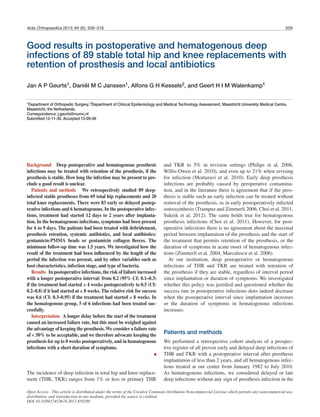
- 5. Acta Orthopaedica 2013; 84 (6): 509–516 513 Risk for failure 0 1.0 0.9 0.8 0.7 0.6 0.5 0.4 0.3 0.2 0.1 0.0 1 2 3 4 5 6 7 8 9 10 Postoperative interval (weeks) Statistics Data are presented as median (total range) or as mean (SD). We analyzed the relationship between the result and the length of the postoperative interval using the following steps. For each of the first 10 postoperative weeks, we distinguished 2 periods: the period including and after (≥) a particular week, and the period before (<) that particular week. For both periods, we then estimated the number of failures and successes. Then we estimated first the risk of failure at or after that week. Secondly, we determined the relative risk (RR) with 95% confidence intervals (CIs) for success comparing the results in the period at or after that week with the results obtained before that week. For these calculations, we used Stata 11 for Windows. Using SPSS version 17.0 for Windows, we calculated RR with CI to determine the influence of the host and wound staging on the result of the treatment. We tested differences between proportions with chi-square test or Fisher’s exact test. We used the Mann-Whitney U test to examine the influence of preoperative body temperature, laboratory values, and the MIC of gentamicin for the causative bacteria on the result of the treatment. Results Of the 89 infected prostheses, 74 infections were treated suc-cessfully with retention and 15 treatments failed. In the group of postoperative infections, 55 of 66 THRs were treated successfully and 11 treatments failed. 10 of these 11 prostheses were removed at a later stage. In TKR patients, 14 of 17 prostheses were successfully treated. In 3 TKRs there was no successful eradication of infection, resulting in removal of the implant in 2 patients. 2 of the 3 hematogenous RR for success 2.0 1.8 1.6 1.4 1.2 1.0 0.8 0.6 0.4 0.2 Figure 5. Relation between the relative risk (RR) for successful treat-ment of an infected prosthesis and the postoperative interval in weeks. The RR is expressed as success if a treatment started after ≥ N weeks, as compared to the period < N weeks. The null hypothesis of RR = 1.0 is represented by a broken line. THR infections were treated successfully, and 1 failed but became infection-free after extraction of the implant. 3 of 3 hematogenous TKR infections were successfully treated with retention of the implant. 4 patients died during the course of treatment, either because of sepsis or poor health: 3 with THR (at 1, 3, and 8 months after the start of treatment), and 1 with TKR (at 8 months). 8 other patients died of other causes 6–17 months after the treat-ment started; none of them had signs of infection, so they had probably resolved. In the first 4 weeks postoperatively, the risk of failed treat-ment remained almost unchanged and gradually increased thereafter, week by week. The risk of failure in the group of patients where the treatment started ≥ 4 weeks was 0.2 (CI: 0.1–0.3), and it was 0.5 (CI: 0.2–0.8) when the treatment started ≥ 8 weeks (Figure 4). Concerning the RR for successful treatment, we found a gradual decrease in the RR when the postoperative interval increased. If treated ≥ 4 weeks, the RR was 1.0 (CI: 0.8–1.2) compared with < 4 weeks. The RR for success if treated ≥ 8 weeks (compared with treatment < 8 weeks) was 0.6 (CI: 0.3–0.95) (Figure 5). In the group of patients where the treatment started ≥ 8 weeks, 7 of 14 infections healed (Table 1). Of the 6 THR infections, 2 infections healed despite retention of the prosthe-sis. In the remaining 4 patients, the THR had to be extracted, resulting in resolution of infection in 2. Of the 8 TKR infec-tions, 5 healed. In the remaining 3, the implant was removed, and 2 of these infections resolved. Thus, altogether, in 11 of 14 prostheses the infection eventually healed despite an interval of more than 8 weeks after implantation. 7 of these 11 infec-tions became infection-free without extraction, even with an interval of almost 1 year postoperatively. Figure 4. Risk (with 95% CI) for failure of the treatment of an infected prosthesis if treated at or after a particular postoperative time interval. 0 0.0 1 2 3 4 5 6 7 8 9 10 Postoperative interval (weeks)
- 6. 514 Acta Orthopaedica 2013; 84 (6): 509–516 In the 6 hematogenous THR and TKR infections, we found no correlation between the duration of symptoms and the results of the treatment with retention of the prosthesis. In the infections that were difficult to treat, more debride-ments were needed, but the failure rate increased (Table 2). 8 of the 11 infections that were debrided for a third time healed, but when a fourth debridement was necessary none of the 3 infections healed. ASA score, type of infection, host and wound staging, number of interventions, or preoperative infection parameters such as fever or laboratory values were similar in the success group and the failure group. We found no relation between the result of the treatment and the causative bacteria. Neither a difference in the result of the treatment between gram posi-tive and gram negative bacteria, or between staphylococci and streptococci. We found no influence of the use of rifampicin, which was added to the treatment protocol since 2004. There was no association between the MIC of gentamicin for the causative bacteria and the success rate of the treatment. Discussion We found good results if we treated deep-infected stable THRs and TKRs by debridement and retention of the prosthesis, in combination with systemic and local antibiotics. Removal of a stable, well-fixed implant is associated with high morbidity and mortality. So the treatment of an infected implant without removal is attractive. Since the results vary greatly, with suc-cess rates between 31% and 100% (Mont et al. 1997, Azzam et al. 2010, Gardner et al. 2011, Koyonos et al. 2011, Sukeik et al. 2012, Fehring et al. 2013, Lee et al. 2013), retention of the implant remains controversial. The controversy is, however, less focused on the treatment with retention as such, and more on the interval after which the results become too bad. We therefore focused on the delay in the start of treatment in relation to the results. We could quantify the risks for failure and success of the treatment postoperatively up to 10 weeks. The treatment had a low and almost unchanged risk of failure up to an interval of 4 weeks, and thereafter it increased every week (Figure 4). The RR for successful treatment showed a gradual decrease in these first weeks, and after 8 weeks there was significantly more risk of failure (as indicated by its CI) (Figure 5). The smaller numbers of infections treated after 10 weeks justifies limitation of our conclusions to only these intervals. When we consider the balance between the disadvantages of the removal of a prosthesis on the one hand and the failure rate of a treatment with retention on the other, we prefer a retention up to 8 weeks postoperatively. Several authors reported a cut-off of only a few days of symptoms for successful retainment of a prosthesis after a deep infection (Brandt et al. 1997, Tattevin et al. 1999, Meehan et al. 2003). Most authors consider a postoperative interval of 2–4 weeks to be the maximum period that a prosthesis can be retained (Theis et al. 2007, Kim et al. 2011). Some stud-ies have suggested that this period could be longer (Schoifet and Morrey 1990, Mont et al. 1997, Zimmerli et al. 2004). Currently, the algorithm by Zimmerli et al. is the one most commonly used (Giulieri et al. 2004, Zimmerli et al. 2004). In their algorithm, they limit the acceptable period of symptoms to a maximum of 3 weeks if the prosthesis is stable, the soft tissues are in good condition, and an antibiotic with activity against biofilm is available. However, confusingly, in the literature 2 different periods are used in protocols: the duration of symptoms and the post-operative period since implantation (“joint age”) (Gardner et al. 2011). The recent guideline of the Infectious Diseases Soci-ety of America uses a limit for in situ treatment of 3 weeks of symptoms, and also a joint age of less than 30 days (Osmon et al. 2013). We regard the postoperative period as a clearer guideline, since the onset of symptoms of a deep infection is very difficult to estimate in clinical practice. Another argu-ment is that these infections must be regarded as having been caused by contamination during the implantation operation. In some patients, an even higher risk of failure with an inter-val of more than 8 weeks might be acceptable. In 14 of our patients who were treated after such a long interval, the infec-tion resolved in 7 cases with retention of the prosthesis, and in 4 after extraction, so the result for healing of infection was 11 out of 14. This result is comparable with results in the litera-ture when the postoperative infection was treated with early extraction, with reimplantation in 1 or 2 stages (Raut et al. 1994, Jämsen et al. 2009). As we do, Kim et al. (2011) also advocated repeated debridement, but their advice was to stop and remove the pros-thesis after 4 attempts. In our patients, no infections healed when debridement was performed more than 3 times, so in our hands extraction after 3 debridements appears to be justified. Comparing our results with those in the literature, they are relatively good, despite an often long postoperative interval. One explanation for this could be the consistent use of local antibiotic carriers in our treatments, with gentamicin-loaded beads or collagen. The high local gentamicin concentration is important, since the infection is probably limited to recently operated tissues, which will be accessible for the debridement and local antibiotic carriers. In 28-year study period, our treatment protocol remained essentially unchanged, focusing on retainment of the implant and on the use of local antibiotic carriers, to supplement sys-temic antibiotics. The main advantage of gentamicin-PMMA beads is a high local antibiotic concentration at the site of the infection, without systemic toxic side effects (Walenkamp et al. 1986). A disadvantage of beads is the space needed, and they have to be removed with an extra operation. The removal operation can, however, be performed with a smaller incision, permitting local inspection, deep cultures, and if necessary a repeated debridement. Gentamicin collagen fleeces have
- 7. Acta Orthopaedica 2013; 84 (6): 509–516 515 the advantage that they are resorbable and have less volume, which makes insertion easier, especially in TKR infections, and removal unnecessary. In our experience, however, a disad-vantage of fleeces is increased wound secretion up to 6 weeks postoperatively, causing difficulties in wound control. Also, they release most of their antibiotics in the first 1–2 hours of implantation (Sørensen et al. 1990). During the study period, we did not replace polyethylene components or modular heads, but we have been doing this routinely since 2010. We found more S. aureus infections than CNS infections. This can be explained since S. aureus causes more acute infections (Gardner et al. 2011), and CNS with a lower virulence are more frequently seen in low-grade and late infections (Giulieri et al. 2004). We found no association between the result of the treatment and the MIC of gentamicin for the bacteria, but even high MIC values are not an absolute contraindication for the use of gen-tamicin beads or fleeces. These MIC values are based on sys-temic gentamicin treatment, and in a treatment with local anti-biotic carriers the local gentamicin concentrations are much higher, up to several hundreds of μg/mL (Wahlig et al. 1978, Hedström et al. 1980, Walenkamp et al. 1986) . The present study had some limitations. It was a retrospec-tively studied cohort, and the treatment was performed by sev-eral orthopedic surgeons. We combined the data on postopera-tive infections of THRs and TKRs, and the cohort included both primary and revision implantations. However, there were also some strong points: the patients were treated at a single center with an almost unchanged protocol for 28 years, treating the infections in the same way with local antibiotics. Although several debridements were performed by different colleagues at the department, a single surgeon was responsible for the treatment of the patients over the whole period. As our depart-ment has a “last-resort function” in treating infections, loss to follow-up was low. We were able to follow the patients for at least 1.5 years if they were still alive. Instead of presenting the results of the treatment as percentages of healing, as in most studies in the literature, we were able to calculate the relative effect of the treatments to show the estimation uncertainty, especially regarding variation in the postoperative interval. In conclusion, treatment of THR or TKR infections can be performed with retention of the prosthesis when the implant is stable. The use of local antibiotics is probably helpful. In post-operative infections, a gradually increased risk of failure of the treatment should be weighed by each surgeon against the disadvantages of removal of the prosthesis. We consider a risk of failure of 50%, if treatment occurs within 8 weeks in most patients, to be acceptable. This approach can still be consid-ered for even longer postoperative intervals in some patients, although we cannot identify these specific patients. JG and GW treated the patients, designed the study and wrote the manuscript. DJ collected the data of the medical records, completed the follow-up, and performed statistical analyses. AK supervised and helped in the statistical analysis. All authors contributed to interpretation of the data and to the revi-sions of the manuscript. No competing interests declared. Azzam K A, Seeley M, Ghanem E, Austin M S, Purtill J J, Parvizi J. Irriga-tion and debridement in the management of prosthetic joint infection: tradi-tional indications revisited. J Arthroplasty 2010; 25 (7): 1022-7. Berbari E F, Hanssen A D, Duffy M C, Steckelberg J M, Ilstrup D M, Harm-sen W S, et al. Risk factors for prosthetic joint infection: case-control study. Clin Inf Dis 1998; 27 (5): 1247-54. Brandt C M, Sistrunk W W, Duffy M C, Hanssen A D, Steckelberg J M, Ilstrup D M, et al. Staphylococcus aureus prosthetic joint infection treated with debridement and prosthesis retention. Clin Inf Dis 1997; 24 (5): 914-9. Choi H-R, von Knoch F, Zurakowski D, Nelson S, Malchau H. Can implant retention be recommended for treatment of infected TKA? Clin Orthop 2011; (469) (4): 961-9. Cierny G, DiPasquale D. Periprosthetic total joint infections: staging, treat-ment, and outcomes. Clin Orthop 2002; (403): 23-8. Fehring T K, Odum S M, Berend K R, Jiranek W A, Parvizi J, Bozic K J, et al. Failure of irrigation and débridement for early postoperative periprosthetic infection. Clin Orthop 2013; (471): 250-7. Gardner J, Gioe T J, Tatman P. Can this prosthesis be saved?: implant salvage attempts in infected primary TKA. Clin Orthop 2011; (469) (4): 970-6. Giulieri S, Graber P, Ochsner P, Zimmerli W. Management of infection asso-ciated with total hip arthroplasty according to a treatment algorithm. Infec-tion 2004; 32 (4): 222-8. Hedström S Å, Lidgren L, Torholm C, Onnerfalt R. Antibiotic containing bone cement beads in the treatment of deep muscle and skeletal infections. Acta Orthop Scand 1980; 51 (6): 863-9. Jämsen E, Stogiannidis I, Malmivaara A, Pajamaki J, Puolakka T, Konttinen Y T. Outcome of prosthesis exchange for infected knee arthroplasty: the effect of treatment approach. Acta Orthop 2009; 80 (1): 67-77. Kim Y H, Kim J S, Park J W, Joo J H. Cementless revision for infected total hip replacements. J Bone Joint Surg (Br) 2011; 93 (1): 19-26. Koyonos L, Zmistowski B, Della Valle C J, Parvizi J. Infection control rate of irrigation and débridement for periprosthetic joint infection. Clin Orthop 2011; (469): 3043-8. Lee Y K, Lee K H, Nho J H, Ha Y C, Koo K H. Retaining well-fixed cement-less stem in the treatment of infected hip arthroplasty. Good results in 19 patients followed for mean 4 years. Acta Orthop 2013; 84 (3): 260-4. Marculescu C E, Berbari E F, Hanssen A D, Steckelberg J M, Harmsen S W, Mandrekar J N, et al. Outcome of prosthetic joint infections treated with debridement and retention of components. Clin Inf Dis 2006; 42 (4): 471-8. McPherson E, Woodson C, Holtom P, Roidis N, Shufelt C, Patzakis M. Peri-prosthetic total hip infection. Clin Orthop 2002; (403): 8-15. Meehan A M, Osmon D R, Duffy M C T, Hansen A D, Keating M R. Outcome of penicillin-susceptible streptococcal prosthetic joint infection treated with debridement and retention of the prosthesis. Clin Inf Dis 2003; 36: 845-9. Mont M, Waldman R, Banerjee C, Pacheco I, Hungerfors D. Multiple irri-gation, debridement, and retention of components in infected total knee arthroplasty. J Arthroplasty 1997; 12 (4): 426-33. Mortazavi S M, Schwartzenberger J, Austin M S, Purtill J J, Parvizi J. Revi-sion total knee arthroplasty infection: incidence and predictors. Clin Orthop 2010; (468) (8): 2052-9. Osmon D R, Berbari E F, Behrendt A R, Lew L, Zimmerli W, Stecklenberg J M, et al. Diagnosis and management of prosthetic joint infection: clinical practice guidelines by the Infectious Diseases Society of America. Clin Inf Dis 2013; 56: 1-25.
- 8. 516 Acta Orthopaedica 2013; 84 (6): 509–516 Philips J, Crane T, Noy M, Elliot T, Grimer R. The incidence of deep pros-thetic infections in a specialist orthopaedic hospital: a 15 year prospective survey. J Bone Joint Surg (Br) 2006; 88: 943-8. Raut V V, Siney P D, Wroblewski B M. One-stage revision of infected total hip replacements with discharging sinuses. J Bone Joint Surg (Br) 1994;76 (5): 721-4. Schoifet S, Morrey B. Treatment of infection after total knee arthroplasty by debridement with retention of the components. J Bone Joint Surg (Am) 1990; 72: 1383-90. Sørensen T S, Sørensen A I, Merser S. Rapid release of gentamicin from col-lagen sponge. In vitro comparison with plastic beads. Acta Orthop Scand 1990; 61 (4): 353-6. Sukeik M, Patel S, Haddad F S. Aggressive early debridement for treatment of acutely infected cemented total hip arthroplasty. Clin Orthop 2012; (470): 3164-70. Tattevin P, Cremieux A C, Pottier P, Huten D, Carbon C. Prosthetic joint infection: when can prosthesis salvage be considered? Clin Infect Dis 1999; 29 (2): 292-5. Theis J C, Gambhir S, White J. Factors affecting implant retention in infected joint replacements. ANZ J Surg 2007; 77 (10): 877-9. Trampuz A, Zimmerli W. Diagnosis and treatment of infections associated with fracture-fixation devices. Injury 2006; 37: S59-S66. Wahlig H, Dingeldein E, Bergmann R, Reuss K. The release of gentamicin from polymethylmethacrylate beads. An experimental and pharmacoki-netic study. J Bone Joint Surg (Br) 1978; 60 (2): 270-5. Walenkamp G H I M, Vree T B, van Rens T J G. Gentamicin-PMMA beads. Pharmacokinetic and nephrotoxicological study. Clin Orthop 1986; (205): 171-83. Willis-Owen C, Konyves A, Martin D. Factors affecting the incidence of infection in hip and knee replacement: an analysis of 5277 cases. J Bone Joint Surg (Br) 2010; 92: 1128-33. Zimmerli W, Trampuz A, Ochsner P. Prosthetic-joint infections. N Engl J Med 2004; 351: 1645-54.