
Recommended
More Related Content
What's hot
What's hot (19)
Similar to Score risk chest pain
Similar to Score risk chest pain (20)
Recently uploaded
Recently uploaded (20)
Score risk chest pain
- 1. Performance of Coronary Risk Scores Among Patients With Chest Pain in the Emergency Department Dustin G. Mark, MD,a,b Jie Huang, PHD,a Uli Chettipally, MD, MPH,c Mamata V. Kene, MD, MPH,d Megan L. Anderson, MD,e Erik P. Hess, MD, MSC,f Dustin W. Ballard, MD, MBE,a,g David R. Vinson, MD,a,e Mary E. Reed, DRPH,a on behalf of the Kaiser Permanente CREST Network Investigators ABSTRACT BACKGROUND Both the modified History, Electrocardiogram, Age, Risk factors and Troponin (HEART) score and the Emergency Department Assessment of Chest pain Score (EDACS) can identify patients with possible acute coronary syndrome (ACS) at low risk (<1%) for major adverse cardiac events (MACE). OBJECTIVES The authors sought to assess the comparative accuracy of the EDACS (original and simplified) and modified HEART risk scores when using cardiac troponin I (cTnI) cutoffs below the 99th percentile, and obtain precise MACE risk estimates. METHODS The authors conducted a retrospective study of adult emergency department (ED) patients evaluated for possible ACS in an integrated health care system between 2013 and 2015. Negative predictive values for MACE (composite of myocardial infarction, cardiogenic shock, cardiac arrest, and all-cause mortality) were determined at 60 days. Reclassification analyses were used to assess the comparative accuracy of risk scores and lower cTnI cutoffs. RESULTS A total of 118,822 patients with possible ACS were included. The 3 risk scores’ accuracies were optimized using the lower limit of cTnI quantitation (<0.02 ng/ml) to define low risk for 60-day MACE, with reclassification yields ranging between 3.4% and 3.9%, while maintaining similar negative predictive values (range 99.49% to 99.55%; p ¼ 0.27). The original EDACS identified the largest proportion of patients as low risk (60.6%; p < 0.0001). CONCLUSIONS Among ED patients with possible ACS, the modified HEART score, original EDACS, and simplified EDACS all predicted a low risk of 60-day MACE with improved accuracy using a cTnI cutoff below the 99th percentile. The original EDACS identified the most low-risk patients, and thus may be the preferred risk score. (J Am Coll Cardiol 2018;71:606–16) © 2018 by the American College of Cardiology Foundation. Chest pain is the second leading reason for emergency department (ED) visits in the United States, with an estimated 7 million visits in 2010 at an estimated cost of $5 billion (1). Patients presenting to EDs with chest pain have among the largest variations in hospital admission rates, but lack clearly associated differences in observed mortality (2,3). Much of the variation in practice is driven by low-grade recommendations to secure functional (exercise electrocardiography, nuclear stress testing, or stress echocardiography) or anatomic (coronary computed tomography angiog- raphy) testing in patients with possible acute coronary syndromes (ACS) before or within 72 h of ISSN 0735-1097/$36.00 https://doi.org/10.1016/j.jacc.2017.11.064 From the a Division of Research, Kaiser Permanente Northern California, Oakland, California; b Departments of Emergency Med- icine and Critical Care, Kaiser Permanente, Oakland, California; c Department of Emergency Medicine, Kaiser Permanente, South San Francisco, California; d Department of Emergency Medicine, Kaiser Permanente, San Leandro, California; e Department of Emergency Medicine, Kaiser Permanente, Sacramento, California; f Department of Emergency Medicine, Mayo Clinic, Rochester, Minnesota; and the g Department of Emergency Medicine, Kaiser Permanente, San Rafael, California. Funded by Kaiser Perma- nente Northern California Delivery Science Grant. The authors have reported that they have no relationships relevant to the contents of this paper to disclose. Manuscript received September 15, 2017; revised manuscript received October 30, 2017, accepted November 27, 2017. Listen to this manuscript’s audio summary by JACC Editor-in-Chief Dr. Valentin Fuster. J O U R N A L O F T H E A M E R I C A N C O L L E G E O F C A R D I O L O G Y V O L . 7 1 , N O . 6 , 2 0 1 8 ª 2 0 1 8 B Y T H E A M E R I C A N C O L L E G E O F C A R D I O L O G Y F O U N D A T I O N P U B L I S H E D B Y E L S E V I E R
- 2. hospital discharge (4,5). However, among those without diagnostic electrocardiograms (ECGs) and/or cardiac biomarkers, only 1% to 4% of these patients have angiographic evidence of significant coronary artery disease (6–8). Given the relatively low yield of this historic approach to possible ACS, researchers have created risk scores to identify patients at low risk of major adverse cardiac events (MACE). Among these, the modified History, Electrocardiogram, Age, Risk fac- tors and Troponin (HEART) score and the Emergency Department Assessment of Chest pain Score (EDACS), both of which treat abnormal troponin values as in- dependent, non–low-risk factors, stand out with the best specificities (ranging from 40% to 60%) in achieving negative predictive value (NPV) estimates >99% for 30- to 45-day MACE, specifically when applied alongside accelerated diagnostic protocols employing cardiac troponin I (cTnI) measurement at ED arrival and 2 to 3 h later (9–12). Used in this fashion, both scores have demonstrated improve- ments in operational efficiency and downstream resource utilization (10,12). A simplified unweighted version of the EDACS, which has minimal reliance on presenting symptoms, performs similarly well in terms of NPV, albeit with lower specificity (13). The use of these risk scores may allow for direct discharge of patients with possible ACS from the ED without further planned cardiac testing, an approach that appears safe, medico-legally acceptable to cli- nicians, and cost-effective (12,14–17). However, adoption in practice is limited by imprecision in risk estimates, as well as uncertainty surrounding the optimal cTnI cutoff, given increased risks of future MACE among patients with cTnI concentra- tions at the high end of the normal range (18–21). Accordingly, we sought to improve both the preci- sion and accuracy of risk estimates by exploring the incorporation of alternative cutoffs for cTnI below the 99th percentile among a large retrospective cohort drawn from the electronic health record of an integrated health system. We contextualized our analysis and findings from a risk-benefit perspective using estimated testing thresholds for functional or anatomic testing in patients with possible ACS, supported by cost-benefit estimates (14,22). We hy- pothesized that improved accuracy and precision in risk estimates could identify a sizeable portion of ED patients with possible ACS for whom routine func- tional or anatomic testing is unlikely beneficial (8,22–25). METHODS STUDY DESIGN, SETTING, AND SUBJECTS. We conducted a retrospective study of pa- tients with ED visits between January 1, 2013, and December 31, 2015, to the 21 medical centers within Kaiser Permanente Northern California, a private, not-for-profit integrated health system of 3.8 million members covering approximately 33% of the region’s insured population. The Kaiser Permanente Northern California Institutional Review Board approved the study. All arenas of care (inpatient, outpatient, emergency) within the system utilize a single integrated electronic health record (Epic, Verona, Wisconsin), including all clinical documentation and comprehensive pharmacy, laboratory, and imaging data. Study inclusion criteria were age >17 years, cTnI testing during the index ED visit, and either of the following: a chief complaint of chest pain or chest discomfort, or a primary or second position Interna- tional Statistical Classification of Diseases and Related Health Problems-9th revision (ICD-9) or 10th revision (ICD-10) coded diagnosis of chest pain by the treating ED physician. For consistent capture of out- comes, continuous health system insurance coverage status was required in the month of the index visit as well as the 2 months following, unless interrupted by death. Patients were excluded if they had a diagnosis of myocardial infarction, cardiac arrest, or cardio- genic shock in the ED or 30 days before, or if they had a cTnI concentration above the 99th percentile in the ED. Patients were also excluded if they had certain chest pain–related diagnoses during the index hos- pitalization (Online Table 1). Only the first qualifying visit for any given patient during the study period was included. cTnI values at all sites were obtained using the Access AccuTnI assay (Beckman-Coulter, Brea, Cali- fornia) from the beginning of the study period through July 14, 2014, and then using the Access AccuTnIþ3 assay (Beckman-Coulter, Brea, California) from July 15, 2014, through the end of the study period. The 99th percentile for both assays is 0.04 ng/ml per local institutional reporting guidelines and reference published reports (26). In terms of imprecision, the coefficient of variation at a concen- tration of 0.04 ng/ml is 14% and 10% for the Access AccuTnI and Access AccuTnIþ3, respectively; at 0.02 ng/ml, it is 20% for both assays, making 0.02 ng/ml the lowest acceptable cutoff value for the exclusion of myocardial infarction (limit of SEE PAGE 617 A B B R E V I A T I O N S A N D A C R O N Y M S ACS = acute coronary syndrome CI = confidence interval cTnI = cardiac troponin I ECG = electrocardiogram ED = emergency department EDACS = Emergency Department Assessment of Chest pain Score HEART = History, Electrocardiogram, Age, Risk factors and Troponin ICD = International Classification of Disease MACE = major adverse cardiac event NPV = negative predictive value J A C C V O L . 7 1 , N O . 6 , 2 0 1 8 Mark et al. F E B R U A R Y 1 3 , 2 0 1 8 : 6 0 6 – 1 6 Comparing the EDACS and Modified HEART Score 607
- 3. quantitation) (27). The limits of blank and detection for both assays are <0.01 ng/ml and 0.01 ng/ml, respectively. DATA COLLECTION AND OUTCOME MEASURES. To identify predictor variables for the risk score calcu- lations (Table 1), we used a combination of electronic extraction of structured data (Online Table 2) and text string processing of unstructured clinical notes and ECG interpretations from the index ED encounter (Online Appendix). Using an iterative process, we developed text string algorithms that categorized key elements of the presenting symptoms as either “pre- sent,” “absent,” or “missing data.” Text string searches were also used to supplement smoking sta- tus and family history of premature coronary artery disease, as well as to categorize final ECG in- terpretations as ischemic, nonspecific, or normal. Six investigators manually abstracted risk score predictor variables from a random sample of 450 charts to calculate measures of agreement for the individual text string algorithms. The primary outcome of interest was the cumula- tive 60-day MACE rate, defined as the composite outcome of myocardial infarction, cardiac arrest, cardiogenic shock, and all-cause mortality. A MACE was considered to have occurred if a corresponding ICD code was the first or second diagnosis listed at an TABLE 1 Modified HEART, Original EDACS, and Simplified EDACS Scores: Data Elements and Weights HEART variables (modified for retrospective review) Low risk 0–3 points; Non-low risk $4 points History Typical symptoms - Exertional chest pain or dyspnea; pain radiating to arm, shoulder, neck or jaw; diaphoresis Atypical symptoms - Pain worse with inspiration; pain reproduced by palpation Typical symptoms only (highly suspicious, 2 points) Both typical and atypical symptoms, (moderately suspicious, 1 point) Only atypical symptoms (slightly suspicious, 0 points) Electrocardiogram findings Ischemia (2 points) Nonspecific abnormalities (i.e., fascicular blocks, 1 point) Normal (0 points) Age >65 (2 points) 45–65 (1 point) #45 (0 points) Risk factors (hypercholesterolemia, hypertension, diabetes, smoking in past 90 days, premature family history of premature coronary artery disease in 1st-degree relative <55 years of age, body mass index $30) 3 risk factors OR any known atherosclerotic disease (coronary revascularization, cerebrovascular accident, myocardial infarction, peripheral artery disease; 2 points) 1–2 risk factors (1 point) 0 risk factors (0 points) Original EDACS variables Low risk 0–15 points; Non-low risk ‡16 points Age 18–45 (2 points) 46–50 (4 points) 51–55 (6 points) 56–60 (8 points) 61–65 (10 points) 66–70 (12 points) 71–75 (14 points) 76–80 (16 points) 81–85 (18 points) 86þ (20 points) Known coronary artery disease (previous myocardial infarction, coronary bypass surgery or percutaneous coronary intervention) OR $3 cardiac risk factors in patients age #50 years (4 points) Premature family history of premature coronary artery disease in 1st-degree relative (age <55 years) Hyperlipidemia Diabetes Smoking within past 90 days Hypertension Male sex (6 points) Typical symptoms Diaphoresis (3 points) Pain radiating to arm, shoulder, neck or jaw (5 points) Atypical symptoms Pain with inspiration (subtract 4 points) Pain reproduced by palpation (subtract 6 points) Continued in the next column TABLE 1 Continued Simplified unweighted EDACS score Low risk 0–3 points; Non-low risk ‡4 points Age 18–39 (0 points) 40–49 (1 point) 50–59 (2 points) 60–69 (3 points) 70–79 (4 points) 80–89 (5 points) 90þ (6 points) Known coronary artery disease (previous myocardial infarction, coronary bypass surgery or percutaneous coronary intervention) OR $3 cardiac risk factors in patients aged #50 years (1 point) Premature family history of premature coronary artery disease in 1st-degree relative (age <55 years) Hyperlipidemia Diabetes Smoking within past 90 days Hypertension Male sex (1 point) Symptoms Pain radiating to arm, shoulder, neck or jaw (1 point) EDACS ¼ Emergency Department Assessment of Chest pain Score; HEART ¼ History, Electrocardiography, Age, Risk Factors, Troponin. Mark et al. J A C C V O L . 7 1 , N O . 6 , 2 0 1 8 Comparing the EDACS and Modified HEART Score F E B R U A R Y 1 3 , 2 0 1 8 : 6 0 6 – 1 6 608
- 4. inpatient or subsequent ED encounter within the in- tegrated health system. Additionally, we queried claims for services provided at facilities outside of the study setting without regard for coding position. The secondary endpoint was MACE inclusive of either percutaneous or surgical coronary revascularization (MACE plus). Revascularization was not included de facto in the primary composite outcome due to the inability to categorize these procedures as either emergent or elective, the latter of which would be inconsistent with consensus agreements on appro- priate MACE endpoints (28). Mortality was deter- mined using a composite death database of internal health system mortality statistics cross-referenced with state (California death index) and federal (so- cial security death index) data. Finally, we collected measures of resource utilization, including ED length of stay, rates of inpatient or observation status admission, and 30-day downstream use of functional or anatomic cardiac testing. Outcome and utilization coding details are provided in Online Table 3. RISK SCORE CALCULATION AND RESULT REPORTING. The modified HEART, original EDACS, and simplified EDACS scores were calculated for each eligible pa- tient, assigning points as per Table 1, with the his- tory component of the modified HEART score calculated as described in the Online Appendix (12,13). We dichotomized each score into low-risk and non–low-risk categories using previously re- ported cutoffs, with non–low-risk designation indi- cated by a modified HEART score $4, an original EDACS $16 or a simplified EDACS $4 (9,12,13). We also further stratified populations based on the highest reported cTnI values (below the 99th percentile) during the ED evaluation. Finally, given published sex- and age-specific differences in the 99th percentile for cTnI, we also assessed stratifi- cation on these variables (26,29). Time-to-event curves were generated to compare MACE rates be- tween identified risk strata. Analysis was performed using SAS version 9.3 (SAS institute, Cary, North Carolina). Because, in terms of disposition decision making, ED physicians are primarily concerned with the pos- terior probability of disease among patients with undifferentiated chest pain, we reported test char- acteristics for the low-risk categories of each respec- tive risk score in terms of either the NPV or outcome rate per 1,000 patients. We elected not to report sensitivity and specificity because we excluded pa- tients with either cTnI concentrations above the 99th percentile or a MACE diagnosis in the ED, recognizing that the resulting lower overall acuity and prevalence of disease will result in lower test sensitivities and higher specificities (spectrum effect), potentially causing unnecessary confusion (30). RECLASSIFICATION YIELD. To quantitatively sum- marize differences in accuracy among the 3 risk scores, as well as between alternative cTnI thresh- olds, we reported the net increase in true positives (patients with a MACE reclassified as non-low risk) and the net increase in false positives (patients without a MACE reclassified as non-low risk) as a proportion (net increase in true positives over the sum of the net increase in both true and false posi- tives), which we term the “reclassification yield.” This allows for an estimate of the event rate among the sum of patients reclassified as non-low risk, which can then be directly compared to a test threshold to determine whether the reclassification scheme was beneficial. We calculated test thresholds using the methods of Pauker and Kassirer (22) with risk-benefit estimates from the available published reports (8,31–35), resulting in testing threshold esti- mates between 0.71% and 0.91%, below which the risks of false-positive testing are outweighed by the potential for harm from untreated disease (36). To simplify interpretation, we designated a reclassifica- tion yield of >1% as clinically significant. To alterna- tively contextualize these testing thresholds, we also extrapolated cost-effectiveness data (14) for exercise treadmill testing and computed tomography coronary angiography, which yielded cost estimates in excess of $100,000 per quality-adjusted life-year gained. Details of these calculations are provided in the Online Appendix. SENSITIVITY ANALYSES. We conducted 8 sensitivity analyses by altering the inclusion, exclusion, and outcome criteria for the cohort (Online Appendix). These analyses were meant to provide a broader range of 60-day MACE and MACE plus estimates by removing certain assumptions (e.g., including pa- tients with comorbid chest pain-related exclusion diagnoses), as well as to examine narrower pop- ulations (e.g., patients with 2 or more cTnI measure- ments in the ED). RESULTS From a total of 3,267,915 encounters within the 21 EDs of the Kaiser Permanente Northern California inte- grated health system during the study period, among those undergoing cTnI testing in the ED, there were a total of 172,304 adult patients (5.27%) who presented with a chief complaint of chest pain or chest discomfort, and a total of 112,691 patients (3.45%) J A C C V O L . 7 1 , N O . 6 , 2 0 1 8 Mark et al. F E B R U A R Y 1 3 , 2 0 1 8 : 6 0 6 – 1 6 Comparing the EDACS and Modified HEART Score 609
- 5. who were given a primary or second position ICD- coded diagnosis of chest pain by their ED physician, resulting in 188,310 (5.76%) unique patient encoun- ters, of which 8.0% had a known 60-day MACE. Exclusion criteria left a total of 118,822 patients with possible ACS for analysis in the study cohort. A CONSORT diagram of the cohort selection is pre- sented in Figure 1. The demographic characteristics of the study cohort are presented in Table 2. The median age was 59 years, 42.70% were male, and 9.88% were hospi- talized at the index ED encounter. The overall 60-day MACE rate was 1.94%, whereas the overall 60-day MACE plus rate was 3.69%. Follow-up cardiac testing (functional or anatomic) was requested for 41.50% of all patients within the next 30 days. Analysis stratified by varying cTnI cutoffs revealed a clinically significant reclassification yield (i.e., >1%) only at the transition between a cTnI concentration of <0.02 ng/ml (lower limit of quantitation) and 0.02 ng/ml, a finding that persisted regardless of sex or age strata (Online Tables 4 and 5). When applied to the 3 risk scores, using a lower cTnI cutoff of <0.02 ng/ml, as compared with the 99th percentile (0.04 ng/ml), resulted in reclassification yields ranging from 3.40% to 3.93%, indicating clinically important improvements in accuracy (Table 3). Accordingly, the NPVs for the risk scores were lower for 60-day MACE (range: 99.49% to 99.55% vs. 99.19% to 99.32%) and 60-day MACE plus (range: 98.88% to 99.08% vs. 98.37% to 98.75%), with the original EDACS identifying the largest proportion of patients as low risk (60.6%; 95% confidence interval [CI]: 60.3% to 60.9%) as compared with the modified HEART (51.8%; 95% CI: 51.6% to 52.1%) or the simplified EDACS (48.1%; 95% CI: 47.8% to 48.3%; p < 0.0001) (Table 4). Building upon these data, we conceptualized 4 strata of risk among patients with possible ACS by combining risk scores with dichotomized peak ED cTnI concentrations within the 99th percentile (<0.02 ng/ml vs. 0.02 to 0.04 ng/ml), graphically represented for each risk score with time-to-event MACE (Figure 2) and MACE plus (Online Figure 1) FIGURE 1 Study Cohort Selection • MACE diagnosis in ED (n = 5,400) • MACE diagnosis in 30 days prior to ED visit (n = 2,874) • Alternative non-ACS diagnoses at index ED visit or admission* (n = 29,472) • No active health plan membership in month of the index ED visit and two months following, except in cases of death (n = 22,863) • Troponin I result >0.04 ng/ml (n = 14,706) Exclusions: 60-day MACE rate 1.9% 60-day MACE rate 8.0% Primary study cohort (n = 118,822) • Age ≥18 years • Troponin I testing during ED visit Initial cohort (n = 188,310) with either chief complaint of chest pain/discomfort (n = 172,304) or ICD-coded ED diagnosis of chest pain (n = 112,691), plus: *Non-ACS diagnoses included pneumonia and other respiratory tract infections, aortic dissection, acute pericarditis, myocarditis or endo- carditis, pneumothorax, traumatic injuries, or external causes of injury and poisoning. ACS ¼ acute coronary syndrome; ED ¼ emergency department; EDACS ¼ Emergency Department Assessment of Chest pain Score; HEART ¼ History, Electrocardiogram, Age, Risk factors and Troponin; ICD ¼ international classification of diseases; MACE ¼ major adverse cardiac event (composite of acute myocardial infarction, cardiac arrest, cardiogenic shock and all-cause mortality). Mark et al. J A C C V O L . 7 1 , N O . 6 , 2 0 1 8 Comparing the EDACS and Modified HEART Score F E B R U A R Y 1 3 , 2 0 1 8 : 6 0 6 – 1 6 610
- 6. survival curves. Overall, 8% to 9% of patients with a low-risk score and 21% to 25% with a non–low-risk score had peak cTnI concentration in the 0.02 to 0.04 ng/ml range while in the ED, which correlated with a roughly 5-fold increase in estimated 60-day MACE rates compared with the same risk score cate- gory with a peak cTnI concentration of <0.02 ng/ml. When comparing accuracy between the 3 low-risk scores for predicting 60-day MACE using a cTnI cut- off of <0.02 ng/ml (Table 5), moving from the original EDACS to either the modified HEART score (reclassi- fication yield: 0.86%; 95% CI: 0.70% to 1.06%) or the simplified EDACS (reclassification yield: 0.66%; 95% CI: 0.54% to 0.80%) did not result in clinically significant improvements. However, the simplified EDACS was inferior when compared to the modified HEART score (reclassification yield: 0.19%; 95% CI: 0.11% to 0.38%), which is well below the lower testing threshold estimate of 0.71%. Sensitivity analyses examining the stability of 60- day MACE and MACE plus rates per 1,000 patients among the 4 conceived strata of risk were largely stable, exceptions being lower event rates when restricting to patients with 2 or more cTnI measure- ments during ED evaluation, excluding patients with MACE diagnoses within the first day of inpatient admission at the index ED visit, or restricting to pa- tients with orders for functional or anatomic cardiac testing. Slightly higher estimates were conversely observed when including patients with chest pain related exclusion diagnoses. Detailed results and accompanying discussion for each sensitivity analysis are available in the supplementary index (Online Tables 6 to 9). Finally, when comparing manually to electroni- cally abstracted variables, percent agreement and unweighted kappa values ranged between 94.4% and 98.8%, and 0.77 and 0.91 for individual risk score variables, respectively, whereas overall dichotomous risk score classification agreement ranged between 96.9% and 99.3%, and 0.94 and 0.99, respectively, signifying that significant nonrandom bias in elec- tronic risk score calculation was highly unlikely (Online Table 10). DISCUSSION In this retrospective study of patients with possible ACS following an ED evaluation, we found that the accuracy of a low-risk classification for 60-day MACE by 3 risk scores (modified HEART, original EDACS, and simplified EDACS) was optimized using a cTnI concentration threshold of <0.02 ng/ml (lower limit of quantitation) (Central Illustration). Of the 3 risk TABLE 2 Cohort Characteristics (N ¼ 118,822) Risk factors Age, yrs 59 (47–71) Male 42.7 Hypertension 52.5 Hypercholesterolemia 53.5 Diabetes mellitus 24.0 Coronary artery disease 18.8 Coronary revascularization 11.3 Myocardial infarction 11.6 Stroke 9.1 Peripheral artery disease 3.9 Estimated glomerular filtration rate <60 ml/min/1.73 m2 17.1 Smoker in past 90 days 11.2 Family history of premature coronary disease 5.8 Obesity (BMI $30 kg/m2 ) 40.9 Red flag initial vital signs (HR >100 or <50 beats/min, SBP <100 mm Hg, RR >20 breaths/min, SaO2 <95%) 13.1 Symptoms Diaphoresis 14.2 Radiating pain 28.3 Palpation pain 12.9 Pleuritic pain 14.8 Exertional symptoms 17.0 Sharp or stabbing pain 21.1 Electrocardiogram Normal 59.1 Nondiagnostic 34.7 Ischemic 4.8 ED disposition Admission 9.9 Transfer 0.6 Observation unit 11.6 Discharge 76.9 Against medical advice 1.0 Expired <0.001 Length of stay, h 3.8 (2.6–5.5) Outcomes at 60 days Diagnosis Acute myocardial infarction 1.25 Cardiac arrest 0.21 Cardiogenic shock 0.08 Percutaneous coronary intervention 2.08 Coronary artery bypass surgery 0.39 Death 0.82 Composite MACE rate 1.94 Composite MACE plus rate 3.69 Utilization at 30 days Cardiac test requested ECG stress treadmill 29.7 Stress myocardial perfusion imaging 12.0 Stress echocardiography 0.7 Computed tomography coronary angiography 0.4 Catheter coronary angiography 4.5 Total composite rate 41.5 Values are median (interquartile range) or %. BMI ¼ body mass index; ECG ¼ electrocardiogram; ED ¼ emergency department; HR ¼ heart rate; MACE ¼ major adverse cardiac events (composite of acute myocardial infarction, cardiac arrest, cardiogenic shock, and all-cause mortality); RR ¼ respiratory rate; SaO2 ¼ oxygen satu- ration; SBP ¼ systolic blood pressure. J A C C V O L . 7 1 , N O . 6 , 2 0 1 8 Mark et al. F E B R U A R Y 1 3 , 2 0 1 8 : 6 0 6 – 1 6 Comparing the EDACS and Modified HEART Score 611
- 7. scores, the original EDACS classified the greatest proportion of patients as low risk and performed similarly when compared when either the modified HEART score or the simplified EDACS in terms of accuracy. The fact that lowering the cTnI threshold for low- risk designation below the 99th percentile resulted in a clinically significant reclassification yield is not surprising. Indeed, using the 99th percentile may be a misguided approach to excluding downstream MACE, because patients with varying levels of car- diac disease (and MACE risk) are unavoidably pre- sent in reference populations. As such, a strategy designed to “rule out” disease will require a lower determinant threshold as compared with a “rule-in” strategy. This phenomenon is evident in the fact that average cTn concentrations are lower than the 99th percentile among similarly aged patients with minimal rates of long-term cardiac outcomes (18,20,21). Even for near-horizon events, studies examining the predictive value of high-sensitivity cTn in practice have shown that thresholds well below the 99th percentile and/or a lack of very small changes in cTn on serial testing offer improved NPV for 30-day MACE over use of the 99th percentile (37–39). Other studies have demonstrated that add- ing clinical risk criteria can reasonably allow the use of the 99th percentile to exclude short-term out- comes (10,12,40,41). As such, a combination of the 2 strategies (use of a risk score with a cTn threshold below the 99th percentile) is likely to offer improved safety and discrimination for longer-term outcomes, as suggested by our findings. Whether further improvements in accuracy can be obtained using lower cTn thresholds in conjunction with risk scores remains unclear, but offers an avenue for further exploration with high sensitivity cTn as- says (42). Regardless, it is arguable that the levels of NPV for 60-day MACE observed among the lowest risk group (low-risk score and cTnI <0.02 ng/ml) are suf- ficiently low to consider forgoing further evaluation with functional or anatomic cardiac testing. Indeed, several observational studies have found a lack of reduction in downstream MACE when pursuing such testing among similarly low risk patients (8,23–25). At the very least, this practice is not likely to be cost- effective, with estimates in excess of $100,000 per quality-adjusted life-year when applied to pop- ulations with MACE rates below 1%, as summarized in the Online Appendix (14). Along these lines, in terms of optimizing resource utilization, the original EDACS may be the preferred single risk score because it classified the greatest proportion of patients as low risk. Two prospective and 2 retrospective validation studies of the original EDACS yielded similar low-risk proportions among ED patients with chest pain which, individually or pooled, also demonstrated NPVs for 30-day MACE (pooled 95% CI: 99.23% to 99.94%) that overlap the 95% CIs seen in our study, further supporting this conclusion (9–11,43). TABLE 3 Reclassification Yield for 60-Day MACE Using a cTnI Cutoff of <0.02 ng/ml Instead of #0.04 ng/ml Correctly Reclassified Patients Falsely Reclassified Patients Reclassification Yield, % (95% CI) Modified HEART low risk 176 5,006 3.40 (2.94–3.93) Original EDACS low risk 275 6,721 3.93 (3.50–4.41) Simplified EDACS low risk 196 4,853 3.88 (3.38–4.45) cTnI ¼ cardiac troponin I; other abbreviations as in Tables 1 and 2. TABLE 4 60-Day Outcomes by Low–Risk-Score Designation Among Patients With a Peak cTnI Level #0.04 ng/ml (99th Percentile) Versus <0.02 ng/ml (Limit of Quantitation) During Evaluation in the ED cTnI Cutoff for Low-Risk Designation NPV for 60-Day MACE (95% CI) NPV for 60-Day MACE Plus (95% CI) Number of Patients Meeting Low-Risk Criteria (%, 95% CI) #0.04 ng/ml Modified HEART, low-risk 99.32 (99.25–99.38) 98.75 (98.66–98.83) 66,765 (56.2, 55.9–56.5) Original EDACS, low-risk 99.19 (99.12–99.25) 98.37 (98.28–98.46) 79,008 (66.5, 66.2–66.8) Simplified EDACS, low-risk 99.25 (99.18–99.32) 98.41 (98.31–98.51) 62,158 (52.3, 52.0–52.6) <0.02 ng/ml Modified HEART, low-risk 99.55 (99.49–99.60) 99.08 (99.01–99.16) 61,583 (51.8, 51.6–52.1) Original EDACS, low-risk 99.49 (99.44–99.54) 98.88 (98.80–98.95) 72,012 (60.6, 60.3–60.9) Simplified EDACS, low-risk 99.53 (99.47–99.58) 98.88 (98.79–98.96) 57,109 (48.1, 47.8–48.3) Chi-square analysis by grouping. For cTnI #0.04: 60-day MACE (p ¼ 0.0012); 60-day MACE plus (p < 0.0001); patients meeting low-risk criteria (p < 0.0001). For cTnI <0.02 ng/ml: 60-day MACE (p ¼ 0.27), 60-day MACE plus (p ¼ 0.0003); patients meeting low-risk criteria (p < 0.0001). CI ¼ confidence interval; MACE plus ¼ major adverse cardiac events including coronary revascularization; NPV ¼ negative predictive value; other abbreviations as in Tables 1, 2, and 3. Mark et al. J A C C V O L . 7 1 , N O . 6 , 2 0 1 8 Comparing the EDACS and Modified HEART Score F E B R U A R Y 1 3 , 2 0 1 8 : 6 0 6 – 1 6 612
- 8. STUDY LIMITATIONS. First, this study was conducted within an integrated health system and thus may not be representative of other ED practice environments. Next is the potential unreliability of retrospectively assigning a risk score using unstructured clinical notes. It was primarily for this reason that we included the simplified EDACS score in our analysis, because its reliance on symptoms is minimal (only uses radiation of pain), allowing us to use it as a control for the original EDACS, given existing data comparing the relative prospective performance of the 2 scores (13). It is reassuring that our results were fully consistent with these prior findings (equivalent NPV between the 2 scores and a 20% relative decrease in low-risk proportion using the simplified EDACS). Also, the retrospective collection of symptom-based categorical data resulted in missing data, which were conservatively imputed as absent symptoms. Similar strategies have been used by other in- vestigators to retrospectively calculate the original EDACS and modified HEART scores, with resulting test characteristics similar to those found in pro- spective studies (11,44). By considering missingness as the equivalent of absence, our study is effectively biased towards misclassifying patients with undocu- mented typical symptoms as low risk, and conversely misclassifying patients with undocumented atypical symptoms as non-low risk. In both cases, the result should be an overestimation of 60-day MACE rates among low-risk patients. Accordingly, we can feel more confident that our NPV estimates for the low- risk subgroups are, at worst, conservatively low estimates. It is also possible that our cohort selection method resulted in dilution of the sample with patients whose clinicians did not truly have concern for ACS (spec- trum bias). To assess for this possibility, we restricted the analysis to patients with either 2 cTnI measure- ments in the ED (sensitivity analysis S2), or orders for functional or anatomic cardiac tests within 30 days (sensitivity analysis S8) as potential surrogate markers for higher clinical concern. We found that the MACE rates in both these groups were up to 2-fold lower than the primary cohort, arguing against such a bias. The lower MACE rate among the S2 group may be due to expedited hospital admission without serial cTnI testing in the ED among patients who later ruled in for ACS, as suggested by similar MACE rates in the S5 analysis, which excluded patients with a MACE within 1 day of hospital admission at the index visit. At the same time, ED clinicians may have further selected out lower-risk patients through the use of serial cTnI testing. Together, these findings support FIGURE 2 Time to MACE, Stratified by Risk Category and cTnI Concentration NegativePredictiveValue 0.96 1.00 0.92 0.88 0.84 0 60504030 Days to Event Days to Event Negative Predictive Value for MACE, Modified HEART 20 Low Risk Score, cTnl <0.02 ng/ml (n = 61,583) Non-Low Risk Score, cTnl <0.02 ng/ml (n = 40,077) Low Risk Score, cTnl 0.02-0.04 ng/ml (n = 5,182) Non-Low Risk Score, cTnl 0.02-0.04 ng/ml (n = 11,980) 10 A Low Risk Score, cTnl <0.02 ng/ml (n = 57,109) Non-Low Risk Score, cTnl <0.02 ng/ml (n = 44,551) Low Risk Score, cTnl 0.02-0.04 ng/ml (n = 5,049) Non-Low Risk Score, cTnl 0.02-0.04 ng/ml (n = 12,113) NegativePredictiveValue 0.96 1.00 0.92 0.88 0.84 0 60504030 Days to Event Negative Predictive Value for MACE, Simplified EDACS 2010 C Low Risk Score, cTnl <0.02 ng/ml (n = 72,012) Non-Low Risk Score, cTnl <0.02 ng/ml (n = 29,648) Low Risk Score, cTnl 0.02-0.04 ng/ml (n = 6,996) Non-Low Risk Score, cTnl 0.02-0.04 ng/ml (n = 10,166) NegativePredictiveValue 0.96 1.00 0.92 0.88 0.84 0 60504030 Negative Predictive Value for MACE, Original EDACS 2010 B The lighter upper and lower lines indicate 95% confidence intervals. cTnI ¼ cardiac troponin I; other abbreviations as in Figure 1. J A C C V O L . 7 1 , N O . 6 , 2 0 1 8 Mark et al. F E B R U A R Y 1 3 , 2 0 1 8 : 6 0 6 – 1 6 Comparing the EDACS and Modified HEART Score 613
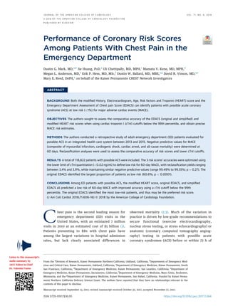
- 9. recommendations to perform serial and/or appropri- ately delayed cTn testing in the ED, as was done in the modified HEART pathway and EDACS accelerated diagnostic protocol studies (9,10,12). It should also be noted that the 2 most recent studies of the EDACS added “red flag” criteria (cre- scendo angina or abnormal vital signs) as stand- alone, non–low-risk criteria, irrespective of the risk score, in order to reach NPV point estimates of 100% (10,11). Although we were unable to fully evaluate the impact of these additional criteria given limitations in assessing for crescendo angina retrospectively, we intentionally did not include vital signs in our pri- mary validation analysis because these were not found to improve diagnostic accuracy in the deriva- tion of the EDACS (9). Regardless, the NPV estimates found in the lowest-risk group need not be 100% to justify forgoing further testing, as discussed in the preceding text. CONCLUSIONS Among ED patients with possible ACS, either the modified HEART score, the original EDACS or the simplified EDACS were accurate in predicting a low risk of 60-day MACE (i.e., >99% NPV), with the original EDACS classifying the greatest pro- portion of patients as low-risk. Accuracy was TABLE 5 Reclassification Yield for 60-Day MACE Between Low-Risk Scores Among Patients With Peak cTnI <0.02 ng/ml Correctly Reclassified Patients Falsely Reclassified Patients Reclassification Yield (%) Original EDACS to modified HEART 90 10,339 0.86 (0.70–1.06) Original EDACS to simplified EDACS 99 14,804 0.66 (0.54–0.80) Modified HEART to simplified EDACS 9 4,465 0.19 (0.11–0.38) Abbreviations as in Tables 1 to 3. CENTRAL ILLUSTRATION Performance of the EDACS Versus Modified HEART Score Among Emergency Department Patients With Chest Pain 60 cTnl <0.02 + LOW RISK SCORE cTnl <0.02 + NON-LOW RISK SCORE cTnl 0.02-0.04 + LOW RISK SCORE cTnl 0.02-0.04 + NON-LOW RISK SCORE 30701.00 0.95 NPV DAYS 0.90 Lower troponin cut-off to define low risk is superior EDACS & mHEART perform similarly in terms of NPV mHEART NPV 99.5 EDACS NPV 99.5 EDACS categorizes more patients as low risk EDACS mHEART 60.6% LOW RISK 51.8% LOW RISK Mark, D.G. et al. J Am Coll Cardiol. 2018;71(6):606–16. The EDACS and mHEART scores provide equivalent NPVs for cardiac events. EDACS classifies more patients as low risk. Risk stratification is improved using a cTnI cutoff below the 99th percentile. cTnI ¼ cardiac troponin I; EDACS ¼ Emergency Department Assessment of Chest pain Score; HEART ¼ History, Electrocardiogram, Age, Risk factors and Troponin; mHEART ¼ modified History, Electrocardiogram, Age, Risk factors and Troponin; NPV ¼ negative predictive value. Mark et al. J A C C V O L . 7 1 , N O . 6 , 2 0 1 8 Comparing the EDACS and Modified HEART Score F E B R U A R Y 1 3 , 2 0 1 8 : 6 0 6 – 1 6 614
- 10. optimized by lowering the cTnI cutoff to the lower limit of quantitation (<0.02 ng/ml), thus identi- fying higher-risk subgroups with cTnI concentra- tions at the upper end of the 99th percentile. Large prospective studies to confirm the safety of ED discharge with deferral of functional or anatomic cardiac testing among low-risk subgroups are needed. ADDRESS FOR CORRESPONDENCE: Dr. Dustin G. Mark, Department of Emergency Medicine, Kaiser Permanente, 275 West MacArthur Boulevard, Oakland, California 94611. E-mail: Dustin.G.Mark@kp.org. R E F E R E N C E S 1. Owens PL, Barrett ML, Gibson TB, Andrews RM, Weinick RM, Mutter RL. Emergency department care in the United States: a profile of national data sources. Ann Emerg Med 2010;56:150–65. 2. Venkatesh AK, Dai Y, Ross JS, Schuur JD, Capp R, Krumholz HM. Variation in US hospital emergency department admission rates by clinical condition. Med Care 2015;53:237–44. 3. Sabbatini AK, Nallamothu BK, Kocher KE. Reducing variation in hospital admissions from the emergency department for low-mortality condi- tions may produce savings. Health Aff (Millwood) 2014;33:1655–63. 4. Amsterdam EA, Kirk JD, Bluemke DA, et al. Testing of low-risk patients presenting to the emergency department with chest pain: a scienti- fic statement from the American Heart Associa- tion. Circulation 2010;122:1756–76. 5. Amsterdam EA, Wenger NK, Brindis RG, et al. 2014 AHA/ACC guideline for the management of patients with non-ST-elevation acute coronary syndromes: a report of the American College of Cardiology/American Heart Association Task Force on Practice Guidelines. J Am Coll Cardiol 2014;64: e139–228. 6. Napoli AM, Arrighi JA, Siket MS, Gibbs FJ. Physician discretion is safe and may lower stress test utilization in emergency department chest pain unit patients. Crit Pathw Cardiol 2012;11:26–31. 7. Aldous S, Richards AM, Cullen L, Pickering JW, Than M. The incremental value of stress testing in patients with acute chest pain beyond serial car- diac troponin testing. Emerg Med J 2016;33: 319–24. 8. Hermann LK, Newman DH, Pleasant WA, et al. Yield of routine provocative cardiac testing among patients in an emergency department-based chest pain unit. JAMA Intern Med 2013;173:1128–33. 9. Than M, Flaws D, Sanders S, et al. Development and validation of the Emergency Department Assessment of Chest pain Score and 2 h acceler- ated diagnostic protocol. Emerg Med Australas 2014;26:34–44. 10. Than MP, Pickering JW, Aldous SJ, et al. Effectiveness of EDACS versus ADAPT accelerated diagnostic pathways for chest pain: a pragmatic randomized controlled trial embedded within practice. Ann Emerg Med 2016;68:93–102.e1. 11. Flaws D, Than M, Scheuermeyer FX, et al. External validation of the emergency department assessment of chest pain score accelerated diag- nostic pathway (EDACS-ADP). Emerg Med J 2016; 33:618–25. 12. Mahler SA, Riley RF, Hiestand BC, et al. The HEART Pathway randomized trial: identifying emergency department patients with acute chest pain for early discharge. Circ Cardiovasc Qual Outcomes 2015;8:195–203. 13. Sanders S, Flaws D, Than M, Pickering JW, Doust J, Glasziou P. Simplification of a scoring system maintained overall accuracy but decreased the proportion classified as low risk. J Clin Epi- demiol 2016;69:32–9. 14. Goodacre S, Thokala P, Carroll C, et al. Sys- tematic review, meta-analysis and economic modelling of diagnostic strategies for suspected acute coronary syndrome. Health Technol Assess 2013;17:v–vi,1–188. 15. Carlton EW, Khattab A, Greaves K. Identifying patients suitable for discharge after a single- presentation high-sensitivity troponin result: a comparison of five established risk scores and two high-sensitivity assays. Ann Emerg Med 2015;66: 635–45.e1. 16. Brooker JA, Hastings JW, Major-Monfried H, et al. The association between medicolegal and professional concerns and chest pain admission rates. Acad Emerg Med 2015;22:883–6. 17. Than M, Herbert M, Flaws D, et al. What is an acceptable risk of major adverse cardiac event in chest pain patients soon after discharge from the emergency department?: a clinical survey. Int J Cardiol 2013;166:752–4. 18. Willeit P, Welsh P, Evans JDW, et al. High- sensitivity cardiac troponin concentration and risk of first-ever cardiovascular outcomes in 154, 052 participants. J Am Coll Cardiol 2017;70: 558–68. 19. Eggers KM, Lagerqvist B, Venge P, Wallentin L, Lindahl B. Persistent cardiac troponin I elevation in stabilized patients after an episode of acute coronary syndrome predicts long-term mortality. Circulation 2007;116:1907–14. 20. Kavsak PA, Newman AM, Lustig V, et al. Long- term health outcomes associated with detectable troponin I concentrations. Clin Chem 2007;53: 220–7. 21. Zethelius B, Johnston N, Venge P. Troponin I as a predictor of coronary heart disease and mortality in 70-year-old men: a community-based cohort study. Circulation 2006;113:1071–8. 22. Pauker SG, Kassirer JP. The threshold approach to clinical decision making. N Engl J Med 1980;302:1109–17. 23. Foy AJ, Liu G, Davidson WR Jr., Sciamanna C, Leslie DL. Comparative effectiveness of diagnostic testing strategies in emergency department pa- tients with chest pain: an analysis of downstream testing, interventions, and outcomes. JAMA Intern Med 2015;175:428–36. 24. Sandhu AT, Heidenreich PA, Bhattacharya J, Bundorf MK. Cardiovascular testing and clinical outcomes in emergency department patients with chest pain. JAMA Intern Med 2017;177:1175–82. 25. Safavi KC, Li SX, Dharmarajan K, et al. Hospital variation in the use of noninvasive cardiac imaging and its association with downstream testing, in- terventions, and outcomes. JAMA Intern Med 2014;174:546–53. 26. Venge P, Lagerqvist B, Diderholm E, Lindahl B, Wallentin L. Clinical performance of three cardiac troponin assays in patients with unstable coronary artery disease (a FRISC II substudy). Am J Cardiol 2002;89:1035–41. 27. Thygesen K, Alpert JS, Jaffe AS, et al. Third universal definition of myocardial infarction. Cir- culation 2012;126:2020–35. 28. Cullen L, Than M, Brown AF, et al. Compre- hensive standardized data definitions for acute coronary syndrome research in emergency de- partments in Australasia. Emerg Med Australas 2010;22:35–55. 29. Greene DN, Leong TK, Collinson PO, et al. Age, sex, and racial influences on the Beckman Coulter AccuTnIþ3 99th percentile. Clin Chim Acta 2015; 444:149–53. PERSPECTIVES COMPETENCY IN PATIENT CARE AND PROCEDURAL SKILLS: When considered in combination with blood levels of cTnI, either the modified HEART score or the EDACS can identify patients with chest pain at low risk (<1%) of MACE. Routine cardiac stress testing may not be necessary in these very–low- risk patients. TRANSLATIONAL OUTLOOK: Prospective studies in which stress testing is omitted from the evaluation of lowest-risk pa- tients are necessary to confirm the safety of this approach. J A C C V O L . 7 1 , N O . 6 , 2 0 1 8 Mark et al. F E B R U A R Y 1 3 , 2 0 1 8 : 6 0 6 – 1 6 Comparing the EDACS and Modified HEART Score 615
- 11. 30. Usher-Smith JA, Sharp SJ, Griffin SJ. The spectrum effect in tests for risk prediction, screening, and diagnosis. BMJ 2016;353:i3139. 31. West R, Ellis G, Brooks N, Joint Audit Com- mittee of the British Cardiac Society and Royal College of Physicians of London. Complications of diagnostic cardiac catheterisation: results from a confidential inquiry into cardiac catheter compli- cations. Heart 2006;92:810–4. 32. Udelson JE, Beshansky JR, Ballin DS, et al. Myocardial perfusion imaging for evaluation and triage of patients with suspected acute cardiac ischemia: a randomized controlled trial. JAMA 2002;288:2693–700. 33. Lewis HD Jr., Davis JW, Archibald DG, et al. Protective effects of aspirin against acute myocar- dial infarction and death in men with unstable angina. Results of a Veterans Administration Coop- erative Study. N Engl J Med 1983;309:396–403. 34. Hage FG. Regadenoson for myocardial perfu- sion imaging: is it safe? J Nucl Cardiol 2014;21: 871–6. 35. Geleijnse ML, Fioretti PM, Roelandt JR. Meth- odology, feasibility, safety and diagnostic accu- racy of dobutamine stress echocardiography. J Am Coll Cardiol 1997;30:595–606. 36. Kline JA, Johnson CL, Pollack CV Jr., et al. Pretest probability assessment derived from attribute matching. BMC Med Inform Decis Mak 2005;5:26. 37. Chapman AR, Anand A, Boeddinghaus J, et al. Comparison of the efficacy and safety of early rule-out pathways for acute myocardial infarction. Circulation 2017;135:1586–96. 38. Boeddinghaus J, Nestelberger T, Twerenbold R, et al. Direct comparison of 4 very early rule-out strategies for acute myocardial infarction using high-sensitivity cardiac troponin I. Circulation 2017;135:1597–611. 39. Sorensen NA, Neumann JT, Ojeda F, et al. Challenging the 99th percentile: a lower troponin cutoff leads to low mortality of chest pain patients. Int J Cardiol 2017;232:289–93. 40. Sandoval Y, Smith SW, Thordsen SE, et al. Diagnostic performance of high sensitivity compared with contemporary cardiac troponin I for the diagnosis of acute myocardial infarction. Clin Chem 2017;63:1594–604. 41. Cullen L, Mueller C, Parsonage WA, et al. Validation of high-sensitivity troponin I in a 2-hour diagnostic strategy to assess 30-day outcomes in emergency department patients with possible acute coronary syndrome. J Am Coll Cardiol 2013; 62:1242–9. 42. Love SA, Sandoval Y, Smith SW, et al. Inci- dence of undetectable, measurable, and increased cardiac troponin I concentrations above the 99th percentile using a high-sensitivity vs a contem- porary assay in patients presenting to the emer- gency department. Clin Chem 2016;62:1115–9. 43. Stopyra JP, Miller CD, Hiestand BC, et al. Performance of the EDACS-accelerated diagnostic pathway in a cohort of US patients with acute chest pain. Crit Pathw Cardiol 2015;14:134–8. 44. Six AJ, Cullen L, Backus BE, et al. The HEART score for the assessment of patients with chest pain in the emergency department: a multina- tional validation study. Crit Pathw Cardiol 2013; 12:121–6. KEY WORDS acute coronary syndrome, myocardial ischemia, risk stratification APPENDIX For an expanded Methods section as well as supplemental tables and a figure, please see the online version of this article. Mark et al. J A C C V O L . 7 1 , N O . 6 , 2 0 1 8 Comparing the EDACS and Modified HEART Score F E B R U A R Y 1 3 , 2 0 1 8 : 6 0 6 – 1 6 616