
Recommended
Recommended
More Related Content
What's hot
What's hot (20)
Similar to Accessory Navicular Bone Removal Recovery Guide
Similar to Accessory Navicular Bone Removal Recovery Guide (20)
Accessory Navicular Bone Removal Recovery Guide
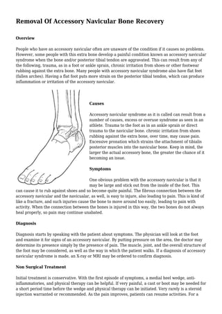
- 1. Removal Of Accessory Navicular Bone Recovery Overview People who have an accessory navicular often are unaware of the condition if it causes no problems. However, some people with this extra bone develop a painful condition known as accessory navicular syndrome when the bone and/or posterior tibial tendon are aggravated. This can result from any of the following, trauma, as in a foot or ankle sprain, chronic irritation from shoes or other footwear rubbing against the extra bone. Many people with accessory navicular syndrome also have flat feet (fallen arches). Having a flat foot puts more strain on the posterior tibial tendon, which can produce inflammation or irritation of the accessory navicular. Causes Accessory navicular syndrome as it is called can result from a number of causes, excess or overuse syndrome as seen in an athlete. Trauma to the foot as in an ankle sprain or direct trauma to the navicular bone. chronic irritation from shoes rubbing against the extra bone, over time, may cause pain. Excessive pronation which strains the attachment of tibialis posterior muscles into the navicular bone. Keep in mind, the larger the actual accessory bone, the greater the chance of it becoming an issue. Symptoms One obvious problem with the accessory navicular is that it may be large and stick out from the inside of the foot. This can cause it to rub against shoes and so become quite painful. The fibrous connection between the accessory navicular and the navicualar, as well, is easy to injure, also leading to pain. This is kind of like a fracture, and such injuries cause the bone to move around too easily, leading to pain with activity. When the connection between the bones is injured in this way, the two bones do not always heal properly, so pain may continue unabated. Diagnosis Diagnosis starts by speaking with the patient about symptoms. The physician will look at the foot and examine it for signs of an accessory navicular. By putting pressure on the area, the doctor may determine its presence simply by the presence of pain. The muscle, joint, and the overall structure of the foot may be considered, as well as the way in which the patient walks. If a diagnosis of accessory navicular syndrome is made, an X-ray or MRI may be ordered to confirm diagnosis. Non Surgical Treatment Initial treatment is conservative. With the first episode of symptoms, a medial heel wedge, anti- inflammatories, and physical therapy can be helpful. If very painful, a cast or boot may be needed for a short period time before the wedge and physical therapy can be initiated. Very rarely is a steroid injection warranted or recommended. As the pain improves, patients can resume activities. For a
- 2. minority of patients, an arch support or custom orthotic can help to take some of the extra pressure off of the accessory navicular and the posterior tibial tendon. Surgical Treatment Fusion of the accessory navicular to the navicular with screws is required when there is a large accessory navicular bone and removal of this bone would reduce the articular surface of the Navicular to the talus (coxa pedis). Fusion will relieve pain without disrupting the tibialis posterior tendon insertion nor narrowing talar head support. In most instances, a patient’s recovery will be as follows. 0-6 weeks: Immobilization (in case or cast boot) non-weight-bearing or touch weight-bearing. 6-10 weeks: Increasing activity in a cast boot. Physical therapy to work on strength and balance. Full recovery after 9 weeks-2 months. In some patients (where the posterior tibial tendon is still intact and functioning) the treating surgeon may allow weight-bearing as tolerated in a cast boot immediately after surgery.