College Call Girls Pune Mira 9907093804 Short 1500 Night 6000 Best call girls...
Cell division-1.ppt
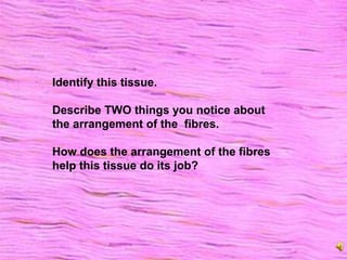
1. Identify this tissue.
Describe TWO things you notice about
the arrangement of the fibres.
How does the arrangement of the fibres
help this tissue do its job?
2. Cells become specialised for the job they do.
Look at the picture above of a neurone (nerve cell).
Discuss with your neighbours about what you notice about the shape of the cell.
What special features do these cells have?
How does the shape of the cell, or its special features, help it carry out its
function (job)?
5. Functions of Cell Division
New cells for growth, and to replace
damaged or diseased cells.
Special cells for reproduction that pass on
genetic information
Somatic cell division: occurs in all cells
except reproductive cells
Reproductive cell division: only occurs in
reproductive cells of the testes and ovaries
9. Interphase – “time for a break”
The chromosomes are not visible, but exist
as a chromatin network.
http://www.life.illinois.edu/ib/102/lectures/08reproduction.html
10. Before MITOSIS happens
Each chromosome is
duplicated
Each chromosome is
attached to its
duplicate at the
centromere
Each of these
identical “sister”
chromosomes is
called a chromatid http://www.uic.edu/classes/bios/bios100/labs/realchromo.jpeg
11. Prophase – “Photocopy”or “produce”
The duplicated chromosomes “appear” and contain two
sets of genetic information
The chromosomes become visible as they shorten and
thicken
Nuclear membrane disappears
13. Anaphase – “Apart”
Chromosomes separate as the spindle
shortens, so one chromatid is at each end of
the cell. Each half of the cell ends up with
original number of chromosomes.
14. Telophase – “Two”
The plasma membrane pinches inwards
New nuclear membrane forms around the
chromosomes
Cytoplasm divides (cytokinesis)
15. Interphase
The two new daughter cells now enter the
“resting phase” before going undergoing
mitosis themselves.
18. Did you know?
Neurons in the brain do not
undergo mitosis.
Therefore we always assumed that
there are never any “new” brain cells.
However, it has recently been
discovered that there are stem cells in
the brain.
Brain cell replacement is possible!
19. What’s a stem cell?
These are unspecialised cells that produce
other cells.
An individual paralysed after a spinal cord injury
donated stem cells from in her nose, and had
them placed within her spinal cord. The stem
cells developed into nerve cells.
20. Some cells differentiate to form sex cells or
gametes, also known as:
Egg or Ova
Sperm or spermatozoa
21. Homologous pairs
Chromosomes in cells occur in
pairs – you get one from each
parent.
These homologous
chromosomes are
DIFFERENT from the
chromatids.
The two homologous
chromosomes are identical in
form – each one has
instructions to make the same
thing e.g. hair colour, eye
colour, but one set of
instructions is Mums, and the
other Dads.
22. The “Poids”
There are 23 pairs (46 chromosomes) in a
cell with a full set of homologous
chromosomes is said to be DIPLOID
Each of the gametes (sex cells) produced by
a parent only has HALF the chromosomes
and is said to be HAPLOID
23. Stages of Meiosis
Meiosis I - Reductional division
Daughter cells have half the number of
chromosomes that the parent cell has
Meiosis II - Equational division
These chromosomes are distributed evenly
among four daughter cells
25. Meiosis I – reductional division
Interphase
In this diagram the
parent cell has 4
chromosomes.
Chromosomes
duplicate before
meiosis begins.
Each homologous
chromosome
consists of two
joined “sister”
chromatids joined
by centromere
28. Crossing over
Crossing over can occur. The chromosomes entangle
and bits can break off, and can be exchanged. This
increases the variation among us!
The nuclear membrane then disappears
Spindle fibres form
Chromosomes line up in the middle
29. The chromosomes segregate.
Then two new daughter cells form with
unseparated chromatids
(only the homologous pairs have separated)
Each of the cells above has 4
chromosomes, or pairs of chromatids.
30. Meiosis II – equational division
The rest of the process is pretty much like
mitosis.
The nuclear membrane disappears again and
the spindle forms.
The chromosomes line up in the middle
The chromatids separate as the centromere
breaks, and chromosomes move to opposite
sides of cell
Nuclear membrane reappears, and cytokinesis
occurs
31. Division is complete – in this diagram, four cells with
haploid numbers of chromosomes.
32. Meiosis II
Same as mitosis in every way except
chromosomes don’t replicate before starting
Spindle forms
Chromosomes line up in centre
Centromere breaks, chromosomes move to
opposite sides of cell
Nuclear membrane reappears
Cytokinesis occurs
4 daughter cells formed each with 23
chromosomes (haploid)
33.
34.
35. Variations in cell division
Most cell growth is orderly. Cells usually
reproduce at the proper rate and align
themselves in the right places.
Sometimes cell growth can become
uncontrolled and disorganised. This process
is experienced by the patient as a lump or a
tumor (swelling).
36. Variations in Cell Division
Very rapid in skin, GI tract, bone marrow and
in tumour cells
Non-existent in skeletal muscle, nervous
tissue, cardiac muscle
37. Tumours
Benign – non-cancerous
Malignant – cancerous
Cancer cells are appropriately named because like a
“crab” the cancer cells send out clawlike extensions
that invade surrounding tissue.
Cancer cells may detach from the original tumour, and
spread throught the body. This is known as
METASTASIS.
38. Cell Transformation
Process of normal cells becoming cancer cells
Autonomy: refers to cancer cell’s
independence from normal cellular controls
Anaplasia: loss of differentiation
Cancer cells are less differentiated than
normal tissues and they replicate faster
Level of differentiation can be used to grade
severity of cancer
39. And what’s in store for our cells?
Cells get larger as we get old, and they can’t divide
as easily.
The ability to repair themselves, also declines as we
age
Cells function less efficiently as there are less
mitochondria and lysosomes