Call Girls In Andheri East Call 9920874524 Book Hot And Sexy Girls
medical exam
1. Lichen planus can affect any part of the body surface, but is most
often seen on the volar aspect of the wrists, the lumbar region and
around the ankles. Flexural sites like axillae, groins and inframam-
mary regions may be rarely involved in typical lichen planus. Reports
on flexural LP in the published work are either associated with LP
pigmentosus or erosive variants.1,2
In most LP cases, the papule
lesions eventually flatten after a few months, often to be replaced by
an area of pigmentation that retains the shape of the papule and
persists for months or years. In this case, we can see three kinds of
different period lesions (violaceous-brown papules, papules with
the pitchy edge and annular dark brownish macules) on the flexural
sites representing the gradual regression.
Follicular lesions usually appear during the course of typical LP,
sometimes as sole manifestation of the disease in the scalp. But
they rarely occur in flexural LP. Gunduz et al.3
reported the first case
of combination of follicular and flexural variants of LP. But there was
a little difference between the two cases because the follicular
lesions localized to the flexures and the waist, respectively.
The infiltrating cells in LP are predominantly T-lymphocytes with
very few B-lymphocytes. The identification of various subtypes of
T-lymphocytes has given contradictory results with regards to the
predominance of CD4+
helper-inducer T-lymphocytes and CD8+
suppressor-cytotoxic T-lymphocytes in the infiltrate. It is likely that
both subsets participate in the immunological reaction.4
Our
immunohistochemical study demonstrated the same result and it
was easy to distinguish with LP-like keratosis because CD4+
lymphocytes were abundant in the dermis as Jang et al.5
observed. Contrasted to the flexural lesion, the follicular lesion
of the waist was characterized by a higher CD4 ⁄ CD8 ratio of
T-lymphocytes.
Our patient did not use any special treatment in the 6-month
course of disease, but we can see the submammary and groin
lesions are undergoing progressive spontaneous regression.
We conclude that the process is benign and tends to resolve
spontaneously. We are now following up the patient without admin-
istering any special treatment.
Han MA, Lei GUAN, Xiang-yang SU, Wei LAI,
Chun LU
Department of Dermatology, The Third Affiliated Hospital of Sun Yat-sen
University, Guangzhou, Guangdong, China
REFERENCES
1 Pock L, Jelinkova L, Drlik L et al. Lichen planus pigmentosus-inversus.
J Eur Acad Dermatol Venereol 2001; 15: 452–454.
2 Eisman S, Orteu CH. Recalcitrant erosive flexural lichen planus, successful
treatment with a combination of thalidomide and 0.1% tacrolimus ointment.
Clin Exp Dermatol 2004; 29: 268–270.
3 Gunduz K, Sacar T, Inanir I et al. Flexural follicular lichen planus. Clin Exp
Dermatol 2009; 34: 297–298.
4 Elder DE, Elenitsas R, Johnson BL et al. Lever’s Histopathology of the Skin.
In: Narciss M, Sonia T, Hideko K, eds. Noninfectious Erythematous, Papu-
lar, and Squamous Diseases, 9nd edn. Philadelphia: Lippincott Williams &
Wilkins Press, 2005; 198–199.
5 Jang KA, Kim SH, Choi JH et al. Lichenoid keratosis: a clinicopathologic
study of 17 patients. J Am Acad Dermatol 2000; 43: 511–516.
Linear lichen planus pigmentosus of the forehead treated
by neodymium:yttrium–aluminum–garnet laser and topical
tacrolimus
Dear Editor,
Lichen planus pigmentosus (LPP) is an uncommon variant of lichen
planus (LP), which is characterized by the insidious onset of dark-
brown macules in sun-exposed areas and flexural folds. It was origi-
nally reported from India but it tends to occur also in other racial and
ethnic groups.1,2
The pattern of pigmentation is mostly diffuse and it
can present rarely in linear or follicular pattern.3
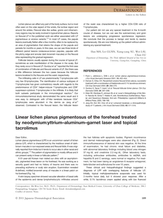
A 61-year-old Korean man visited our clinic with an asymptom-
atic, pigmented linear lesion on his forehead. He was working as a
security guard and had no history of trauma and there was no
preceding erythema or scaly skin eruption. Examination revealed
ill-defined, mottled brownish array of macules in a linear patch on
his forehead (Fig. 1a).
A skin biopsy specimen showed vacuolar alteration of basal cells
of the epidermis and dense lymphohistiocytic infiltrates around
the hair follicles with apoptotic bodies. Pigment incontinence
and dermal melanophages were also observed (Fig. 2). Direct
immunofluorescence of lesional skin was negative. At the time
of examination, he had chronic renal failure and diabetes,
with abnormal laboratory findings including blood urea nitrogen
37 mg ⁄ dL and creatinine 2.3 mg ⁄ dL. Other laboratory tests,
including antinuclear antibodies, antibodies to dsDNA, and
hepatitis B and C serology, were normal or negative. For treat-
ment, he had been taking an angiotensin II receptor antagonist,
beta-blocker and sulfonylurea for over 10 years.
These clinical and histopathological findings suggested a
diagnosis of LPP with overlapping features of follicular LP.
Initially, topical methylprednisolone aceponate was used for
3 months twice daily but it showed poor response. After a
while, 0.1% tacrolimus cream applied along with low fluenced
Correspondence: Mi-Woo Lee, M.D., Ph.D., Department of Dermatology, Asan Medical Center, University of Ulsan College of Medicine, 388-1
Pungnapdong, Songpagu, 138-736 Seoul, Korea. Email: dermakim@gmail.com
Conflict of interest: None.
Letters to the Editor
Ó 2011 Japanese Dermatological Association 189
2. (1.8 J ⁄ cm2
) 1064-nm Q-switched neodymium:yttrium–aluminum–
garnet laser (QSNY) (Spectra VRM; Lutronic, Gyeonggi, South
Korea) every 3 weeks. Six weeks later, the lesions were much
lighter and after 4 months, the lesion had cleared without a scar
and there has been no evidence of recurrence for over 6 months
(Fig. 1b–d).
(a) (b)
(c) (d)
Figure 1. Photographs showing (a) brownish linear patch on the forehead before treatment, (b) partial improvement after 6 weeks of treatment
with a 0.1% topical tacrolimus and 1064-nm Q-switched neodymium:yttrium–aluminum–garnet laser, (c) resolution without a scar after 4 months
of treatment and (d) maintenance without recurrence 6 months after ceasing treatment.
(a) (b)
Figure 2. Histopathology showing epidermal basal cell vacuolar degeneration, lymphohistiocytic infiltration and basal cell liquefaction around
the hair follicle, as well as pigment incontinence (hematoxylin–eosin, original magnifications: [a] ·100, [b] ·200).
Letters to the Editor
190 Ó 2011 Japanese Dermatological Association
3. The histopathological changes of LPP consisted of vacuolar
degeneration of the basal layer in the epidermis. In the dermis, peri-
vascular or lichenoid infiltrate and the presence of melanin inconti-
nence were the predominant changes noted. A recently developed
lesion tends to show more predominant band-like lymphocytic
infiltration and epidermal vacuolization rather than epidermal
atrophy.3,4
Linear lesions can frequently occur at sites of scratching or
trauma in patients with LP as a result of Koebner’s phenomenon, or,
as in our case, they may appear spontaneously within the lines of
Blaschko on the face.5
In acquired Blaschko linear inflammatory
dermatosis, cutaneous antigenic mosaicism could be responsible
for the susceptibility to induce mosaic T-cell responses. Because
drugs had not been changed in type or dosage over several years
of treatment, and underlying medical diseases had been well con-
trolled, the possibility of drug-related reaction was thought to be
low. Considering the clinical features in our patient, and the fact
that exposed sites were frequently the first to be involved, it can be
suggested that exposure to sunlight (even in a casual dose) may be
a kind of stimuli to induce the lesion of LPP in a genetically suscepti-
ble patient.4
Usually the course is chronic and treatments are less effective
for follicular LP or LPP than for classical LP.3–7
Topical tacrolimus,
a member of the immunosuppressive macrolide family that sup-
presses T-cell activation, has been shown to be effective in the
treatment of some mucosal and follicular LP.3,6,7
There is only one
article about the successful treatment of LPP with topical tacroli-
mus.3
Although they showed over 50% improvement in seven of
13 patients after 4 months of treatment, the authors did not men-
tion any case of complete clearance in their article. Moreover,
the other six of the 13 patients did not show improvement in
pigmentation.
Therefore, in the present case, 1064-nm QSNY with low fluence
treatment was chosen for treating pigmentation. The 1064-nm
QSNY in nanosecond (ns) domain is strongly absorbed by the
finely distributed melanin in dermal pigmented lesions. Moreover,
1064-nm QSNY with low fluence, which in a ‘‘top-hat’’ beam
mode can evenly distribute energy density throughout the whole
spot, is now widely used when treating darker skin types, because
it greatly reduces the risk of epidermal injury and post-therapy
dyschromia.8,9
In our patient, because of poor response to topical
steroid, we started tacrolimus ointment for mainly targeting T cells,
and for the treatment of pigmentation, we added QSNY treatment.
It suggests that the combination treatment of 1064-nm low flu-
enced QSNY with topical tacrolimus may be a good therapeutic
option for patients with recalcitrant facial LPP in dark-skinned
individuals.
Jeong-Eun KIM, Chong-Hyun WON,
Sungeun CHANG, Mi-Woo LEE, Jee-Ho CHOI,
Kee-Chan MOON
Department of Dermatology, Asan Medical Center,
University of Ulsan College of Medicine, Seoul, Korea
REFERENCES
1 Bhutani LK, Bedi TR, Pandhi RK, Nayak NC. Lichen planus pigmentosus.
Dermatologica 1974; 149: 43–50.
2 Kanwar AJ, Kaur S. Lichen planus pigmentosus. J Am Acad Dermatol
1989; 21: 815.
3 Al-Mutairi N, El-Khalawany M. Clinicopathological characteristics of lichen
planus pigmentosus and its response to tacrolimus ointment: an open
label, non-randomized, prospective study. J Eur Acad Dermatol Venereol
2010; 24: 535–540.
4 Kanwar AJ, Dogra S, Handa S et al. A study of 124 Indian patients with
lichen planus pigmentosus. Clin Exp Dermatol 2003; 28: 481–485.
5 Ezzedine K, Simonart T, Vereecken P et al. Facial actinic lichen planus
following the Blaschko’s line: successful treatment with topical 0.1%
pimecrolimus cream. J Eur Acad Dermatol Venereol 2009; 23: 458–
459.
6 Volz T, Caroli U, Ludtke H et al. Pimecrolimus cream 1% in erosive oral
lichen planus – a prospective randomized double-blind vehicle-controlled
study. Br J Dermatol 2008; 159: 936–941.
7 Blazek C, Megahed M. Lichen planopilaris. Successful treatment with
tacrolimus. Hautarzt 2008; 59: 874–877.
8 Cho SB, Park SJ, Kim SJ et al. Treatment of post-inflammatory hyperpig-
mentation using 1064-nm Q-switched Nd:YAG laser with low fluence:
report of three cases. J Eur Acad Dermatol Venereol 2009; 23: 1206–
1207.
9 Kim JH, Kim H, Park HC et al. Subcellular selective photothermolysis of
melanosomes in adult zebrafish skin following 1064-nm Q-switched
Nd:YAG laser irradiation. J Invest Dermatol 2010; 130: 2333–2335.
Granuloma annulare within the red dye of a tattoo
Dear Editor,
Eczematous, lymphohistiocytic, lichenoid, granulomatous, sarcoid-
osis-like or pseudolymphomatous reactions may occur in tattoos
with highly variable delays.1,2
Granuloma annulare (GA) is a com-
mon dermatosis that has seldom been reported in tattoos.3–5
We
report a new case within the red dye of a tattoo.
A 36-year-old otherwise healthy Caucasian man was referred for
an inflamed and infiltrated itchy reaction restricted to the red part of
a leg tattoo that had developed 6 months earlier. He had been tat-
tooed on three different occasions without any complication. One
tattoo depicting a dragon was performed on the left lower leg in
2000 in a tattoo parlor. The healing phase had been unremarkable.
Ten years later, he developed an itchy infiltrated reaction restricted
to its red part (Fig. 1). The rest of the tattoo was spared, as were
two prior black-colored tattoos. Examination was otherwise
unremarkable with no sign of systemic disease. A 3-mm punch
biopsy of the inflamed tattoo revealed large areas of necrobiosis
surrounded by a heavy interstitial and perivascular inflammatory
Correspondence: Nicolas Kluger, M.D., Departments of Dermatology, Allergology and Venereology, Institute of Clinical Medicine, University of
Helsinki, Helsinki University Central Hospital, Meilahdentie 2, PO Box 160, 00029 HUS, Finland. Email: nicolaskluger@yahoo.fr
Letters to the Editor
Ó 2011 Japanese Dermatological Association 191
4. Copyright of Journal of Dermatology is the property of Wiley-Blackwell and its content may not be copied or
emailed to multiple sites or posted to a listserv without the copyright holder's express written permission.
However, users may print, download, or email articles for individual use.
![(1.8 J ⁄ cm2
) 1064-nm Q-switched neodymium:yttrium–aluminum–
garnet laser (QSNY) (Spectra VRM; Lutronic, Gyeonggi, South
Korea) every 3 weeks. Six weeks later, the lesions were much
lighter and after 4 months, the lesion had cleared without a scar
and there has been no evidence of recurrence for over 6 months
(Fig. 1b–d).
(a) (b)
(c) (d)
Figure 1. Photographs showing (a) brownish linear patch on the forehead before treatment, (b) partial improvement after 6 weeks of treatment
with a 0.1% topical tacrolimus and 1064-nm Q-switched neodymium:yttrium–aluminum–garnet laser, (c) resolution without a scar after 4 months
of treatment and (d) maintenance without recurrence 6 months after ceasing treatment.
(a) (b)
Figure 2. Histopathology showing epidermal basal cell vacuolar degeneration, lymphohistiocytic infiltration and basal cell liquefaction around
the hair follicle, as well as pigment incontinence (hematoxylin–eosin, original magnifications: [a] ·100, [b] ·200).
Letters to the Editor
190 Ó 2011 Japanese Dermatological Association](data:image/gif;base64,R0lGODlhAQABAIAAAAAAAP///yH5BAEAAAAALAAAAAABAAEAAAIBRAA7)