SlideShare a Scribd company logo
1 of 10
Download to read offline
State of the Art Review
Anesthetic management of the head trauma patient
Elizabeth A. Armitage-Chan, MA, VetMB, DACVA, MRCVS, Lois A. Wetmore, DVM, ScD, DACVA
and Daniel L. Chan, DVM, DACVECC, DACVN, MRCVS
Abstract
Objective: To describe the optimal anesthetic management of patients with brain injury, with emphasis on the
support of oxygen delivery to the brain, and the effects of anesthetic agents on cerebral perfusion.
Data sources: Clinical and experimental studies from both the human and veterinary neuroanesthesia
literature.
Summary: The management of patients following primary traumatic brain injury (TBI) significantly impacts
outcome. Outcome can be improved by strategies that improve oxygen delivery to the brain and prevent
cerebral ischemia. Anesthetic agents have widely variable effects on the blood supply to the brain and,
therefore, choice of anesthetic agent can influence neurological outcome. Although in the past, anesthetic
agents have been selected for their neuroprotective properties, it is increasingly being recognized that the
support of cerebral perfusion during anesthesia contributes more significantly to a positive outcome for these
patients. Support of cardiorespiratory function is, therefore, highly important when anesthetizing patients
with TBI.
Conclusion: Choice of anesthetic agent is determined by the extent of brain injury and intracranial pressure
(ICP) elevation. Factors that should be considered when anesthetizing head trauma patients include the effects
of anesthetic agents on the cardiac and respiratory systems, their effects on cerebral blood flow (CBF), ICP, and
possible neuroprotective benefits offered by certain agents.
(J Vet Emerg Crit Care 2007; 17(1): 5–14) doi: 10.1111/j.1476-4431.2006.00194.x
Keywords: cerebral blood flow, dog, intracranial pressure, neuroanesthesia, neuroprotection
Introduction
Head trauma is seen frequently in dogs and cats. Ve-
hicular trauma, kick or bite injuries, ‘high-rise’ injuries,
and penetrating wounds are all reported causes.1,2
General anesthesia is often required during the man-
agement of head trauma patients, for purposes such as
surgery, diagnostic imaging, or mechanical ventilation.
Surgical intervention may be required to repair frac-
tures, thoracic trauma or large skin lacerations, or for
investigation and treatment of abdominal hemorrhage.
In addition, more severely affected cases may require
anesthesia for decompressive craniectomy, which is be-
coming increasingly common in veterinary medicine
for the management of head trauma.2,3
Analgesics are
also usually indicated in head trauma patients. Many
anesthetic and analgesic drugs have effects on intra-
cranial physiology, which under certain circumstances
may result in further neuronal insult.4,5
In contrast,
agents such as the barbiturates are frequently used
therapeutically in head trauma patients to reduce sei-
zure activity and protect neuronal function.6,7
An un-
derstanding of the mechanisms by which anesthetics
influence the injured brain is therefore beneficial in the
management of patients with head injury.
Intracranial Physiology
When planning an anesthetic regimen for patients with
traumatic brain injury (TBI) an understanding of cer-
ebral blood flow (CBF) physiology is beneficial. Many
anesthetic agents cause alterations in blood flow to the
brain and therefore have the potential to cause further
insult. The physiology of intracranial hemodynamics
and the effects of head trauma have been reviewed ex-
tensively elsewhere.8–10
However, a review of intracra-
nial physiology as it pertains to brain injury and the
implication to anesthetic management are briefly
discussed.
Address correspondence and reprint requests to:
Elizabeth A. Armitage-Chan, Department of Veterinary Clinical Sciences,
The Royal Veterinary College, Hawkshead Lane, North Mymms, Hatfield,
Hertfordshire AL9 7TA, UK.
E-mail: echan@rvc.ac.uk
From the Department of Clinical Sciences, Section of Anesthesia (Armitage-
Chan, Wetmore), Section of Emergency & Critical Care (Chan), Cummings
School of Veterinary Medicine at Tufts University, North Grafton, MA.
Journal of Veterinary Emergency and Critical Care 17(1) 2007, pp 5–14
doi:10.1111/j.1476-4431.2006.00194.x
& Veterinary Emergency and Critical Care Society 2006 5
Changes in intracranial blood flow are a significant
cause of cerebral injury after head trauma, and pre-
vention of ischemic damage by maintaining oxygen
delivery to the brain contributes significantly to out-
come.11–13
An understanding of the protective mecha-
nisms supporting blood flow to the brain is therefore
important in head trauma management. In order to
support a high oxygen requirement, the brain normally
receives a large percentage of the cardiac output, and
CBF is tightly regulated to prevent decreases in perfu-
sion. In a normal brain, constant CBF is maintained by
alterations in vasomotor tone regulated according to
changes in arterial oxygen (PaO2) and carbon dioxide
(PaCO2) partial pressures, and systemic blood pressure
(Figure 1). In the injured brain, these protective mech-
anisms are lost. A decrease in arteriolar pH caused by
an increase in PaCO2 results in vasodilation, reduction
of cerebral vascular resistance, and an increase in CBF.14
In contrast, hypocapnia results in intracranial vasocon-
striction and a decrease in cerebral perfusion.15
Alter-
ations of arterial oxygenation have a lesser effect on
intracranial hemodynamics unless severe hypoxemia
occurs. When PaO2 decreases below 50 mmHg, vascu-
lar resistance decreases in order to increase CBF and
maintain cerebral oxygen delivery.15
Changes in sys-
temic blood pressure are usually prevented from caus-
ing alterations in CBF by cerebral pressure
autoregulation. Pressure autoregulation maintains con-
stant CBF when mean arterial blood pressure (MABP)
varies within a physiological range (50–150 mmHg).15
Following TBI, pressure autoregulation may be lost
causing mildly decreased systemic blood pressures that
otherwise might be considered safe to result in mark-
edly reduced CBF. The increased dependency of CBF
on MAP may explain the worsened outcome associated
with hypotension in patients with TBI.13–17
Clinical assessment of CBF is difficult and as a result,
cerebral perfusion pressure (CPP), a variable that is
more easily ascertained and a clinical correlate of CBF,
is used to predict a patient’s risk for cerebral ischemia.18
CPP is the pressure gradient driving CBF. It is calcu-
lated as the difference between MABP and intracranial
pressure (ICP):
CPP ¼ MAP  ICP
ICP is defined as the pressure exerted between the skull
and the intracranial tissues.7
Since the skull is rigid and
poorly compliant, an increase in volume by any of its
contents without a concomitant reduction in the other
components results in an increase in pressure. Increases
in intracranial volume and pressure may result from an
increase in volume of brain tissue (e.g., by formation of
cerebral edema), blood (e.g., due to hemorrhage or
vasodilation), or cerebrospinal fluid (e.g., by obstruc-
tion to fluid outflow). As ICP increases, systemic blood
pressure must increase to prevent a decrease in CPP
and a resultant decrease in CBF. Excessive unregulated
cerebral vasodilation such as that which may arise dur-
ing anesthesia of TBI patients may also increase ICP,
reduce CPP, and may lead to cerebral ischemia.
Under normal circumstances, vasomotor tone is cou-
pled to the oxygen requirement of the brain by flow-
metabolism coupling. This phenomenon describes the
ability of the cerebral vasculature to respond appropri-
ately to changes in oxygen demand. Provided flow-
metabolism coupling remains intact, a reduction of cer-
ebral metabolic activity leads to a decrease in oxygen
demand, followed by vasoconstriction and a subse-
quent decrease in ICP. Because anesthetic agents reduce
brain metabolism when a state of unconsciousness is
reached, oxygen demand is reduced, minimizing the
risk of ischemia.4
Flow-metabolism coupling is disrupt-
ed by any event causing a change in vasomotor tone
that is not reflected by a similar alteration in cerebral
metabolism. Therefore, the induction of vasoconstric-
tion without a parallel decrease in the cerebral meta-
bolic rate, for example by hyperventilation, can actually
increase the risk of cerebral hypoxia.19
The high met-
abolic demands of the brain result in poor tolerance to
reduced oxygen delivery. Ischemic injury leads to ab-
normal nerve function (e.g., unregulated sodium and
calcium flux across cell membranes), release of the
excitatory neurotransmitter glutamate (causing an
increase in oxygen requirement and generation of
seizures), and neuronal cell death.20,21
Selection of Anesthetic Agent
In selecting anesthetic agents for use in patients with
TBI, specific properties of the agent that must be
CBF
(mL
/100g/min)
MABP
PaO2
PaCO2
50 150
20
40
60
80
100
mm Hg
Figure 1: Schematic depiction of the effects of arterial partial
pressure of oxygen (PaO2), carbon dioxide (PaCO2), and mean
arterial blood pressure (MABP) [same scale and measured in
mmHg] on cerebral blood flow (CBF) [measured in mL/100 g/
min]. (Adapted from Hopkins AL. Head Trauma. Vet Clin N
Amer Small Anim Pract. 1996;26(4):875–891, with permission.)b
 Veterinary Emergency and Critical Care Society 2006, doi: 10.1111/j.1476-4431.2006.00194.x
6
E.A. Armitage-Chan et al.
considered include the effects on cardiovascular and
ventilatory function, effects on intracranial hemody-
namics, and potential neuroprotective properties. These
neuroprotective properties include reduction of brain
ischemia by decreasing cerebral oxygen demand and
enhancing cerebral pressure autoregulation.22,23
Agents
that have been investigated for their potential neuro-
protective properties include volatile anesthetics, bar-
biturates, propofol, benzodiazepines, and ketamine.21,24
However, despite much research, evidence for long-
term beneficial anesthetic-induced brain protection is
scarce.24
A simple algorithm for selection of anesthetic
agent in head trauma patients is shown in Figure 2. The
importance of minimizing cardiovascular and respira-
tory compromise cannot be overemphasized, and main-
taining CPP remains a top priority when selecting an
anesthetic protocol.24
Volatile anesthetics
Most volatile anesthetics, including halothane, isoflu-
rane, sevoflurane, and desflurane have dose-related
effects on ICP. Low concentrations of all these agents
reduce cerebral metabolism; and if flow-metabolism
coupling is intact, there is a resultant decrease in cer-
ebral blood volume and a reduction in ICP.25
As the
dose is increased over 1–1.5 MAC (minimum effective
alveolar concentration), suppression of metabolic activ-
ity persists; however, the predominant effect becomes
increased ICP and decreased CPP.26
This is primarily
caused by a direct vasodilatory effect; however, it is
augmented by anesthetic-induced hypoventilation and
hypercapnia. The systemic hypotensive effects of vol-
atile anesthetics cause an additional detrimental effect
on cerebral perfusion. Additionally, at higher alveolar
concentrations, cerebral pressure autoregulation is dis-
rupted. Perfusion therefore becomes dependent on sys-
temic blood pressure and is decreased if blood pressure
is not supported.27
The dose/effect response varies between the different
inhalant agents. Most of the detrimental effects are sig-
nificant at 1.0 MAC, although the increase in ICP seen
with halothane is greater than that observed with new-
Preanesthetic Evaluation: Is there evidence of ICP elevation?
No Yes
Preanesthetic: Opioid + benzodiazepine
Induction: Thiopental or propofol
Maintenance: Sevoflurane or Isoflurane
Keep concentration low: add opioid,
benzodiazepine, or propofol infusion
Attempt ICP reduction prior to anesthesia:
Mannitol, hypertonic saline, temporary hyperventilation
Preanesthetic: Benzodiazepine ± low dose opioid
Induction: Thiopental or propofol
Ensure smooth intubation
Maintenance: Propofol, barbiturate, or benzodiazepine
Supportive measures
Hypovolemia?
MABP  60 – 80 mm Hg?
PaCO  40 mm Hg?
Or PaO  80 mm Hg?
Signs of elevated ICP?
No
No
No
No
Yes
Yes
Yes
Yes
Aliquots of isotonic crystalloids, colloids, or
hypertonic saline; plus  10 ml/kg/hr IV crystalloids
IV isotonic crystalloids
(5 –10 mL/kg/hr)
Continue monitoring
Consider supplemental O
to increase O delivery
Continue monitoring
Aliquots of isotonic crystalloids, colloids, or
hypertonic saline; consider dopamine
IPPV: Peak inspiratory pressure  25 cm H O
Positive end expiratory pressure  5 cm H O
Temporary hyperventilation to PaCO of 30 mm Hg,
mannitol, hypertonic saline
Figure 2: Selection of anesthetic protocol for use in head trauma patients.
 Veterinary Emergency and Critical Care Society 2006, doi: 10.1111/j.1476-4431.2006.00194.x 7
Head trauma anesthesia
er inhalant anesthetics.26
For example, sevoflurane does
not impair pressure autoregulation until concentrations
exceeds 1.5 MAC (3.3%), while isoflurane disrupts au-
toregulation at 1.0 MAC (1.3%).28
Other studies, how-
ever, comparing sevoflurane to isoflurane at up to 1.5
MAC have failed to show any benefit to the use of se-
voflurane in patients with intracranial disease.29,30
These differences may emphasize the fact that disease
states may influence drug responses. An additional
benefit of sevoflurane includes its lower solubility, pro-
ducing a more rapid anesthetic recovery compared to
the use of isoflurane, permiting earlier neurological as-
sessment following anesthesia. This has been demon-
strated in people even after prolonged neurosurgical
procedures lasting longer than 6 hours.31
Effects of
desflurane on intracranial blood flow and CO2 vasore-
activity in dogs and pigs have also been investigated.
Desflurane use was associated with higher ICP, greater
degree of vasodilation, and decreased responsiveness
to hypocapnia when compared to isoflurane and sevo-
flurane.32
This agent may therefore be less suitable for
use in patients with intracranial disease.
The intracranial effects of inhalant anesthetics can be
minimized when low anesthetic concentrations are
used and with appropriate support of ventilation and
blood pressure. In the absence of ICP elevation, the
vasodilatory effects of these agents may even improve
cerebral perfusion.33
However, if ICP is already elevat-
ed, an anesthetic protocol that does not include volatile
anesthetics is recommended.34
Injectable anesthetics and analgesics
Injectable anesthetic agents such as propofol and the
barbiturates are widely used in people following head
trauma.22,35
Beneficial properties of barbiturates in-
clude neuroprotection conferred by their reduction of
cerebral oxygen requirements, cerebral vasoconstric-
tion, reduction of ICP, and increased protection from
excitatory neurotransmitter-induced neuronal dam-
age.24,36
Other potential neuroprotective properties in-
clude reduction of sodium channel conduction and
intracellular calcium entry, enhancement of cAMP
production, and antioxidant effects.21
Their main dis-
advantages include delayed anesthetic recovery, hypo-
tension, and potent respiratory depressant effects,
which are detrimental in TBI patients, particularly
those with disrupted pressure autoregulation.
Neuroprotective properties of propofol under inves-
tigation include modulation of GABA receptors and
antioxidant effects.21
Clinically, advantages to propofol
in head injured patients include more rapid recovery
compared to thiopental, allowing earlier assessment of
neurological status and easier titration to desired anes-
thetic depth. In the authors’ experience, however, a
small percentage of cats (some with and some without
evidence of intracranial disease, based on cerebrospinal
fluid analysis and magnetic resonance imaging) have
had prolonged recoveries after propofol anesthesia, and
therefore care should be taken when this drug is used
for anesthetic maintenance in this species. Because of
the negative cardiovascular effects of propofol, blood
pressure should be supported during its use. Although
propofol does not directly disrupt pressure autoregu-
lation, this may be absent in TBI patients, making cer-
ebral perfusion dependent on systemic blood pressure.
Propofol can cause respiratory depression and there-
fore care should be taken to avoid hypercapnia and
hypoxemia. A recent investigation in patients at risk for
cerebral hypoperfusion indicated that propofol use
leads to disruption of flow-metabolism coupling and
vasoconstriction in excess of that resulting from sup-
pression of brain activity.23
The significance of this is
unclear, although increased cerebral ischemia has been
associated with propofol use when compared to both
isoflurane and sevoflurane.37,38
Until more is known
about the effects of propofol on cerebral vasculature,
long periods of high doses of propofol should be used
cautiously in patients at risk for ischemic brain injury.
Despite the controversy associated with the use of
some injectable anesthetics, propofol and barbiturates
offer a number of advantages over the use of volatile
anesthetics when ICP elevation is present. Compared to
volatile anesthetics, barbiturates have been shown to
produce superior reduction of cerebral edema and
ICP.39,40
In patients with intracranial disease, compar-
isons of propofol and volatile anesthetics (sevoflurane
and isoflurane) have demonstrated improved cerebral
perfusion and better maintenance of pressure autoreg-
ulation when total intravenous anesthesia was
used.28,34,41
In addition, in contrast to volatile anesthe-
tics, cerebral pressure autoregulation is maintained by
the use of these agents.42
However, it is important to
remain cognizant that pressure autoregulation may be
disrupted focally or globally in cases of intracranial
disease and that in such cases, hypotension may not be
tolerated. Under conditions of preexisting ICP eleva-
tion, total intravenous anesthesia, such as that achieved
with propofol or fentanyl is recommended.
Other anesthetic and sedative agents available for use
in head trauma patients include benzodiazepines, keta-
mine, and etomidate. Benzodiazepines (midazolam and
diazepam) are advantageous due to their lack of ad-
verse intracranial effects and lack of adverse effects on
cardiovascular and respiratory function. Although they
do not appear to decrease ICP, mild reductions in cer-
ebral oxygen requirement are reported.43
Their use also
enables dose reduction of other agents, such as prop-
ofol or barbiturates, thereby reducing depression of
 Veterinary Emergency and Critical Care Society 2006, doi: 10.1111/j.1476-4431.2006.00194.x
8
E.A. Armitage-Chan et al.
cardiovascular and respiratory systems. Etomidate is
another agent that is frequently selected for cardiovas-
cular and respiratory stability and has previously been
thought to produce neuroprotection by decreasing cer-
ebral metabolism.44
However, in contrast to the ben-
zodiazepines, use of etomidate has been associated
with cerebral hypoxia and ischemic injury.44
The mech-
anism by which etomidate decreases brain tissue oxy-
gen tension is not known; however, the changes
observed are consistent with cerebral vasoconstriction,
possibly due to hemolysis and nitric oxide scavenging
by free hemoglobin.44
It is, therefore, suggested that
etomidate be avoided in patients with head injury.
Ketamine is an alternative anesthetic and analgesic
agent that has recently gained interest for use in neu-
rosurgical patients. It is typically avoided in the pres-
ence of intracranial disease, since the sympathetic
stimulation it produces may increase ICP. However,
studies in head trauma patients have demonstrated that
administration of ketamine under propofol sedation
decreases ICP.45
This agent, unlike other commonly
used anesthetics, acts by inhibiting the NMDA receptor.
Because this is the predominant receptor type respon-
sible for ischemic injury, ketamine use may theoretically
have beneficial neuroprotective properties. An addi-
tional advantage is the lack of cardiovascular or respi-
ratory depressant effects. Ketamine administration,
however, has also been demonstrated to increase cer-
ebral oxygen consumption, possibly by inhibition of the
GABA receptor (the major inhibitory neurotransmitter
system within the brain).46
It is possible that the det-
rimental effects of ketamine on cerebral activity may be
reduced by co-administration of a GABA agonist such
as propofol. Further investigation of the beneficial and
detrimental effects of ketamine and other NMDA an-
tagonists is required before their use can be recom-
mended for use in head injured patients.
The provision of adequate analgesia is essential to
prevent further ICP elevation. Opioids are widely used
to provide analgesia for critically ill patients due to
their relative lack of adverse cardiovascular effects and
ease of reversal. Adverse effects of opioids, such as
respiratory depression and hypotension, have greater
significance in the presence of ICP elevation, especially
when used at high doses. As a result opioids were pre-
viously withheld from head trauma patients. However
when carefully titrated to patient analgesia and when
ventilation is supported, opioids are safe to use in cases
of intracranial hypertension.47
In the presence of cardio-
vascular shock or damage to the blood–brain barrier
(BBB), dose requirements may be decreased and so care
should be taken to avoid overdose. Opioid agonists,
such as fentanyl and morphine, can be administered as
a continuous rate infusion (CRI) to avoid peaks and
troughs in analgesia and the adverse effects seen at
higher blood levels. Recommended CRI dosages for
fentanyl include 0.2–0.7 mg/kg/min and 0.1–0.5 mg/
kg/hr for morphine. These drugs may be reversed us-
ing an opioid antagonist, such as naloxone, if signifi-
cant respiratory or cardiovascular depression occurs.
Opioid agonist/antagonists such as butorphanol and
buprenorphine are analgesics used to treat mild to
moderate pain. They are generally thought to be safer
than opioid agonists because they cause less cardio-
vascular and respiratory depression.48,49
When consid-
ering administering these agents to patients with TBI at
risk for rapid changes in neurological status, it is im-
portant to consider that the effects of buprenorphine are
difficult to reverse with standard doses of naloxone.48
It
is also important to remember that the duration of
analgesia from butorphanol is relatively short and,
if used, should be repeated every 2 hours.49
Medetomidine, an a2 agonist used for sedation and
analgesia, appears not to influence ICP in dogs.50
Re-
duction in heart rate and cardiac output may impair
cerebral perfusion however, and therefore it should
only be administered at a very low dose (1–2 mg/kg/hr)
and only used if analgesics with less adverse cardio-
vascular effects are unavailable or are providing insuf-
ficient pain relief.
New Neuroprotective Anesthetic Adjuncts
A number of drugs are under investigation for their
possible neuroprotective properties. Lidocaine may re-
duce secondary brain injury by preventing sodium in-
flux into ischemic neurons.17,51
There is some
experimental evidence that infusion of antiarrythmic
doses (1.5–2 mg/kg) of lidocaine after the onset of brain
ischemia reduces neuronal death and improves neurol-
ogic outcome.52
Xenon is another agent that is gaining
interest as a potential neuroprotective agent.52
This is a
volatile anesthetic, but unlike other inhalant agents, it
produces its effect via NMDA receptor antagonism and
produces no adverse hemodynamic effects. Finally, am-
antadine, also an NMDA antagonist, may prove to be
beneficial in head trauma. A small population of head
injury patients showed a significant improvement in
neurological outcome and mortality when adminis-
tered amantadine compared to a group which did not
receive this drug.53
More studies are necessary, how-
ever, before amantadine can be recommended for use in
a clinical setting.
Supportive Care for the Anesthetized Patient
Of equal importance to the selection of an anesthetic
agent is the support of cardiovascular and respiratory
 Veterinary Emergency and Critical Care Society 2006, doi: 10.1111/j.1476-4431.2006.00194.x 9
Head trauma anesthesia
function. Prevention of cerebral ischemia during an-
esthesia is vital to a successful outcome for a patient
with head injury. To ensure adequate oxygen delivery
to the brain, PaO2, PaCO2, hemoglobin concentration,
and systemic blood pressure must be maintained with-
in normal ranges.
Blood pressure support
In head injured patients, where cerebral pressure auto-
regulation may be impaired by disease or the effect of
anesthetic agents, systemic blood pressure should be
supported to maintain a CPP between 60 and
70 mmHg.54,a
Normal ICP in dogs and cats is bet-
ween 7 and 12 mmHg.55–57
The necessary MABP re-
quired to support a CPP of 60–70 mmHg in the absence
of ICP elevation can, therefore, be calculated as ap-
proximately 70–80 mmHg. Without the benefit of ICP
monitoring devices, exact values of CPP cannot be cal-
culated. However, blood pressure targets should be in-
creased if signs of severe ICP elevation, such as cranial
nerve deficits (e.g., nonresponsive pupils, strabismus,
lack of menace response), changes in mental status or
seizures become apparent. Improved brain oxygenation
in head trauma patients has been demonstrated by
maintaining MABP above 90 mmHg, compared to pa-
tients managed similarly but using 70 mmHg as the
minimum acceptable blood pressure.54
However, use of
vasopressors to achieve targeted blood pressures in
head trauma patients have also been associated in in-
creased risk of developing adult respiratory distress
syndrome and therefore should be used judiciously.58,a
During anesthetic procedures, hypotension is avoided
by the selection of anesthetic agents that do not reduce
cardiac output, the use of intravenous fluid therapy,
and the careful administration of vasopressors. Care
should be taken to avoid inducing excessive intracra-
nial vasoconstriction, which may negatively impact
cerebral perfusion. Dopamine has been shown to im-
prove CBF after head trauma without causing detri-
mental vasoconstriction.59
In the authors’ experience,
dopamine infusions of 5–10 mg/kg/min effectively im-
prove blood pressure. Vasopressin has also been used
successfully in acute brain injury, although widespread
clinical use in head injury is not reported.60
Reports of
the use of norepinephrine in TBI are variable. Its use
has been associated with detrimental effects on CBF
after damage to the BBB.61
In contrast, more recent re-
ports suggest that norepinephrine use is not associated
with cerebral perfusion compromise.62
Because of this
controversy, dopamine is the vasopressor agent most
frequently recommended for use in head trauma
patients.
Intravenous fluid management
Intravenous fluid administration during anesthesia is
necessary to maintain blood volume and promote cer-
ebral and systemic perfusion, but should be performed
judiciously as fluid overload may exacerbate vasogenic
cerebral edema.63
Vasogenic edema is formed by leak-
age of protein and fluid across blood vessel walls and
can be reduced by maintaining serum osmolarity and
colloid osmotic pressure (COP).64
Selection of fluid type
may, therefore, be guided by measurements of serum
osmolarity and COP, as well as sodium and total pro-
tein levels. Isotonic crystalloids (e.g., lactated Ringer’s
solution), hypertonic fluids (e.g., 3–7% hypertonic
saline), and artificial colloids (e.g., 6% hetastarch) are
all suitable fluid choices; hypotonic fluids (e.g., 0.45%
saline) should be avoided as these may contribute to
edema formation.64
Glucose-containing fluids should
also be avoided, unless there is significant hypo-
glycemia, since hyperglycemia drives cerebral lactate
production and has been associated with worse neuro-
logical outcome.1,13,14,65,66
The use of hypertonic saline
(e.g., 3–7%) for blood volume support is being increas-
ingly described in the treatment of head trauma in
people.67–71
This has been associated with greater
ICP reduction, thereby improving CPP, when com-
pared to the use of lactated Ringer’s solution or man-
nitol in both human head trauma patients and in
dogs.68,72–75
The volume of fluid administered should be carefully
considered, because of a possible association between
excessive hydrostatic pressure (i.e., overhydration) and
edema formation. In the past, fluid restriction and re-
duction of systemic blood pressure were advocated to
decrease formation of vasogenic edema.76
However,
negative fluid balance has since been associated with
poor outcome, and more aggressive fluid resuscitation
to support intravascular volume is now recommend-
ed.63,77
It is possible that in certain, well-hydrated
euvolemic patients with no evidence of ongoing blood
loss, the commonly recommended anesthetic mainte-
nance fluid rate of 10 mL/kg/hr of isotonic crystalloid
may be excessive and lead to fluid overload. Converse-
ly, care should be taken to avoid compromising cardiac
output by inadvertent fluid restriction. Fluid therapy
should be adjusted according to markers of systemic
perfusion and cardiac output. Parameters such as heart
rate, pulse quality, mucous membrane color, urine
output, and serum lactate concentration should be
monitored frequently and used to guide fluid admin-
istration. In the absence of clinical parameters suggest-
ing preanesthetic hypovolemia (tachycardia, weak or
bounding pulses, pale mucous membranes, and oligur-
ia), infusion of isotonic crystalloids at 5 mL/kg/hr
during the anesthetic period is likely sufficient to
 Veterinary Emergency and Critical Care Society 2006, doi: 10.1111/j.1476-4431.2006.00194.x
10
E.A. Armitage-Chan et al.
meet anesthetic fluid requirements, although monitor-
ing parameters of intravascular volume should be con-
tinued. Preoperative administration of osmotic
diuretics should also prompt titration of fluid rates to
maintain euvolemia. The presence or development of
hypotension or other indicators of hypovolemia during
anesthesia should be aggressively treated with careful
titration of intravenous fluids (e.g., isotonic, hypertonic
crystalloids, and colloids) until acceptable clinical pa-
rameters are achieved. Aliquots of isotonic crystalloids
(10–20 mL/kg) or 6% hetastarch (5 mL/kg) should be
given to effect.
Ventilatory support
Assisted ventilation is often required for head trauma
patients under anesthesia. Since many anesthetic agents
cause hypoventilation, either manual or mechanical
positive pressure ventilation is necessary to prevent
hypercapnia-induced cerebral vasodilation and in-
creased ICP. Although the application of positive pres-
sure ventilation may increase ICP by decreasing venous
return from the head, studies have shown that main-
taining peak inspiratory pressure below 25 cmH2O and
positive end expiratory pressure less than 5 cmH2O
prevents a clinically significant increase in ICP.78
In-
creasing arterial oxygenation by the provision of sup-
plemental oxygen also helps support cerebral oxygen
delivery. Previously, hyperventilation leading to hypo-
capnia was recommended as a method of causing
vasoconstriction and prophylactically reducing ICP.
However, this strategy reduces cerebral perfusion, in-
creases the risk of ischemia, and is no longer recom-
mended for routine use in anesthetized head trauma
patients.19,22,79
Additionally, cerebrovascular CO2 reac-
tivity following severe traumatic brain is variably de-
pressed, potentially limiting the usefulness of
hyperventilatory therapy in TBI patients.80
Recent stud-
ies indicate that there is an increased risk of ischemic
damage with even mild hypocapnia (PaCO2 5 30–
35 mmHg).80
Head trauma patients under anesthesia
should, therefore, be ventilated to eucapnia (Pa-
CO2 5 40 mmHg) to avoid inducing either vasodilation
or vasoconstriction.
Management of ICP Elevation During Anesthesia
Despite careful patient management, acute increases in
ICP may occur during anesthesia, necessitating emer-
gency treatment to prevent decreased CPP, cerebral is-
chemia, and ultimately, herniation of brain tissue.
Timely identification of ICP elevation during anesthesia
is essential, but hampered by the effects of the anes-
thetic agent. In particular, proper assessments of mental
status and many cranial nerve reflexes are often
impossible. Clinical parameters indicative of ICP ele-
vation that remain detectable in the anesthetized pa-
tient include miotic pupils, pupil asymmetry, and loss
of palpebral and corneal reflexes.7
As the palpebral re-
flex may be lost in patients at a surgical plane of an-
esthesia, this may not be a reliable indicator of
increasing ICP. In addition, the perianesthetic use of
anticholinergics may interfere with assessment of pupil
size by causing pupil dilation, and therefore pupil size
must be interpreted in light of anticholinergics use.
These difficulties can be overcome by lightening the
plane of anesthesia or discontinuing administration of
anesthetic agents if increasing ICP is suspected. If the
patient is not being ventilated, altered breathing pat-
terns such as apneustic or Cheyne–Stokes breathing
may also be seen in the presence of rising ICP. The
Cushing’s reflex is a cardiovascular phenomenon asso-
ciated with increased ICP. In response to ICP elevation,
systemic blood pressure increases, often to a systolic
pressure greater than 200 mmHg, to maintain CPP. Re-
flex bradycardia prevents tissue damage resulting from
such severe hypertension. This is a protective mecha-
nism and treatment with anticholinergics (atropine or
glycopyrrolate) may cause further ICP elevation and
increase ischemic brain injury. Rather than treating the
bradycardia, the anesthetist should therefore consider
the possibility of rising ICP, and perform the treatments
described below to reduce ICP and avoid further brain
swelling and ischemia.
Methods to rapidly reduce ICP include hyperventi-
lation and administration of hypermolar agents. Hy-
perventilation is one of the most rapid and effective
methods of reducing ICP. Decreasing PaCO2 by
10 mmHg can reduce ICP by up to 30% within 15 sec-
onds.81
Because of the adverse effects of hypoventila-
tion on cerebral perfusion, hyperventilation is regarded
as an emergency therapy only, and should be discon-
tinued when clinical signs of ICP elevation improve.
Hyperventilation to maintain a PaCO2 of 30 mmHg for
up to 30 minutes, with concomitant administration of
other therapies can be attempted initially. In the
absence of significant pulmonary disease or reduction
in cardiac output, the difference between PaCO2
and end-tidal CO2 (ETCO2) will be less than 5 mmHg
and ETCO2 values can be used as a noninvasive
measure of arterial CO2. When using ETCO2 to guide
hyperventilatory therapy, a target of 35 mmHg is
recommended. Hyperventilation to a PaCO2 less than
30 mmHg should be only be used for intractable intra-
cranial hypertension and for the shortest duration
possible.
Hyperosmolar agents such as mannitol can be used
to decrease ICP. Mannitol acts as an osmotic diuretic
to reduction cerebral edema. Other benefits attributed
 Veterinary Emergency and Critical Care Society 2006, doi: 10.1111/j.1476-4431.2006.00194.x 11
Head trauma anesthesia
to mannitol include reduction in blood viscosity,
improved perfusion of ischemic regions, free radical
scavenging properties, and possible reduction of sub-
sequent edema formation.81
Twenty-five percent man-
nitol (osmolarity 5 1372 mOsm/L) is administered at a
dose of 0.25–1 g/kg over 20 minutes.6
Because the os-
motic effects of mannitol are dependant on an intact
BBB, its use when the BBB is disrupted, for example,
following intracranial hemorrhage, may worsen cere-
bral edema. When the BBB is no longer intact, mannitol
may leak into brain interstitium, increase tissue os-
molarity, and therefore increase fluid accumulation.
While a disruption of the BBB will increase its perme-
ability to all ions, the higher membrane reflection co-
efficient for sodium chloride (s 5 1) compared to
mannitol (s 5 0.9) suggests that the use of saline-based
hyperosmolar agents may be preferable over mannitol
in certain intracranial pathologies such as intracerebral
hemorrhage.72,82
Additionally, because of the tendency
of mannitol to deplete the intravascular volume,
repeated doses are not recommended and use of alter-
native hyperosmolar agents may be preferred. Hyper-
tonic saline (7.2% hypertonic saline; osmola-
rity 5 2464 mOsm/L), given at a dose of 4 mL/kg ad-
ministered slowly IV, can be used to reduce ICP and
cerebral edema.
Other management strategies including the preven-
tion of hyperthermia, head elevation, and avoiding oc-
clusion of jugular veins may also be employed to
prevent increases in ICP. In experimental models, mod-
erate hypothermia (31–34 1C) reduces the effects of glo-
bal ischemia, decreases cerebral metabolic rate, and
decreases ICP.83
While clinical induction of hypother-
mia in patients with TBI cannot be recommended
currently, avoidance of hyperthermia may be pru-
dent.13,24,84
Elevation of the head by 15–301 may
limit venous congestion and thereby reduce ICP with-
out decreasing CPP or CBF.85
Jugular vein occlusion
impairs venous drainage from the head and can
cause increased intracranial blood volume; jugular
catheters, twisting the neck, and tight neck bandages
should be avoided in head trauma patients. Coughing
and gagging during endotracheal intubation can
also contribute to ICP elevation, and therefore a smooth
anesthetic induction is beneficial. This is easily accom-
plished by administering a premedication that causes
sedation (e.g., a benzodiapine or opioid), applying
lidocaine to the larynx and administering sufficient
anesthetic induction agent that laryngeal reflexes are
suppressed prior to attempting intubation. Additional-
ly, rough anesthetic recovery may cause sympa-
thetic stimulation elevating ICP; therefore, providing
sedation during the anesthetic recovery phase is
recommended.
Summary
The effects of anesthetic agents on intracranial hemo-
dynamics and neuronal injury are complex. The overall
effect on CPP is a result of a number of specific effects
that include ICP elevation, vasomotor effects, disrup-
tion of autoregulation, and secondary effects via alter-
ations of cardiovascular and respiratory function. Low
concentrations of isoflurane and sevoflurane are likely
to have minimal effects on cerebral perfusion as long as
blood pressure and ventilation are supported; however,
their tendency to increase ICP is a concern, especially if
the patient shows signs of ICP elevation prior to an-
esthesia. Barbiturates produce minimal adverse intra-
cranial effects and therefore are suitable agents for use
in head trauma patients if blood pressure is supported;
however, delayed anesthetic recovery can complicate
neurological assessment. Propofol has been a useful al-
ternative to barbiturates due to its short half-life leading
to rapid recovery from anesthesia. It remains a useful
agent in neuroanesthesia with appropriate physiologic
support; however, it is probable that it has less neuro-
protective properties than barbiturates, and there is a
concern it may exacerbate ischemic injury. Use of agents
such as opioids and benzodiazepines allows the dose of
the selected anesthetic maintenance agent to be reduced.
This minimizes adverse cardiovascular, respiratory, and
neurological effects and can provide an anesthetic pro-
tocol which is less likely to cause further neuronal dam-
age. More important than the anesthetic drugs selected,
careful monitoring and support of cardiovascular and
respiratory functions remains of primary importance
when managing an anesthetized head trauma patient.
Footnotes
a
Brain Trauma Foundation, American Assoc Neurological Surgeons,
Congress of Neurological Surgeons, Joint Section on Neurotrauma and
Critical Care. Management and prognosis of severe traumatic brain
injury: Cerebral perfusion pressure – update 2003. http://www2.
braintrauma.org/guidelines/index.php
b
Adapted from Hopkins AL. Head trauma. Vet Clin North Am: Small
Anim Pract 26(4):876. Copyright (1996), with permission from Elsevier.
References
1. Syring RS, Otto CM, Drobatz KJ. Hyperglycemia in dogs and cats
with head trauma: 122 cases (1997–1999). J Am Vet Med Assoc
2001; 218(7):1124–1129.
2. Hopkins AL, Wheeler SJ. Subdural hematoma in a dog. Vet Surg
1991; 20(6):413–417.
3. Niebauer GW, Dayrell-Hart BL, Speciale J. Evaluation of cranio-
tomy in dogs and cats. J Am Vet Med Assoc 1991; 198(1):89–95.
4. Shores A. A review of the effects of anesthetic agents on cerebral
blood flow and intracranial pressure in the dog. Vet Surg 1985;
14(3):257–263.
5. Cornick JL. Anesthetic management of patients with neurologic
abnormalities. Compend Contin Edu Pract Vet 1992; 14(2):163–172.
6. Proulx J, Dhupa N. Severe brain injury. Part 2. Therapy. Compend
Contin Edu Pract Vet 1998; 20(9):993–1005.
 Veterinary Emergency and Critical Care Society 2006, doi: 10.1111/j.1476-4431.2006.00194.x
12
E.A. Armitage-Chan et al.
7. Bagley RS. Pathophysiologic sequelae of intracranial disease. Vet
Clin N Am Small Anim Pract 1996; 26(4):711–733.
8. Proulx J, Dhupa N. Severe brain injury. Part 1. Pathophysiology.
Compend Contin Edu Pract Vet 1998; 20(8):897–905.
9. Hopkins AL. Head trauma. Vet Clin N Am Small Anim Pract 1996;
26(4):875–891.
10. Tonnesen AS. Hemodynamic management of brain-injured pa-
tients. New Horiz 1995; 3(3):499–505.
11. Chesnut RM, Marshall LF, Klauber MR. The role of secondary
brain injury in determining outcome from severe head injury. J
Trauma 1993; 34(2):216–222.
12. Stocchetti N, Fn A, Volta F. Hypoxemia and arterial hypotension
at the accident scene in head injury. J Trauma 1996; 40(5):
764–767.
13. Jeremitsky E, Omert L, Dunham CM, et al. Harbingers of poor
outcome the day after severe brain injury: hypothermia, hypoxia
and hypoperfusion. J Trauma 2003; 54(2):312–319.
14. Bedell E, Prough DS. Anesthetic management of traumatic brain
injury. Anesthesiol Clin N Am 2002; 20(2):417–439.
15. Shapiro HM. Intracranial hypertension: therapeutic and anesthetic
considerations. Anesthesiology 1975; 43(4):445–471.
16. Dewitt DS, Prough DS, Taylor CL, et al. Regional cerebrovascular
responses to progressive hypotension after traumatic brain injury
in cats. Am J Physiol 1992; 263(4):H1276–H1284.
17. Pietropaoli JA, Rogers FB, Shackford SR, et al. The deleterious
effects of intraoperative hypotension on outcome in patients with
severe head injuries. J Trauma 1992; 33(3):403–407.
18. Guidelines for the acute medical management of severe traumatic
brain injury in infants, children, and adolescents. Chapter 8. Cer-
ebral perfusion pressure. Pediatr Crit Care Med 2003; 4(3 Sup-
pl.):S31–S33.
19. Laffey JG, Kavanagh BP. Medical progress: hypocapnia. N Engl J
Med 2002; 347(1):43–53.
20. Bayir H, Clark RS, Kochanek PM. Promising strategies to mini-
mize secondary brain injury after head trauma. Crit Care Med
2003; 31(1 Suppl.):S112–S117.
21. Hans P, Bonhomme V. Neuroprotection with anesthetic agents.
Curr Opin Anaesthesiol 2001; 14:491–496.
22. Brain Trauma Foundation, American Association of Neurological
Surgeons, Joint Section on Neurotrauma and Critical Care. J Ne-
urotrauma 2000; 17(6–7):451–627.
23. Steiner LA, Johnston AJ, Chatfield DA, et al. The effects of large-
dose propofol on cerebrovascular pressure autoregulation in head-
injured patients. Anesth Analg 2003; 97:572–576.
24. Hans P, Bonhomme V. The rationale for perioperative brain
protection. Eur J Anaesthesiol 2004; 21(1):1–5.
25. Newberg LA, Milde JH, Michenfelder JD. The cerebral metabolic
effects of isoflurane at and above concentrations that suppress
cortical electrical activity. Anesthesiology 1983; 59(1):23–28.
26. Artru AA. Relationship between cerebral blood volume and CSF
pressure during anesthesia with isoflurane or fentanyl in dogs.
Anesthesiology 1984; 60(6):575–579.
27. McPherson RW, Traystman RJ. Effects of isoflurane on cerebral
autoregulation in dogs. Anesthesiology 1988; 69(4):493–499.
28. McCulloch TJ, Visco E, Lam AM. Graded hypercapnia and cere-
bral autoregulation during sevoflurane or propofol anesthesia.
Anesthesiology 2000; 93(5):1205–1209.
29. Petersen KD, Landsfeldt U, Cold GE, et al. Intracranial pressure
and cerebral hemodynamics in patients with cerebral tumors.
Anesthesiology 2003; 98(2):329–336.
30. Sponheim S, Skraastad Ø, Helseth E, et al. Effects of 0.5 and 1.0
MAC isoflurane, sevoflurane and desflurane on intracranial and
cerebral perfusion pressures in children. Acta Anaesthesiol Scand
2003; 47(8):932–938.
31. Gauthier A, Girard F, Boudreault D, et al. Sevoflurane provides
faster recovery and postoperative neurological assessment than
isoflurane in long-duration neurosurgical cases. Anesth Analg
2002; 95(5):1384–1388.
32. Holmstrom A, Rosen I, Akeson J. Desflurane results in higher
cerebral blood flow than sevoflurane or isoflurane at hypocapnia
in pigs. Acta Anaesthesiol Scand 2004; 48(4):400–404.
33. Statler KD, Janesko KL, Melick JA, et al. Hyperglycolysis is ex-
acerbated after traumatic brain injury with fentanyl vs isoflurane
anesthesia in rats. Brain Res 2003; 994(1):37–43.
34. Cenic A, Craen RA, Lee T-Y, et al. Cerebral blood volume
and blood flow responses to hyperventilation in brain tumors
during isoflurane or propofol anesthesia. Anesth Analg 2002;
94(3):661–666.
35. Johnston AJ, Steiner LA, Chatfield DA, et al. Effects of propofol on
cerebral oxygenation and metabolism after head injury. Br J Ana-
esth 2003; 91(6):781–786.
36. Zhu H, Cottrell JE, Kass IS. The effect of thiopental and propofol
on NMDA- and AMPA-mediated glutamate excitotoxicity.
Anesthesiology 1997; 87(4):944–951.
37. Jansen GFA, van Praagh BH, Kedaria MB, et al. Jugular bulb ox-
ygen saturation during propofol and isoflurane/nitrous oxide an-
esthesia in patients undergoing brain tumor surgery. Anesth
Analg 1999; 89(2):358–363.
38. Kawano Y, Kawaguchi M, Inoue S, et al. Jugular bulb oxygen sat-
uration under propofol or sevoflurane/nitrous oxide anesthesia
during mild hypothermia in neurosurgical patients. J Neurosur-
gical Anesthesiol 2004; 16(1):6–10.
39. Drummond JC, Cole DJ, Patel PM, et al. Focal cerebral ischemia
during anesthesia with etomidate, isoflurane, or thiopental: a
comparison of the extent of cerebral injury. Neurosurgery 1995;
37(4):742–748.
40. Smith AL, Marque JJ. Anesthetics and cerebral edema. Anesthesio-
logy 1976; 45(1):64–72.
41. Holzer A, Winter W, Greher M, et al. A comparison of propofol
and sevoflurane anaesthesia: effects on aortic blood flow velocity
and middle cerebral artery blood flow velocity. Anaesthesia 2003;
58(3):217–222.
42. Strebel S, Lam A, Matta B, et al. Dynamic and static cerebral
autoregulation during isoflurane, desflurane and propofol
anesthesia. Anesthesiology 1995; 83(1):66–76.
43. Schulte am Esch J, Kochs E. Midazolam and flumazenil in neuro-
anaesthesia. Acta Anaesthesiol Scand 1990; 92:96–102.
44. Edelman GJ, Hoffman WE, Charbel FT. Cerebral hypoxia after
etomidate administration and temporary cerebral artery occlusion.
Anesth Analg 1997; 85(4):821–825.
45. Albanese J, Arnaud S, Rey M, et al. Ketamine decreases intracra-
nial pressure and electroencephalographic activity in traumatic
brain injury patients during propofol sedation. Anesthesiology
1997; 87(6):1328–1334.
46. Långsjöjw JW, Salmi E, Kaisti KK, et al. Effects of subanesthetic
ketamine on regional cerebral glucose metabolism in humans.
Anesthesiology 2004; 100(5):1065–1071.
47. Lauer KK, Connolly LA, Schmeling WT. Opioid sedation does not
alter intracranial pressure in head injured patients. Can J
Anesthesia 1997; 44(9):929–933.
48. Pascoe PJ. Opioid analgesics. Vet Clin North Am Small Anim Pract
2000; 30(4):757–769.
49. Hansen B. Acute pain management. Vet Clin North Am Small
Anim Pract 2000; 30(4):899–916.
50. Keegan RD, Greene SA, Bagley RS, et al. Effects of medetomidine
administration on intracranial pressure and cardiovascular vari-
ables of isoflurane-anesthetized dogs. Am J Vet Res 1996; 56(2):
193–198.
51. Hemmings HC. Neuroprotection by Na1 channel blockade. J Ne-
urosurg Anesthesiol 2004; 16(1):100–101.
52. Lei B, Popp S, Capuano-Waters C, et al. Effects of delayed
administration of low-dose lidocaine on transient focal
cerebral ischemia in rats. Anesthesiology 2002; 97(6):1534–
1540.
53. Saniova B, Drobny M, Kneslova L, Minarik M. The outcome
of patients with severe head injuries treated with amantadine
sulphate. J Neural Transm 2004; 111:511–514.
54. Narayan R, Michel ME, Ansell B, et al. Clinical trials in head
injury. J Neurotrauma 2002; 19(5):503–557.
55. Dewey CW, Bailey CS, Haskins SC, et al. Evaluation of an epidural
intracranial pressure monitoring system in cats. J Vet Emerg Crit
Care 1997; 6(2):20–33.
 Veterinary Emergency and Critical Care Society 2006, doi: 10.1111/j.1476-4431.2006.00194.x 13
Head trauma anesthesia
56. Harrington ML, Bagley RS, Moore MP, et al. Effect of craniectomy,
durotomy and wound closure on intracranial pressure in healthy
cats. Am J Vet Res 1996; 57(11):1659–1661.
57. Bagley RS, Keegan RD, Greene SA, et al. Pathologic effects in brain
after intracranial pressure monitoring in clinically normal dogs,
using a fiberoptic monitoring system. Am J Vet Res 1995;
56(11):1475–1478.
58. Constant CF, Valadka AB, Gopinath SP, et al. Adult respiratory
distress syndrome: a complication of induced hypertension after
severe head injury. J Neurosurg 2001; 95:560–568.
59. Kroppenstedt SN, Stover JF, Unterberg AW. Effects of dopamine
on posttraumatic cerebral blood flow, brain edema and cerebro-
spinal fluid glutamate and hypoxanthine concentrations. Crit Care
Med 2000; 28(12):3792–3798.
60. Yeh CC, Wu CT, Lu CH, et al. Early use of small-dose vasopressin
for unstable hemodynamics in an acute brain injury patient re-
fractory to catecholamine treatment: a case report. Anesth Analg
2003; 97(2):577–579.
61. MacKenzie ET, McCulloch J, O’Kean M, et al. Cerebral circulation
and norepinephrine: relevance of the blood-brain barrier. Am J
Physiol 1976; 231(2):483–488.
62. Johnston AJ, Steiner LA, Chatfield DA, et al. Effect of cerebral
perfusion pressure augmentation with dopamine and nor-
epinephrine on global and focal brain oxygenation after traumat-
ic brain injury. Intensive Care Med 2004; 30(5):791–797.
63. Clifton GL, Miller ER, Choi SC, et al. Fluid thresholds and out-
come from severe brain injury. Crit Care Med 2002; 30(4):739–745.
64. Drummond JC, Patel PM, Cole DJ, et al. The effect of the reduction
of colloid reduction of oncotic pressure, with and without reduc-
tion of osmolality, on post-traumatic cerebral edema. Anesthesio-
logy 1998; 88(4):993–1002.
65. Jeremitsky E, Omert LA, Dunham CM, et al. The impact of hyper-
glycemia on patients with severe brain injury. J Trauma 2005;
58(1):47–50.
66. Lam AM, Winn HR, Cullen BF, et al. Hyperglycemia and neuro-
logical outcome in patients with head injury. J Neurosurg 1991;
75:545–551.
67. Khanna S, Davis D, Peterson B, et al. Use of hypertonic saline in
the treatment of severe refractory posttraumatic intracranial hy-
pertension in pediatric traumatic brain injury. Crit Care Med 2000;
28(4):1144–1151.
68. Simma B, Burger R, Falk M, Sacher P, et al. A prospective, ran-
domized and controlled study of fluid management in children
with severe head injury: lactated Ringer’s solution versus hyper-
tonic saline. Crit Care Med 1998; 26(7):1265–1270.
69. Doyle JA, Davis DP, Hoyt DB. The use of hypertonic saline in the
treatment of traumatic brain injury. J Trauma 2001; 50:367–383.
70. Holcroft JW, Vassar MJ, Turner JE, et al. 3% NaCl and 7.5% NaCl
dextran for resuscitation of severely injured patients. Ann Surg
1987; 206:278–288.
71. Vassar MJ, Perry CA, Gannaway WL, et al. 7.5% sodium chloride/
dextran for resuscitation of trauma patients undergoing helicopter
transport. Arch Surg 1991; 126:1065–1072.
72. Quereshi AI, Wilson DA, Traystman RJ. Treatment of elevated in-
tracranial pressure in experimental intracerebral hemorrhage:
comparison between mannitol and hypertonic saline. Neurosur-
gery 1999; 44(5):1055–1063.
73. Prough DS, Johnson JC, Poole GV, et al. Effects on intracranial
pressure of resuscitation from hemorrhagic shock with hypertonic
saline versus lactated Ringer’s solution. Crit Care Med 1985;
13(5):407–411.
74. Prough DS, Johnson JC, Stump Da, et al. Effects of hypertonic
saline versus lactated Ringer’s solution on cerebral oxygen trans-
port during resuscitation from hemorrhagic shock. J Neurosurg
1986; 64(4):627–632.
75. De Vivo P, Del Gaudio A, Ciritella P, et al. Hypertonic saline
solution: a safe alternative to mannitol 18% in neurosurgery.
Minerva Anestesiol 2001; 67(9):603–611.
76. Eker C, Asgeirsson B, Grande PO, et al. Improved outcome after
severe head injury with a new therapy based on principles for
brain volume regulation and preserved microcirculation. Crit Care
Med 1998; 26(11):1881–1886.
77. York J, Arrillaga A, Graham R, Miller R. Fluid resuscitation of
patients with multiple injuries and severe closed head injury: ex-
perience with an aggressive fluid resuscitation strategy. J Trauma
2000; 48(3):376–379.
78. Huynh T, Messer M, Sing R, et al. Positive end-expiratory pressure
alters intracranial and cerebral perfusion pressure in severe trau-
matic brain injury. J Trauma 2002; 53(3):488–493.
79. Coles JP, Minhas PS, Fryer TD, et al. Effect of hyperventilation on
cerebral blood flow in traumatic head injury: clinical relevance
and monitoring correlates. Crit Care Med 2002; 30(9):1950–1959.
80. McLaughlin MR, Marion DW. Cerebral blood flow and vasoreac-
tivity within and around cerebral contusions. J Neurosurg 1996;
85(5):871–876.
81. Reilly P. Management of intracranial pressure and cerebral perfu-
sion, In Reilly P, Bullock R. eds. Head Injury. London: Chapman 
Hall; 1997, pp. 385–407.
82. Qureshi AI, Suarez JI. Use of hypertonic saline solutions in treat-
ment of cerebral edema and intracranial hypertension. Crit Care
Med 2000; 28(9):3301–3313.
83. Ebmeyer U, Safar P, Radovsky A, et al. Moderate hypothermia for
48 hours after temporary epidural brain compression injury in
canine outcome model. J Neurotrauma 1998; 15(5):323–336.
84. Sahuquillo J, Mena MP, Vilalta A, et al. Moderate hypothermia in
the management of severe traumatic brain injury: a good idea
proved ineffective? Curr Pharm Des 2004; 19(18):2193–2204.
85. Feldman Z, Kanter MJ, Robertson CS, et al. Effect of head elevation
on intracranial pressure, cerebral perfusion pressure, and cerebral
blood flow in head-injured patients. J Neurosurg 1992; 76:207–211.
 Veterinary Emergency and Critical Care Society 2006, doi: 10.1111/j.1476-4431.2006.00194.x
14
E.A. Armitage-Chan et al.

More Related Content

Similar to oski.pdf

Management of Cerebral edema 1 [Autosaved].pptx
Management of Cerebral edema 1 [Autosaved].pptxManagement of Cerebral edema 1 [Autosaved].pptx
Management of Cerebral edema 1 [Autosaved].pptxChirayuRegmi2
 
Intra-cranial pressur (ICP)
Intra-cranial pressur (ICP)Intra-cranial pressur (ICP)
Intra-cranial pressur (ICP)Murad Aamar
 
Nursing Case Study of a Patient with Severe Traumatic Brain Injury
Nursing Case Study of a Patient with Severe Traumatic Brain InjuryNursing Case Study of a Patient with Severe Traumatic Brain Injury
Nursing Case Study of a Patient with Severe Traumatic Brain Injuryrubielis
 
Neurological management of severely injured patient
Neurological management of severely injured patientNeurological management of severely injured patient
Neurological management of severely injured patientMohammed Al Siraj IBRAHIM
 
Managementof cerebral edema
Managementof cerebral edemaManagementof cerebral edema
Managementof cerebral edemaTejasvi Charan
 
Mechanisms of cerebral injury and cerebral protection
Mechanisms of cerebral injury and cerebral protectionMechanisms of cerebral injury and cerebral protection
Mechanisms of cerebral injury and cerebral protectionDr Kumar
 
Management of cerebral vasospasm after aSAH.pptx
Management of cerebral vasospasm after aSAH.pptxManagement of cerebral vasospasm after aSAH.pptx
Management of cerebral vasospasm after aSAH.pptxmohamed abuelnaga
 
Mannitol Vs Hypertonic Saline
Mannitol Vs Hypertonic SalineMannitol Vs Hypertonic Saline
Mannitol Vs Hypertonic SalineRashidi Ahmad
 
Anemia management after acute brain injury
Anemia management after acute brain injuryAnemia management after acute brain injury
Anemia management after acute brain injuryAsri Anti
 
Decompressive craniectomy for_elevated.23
Decompressive craniectomy for_elevated.23Decompressive craniectomy for_elevated.23
Decompressive craniectomy for_elevated.23Ruben Briceño
 
Increased intracranial pressure
Increased intracranial pressureIncreased intracranial pressure
Increased intracranial pressuregslister
 
Pediatric traumatic brain injury presentation
Pediatric traumatic brain injury presentation Pediatric traumatic brain injury presentation
Pediatric traumatic brain injury presentation Robert Parker
 
Perinatal asphyxia
Perinatal asphyxiaPerinatal asphyxia
Perinatal asphyxiaTai Alakawy
 

Similar to oski.pdf (20)

mannitolvssaline.pdf
mannitolvssaline.pdfmannitolvssaline.pdf
mannitolvssaline.pdf
 
ICP management.pptx
ICP management.pptxICP management.pptx
ICP management.pptx
 
Art. hemodinamia
Art. hemodinamiaArt. hemodinamia
Art. hemodinamia
 
Management of Cerebral edema 1 [Autosaved].pptx
Management of Cerebral edema 1 [Autosaved].pptxManagement of Cerebral edema 1 [Autosaved].pptx
Management of Cerebral edema 1 [Autosaved].pptx
 
Intra-cranial pressur (ICP)
Intra-cranial pressur (ICP)Intra-cranial pressur (ICP)
Intra-cranial pressur (ICP)
 
Nursing Case Study of a Patient with Severe Traumatic Brain Injury
Nursing Case Study of a Patient with Severe Traumatic Brain InjuryNursing Case Study of a Patient with Severe Traumatic Brain Injury
Nursing Case Study of a Patient with Severe Traumatic Brain Injury
 
Neurological management of severely injured patient
Neurological management of severely injured patientNeurological management of severely injured patient
Neurological management of severely injured patient
 
Managementof cerebral edema
Managementof cerebral edemaManagementof cerebral edema
Managementof cerebral edema
 
Mechanisms of cerebral injury and cerebral protection
Mechanisms of cerebral injury and cerebral protectionMechanisms of cerebral injury and cerebral protection
Mechanisms of cerebral injury and cerebral protection
 
Head trauma
Head traumaHead trauma
Head trauma
 
Management of cerebral vasospasm after aSAH.pptx
Management of cerebral vasospasm after aSAH.pptxManagement of cerebral vasospasm after aSAH.pptx
Management of cerebral vasospasm after aSAH.pptx
 
Mannitol Vs Hypertonic Saline
Mannitol Vs Hypertonic SalineMannitol Vs Hypertonic Saline
Mannitol Vs Hypertonic Saline
 
3a. dr. sri revisi makasar joint symposium
3a. dr. sri revisi makasar joint symposium3a. dr. sri revisi makasar joint symposium
3a. dr. sri revisi makasar joint symposium
 
Heart failure
Heart failureHeart failure
Heart failure
 
Stroke.pdf
Stroke.pdfStroke.pdf
Stroke.pdf
 
Anemia management after acute brain injury
Anemia management after acute brain injuryAnemia management after acute brain injury
Anemia management after acute brain injury
 
Decompressive craniectomy for_elevated.23
Decompressive craniectomy for_elevated.23Decompressive craniectomy for_elevated.23
Decompressive craniectomy for_elevated.23
 
Increased intracranial pressure
Increased intracranial pressureIncreased intracranial pressure
Increased intracranial pressure
 
Pediatric traumatic brain injury presentation
Pediatric traumatic brain injury presentation Pediatric traumatic brain injury presentation
Pediatric traumatic brain injury presentation
 
Perinatal asphyxia
Perinatal asphyxiaPerinatal asphyxia
Perinatal asphyxia
 

More from leroleroero1

reparación septal.pdf
reparación septal.pdfreparación septal.pdf
reparación septal.pdfleroleroero1
 
anest español.pdf
anest español.pdfanest español.pdf
anest español.pdfleroleroero1
 
Guia Practica Analgesia y Anestesia.pdf
Guia Practica Analgesia y Anestesia.pdfGuia Practica Analgesia y Anestesia.pdf
Guia Practica Analgesia y Anestesia.pdfleroleroero1
 
buprenorfina y medetomidina en gatos.pdf
buprenorfina y medetomidina en gatos.pdfbuprenorfina y medetomidina en gatos.pdf
buprenorfina y medetomidina en gatos.pdfleroleroero1
 
Protocolos Anestesia.pdf
Protocolos Anestesia.pdfProtocolos Anestesia.pdf
Protocolos Anestesia.pdfleroleroero1
 
complicaciones en toracotmías en ghatos.pdf
complicaciones en toracotmías en ghatos.pdfcomplicaciones en toracotmías en ghatos.pdf
complicaciones en toracotmías en ghatos.pdfleroleroero1
 
intratecal morfina.pdf
intratecal morfina.pdfintratecal morfina.pdf
intratecal morfina.pdfleroleroero1
 
Keta a ic en mastectomía.pdf
Keta a ic en mastectomía.pdfKeta a ic en mastectomía.pdf
Keta a ic en mastectomía.pdfleroleroero1
 
uretor en gatos.pdf
uretor en gatos.pdfuretor en gatos.pdf
uretor en gatos.pdfleroleroero1
 
acepromacina y anest.pdf
acepromacina y anest.pdfacepromacina y anest.pdf
acepromacina y anest.pdfleroleroero1
 
recuperación en hipotermia anestesia.pdf
recuperación en hipotermia anestesia.pdfrecuperación en hipotermia anestesia.pdf
recuperación en hipotermia anestesia.pdfleroleroero1
 
La Agenda del Anestesi.doc
La Agenda del Anestesi.docLa Agenda del Anestesi.doc
La Agenda del Anestesi.docleroleroero1
 
efedrina y dopa en isofluorano.pdf
efedrina y dopa en isofluorano.pdfefedrina y dopa en isofluorano.pdf
efedrina y dopa en isofluorano.pdfleroleroero1
 

More from leroleroero1 (20)

2007.pdf
2007.pdf2007.pdf
2007.pdf
 
reparación septal.pdf
reparación septal.pdfreparación septal.pdf
reparación septal.pdf
 
anest español.pdf
anest español.pdfanest español.pdf
anest español.pdf
 
Guia Practica Analgesia y Anestesia.pdf
Guia Practica Analgesia y Anestesia.pdfGuia Practica Analgesia y Anestesia.pdf
Guia Practica Analgesia y Anestesia.pdf
 
buprenorfina y medetomidina en gatos.pdf
buprenorfina y medetomidina en gatos.pdfbuprenorfina y medetomidina en gatos.pdf
buprenorfina y medetomidina en gatos.pdf
 
Protocolos Anestesia.pdf
Protocolos Anestesia.pdfProtocolos Anestesia.pdf
Protocolos Anestesia.pdf
 
complicaciones en toracotmías en ghatos.pdf
complicaciones en toracotmías en ghatos.pdfcomplicaciones en toracotmías en ghatos.pdf
complicaciones en toracotmías en ghatos.pdf
 
Flap facial.pdf
Flap facial.pdfFlap facial.pdf
Flap facial.pdf
 
Vademecum.pdf
Vademecum.pdfVademecum.pdf
Vademecum.pdf
 
intratecal morfina.pdf
intratecal morfina.pdfintratecal morfina.pdf
intratecal morfina.pdf
 
Keta a ic en mastectomía.pdf
Keta a ic en mastectomía.pdfKeta a ic en mastectomía.pdf
Keta a ic en mastectomía.pdf
 
uretor en gatos.pdf
uretor en gatos.pdfuretor en gatos.pdf
uretor en gatos.pdf
 
acepromacina y anest.pdf
acepromacina y anest.pdfacepromacina y anest.pdf
acepromacina y anest.pdf
 
recuperación en hipotermia anestesia.pdf
recuperación en hipotermia anestesia.pdfrecuperación en hipotermia anestesia.pdf
recuperación en hipotermia anestesia.pdf
 
cX E ivm.pdf
cX E ivm.pdfcX E ivm.pdf
cX E ivm.pdf
 
La Agenda del Anestesi.doc
La Agenda del Anestesi.docLa Agenda del Anestesi.doc
La Agenda del Anestesi.doc
 
efedrina y dopa en isofluorano.pdf
efedrina y dopa en isofluorano.pdfefedrina y dopa en isofluorano.pdf
efedrina y dopa en isofluorano.pdf
 
jsap_0.pdf
jsap_0.pdfjsap_0.pdf
jsap_0.pdf
 
10[1].pdf
10[1].pdf10[1].pdf
10[1].pdf
 
iso vs sevo.pdf
iso vs sevo.pdfiso vs sevo.pdf
iso vs sevo.pdf
 

Recently uploaded

Top Rated Bangalore Call Girls Richmond Circle ⟟ 8250192130 ⟟ Call Me For Gen...
Top Rated Bangalore Call Girls Richmond Circle ⟟ 8250192130 ⟟ Call Me For Gen...Top Rated Bangalore Call Girls Richmond Circle ⟟ 8250192130 ⟟ Call Me For Gen...
Top Rated Bangalore Call Girls Richmond Circle ⟟ 8250192130 ⟟ Call Me For Gen...narwatsonia7
 
Call Girls Cuttack Just Call 9907093804 Top Class Call Girl Service Available
Call Girls Cuttack Just Call 9907093804 Top Class Call Girl Service AvailableCall Girls Cuttack Just Call 9907093804 Top Class Call Girl Service Available
Call Girls Cuttack Just Call 9907093804 Top Class Call Girl Service AvailableDipal Arora
 
High Profile Call Girls Coimbatore Saanvi☎️ 8250192130 Independent Escort Se...
High Profile Call Girls Coimbatore Saanvi☎️  8250192130 Independent Escort Se...High Profile Call Girls Coimbatore Saanvi☎️  8250192130 Independent Escort Se...
High Profile Call Girls Coimbatore Saanvi☎️ 8250192130 Independent Escort Se...narwatsonia7
 
Book Paid Powai Call Girls Mumbai 𖠋 9930245274 𖠋Low Budget Full Independent H...
Book Paid Powai Call Girls Mumbai 𖠋 9930245274 𖠋Low Budget Full Independent H...Book Paid Powai Call Girls Mumbai 𖠋 9930245274 𖠋Low Budget Full Independent H...
Book Paid Powai Call Girls Mumbai 𖠋 9930245274 𖠋Low Budget Full Independent H...Call Girls in Nagpur High Profile
 
VIP Mumbai Call Girls Hiranandani Gardens Just Call 9920874524 with A/C Room ...
VIP Mumbai Call Girls Hiranandani Gardens Just Call 9920874524 with A/C Room ...VIP Mumbai Call Girls Hiranandani Gardens Just Call 9920874524 with A/C Room ...
VIP Mumbai Call Girls Hiranandani Gardens Just Call 9920874524 with A/C Room ...Garima Khatri
 
VIP Service Call Girls Sindhi Colony 📳 7877925207 For 18+ VIP Call Girl At Th...
VIP Service Call Girls Sindhi Colony 📳 7877925207 For 18+ VIP Call Girl At Th...VIP Service Call Girls Sindhi Colony 📳 7877925207 For 18+ VIP Call Girl At Th...
VIP Service Call Girls Sindhi Colony 📳 7877925207 For 18+ VIP Call Girl At Th...jageshsingh5554
 
Bangalore Call Girl Whatsapp Number 100% Complete Your Sexual Needs
Bangalore Call Girl Whatsapp Number 100% Complete Your Sexual NeedsBangalore Call Girl Whatsapp Number 100% Complete Your Sexual Needs
Bangalore Call Girl Whatsapp Number 100% Complete Your Sexual NeedsGfnyt
 
Call Girls Darjeeling Just Call 9907093804 Top Class Call Girl Service Available
Call Girls Darjeeling Just Call 9907093804 Top Class Call Girl Service AvailableCall Girls Darjeeling Just Call 9907093804 Top Class Call Girl Service Available
Call Girls Darjeeling Just Call 9907093804 Top Class Call Girl Service AvailableDipal Arora
 
Call Girl Number in Panvel Mumbai📲 9833363713 💞 Full Night Enjoy
Call Girl Number in Panvel Mumbai📲 9833363713 💞 Full Night EnjoyCall Girl Number in Panvel Mumbai📲 9833363713 💞 Full Night Enjoy
Call Girl Number in Panvel Mumbai📲 9833363713 💞 Full Night Enjoybabeytanya
 
Call Girls Service Jaipur Grishma WhatsApp ❤8445551418 VIP Call Girls Jaipur
Call Girls Service Jaipur Grishma WhatsApp ❤8445551418 VIP Call Girls JaipurCall Girls Service Jaipur Grishma WhatsApp ❤8445551418 VIP Call Girls Jaipur
Call Girls Service Jaipur Grishma WhatsApp ❤8445551418 VIP Call Girls Jaipurparulsinha
 
Vip Call Girls Anna Salai Chennai 👉 8250192130 ❣️💯 Top Class Girls Available
Vip Call Girls Anna Salai Chennai 👉 8250192130 ❣️💯 Top Class Girls AvailableVip Call Girls Anna Salai Chennai 👉 8250192130 ❣️💯 Top Class Girls Available
Vip Call Girls Anna Salai Chennai 👉 8250192130 ❣️💯 Top Class Girls AvailableNehru place Escorts
 
Lucknow Call girls - 8800925952 - 24x7 service with hotel room
Lucknow Call girls - 8800925952 - 24x7 service with hotel roomLucknow Call girls - 8800925952 - 24x7 service with hotel room
Lucknow Call girls - 8800925952 - 24x7 service with hotel roomdiscovermytutordmt
 
Call Girl Coimbatore Prisha☎️ 8250192130 Independent Escort Service Coimbatore
Call Girl Coimbatore Prisha☎️  8250192130 Independent Escort Service CoimbatoreCall Girl Coimbatore Prisha☎️  8250192130 Independent Escort Service Coimbatore
Call Girl Coimbatore Prisha☎️ 8250192130 Independent Escort Service Coimbatorenarwatsonia7
 
Artifacts in Nuclear Medicine with Identifying and resolving artifacts.
Artifacts in Nuclear Medicine with Identifying and resolving artifacts.Artifacts in Nuclear Medicine with Identifying and resolving artifacts.
Artifacts in Nuclear Medicine with Identifying and resolving artifacts.MiadAlsulami
 
Premium Call Girls Cottonpet Whatsapp 7001035870 Independent Escort Service
Premium Call Girls Cottonpet Whatsapp 7001035870 Independent Escort ServicePremium Call Girls Cottonpet Whatsapp 7001035870 Independent Escort Service
Premium Call Girls Cottonpet Whatsapp 7001035870 Independent Escort Servicevidya singh
 
Call Girl Number in Vashi Mumbai📲 9833363713 💞 Full Night Enjoy
Call Girl Number in Vashi Mumbai📲 9833363713 💞 Full Night EnjoyCall Girl Number in Vashi Mumbai📲 9833363713 💞 Full Night Enjoy
Call Girl Number in Vashi Mumbai📲 9833363713 💞 Full Night Enjoybabeytanya
 
Call Girls Colaba Mumbai ❤️ 9920874524 👈 Cash on Delivery
Call Girls Colaba Mumbai ❤️ 9920874524 👈 Cash on DeliveryCall Girls Colaba Mumbai ❤️ 9920874524 👈 Cash on Delivery
Call Girls Colaba Mumbai ❤️ 9920874524 👈 Cash on Deliverynehamumbai
 
💎VVIP Kolkata Call Girls Parganas🩱7001035870🩱Independent Girl ( Ac Rooms Avai...
💎VVIP Kolkata Call Girls Parganas🩱7001035870🩱Independent Girl ( Ac Rooms Avai...💎VVIP Kolkata Call Girls Parganas🩱7001035870🩱Independent Girl ( Ac Rooms Avai...
💎VVIP Kolkata Call Girls Parganas🩱7001035870🩱Independent Girl ( Ac Rooms Avai...Taniya Sharma
 
Bangalore Call Girls Hebbal Kempapura Number 7001035870 Meetin With Bangalor...
Bangalore Call Girls Hebbal Kempapura Number 7001035870  Meetin With Bangalor...Bangalore Call Girls Hebbal Kempapura Number 7001035870  Meetin With Bangalor...
Bangalore Call Girls Hebbal Kempapura Number 7001035870 Meetin With Bangalor...narwatsonia7
 
The Most Attractive Hyderabad Call Girls Kothapet 𖠋 6297143586 𖠋 Will You Mis...
The Most Attractive Hyderabad Call Girls Kothapet 𖠋 6297143586 𖠋 Will You Mis...The Most Attractive Hyderabad Call Girls Kothapet 𖠋 6297143586 𖠋 Will You Mis...
The Most Attractive Hyderabad Call Girls Kothapet 𖠋 6297143586 𖠋 Will You Mis...chandars293
 

Recently uploaded (20)

Top Rated Bangalore Call Girls Richmond Circle ⟟ 8250192130 ⟟ Call Me For Gen...
Top Rated Bangalore Call Girls Richmond Circle ⟟ 8250192130 ⟟ Call Me For Gen...Top Rated Bangalore Call Girls Richmond Circle ⟟ 8250192130 ⟟ Call Me For Gen...
Top Rated Bangalore Call Girls Richmond Circle ⟟ 8250192130 ⟟ Call Me For Gen...
 
Call Girls Cuttack Just Call 9907093804 Top Class Call Girl Service Available
Call Girls Cuttack Just Call 9907093804 Top Class Call Girl Service AvailableCall Girls Cuttack Just Call 9907093804 Top Class Call Girl Service Available
Call Girls Cuttack Just Call 9907093804 Top Class Call Girl Service Available
 
High Profile Call Girls Coimbatore Saanvi☎️ 8250192130 Independent Escort Se...
High Profile Call Girls Coimbatore Saanvi☎️  8250192130 Independent Escort Se...High Profile Call Girls Coimbatore Saanvi☎️  8250192130 Independent Escort Se...
High Profile Call Girls Coimbatore Saanvi☎️ 8250192130 Independent Escort Se...
 
Book Paid Powai Call Girls Mumbai 𖠋 9930245274 𖠋Low Budget Full Independent H...
Book Paid Powai Call Girls Mumbai 𖠋 9930245274 𖠋Low Budget Full Independent H...Book Paid Powai Call Girls Mumbai 𖠋 9930245274 𖠋Low Budget Full Independent H...
Book Paid Powai Call Girls Mumbai 𖠋 9930245274 𖠋Low Budget Full Independent H...
 
VIP Mumbai Call Girls Hiranandani Gardens Just Call 9920874524 with A/C Room ...
VIP Mumbai Call Girls Hiranandani Gardens Just Call 9920874524 with A/C Room ...VIP Mumbai Call Girls Hiranandani Gardens Just Call 9920874524 with A/C Room ...
VIP Mumbai Call Girls Hiranandani Gardens Just Call 9920874524 with A/C Room ...
 
VIP Service Call Girls Sindhi Colony 📳 7877925207 For 18+ VIP Call Girl At Th...
VIP Service Call Girls Sindhi Colony 📳 7877925207 For 18+ VIP Call Girl At Th...VIP Service Call Girls Sindhi Colony 📳 7877925207 For 18+ VIP Call Girl At Th...
VIP Service Call Girls Sindhi Colony 📳 7877925207 For 18+ VIP Call Girl At Th...
 
Bangalore Call Girl Whatsapp Number 100% Complete Your Sexual Needs
Bangalore Call Girl Whatsapp Number 100% Complete Your Sexual NeedsBangalore Call Girl Whatsapp Number 100% Complete Your Sexual Needs
Bangalore Call Girl Whatsapp Number 100% Complete Your Sexual Needs
 
Call Girls Darjeeling Just Call 9907093804 Top Class Call Girl Service Available
Call Girls Darjeeling Just Call 9907093804 Top Class Call Girl Service AvailableCall Girls Darjeeling Just Call 9907093804 Top Class Call Girl Service Available
Call Girls Darjeeling Just Call 9907093804 Top Class Call Girl Service Available
 
Call Girl Number in Panvel Mumbai📲 9833363713 💞 Full Night Enjoy
Call Girl Number in Panvel Mumbai📲 9833363713 💞 Full Night EnjoyCall Girl Number in Panvel Mumbai📲 9833363713 💞 Full Night Enjoy
Call Girl Number in Panvel Mumbai📲 9833363713 💞 Full Night Enjoy
 
Call Girls Service Jaipur Grishma WhatsApp ❤8445551418 VIP Call Girls Jaipur
Call Girls Service Jaipur Grishma WhatsApp ❤8445551418 VIP Call Girls JaipurCall Girls Service Jaipur Grishma WhatsApp ❤8445551418 VIP Call Girls Jaipur
Call Girls Service Jaipur Grishma WhatsApp ❤8445551418 VIP Call Girls Jaipur
 
Vip Call Girls Anna Salai Chennai 👉 8250192130 ❣️💯 Top Class Girls Available
Vip Call Girls Anna Salai Chennai 👉 8250192130 ❣️💯 Top Class Girls AvailableVip Call Girls Anna Salai Chennai 👉 8250192130 ❣️💯 Top Class Girls Available
Vip Call Girls Anna Salai Chennai 👉 8250192130 ❣️💯 Top Class Girls Available
 
Lucknow Call girls - 8800925952 - 24x7 service with hotel room
Lucknow Call girls - 8800925952 - 24x7 service with hotel roomLucknow Call girls - 8800925952 - 24x7 service with hotel room
Lucknow Call girls - 8800925952 - 24x7 service with hotel room
 
Call Girl Coimbatore Prisha☎️ 8250192130 Independent Escort Service Coimbatore
Call Girl Coimbatore Prisha☎️  8250192130 Independent Escort Service CoimbatoreCall Girl Coimbatore Prisha☎️  8250192130 Independent Escort Service Coimbatore
Call Girl Coimbatore Prisha☎️ 8250192130 Independent Escort Service Coimbatore
 
Artifacts in Nuclear Medicine with Identifying and resolving artifacts.
Artifacts in Nuclear Medicine with Identifying and resolving artifacts.Artifacts in Nuclear Medicine with Identifying and resolving artifacts.
Artifacts in Nuclear Medicine with Identifying and resolving artifacts.
 
Premium Call Girls Cottonpet Whatsapp 7001035870 Independent Escort Service
Premium Call Girls Cottonpet Whatsapp 7001035870 Independent Escort ServicePremium Call Girls Cottonpet Whatsapp 7001035870 Independent Escort Service
Premium Call Girls Cottonpet Whatsapp 7001035870 Independent Escort Service
 
Call Girl Number in Vashi Mumbai📲 9833363713 💞 Full Night Enjoy
Call Girl Number in Vashi Mumbai📲 9833363713 💞 Full Night EnjoyCall Girl Number in Vashi Mumbai📲 9833363713 💞 Full Night Enjoy
Call Girl Number in Vashi Mumbai📲 9833363713 💞 Full Night Enjoy
 
Call Girls Colaba Mumbai ❤️ 9920874524 👈 Cash on Delivery
Call Girls Colaba Mumbai ❤️ 9920874524 👈 Cash on DeliveryCall Girls Colaba Mumbai ❤️ 9920874524 👈 Cash on Delivery
Call Girls Colaba Mumbai ❤️ 9920874524 👈 Cash on Delivery
 
💎VVIP Kolkata Call Girls Parganas🩱7001035870🩱Independent Girl ( Ac Rooms Avai...
💎VVIP Kolkata Call Girls Parganas🩱7001035870🩱Independent Girl ( Ac Rooms Avai...💎VVIP Kolkata Call Girls Parganas🩱7001035870🩱Independent Girl ( Ac Rooms Avai...
💎VVIP Kolkata Call Girls Parganas🩱7001035870🩱Independent Girl ( Ac Rooms Avai...
 
Bangalore Call Girls Hebbal Kempapura Number 7001035870 Meetin With Bangalor...
Bangalore Call Girls Hebbal Kempapura Number 7001035870  Meetin With Bangalor...Bangalore Call Girls Hebbal Kempapura Number 7001035870  Meetin With Bangalor...
Bangalore Call Girls Hebbal Kempapura Number 7001035870 Meetin With Bangalor...
 
The Most Attractive Hyderabad Call Girls Kothapet 𖠋 6297143586 𖠋 Will You Mis...
The Most Attractive Hyderabad Call Girls Kothapet 𖠋 6297143586 𖠋 Will You Mis...The Most Attractive Hyderabad Call Girls Kothapet 𖠋 6297143586 𖠋 Will You Mis...
The Most Attractive Hyderabad Call Girls Kothapet 𖠋 6297143586 𖠋 Will You Mis...
 

oski.pdf

  • 1. State of the Art Review Anesthetic management of the head trauma patient Elizabeth A. Armitage-Chan, MA, VetMB, DACVA, MRCVS, Lois A. Wetmore, DVM, ScD, DACVA and Daniel L. Chan, DVM, DACVECC, DACVN, MRCVS Abstract Objective: To describe the optimal anesthetic management of patients with brain injury, with emphasis on the support of oxygen delivery to the brain, and the effects of anesthetic agents on cerebral perfusion. Data sources: Clinical and experimental studies from both the human and veterinary neuroanesthesia literature. Summary: The management of patients following primary traumatic brain injury (TBI) significantly impacts outcome. Outcome can be improved by strategies that improve oxygen delivery to the brain and prevent cerebral ischemia. Anesthetic agents have widely variable effects on the blood supply to the brain and, therefore, choice of anesthetic agent can influence neurological outcome. Although in the past, anesthetic agents have been selected for their neuroprotective properties, it is increasingly being recognized that the support of cerebral perfusion during anesthesia contributes more significantly to a positive outcome for these patients. Support of cardiorespiratory function is, therefore, highly important when anesthetizing patients with TBI. Conclusion: Choice of anesthetic agent is determined by the extent of brain injury and intracranial pressure (ICP) elevation. Factors that should be considered when anesthetizing head trauma patients include the effects of anesthetic agents on the cardiac and respiratory systems, their effects on cerebral blood flow (CBF), ICP, and possible neuroprotective benefits offered by certain agents. (J Vet Emerg Crit Care 2007; 17(1): 5–14) doi: 10.1111/j.1476-4431.2006.00194.x Keywords: cerebral blood flow, dog, intracranial pressure, neuroanesthesia, neuroprotection Introduction Head trauma is seen frequently in dogs and cats. Ve- hicular trauma, kick or bite injuries, ‘high-rise’ injuries, and penetrating wounds are all reported causes.1,2 General anesthesia is often required during the man- agement of head trauma patients, for purposes such as surgery, diagnostic imaging, or mechanical ventilation. Surgical intervention may be required to repair frac- tures, thoracic trauma or large skin lacerations, or for investigation and treatment of abdominal hemorrhage. In addition, more severely affected cases may require anesthesia for decompressive craniectomy, which is be- coming increasingly common in veterinary medicine for the management of head trauma.2,3 Analgesics are also usually indicated in head trauma patients. Many anesthetic and analgesic drugs have effects on intra- cranial physiology, which under certain circumstances may result in further neuronal insult.4,5 In contrast, agents such as the barbiturates are frequently used therapeutically in head trauma patients to reduce sei- zure activity and protect neuronal function.6,7 An un- derstanding of the mechanisms by which anesthetics influence the injured brain is therefore beneficial in the management of patients with head injury. Intracranial Physiology When planning an anesthetic regimen for patients with traumatic brain injury (TBI) an understanding of cer- ebral blood flow (CBF) physiology is beneficial. Many anesthetic agents cause alterations in blood flow to the brain and therefore have the potential to cause further insult. The physiology of intracranial hemodynamics and the effects of head trauma have been reviewed ex- tensively elsewhere.8–10 However, a review of intracra- nial physiology as it pertains to brain injury and the implication to anesthetic management are briefly discussed. Address correspondence and reprint requests to: Elizabeth A. Armitage-Chan, Department of Veterinary Clinical Sciences, The Royal Veterinary College, Hawkshead Lane, North Mymms, Hatfield, Hertfordshire AL9 7TA, UK. E-mail: echan@rvc.ac.uk From the Department of Clinical Sciences, Section of Anesthesia (Armitage- Chan, Wetmore), Section of Emergency & Critical Care (Chan), Cummings School of Veterinary Medicine at Tufts University, North Grafton, MA. Journal of Veterinary Emergency and Critical Care 17(1) 2007, pp 5–14 doi:10.1111/j.1476-4431.2006.00194.x & Veterinary Emergency and Critical Care Society 2006 5
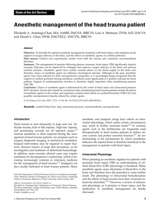
  • 2. Changes in intracranial blood flow are a significant cause of cerebral injury after head trauma, and pre- vention of ischemic damage by maintaining oxygen delivery to the brain contributes significantly to out- come.11–13 An understanding of the protective mecha- nisms supporting blood flow to the brain is therefore important in head trauma management. In order to support a high oxygen requirement, the brain normally receives a large percentage of the cardiac output, and CBF is tightly regulated to prevent decreases in perfu- sion. In a normal brain, constant CBF is maintained by alterations in vasomotor tone regulated according to changes in arterial oxygen (PaO2) and carbon dioxide (PaCO2) partial pressures, and systemic blood pressure (Figure 1). In the injured brain, these protective mech- anisms are lost. A decrease in arteriolar pH caused by an increase in PaCO2 results in vasodilation, reduction of cerebral vascular resistance, and an increase in CBF.14 In contrast, hypocapnia results in intracranial vasocon- striction and a decrease in cerebral perfusion.15 Alter- ations of arterial oxygenation have a lesser effect on intracranial hemodynamics unless severe hypoxemia occurs. When PaO2 decreases below 50 mmHg, vascu- lar resistance decreases in order to increase CBF and maintain cerebral oxygen delivery.15 Changes in sys- temic blood pressure are usually prevented from caus- ing alterations in CBF by cerebral pressure autoregulation. Pressure autoregulation maintains con- stant CBF when mean arterial blood pressure (MABP) varies within a physiological range (50–150 mmHg).15 Following TBI, pressure autoregulation may be lost causing mildly decreased systemic blood pressures that otherwise might be considered safe to result in mark- edly reduced CBF. The increased dependency of CBF on MAP may explain the worsened outcome associated with hypotension in patients with TBI.13–17 Clinical assessment of CBF is difficult and as a result, cerebral perfusion pressure (CPP), a variable that is more easily ascertained and a clinical correlate of CBF, is used to predict a patient’s risk for cerebral ischemia.18 CPP is the pressure gradient driving CBF. It is calcu- lated as the difference between MABP and intracranial pressure (ICP): CPP ¼ MAP ICP ICP is defined as the pressure exerted between the skull and the intracranial tissues.7 Since the skull is rigid and poorly compliant, an increase in volume by any of its contents without a concomitant reduction in the other components results in an increase in pressure. Increases in intracranial volume and pressure may result from an increase in volume of brain tissue (e.g., by formation of cerebral edema), blood (e.g., due to hemorrhage or vasodilation), or cerebrospinal fluid (e.g., by obstruc- tion to fluid outflow). As ICP increases, systemic blood pressure must increase to prevent a decrease in CPP and a resultant decrease in CBF. Excessive unregulated cerebral vasodilation such as that which may arise dur- ing anesthesia of TBI patients may also increase ICP, reduce CPP, and may lead to cerebral ischemia. Under normal circumstances, vasomotor tone is cou- pled to the oxygen requirement of the brain by flow- metabolism coupling. This phenomenon describes the ability of the cerebral vasculature to respond appropri- ately to changes in oxygen demand. Provided flow- metabolism coupling remains intact, a reduction of cer- ebral metabolic activity leads to a decrease in oxygen demand, followed by vasoconstriction and a subse- quent decrease in ICP. Because anesthetic agents reduce brain metabolism when a state of unconsciousness is reached, oxygen demand is reduced, minimizing the risk of ischemia.4 Flow-metabolism coupling is disrupt- ed by any event causing a change in vasomotor tone that is not reflected by a similar alteration in cerebral metabolism. Therefore, the induction of vasoconstric- tion without a parallel decrease in the cerebral meta- bolic rate, for example by hyperventilation, can actually increase the risk of cerebral hypoxia.19 The high met- abolic demands of the brain result in poor tolerance to reduced oxygen delivery. Ischemic injury leads to ab- normal nerve function (e.g., unregulated sodium and calcium flux across cell membranes), release of the excitatory neurotransmitter glutamate (causing an increase in oxygen requirement and generation of seizures), and neuronal cell death.20,21 Selection of Anesthetic Agent In selecting anesthetic agents for use in patients with TBI, specific properties of the agent that must be CBF (mL /100g/min) MABP PaO2 PaCO2 50 150 20 40 60 80 100 mm Hg Figure 1: Schematic depiction of the effects of arterial partial pressure of oxygen (PaO2), carbon dioxide (PaCO2), and mean arterial blood pressure (MABP) [same scale and measured in mmHg] on cerebral blood flow (CBF) [measured in mL/100 g/ min]. (Adapted from Hopkins AL. Head Trauma. Vet Clin N Amer Small Anim Pract. 1996;26(4):875–891, with permission.)b Veterinary Emergency and Critical Care Society 2006, doi: 10.1111/j.1476-4431.2006.00194.x 6 E.A. Armitage-Chan et al.
  • 3. considered include the effects on cardiovascular and ventilatory function, effects on intracranial hemody- namics, and potential neuroprotective properties. These neuroprotective properties include reduction of brain ischemia by decreasing cerebral oxygen demand and enhancing cerebral pressure autoregulation.22,23 Agents that have been investigated for their potential neuro- protective properties include volatile anesthetics, bar- biturates, propofol, benzodiazepines, and ketamine.21,24 However, despite much research, evidence for long- term beneficial anesthetic-induced brain protection is scarce.24 A simple algorithm for selection of anesthetic agent in head trauma patients is shown in Figure 2. The importance of minimizing cardiovascular and respira- tory compromise cannot be overemphasized, and main- taining CPP remains a top priority when selecting an anesthetic protocol.24 Volatile anesthetics Most volatile anesthetics, including halothane, isoflu- rane, sevoflurane, and desflurane have dose-related effects on ICP. Low concentrations of all these agents reduce cerebral metabolism; and if flow-metabolism coupling is intact, there is a resultant decrease in cer- ebral blood volume and a reduction in ICP.25 As the dose is increased over 1–1.5 MAC (minimum effective alveolar concentration), suppression of metabolic activ- ity persists; however, the predominant effect becomes increased ICP and decreased CPP.26 This is primarily caused by a direct vasodilatory effect; however, it is augmented by anesthetic-induced hypoventilation and hypercapnia. The systemic hypotensive effects of vol- atile anesthetics cause an additional detrimental effect on cerebral perfusion. Additionally, at higher alveolar concentrations, cerebral pressure autoregulation is dis- rupted. Perfusion therefore becomes dependent on sys- temic blood pressure and is decreased if blood pressure is not supported.27 The dose/effect response varies between the different inhalant agents. Most of the detrimental effects are sig- nificant at 1.0 MAC, although the increase in ICP seen with halothane is greater than that observed with new- Preanesthetic Evaluation: Is there evidence of ICP elevation? No Yes Preanesthetic: Opioid + benzodiazepine Induction: Thiopental or propofol Maintenance: Sevoflurane or Isoflurane Keep concentration low: add opioid, benzodiazepine, or propofol infusion Attempt ICP reduction prior to anesthesia: Mannitol, hypertonic saline, temporary hyperventilation Preanesthetic: Benzodiazepine ± low dose opioid Induction: Thiopental or propofol Ensure smooth intubation Maintenance: Propofol, barbiturate, or benzodiazepine Supportive measures Hypovolemia? MABP 60 – 80 mm Hg? PaCO 40 mm Hg? Or PaO 80 mm Hg? Signs of elevated ICP? No No No No Yes Yes Yes Yes Aliquots of isotonic crystalloids, colloids, or hypertonic saline; plus 10 ml/kg/hr IV crystalloids IV isotonic crystalloids (5 –10 mL/kg/hr) Continue monitoring Consider supplemental O to increase O delivery Continue monitoring Aliquots of isotonic crystalloids, colloids, or hypertonic saline; consider dopamine IPPV: Peak inspiratory pressure 25 cm H O Positive end expiratory pressure 5 cm H O Temporary hyperventilation to PaCO of 30 mm Hg, mannitol, hypertonic saline Figure 2: Selection of anesthetic protocol for use in head trauma patients. Veterinary Emergency and Critical Care Society 2006, doi: 10.1111/j.1476-4431.2006.00194.x 7 Head trauma anesthesia
  • 4. er inhalant anesthetics.26 For example, sevoflurane does not impair pressure autoregulation until concentrations exceeds 1.5 MAC (3.3%), while isoflurane disrupts au- toregulation at 1.0 MAC (1.3%).28 Other studies, how- ever, comparing sevoflurane to isoflurane at up to 1.5 MAC have failed to show any benefit to the use of se- voflurane in patients with intracranial disease.29,30 These differences may emphasize the fact that disease states may influence drug responses. An additional benefit of sevoflurane includes its lower solubility, pro- ducing a more rapid anesthetic recovery compared to the use of isoflurane, permiting earlier neurological as- sessment following anesthesia. This has been demon- strated in people even after prolonged neurosurgical procedures lasting longer than 6 hours.31 Effects of desflurane on intracranial blood flow and CO2 vasore- activity in dogs and pigs have also been investigated. Desflurane use was associated with higher ICP, greater degree of vasodilation, and decreased responsiveness to hypocapnia when compared to isoflurane and sevo- flurane.32 This agent may therefore be less suitable for use in patients with intracranial disease. The intracranial effects of inhalant anesthetics can be minimized when low anesthetic concentrations are used and with appropriate support of ventilation and blood pressure. In the absence of ICP elevation, the vasodilatory effects of these agents may even improve cerebral perfusion.33 However, if ICP is already elevat- ed, an anesthetic protocol that does not include volatile anesthetics is recommended.34 Injectable anesthetics and analgesics Injectable anesthetic agents such as propofol and the barbiturates are widely used in people following head trauma.22,35 Beneficial properties of barbiturates in- clude neuroprotection conferred by their reduction of cerebral oxygen requirements, cerebral vasoconstric- tion, reduction of ICP, and increased protection from excitatory neurotransmitter-induced neuronal dam- age.24,36 Other potential neuroprotective properties in- clude reduction of sodium channel conduction and intracellular calcium entry, enhancement of cAMP production, and antioxidant effects.21 Their main dis- advantages include delayed anesthetic recovery, hypo- tension, and potent respiratory depressant effects, which are detrimental in TBI patients, particularly those with disrupted pressure autoregulation. Neuroprotective properties of propofol under inves- tigation include modulation of GABA receptors and antioxidant effects.21 Clinically, advantages to propofol in head injured patients include more rapid recovery compared to thiopental, allowing earlier assessment of neurological status and easier titration to desired anes- thetic depth. In the authors’ experience, however, a small percentage of cats (some with and some without evidence of intracranial disease, based on cerebrospinal fluid analysis and magnetic resonance imaging) have had prolonged recoveries after propofol anesthesia, and therefore care should be taken when this drug is used for anesthetic maintenance in this species. Because of the negative cardiovascular effects of propofol, blood pressure should be supported during its use. Although propofol does not directly disrupt pressure autoregu- lation, this may be absent in TBI patients, making cer- ebral perfusion dependent on systemic blood pressure. Propofol can cause respiratory depression and there- fore care should be taken to avoid hypercapnia and hypoxemia. A recent investigation in patients at risk for cerebral hypoperfusion indicated that propofol use leads to disruption of flow-metabolism coupling and vasoconstriction in excess of that resulting from sup- pression of brain activity.23 The significance of this is unclear, although increased cerebral ischemia has been associated with propofol use when compared to both isoflurane and sevoflurane.37,38 Until more is known about the effects of propofol on cerebral vasculature, long periods of high doses of propofol should be used cautiously in patients at risk for ischemic brain injury. Despite the controversy associated with the use of some injectable anesthetics, propofol and barbiturates offer a number of advantages over the use of volatile anesthetics when ICP elevation is present. Compared to volatile anesthetics, barbiturates have been shown to produce superior reduction of cerebral edema and ICP.39,40 In patients with intracranial disease, compar- isons of propofol and volatile anesthetics (sevoflurane and isoflurane) have demonstrated improved cerebral perfusion and better maintenance of pressure autoreg- ulation when total intravenous anesthesia was used.28,34,41 In addition, in contrast to volatile anesthe- tics, cerebral pressure autoregulation is maintained by the use of these agents.42 However, it is important to remain cognizant that pressure autoregulation may be disrupted focally or globally in cases of intracranial disease and that in such cases, hypotension may not be tolerated. Under conditions of preexisting ICP eleva- tion, total intravenous anesthesia, such as that achieved with propofol or fentanyl is recommended. Other anesthetic and sedative agents available for use in head trauma patients include benzodiazepines, keta- mine, and etomidate. Benzodiazepines (midazolam and diazepam) are advantageous due to their lack of ad- verse intracranial effects and lack of adverse effects on cardiovascular and respiratory function. Although they do not appear to decrease ICP, mild reductions in cer- ebral oxygen requirement are reported.43 Their use also enables dose reduction of other agents, such as prop- ofol or barbiturates, thereby reducing depression of Veterinary Emergency and Critical Care Society 2006, doi: 10.1111/j.1476-4431.2006.00194.x 8 E.A. Armitage-Chan et al.
  • 5. cardiovascular and respiratory systems. Etomidate is another agent that is frequently selected for cardiovas- cular and respiratory stability and has previously been thought to produce neuroprotection by decreasing cer- ebral metabolism.44 However, in contrast to the ben- zodiazepines, use of etomidate has been associated with cerebral hypoxia and ischemic injury.44 The mech- anism by which etomidate decreases brain tissue oxy- gen tension is not known; however, the changes observed are consistent with cerebral vasoconstriction, possibly due to hemolysis and nitric oxide scavenging by free hemoglobin.44 It is, therefore, suggested that etomidate be avoided in patients with head injury. Ketamine is an alternative anesthetic and analgesic agent that has recently gained interest for use in neu- rosurgical patients. It is typically avoided in the pres- ence of intracranial disease, since the sympathetic stimulation it produces may increase ICP. However, studies in head trauma patients have demonstrated that administration of ketamine under propofol sedation decreases ICP.45 This agent, unlike other commonly used anesthetics, acts by inhibiting the NMDA receptor. Because this is the predominant receptor type respon- sible for ischemic injury, ketamine use may theoretically have beneficial neuroprotective properties. An addi- tional advantage is the lack of cardiovascular or respi- ratory depressant effects. Ketamine administration, however, has also been demonstrated to increase cer- ebral oxygen consumption, possibly by inhibition of the GABA receptor (the major inhibitory neurotransmitter system within the brain).46 It is possible that the det- rimental effects of ketamine on cerebral activity may be reduced by co-administration of a GABA agonist such as propofol. Further investigation of the beneficial and detrimental effects of ketamine and other NMDA an- tagonists is required before their use can be recom- mended for use in head injured patients. The provision of adequate analgesia is essential to prevent further ICP elevation. Opioids are widely used to provide analgesia for critically ill patients due to their relative lack of adverse cardiovascular effects and ease of reversal. Adverse effects of opioids, such as respiratory depression and hypotension, have greater significance in the presence of ICP elevation, especially when used at high doses. As a result opioids were pre- viously withheld from head trauma patients. However when carefully titrated to patient analgesia and when ventilation is supported, opioids are safe to use in cases of intracranial hypertension.47 In the presence of cardio- vascular shock or damage to the blood–brain barrier (BBB), dose requirements may be decreased and so care should be taken to avoid overdose. Opioid agonists, such as fentanyl and morphine, can be administered as a continuous rate infusion (CRI) to avoid peaks and troughs in analgesia and the adverse effects seen at higher blood levels. Recommended CRI dosages for fentanyl include 0.2–0.7 mg/kg/min and 0.1–0.5 mg/ kg/hr for morphine. These drugs may be reversed us- ing an opioid antagonist, such as naloxone, if signifi- cant respiratory or cardiovascular depression occurs. Opioid agonist/antagonists such as butorphanol and buprenorphine are analgesics used to treat mild to moderate pain. They are generally thought to be safer than opioid agonists because they cause less cardio- vascular and respiratory depression.48,49 When consid- ering administering these agents to patients with TBI at risk for rapid changes in neurological status, it is im- portant to consider that the effects of buprenorphine are difficult to reverse with standard doses of naloxone.48 It is also important to remember that the duration of analgesia from butorphanol is relatively short and, if used, should be repeated every 2 hours.49 Medetomidine, an a2 agonist used for sedation and analgesia, appears not to influence ICP in dogs.50 Re- duction in heart rate and cardiac output may impair cerebral perfusion however, and therefore it should only be administered at a very low dose (1–2 mg/kg/hr) and only used if analgesics with less adverse cardio- vascular effects are unavailable or are providing insuf- ficient pain relief. New Neuroprotective Anesthetic Adjuncts A number of drugs are under investigation for their possible neuroprotective properties. Lidocaine may re- duce secondary brain injury by preventing sodium in- flux into ischemic neurons.17,51 There is some experimental evidence that infusion of antiarrythmic doses (1.5–2 mg/kg) of lidocaine after the onset of brain ischemia reduces neuronal death and improves neurol- ogic outcome.52 Xenon is another agent that is gaining interest as a potential neuroprotective agent.52 This is a volatile anesthetic, but unlike other inhalant agents, it produces its effect via NMDA receptor antagonism and produces no adverse hemodynamic effects. Finally, am- antadine, also an NMDA antagonist, may prove to be beneficial in head trauma. A small population of head injury patients showed a significant improvement in neurological outcome and mortality when adminis- tered amantadine compared to a group which did not receive this drug.53 More studies are necessary, how- ever, before amantadine can be recommended for use in a clinical setting. Supportive Care for the Anesthetized Patient Of equal importance to the selection of an anesthetic agent is the support of cardiovascular and respiratory Veterinary Emergency and Critical Care Society 2006, doi: 10.1111/j.1476-4431.2006.00194.x 9 Head trauma anesthesia
  • 6. function. Prevention of cerebral ischemia during an- esthesia is vital to a successful outcome for a patient with head injury. To ensure adequate oxygen delivery to the brain, PaO2, PaCO2, hemoglobin concentration, and systemic blood pressure must be maintained with- in normal ranges. Blood pressure support In head injured patients, where cerebral pressure auto- regulation may be impaired by disease or the effect of anesthetic agents, systemic blood pressure should be supported to maintain a CPP between 60 and 70 mmHg.54,a Normal ICP in dogs and cats is bet- ween 7 and 12 mmHg.55–57 The necessary MABP re- quired to support a CPP of 60–70 mmHg in the absence of ICP elevation can, therefore, be calculated as ap- proximately 70–80 mmHg. Without the benefit of ICP monitoring devices, exact values of CPP cannot be cal- culated. However, blood pressure targets should be in- creased if signs of severe ICP elevation, such as cranial nerve deficits (e.g., nonresponsive pupils, strabismus, lack of menace response), changes in mental status or seizures become apparent. Improved brain oxygenation in head trauma patients has been demonstrated by maintaining MABP above 90 mmHg, compared to pa- tients managed similarly but using 70 mmHg as the minimum acceptable blood pressure.54 However, use of vasopressors to achieve targeted blood pressures in head trauma patients have also been associated in in- creased risk of developing adult respiratory distress syndrome and therefore should be used judiciously.58,a During anesthetic procedures, hypotension is avoided by the selection of anesthetic agents that do not reduce cardiac output, the use of intravenous fluid therapy, and the careful administration of vasopressors. Care should be taken to avoid inducing excessive intracra- nial vasoconstriction, which may negatively impact cerebral perfusion. Dopamine has been shown to im- prove CBF after head trauma without causing detri- mental vasoconstriction.59 In the authors’ experience, dopamine infusions of 5–10 mg/kg/min effectively im- prove blood pressure. Vasopressin has also been used successfully in acute brain injury, although widespread clinical use in head injury is not reported.60 Reports of the use of norepinephrine in TBI are variable. Its use has been associated with detrimental effects on CBF after damage to the BBB.61 In contrast, more recent re- ports suggest that norepinephrine use is not associated with cerebral perfusion compromise.62 Because of this controversy, dopamine is the vasopressor agent most frequently recommended for use in head trauma patients. Intravenous fluid management Intravenous fluid administration during anesthesia is necessary to maintain blood volume and promote cer- ebral and systemic perfusion, but should be performed judiciously as fluid overload may exacerbate vasogenic cerebral edema.63 Vasogenic edema is formed by leak- age of protein and fluid across blood vessel walls and can be reduced by maintaining serum osmolarity and colloid osmotic pressure (COP).64 Selection of fluid type may, therefore, be guided by measurements of serum osmolarity and COP, as well as sodium and total pro- tein levels. Isotonic crystalloids (e.g., lactated Ringer’s solution), hypertonic fluids (e.g., 3–7% hypertonic saline), and artificial colloids (e.g., 6% hetastarch) are all suitable fluid choices; hypotonic fluids (e.g., 0.45% saline) should be avoided as these may contribute to edema formation.64 Glucose-containing fluids should also be avoided, unless there is significant hypo- glycemia, since hyperglycemia drives cerebral lactate production and has been associated with worse neuro- logical outcome.1,13,14,65,66 The use of hypertonic saline (e.g., 3–7%) for blood volume support is being increas- ingly described in the treatment of head trauma in people.67–71 This has been associated with greater ICP reduction, thereby improving CPP, when com- pared to the use of lactated Ringer’s solution or man- nitol in both human head trauma patients and in dogs.68,72–75 The volume of fluid administered should be carefully considered, because of a possible association between excessive hydrostatic pressure (i.e., overhydration) and edema formation. In the past, fluid restriction and re- duction of systemic blood pressure were advocated to decrease formation of vasogenic edema.76 However, negative fluid balance has since been associated with poor outcome, and more aggressive fluid resuscitation to support intravascular volume is now recommend- ed.63,77 It is possible that in certain, well-hydrated euvolemic patients with no evidence of ongoing blood loss, the commonly recommended anesthetic mainte- nance fluid rate of 10 mL/kg/hr of isotonic crystalloid may be excessive and lead to fluid overload. Converse- ly, care should be taken to avoid compromising cardiac output by inadvertent fluid restriction. Fluid therapy should be adjusted according to markers of systemic perfusion and cardiac output. Parameters such as heart rate, pulse quality, mucous membrane color, urine output, and serum lactate concentration should be monitored frequently and used to guide fluid admin- istration. In the absence of clinical parameters suggest- ing preanesthetic hypovolemia (tachycardia, weak or bounding pulses, pale mucous membranes, and oligur- ia), infusion of isotonic crystalloids at 5 mL/kg/hr during the anesthetic period is likely sufficient to Veterinary Emergency and Critical Care Society 2006, doi: 10.1111/j.1476-4431.2006.00194.x 10 E.A. Armitage-Chan et al.
  • 7. meet anesthetic fluid requirements, although monitor- ing parameters of intravascular volume should be con- tinued. Preoperative administration of osmotic diuretics should also prompt titration of fluid rates to maintain euvolemia. The presence or development of hypotension or other indicators of hypovolemia during anesthesia should be aggressively treated with careful titration of intravenous fluids (e.g., isotonic, hypertonic crystalloids, and colloids) until acceptable clinical pa- rameters are achieved. Aliquots of isotonic crystalloids (10–20 mL/kg) or 6% hetastarch (5 mL/kg) should be given to effect. Ventilatory support Assisted ventilation is often required for head trauma patients under anesthesia. Since many anesthetic agents cause hypoventilation, either manual or mechanical positive pressure ventilation is necessary to prevent hypercapnia-induced cerebral vasodilation and in- creased ICP. Although the application of positive pres- sure ventilation may increase ICP by decreasing venous return from the head, studies have shown that main- taining peak inspiratory pressure below 25 cmH2O and positive end expiratory pressure less than 5 cmH2O prevents a clinically significant increase in ICP.78 In- creasing arterial oxygenation by the provision of sup- plemental oxygen also helps support cerebral oxygen delivery. Previously, hyperventilation leading to hypo- capnia was recommended as a method of causing vasoconstriction and prophylactically reducing ICP. However, this strategy reduces cerebral perfusion, in- creases the risk of ischemia, and is no longer recom- mended for routine use in anesthetized head trauma patients.19,22,79 Additionally, cerebrovascular CO2 reac- tivity following severe traumatic brain is variably de- pressed, potentially limiting the usefulness of hyperventilatory therapy in TBI patients.80 Recent stud- ies indicate that there is an increased risk of ischemic damage with even mild hypocapnia (PaCO2 5 30– 35 mmHg).80 Head trauma patients under anesthesia should, therefore, be ventilated to eucapnia (Pa- CO2 5 40 mmHg) to avoid inducing either vasodilation or vasoconstriction. Management of ICP Elevation During Anesthesia Despite careful patient management, acute increases in ICP may occur during anesthesia, necessitating emer- gency treatment to prevent decreased CPP, cerebral is- chemia, and ultimately, herniation of brain tissue. Timely identification of ICP elevation during anesthesia is essential, but hampered by the effects of the anes- thetic agent. In particular, proper assessments of mental status and many cranial nerve reflexes are often impossible. Clinical parameters indicative of ICP ele- vation that remain detectable in the anesthetized pa- tient include miotic pupils, pupil asymmetry, and loss of palpebral and corneal reflexes.7 As the palpebral re- flex may be lost in patients at a surgical plane of an- esthesia, this may not be a reliable indicator of increasing ICP. In addition, the perianesthetic use of anticholinergics may interfere with assessment of pupil size by causing pupil dilation, and therefore pupil size must be interpreted in light of anticholinergics use. These difficulties can be overcome by lightening the plane of anesthesia or discontinuing administration of anesthetic agents if increasing ICP is suspected. If the patient is not being ventilated, altered breathing pat- terns such as apneustic or Cheyne–Stokes breathing may also be seen in the presence of rising ICP. The Cushing’s reflex is a cardiovascular phenomenon asso- ciated with increased ICP. In response to ICP elevation, systemic blood pressure increases, often to a systolic pressure greater than 200 mmHg, to maintain CPP. Re- flex bradycardia prevents tissue damage resulting from such severe hypertension. This is a protective mecha- nism and treatment with anticholinergics (atropine or glycopyrrolate) may cause further ICP elevation and increase ischemic brain injury. Rather than treating the bradycardia, the anesthetist should therefore consider the possibility of rising ICP, and perform the treatments described below to reduce ICP and avoid further brain swelling and ischemia. Methods to rapidly reduce ICP include hyperventi- lation and administration of hypermolar agents. Hy- perventilation is one of the most rapid and effective methods of reducing ICP. Decreasing PaCO2 by 10 mmHg can reduce ICP by up to 30% within 15 sec- onds.81 Because of the adverse effects of hypoventila- tion on cerebral perfusion, hyperventilation is regarded as an emergency therapy only, and should be discon- tinued when clinical signs of ICP elevation improve. Hyperventilation to maintain a PaCO2 of 30 mmHg for up to 30 minutes, with concomitant administration of other therapies can be attempted initially. In the absence of significant pulmonary disease or reduction in cardiac output, the difference between PaCO2 and end-tidal CO2 (ETCO2) will be less than 5 mmHg and ETCO2 values can be used as a noninvasive measure of arterial CO2. When using ETCO2 to guide hyperventilatory therapy, a target of 35 mmHg is recommended. Hyperventilation to a PaCO2 less than 30 mmHg should be only be used for intractable intra- cranial hypertension and for the shortest duration possible. Hyperosmolar agents such as mannitol can be used to decrease ICP. Mannitol acts as an osmotic diuretic to reduction cerebral edema. Other benefits attributed Veterinary Emergency and Critical Care Society 2006, doi: 10.1111/j.1476-4431.2006.00194.x 11 Head trauma anesthesia
  • 8. to mannitol include reduction in blood viscosity, improved perfusion of ischemic regions, free radical scavenging properties, and possible reduction of sub- sequent edema formation.81 Twenty-five percent man- nitol (osmolarity 5 1372 mOsm/L) is administered at a dose of 0.25–1 g/kg over 20 minutes.6 Because the os- motic effects of mannitol are dependant on an intact BBB, its use when the BBB is disrupted, for example, following intracranial hemorrhage, may worsen cere- bral edema. When the BBB is no longer intact, mannitol may leak into brain interstitium, increase tissue os- molarity, and therefore increase fluid accumulation. While a disruption of the BBB will increase its perme- ability to all ions, the higher membrane reflection co- efficient for sodium chloride (s 5 1) compared to mannitol (s 5 0.9) suggests that the use of saline-based hyperosmolar agents may be preferable over mannitol in certain intracranial pathologies such as intracerebral hemorrhage.72,82 Additionally, because of the tendency of mannitol to deplete the intravascular volume, repeated doses are not recommended and use of alter- native hyperosmolar agents may be preferred. Hyper- tonic saline (7.2% hypertonic saline; osmola- rity 5 2464 mOsm/L), given at a dose of 4 mL/kg ad- ministered slowly IV, can be used to reduce ICP and cerebral edema. Other management strategies including the preven- tion of hyperthermia, head elevation, and avoiding oc- clusion of jugular veins may also be employed to prevent increases in ICP. In experimental models, mod- erate hypothermia (31–34 1C) reduces the effects of glo- bal ischemia, decreases cerebral metabolic rate, and decreases ICP.83 While clinical induction of hypother- mia in patients with TBI cannot be recommended currently, avoidance of hyperthermia may be pru- dent.13,24,84 Elevation of the head by 15–301 may limit venous congestion and thereby reduce ICP with- out decreasing CPP or CBF.85 Jugular vein occlusion impairs venous drainage from the head and can cause increased intracranial blood volume; jugular catheters, twisting the neck, and tight neck bandages should be avoided in head trauma patients. Coughing and gagging during endotracheal intubation can also contribute to ICP elevation, and therefore a smooth anesthetic induction is beneficial. This is easily accom- plished by administering a premedication that causes sedation (e.g., a benzodiapine or opioid), applying lidocaine to the larynx and administering sufficient anesthetic induction agent that laryngeal reflexes are suppressed prior to attempting intubation. Additional- ly, rough anesthetic recovery may cause sympa- thetic stimulation elevating ICP; therefore, providing sedation during the anesthetic recovery phase is recommended. Summary The effects of anesthetic agents on intracranial hemo- dynamics and neuronal injury are complex. The overall effect on CPP is a result of a number of specific effects that include ICP elevation, vasomotor effects, disrup- tion of autoregulation, and secondary effects via alter- ations of cardiovascular and respiratory function. Low concentrations of isoflurane and sevoflurane are likely to have minimal effects on cerebral perfusion as long as blood pressure and ventilation are supported; however, their tendency to increase ICP is a concern, especially if the patient shows signs of ICP elevation prior to an- esthesia. Barbiturates produce minimal adverse intra- cranial effects and therefore are suitable agents for use in head trauma patients if blood pressure is supported; however, delayed anesthetic recovery can complicate neurological assessment. Propofol has been a useful al- ternative to barbiturates due to its short half-life leading to rapid recovery from anesthesia. It remains a useful agent in neuroanesthesia with appropriate physiologic support; however, it is probable that it has less neuro- protective properties than barbiturates, and there is a concern it may exacerbate ischemic injury. Use of agents such as opioids and benzodiazepines allows the dose of the selected anesthetic maintenance agent to be reduced. This minimizes adverse cardiovascular, respiratory, and neurological effects and can provide an anesthetic pro- tocol which is less likely to cause further neuronal dam- age. More important than the anesthetic drugs selected, careful monitoring and support of cardiovascular and respiratory functions remains of primary importance when managing an anesthetized head trauma patient. Footnotes a Brain Trauma Foundation, American Assoc Neurological Surgeons, Congress of Neurological Surgeons, Joint Section on Neurotrauma and Critical Care. Management and prognosis of severe traumatic brain injury: Cerebral perfusion pressure – update 2003. http://www2. braintrauma.org/guidelines/index.php b Adapted from Hopkins AL. Head trauma. Vet Clin North Am: Small Anim Pract 26(4):876. Copyright (1996), with permission from Elsevier. References 1. Syring RS, Otto CM, Drobatz KJ. Hyperglycemia in dogs and cats with head trauma: 122 cases (1997–1999). J Am Vet Med Assoc 2001; 218(7):1124–1129. 2. Hopkins AL, Wheeler SJ. Subdural hematoma in a dog. Vet Surg 1991; 20(6):413–417. 3. Niebauer GW, Dayrell-Hart BL, Speciale J. Evaluation of cranio- tomy in dogs and cats. J Am Vet Med Assoc 1991; 198(1):89–95. 4. Shores A. A review of the effects of anesthetic agents on cerebral blood flow and intracranial pressure in the dog. Vet Surg 1985; 14(3):257–263. 5. Cornick JL. Anesthetic management of patients with neurologic abnormalities. Compend Contin Edu Pract Vet 1992; 14(2):163–172. 6. Proulx J, Dhupa N. Severe brain injury. Part 2. Therapy. Compend Contin Edu Pract Vet 1998; 20(9):993–1005. Veterinary Emergency and Critical Care Society 2006, doi: 10.1111/j.1476-4431.2006.00194.x 12 E.A. Armitage-Chan et al.
  • 9. 7. Bagley RS. Pathophysiologic sequelae of intracranial disease. Vet Clin N Am Small Anim Pract 1996; 26(4):711–733. 8. Proulx J, Dhupa N. Severe brain injury. Part 1. Pathophysiology. Compend Contin Edu Pract Vet 1998; 20(8):897–905. 9. Hopkins AL. Head trauma. Vet Clin N Am Small Anim Pract 1996; 26(4):875–891. 10. Tonnesen AS. Hemodynamic management of brain-injured pa- tients. New Horiz 1995; 3(3):499–505. 11. Chesnut RM, Marshall LF, Klauber MR. The role of secondary brain injury in determining outcome from severe head injury. J Trauma 1993; 34(2):216–222. 12. Stocchetti N, Fn A, Volta F. Hypoxemia and arterial hypotension at the accident scene in head injury. J Trauma 1996; 40(5): 764–767. 13. Jeremitsky E, Omert L, Dunham CM, et al. Harbingers of poor outcome the day after severe brain injury: hypothermia, hypoxia and hypoperfusion. J Trauma 2003; 54(2):312–319. 14. Bedell E, Prough DS. Anesthetic management of traumatic brain injury. Anesthesiol Clin N Am 2002; 20(2):417–439. 15. Shapiro HM. Intracranial hypertension: therapeutic and anesthetic considerations. Anesthesiology 1975; 43(4):445–471. 16. Dewitt DS, Prough DS, Taylor CL, et al. Regional cerebrovascular responses to progressive hypotension after traumatic brain injury in cats. Am J Physiol 1992; 263(4):H1276–H1284. 17. Pietropaoli JA, Rogers FB, Shackford SR, et al. The deleterious effects of intraoperative hypotension on outcome in patients with severe head injuries. J Trauma 1992; 33(3):403–407. 18. Guidelines for the acute medical management of severe traumatic brain injury in infants, children, and adolescents. Chapter 8. Cer- ebral perfusion pressure. Pediatr Crit Care Med 2003; 4(3 Sup- pl.):S31–S33. 19. Laffey JG, Kavanagh BP. Medical progress: hypocapnia. N Engl J Med 2002; 347(1):43–53. 20. Bayir H, Clark RS, Kochanek PM. Promising strategies to mini- mize secondary brain injury after head trauma. Crit Care Med 2003; 31(1 Suppl.):S112–S117. 21. Hans P, Bonhomme V. Neuroprotection with anesthetic agents. Curr Opin Anaesthesiol 2001; 14:491–496. 22. Brain Trauma Foundation, American Association of Neurological Surgeons, Joint Section on Neurotrauma and Critical Care. J Ne- urotrauma 2000; 17(6–7):451–627. 23. Steiner LA, Johnston AJ, Chatfield DA, et al. The effects of large- dose propofol on cerebrovascular pressure autoregulation in head- injured patients. Anesth Analg 2003; 97:572–576. 24. Hans P, Bonhomme V. The rationale for perioperative brain protection. Eur J Anaesthesiol 2004; 21(1):1–5. 25. Newberg LA, Milde JH, Michenfelder JD. The cerebral metabolic effects of isoflurane at and above concentrations that suppress cortical electrical activity. Anesthesiology 1983; 59(1):23–28. 26. Artru AA. Relationship between cerebral blood volume and CSF pressure during anesthesia with isoflurane or fentanyl in dogs. Anesthesiology 1984; 60(6):575–579. 27. McPherson RW, Traystman RJ. Effects of isoflurane on cerebral autoregulation in dogs. Anesthesiology 1988; 69(4):493–499. 28. McCulloch TJ, Visco E, Lam AM. Graded hypercapnia and cere- bral autoregulation during sevoflurane or propofol anesthesia. Anesthesiology 2000; 93(5):1205–1209. 29. Petersen KD, Landsfeldt U, Cold GE, et al. Intracranial pressure and cerebral hemodynamics in patients with cerebral tumors. Anesthesiology 2003; 98(2):329–336. 30. Sponheim S, Skraastad Ø, Helseth E, et al. Effects of 0.5 and 1.0 MAC isoflurane, sevoflurane and desflurane on intracranial and cerebral perfusion pressures in children. Acta Anaesthesiol Scand 2003; 47(8):932–938. 31. Gauthier A, Girard F, Boudreault D, et al. Sevoflurane provides faster recovery and postoperative neurological assessment than isoflurane in long-duration neurosurgical cases. Anesth Analg 2002; 95(5):1384–1388. 32. Holmstrom A, Rosen I, Akeson J. Desflurane results in higher cerebral blood flow than sevoflurane or isoflurane at hypocapnia in pigs. Acta Anaesthesiol Scand 2004; 48(4):400–404. 33. Statler KD, Janesko KL, Melick JA, et al. Hyperglycolysis is ex- acerbated after traumatic brain injury with fentanyl vs isoflurane anesthesia in rats. Brain Res 2003; 994(1):37–43. 34. Cenic A, Craen RA, Lee T-Y, et al. Cerebral blood volume and blood flow responses to hyperventilation in brain tumors during isoflurane or propofol anesthesia. Anesth Analg 2002; 94(3):661–666. 35. Johnston AJ, Steiner LA, Chatfield DA, et al. Effects of propofol on cerebral oxygenation and metabolism after head injury. Br J Ana- esth 2003; 91(6):781–786. 36. Zhu H, Cottrell JE, Kass IS. The effect of thiopental and propofol on NMDA- and AMPA-mediated glutamate excitotoxicity. Anesthesiology 1997; 87(4):944–951. 37. Jansen GFA, van Praagh BH, Kedaria MB, et al. Jugular bulb ox- ygen saturation during propofol and isoflurane/nitrous oxide an- esthesia in patients undergoing brain tumor surgery. Anesth Analg 1999; 89(2):358–363. 38. Kawano Y, Kawaguchi M, Inoue S, et al. Jugular bulb oxygen sat- uration under propofol or sevoflurane/nitrous oxide anesthesia during mild hypothermia in neurosurgical patients. J Neurosur- gical Anesthesiol 2004; 16(1):6–10. 39. Drummond JC, Cole DJ, Patel PM, et al. Focal cerebral ischemia during anesthesia with etomidate, isoflurane, or thiopental: a comparison of the extent of cerebral injury. Neurosurgery 1995; 37(4):742–748. 40. Smith AL, Marque JJ. Anesthetics and cerebral edema. Anesthesio- logy 1976; 45(1):64–72. 41. Holzer A, Winter W, Greher M, et al. A comparison of propofol and sevoflurane anaesthesia: effects on aortic blood flow velocity and middle cerebral artery blood flow velocity. Anaesthesia 2003; 58(3):217–222. 42. Strebel S, Lam A, Matta B, et al. Dynamic and static cerebral autoregulation during isoflurane, desflurane and propofol anesthesia. Anesthesiology 1995; 83(1):66–76. 43. Schulte am Esch J, Kochs E. Midazolam and flumazenil in neuro- anaesthesia. Acta Anaesthesiol Scand 1990; 92:96–102. 44. Edelman GJ, Hoffman WE, Charbel FT. Cerebral hypoxia after etomidate administration and temporary cerebral artery occlusion. Anesth Analg 1997; 85(4):821–825. 45. Albanese J, Arnaud S, Rey M, et al. Ketamine decreases intracra- nial pressure and electroencephalographic activity in traumatic brain injury patients during propofol sedation. Anesthesiology 1997; 87(6):1328–1334. 46. Långsjöjw JW, Salmi E, Kaisti KK, et al. Effects of subanesthetic ketamine on regional cerebral glucose metabolism in humans. Anesthesiology 2004; 100(5):1065–1071. 47. Lauer KK, Connolly LA, Schmeling WT. Opioid sedation does not alter intracranial pressure in head injured patients. Can J Anesthesia 1997; 44(9):929–933. 48. Pascoe PJ. Opioid analgesics. Vet Clin North Am Small Anim Pract 2000; 30(4):757–769. 49. Hansen B. Acute pain management. Vet Clin North Am Small Anim Pract 2000; 30(4):899–916. 50. Keegan RD, Greene SA, Bagley RS, et al. Effects of medetomidine administration on intracranial pressure and cardiovascular vari- ables of isoflurane-anesthetized dogs. Am J Vet Res 1996; 56(2): 193–198. 51. Hemmings HC. Neuroprotection by Na1 channel blockade. J Ne- urosurg Anesthesiol 2004; 16(1):100–101. 52. Lei B, Popp S, Capuano-Waters C, et al. Effects of delayed administration of low-dose lidocaine on transient focal cerebral ischemia in rats. Anesthesiology 2002; 97(6):1534– 1540. 53. Saniova B, Drobny M, Kneslova L, Minarik M. The outcome of patients with severe head injuries treated with amantadine sulphate. J Neural Transm 2004; 111:511–514. 54. Narayan R, Michel ME, Ansell B, et al. Clinical trials in head injury. J Neurotrauma 2002; 19(5):503–557. 55. Dewey CW, Bailey CS, Haskins SC, et al. Evaluation of an epidural intracranial pressure monitoring system in cats. J Vet Emerg Crit Care 1997; 6(2):20–33. Veterinary Emergency and Critical Care Society 2006, doi: 10.1111/j.1476-4431.2006.00194.x 13 Head trauma anesthesia
  • 10. 56. Harrington ML, Bagley RS, Moore MP, et al. Effect of craniectomy, durotomy and wound closure on intracranial pressure in healthy cats. Am J Vet Res 1996; 57(11):1659–1661. 57. Bagley RS, Keegan RD, Greene SA, et al. Pathologic effects in brain after intracranial pressure monitoring in clinically normal dogs, using a fiberoptic monitoring system. Am J Vet Res 1995; 56(11):1475–1478. 58. Constant CF, Valadka AB, Gopinath SP, et al. Adult respiratory distress syndrome: a complication of induced hypertension after severe head injury. J Neurosurg 2001; 95:560–568. 59. Kroppenstedt SN, Stover JF, Unterberg AW. Effects of dopamine on posttraumatic cerebral blood flow, brain edema and cerebro- spinal fluid glutamate and hypoxanthine concentrations. Crit Care Med 2000; 28(12):3792–3798. 60. Yeh CC, Wu CT, Lu CH, et al. Early use of small-dose vasopressin for unstable hemodynamics in an acute brain injury patient re- fractory to catecholamine treatment: a case report. Anesth Analg 2003; 97(2):577–579. 61. MacKenzie ET, McCulloch J, O’Kean M, et al. Cerebral circulation and norepinephrine: relevance of the blood-brain barrier. Am J Physiol 1976; 231(2):483–488. 62. Johnston AJ, Steiner LA, Chatfield DA, et al. Effect of cerebral perfusion pressure augmentation with dopamine and nor- epinephrine on global and focal brain oxygenation after traumat- ic brain injury. Intensive Care Med 2004; 30(5):791–797. 63. Clifton GL, Miller ER, Choi SC, et al. Fluid thresholds and out- come from severe brain injury. Crit Care Med 2002; 30(4):739–745. 64. Drummond JC, Patel PM, Cole DJ, et al. The effect of the reduction of colloid reduction of oncotic pressure, with and without reduc- tion of osmolality, on post-traumatic cerebral edema. Anesthesio- logy 1998; 88(4):993–1002. 65. Jeremitsky E, Omert LA, Dunham CM, et al. The impact of hyper- glycemia on patients with severe brain injury. J Trauma 2005; 58(1):47–50. 66. Lam AM, Winn HR, Cullen BF, et al. Hyperglycemia and neuro- logical outcome in patients with head injury. J Neurosurg 1991; 75:545–551. 67. Khanna S, Davis D, Peterson B, et al. Use of hypertonic saline in the treatment of severe refractory posttraumatic intracranial hy- pertension in pediatric traumatic brain injury. Crit Care Med 2000; 28(4):1144–1151. 68. Simma B, Burger R, Falk M, Sacher P, et al. A prospective, ran- domized and controlled study of fluid management in children with severe head injury: lactated Ringer’s solution versus hyper- tonic saline. Crit Care Med 1998; 26(7):1265–1270. 69. Doyle JA, Davis DP, Hoyt DB. The use of hypertonic saline in the treatment of traumatic brain injury. J Trauma 2001; 50:367–383. 70. Holcroft JW, Vassar MJ, Turner JE, et al. 3% NaCl and 7.5% NaCl dextran for resuscitation of severely injured patients. Ann Surg 1987; 206:278–288. 71. Vassar MJ, Perry CA, Gannaway WL, et al. 7.5% sodium chloride/ dextran for resuscitation of trauma patients undergoing helicopter transport. Arch Surg 1991; 126:1065–1072. 72. Quereshi AI, Wilson DA, Traystman RJ. Treatment of elevated in- tracranial pressure in experimental intracerebral hemorrhage: comparison between mannitol and hypertonic saline. Neurosur- gery 1999; 44(5):1055–1063. 73. Prough DS, Johnson JC, Poole GV, et al. Effects on intracranial pressure of resuscitation from hemorrhagic shock with hypertonic saline versus lactated Ringer’s solution. Crit Care Med 1985; 13(5):407–411. 74. Prough DS, Johnson JC, Stump Da, et al. Effects of hypertonic saline versus lactated Ringer’s solution on cerebral oxygen trans- port during resuscitation from hemorrhagic shock. J Neurosurg 1986; 64(4):627–632. 75. De Vivo P, Del Gaudio A, Ciritella P, et al. Hypertonic saline solution: a safe alternative to mannitol 18% in neurosurgery. Minerva Anestesiol 2001; 67(9):603–611. 76. Eker C, Asgeirsson B, Grande PO, et al. Improved outcome after severe head injury with a new therapy based on principles for brain volume regulation and preserved microcirculation. Crit Care Med 1998; 26(11):1881–1886. 77. York J, Arrillaga A, Graham R, Miller R. Fluid resuscitation of patients with multiple injuries and severe closed head injury: ex- perience with an aggressive fluid resuscitation strategy. J Trauma 2000; 48(3):376–379. 78. Huynh T, Messer M, Sing R, et al. Positive end-expiratory pressure alters intracranial and cerebral perfusion pressure in severe trau- matic brain injury. J Trauma 2002; 53(3):488–493. 79. Coles JP, Minhas PS, Fryer TD, et al. Effect of hyperventilation on cerebral blood flow in traumatic head injury: clinical relevance and monitoring correlates. Crit Care Med 2002; 30(9):1950–1959. 80. McLaughlin MR, Marion DW. Cerebral blood flow and vasoreac- tivity within and around cerebral contusions. J Neurosurg 1996; 85(5):871–876. 81. Reilly P. Management of intracranial pressure and cerebral perfu- sion, In Reilly P, Bullock R. eds. Head Injury. London: Chapman Hall; 1997, pp. 385–407. 82. Qureshi AI, Suarez JI. Use of hypertonic saline solutions in treat- ment of cerebral edema and intracranial hypertension. Crit Care Med 2000; 28(9):3301–3313. 83. Ebmeyer U, Safar P, Radovsky A, et al. Moderate hypothermia for 48 hours after temporary epidural brain compression injury in canine outcome model. J Neurotrauma 1998; 15(5):323–336. 84. Sahuquillo J, Mena MP, Vilalta A, et al. Moderate hypothermia in the management of severe traumatic brain injury: a good idea proved ineffective? Curr Pharm Des 2004; 19(18):2193–2204. 85. Feldman Z, Kanter MJ, Robertson CS, et al. Effect of head elevation on intracranial pressure, cerebral perfusion pressure, and cerebral blood flow in head-injured patients. J Neurosurg 1992; 76:207–211. Veterinary Emergency and Critical Care Society 2006, doi: 10.1111/j.1476-4431.2006.00194.x 14 E.A. Armitage-Chan et al.