
Recommended
More Related Content
What's hot
What's hot (20)
Similar to PAS.pptx
Similar to PAS.pptx (20)
Recently uploaded
Recently uploaded (20)
PAS.pptx
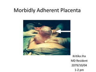
- 1. Morbidly Adherent Placenta Kritika Jha MD Resident 2079/10/04 1-2 pm
- 2. Introduction • Derived from latin “ ac-+ crescere “ : to grow from adhesion to adhere • Abnormal trophoblastic invasion of part or all of the placenta into the myometrium of the uterine wall ACOG 2018 • Term revised as Placenta Accreta Spectrum
- 3. Introduction Incidence • 1 in 2,510 births : 1980s • 1 in 731 births : 2015 • 1 in 22000 of vaginal delivery in absence of placenta previa • 50 % recognised preoperatively • Mortality rate : 4 – 7 % in placenta percreta James High Risk Pregnancy 5th ed
- 4. Incidence Number of CS Incidence of PAS (%) Rate of PAS if placenta previa (%) Rate of PAS if no placenta previa (%) 1 0.24 3.3 0.03 2 0.31 11 0.2 3 0.5 40 0.1 4 2.13 61 0.8 5 2.33 67 0.8 >/= 6 6.74 67 4.7 Arias Practical Guide to High Risk Pregnancy
- 5. FIGO Classification (2019) • Grade 1 : Abormally adherent placenta: Placenta adherent or acreta 80% • Grade 2 : Abnormally invasive placenta: increta 15% • Grade 3 : Abnormally invasive placenta 5% 3a: limited to uterine serosa 3b: urinary bladder invasion 3c: invasion of other pelvic tissue/ organs
- 6. Cesarean scar pregnancy • 1 in 2000 pregnancies • If pregnancy continued 100 % chances for PAS
- 7. Pathogenesis • Damage to decidua basalis and nitubach layer • Failure of normal decidualisation and loss of normal subdecidual myometrial layer • Upregulation of VEGF and angiopoietin -2 and downregulation of VEGF and angiopoietin receptor High volume, high velocity flow placental lacunae Fibrinoid deposition around implanted villi at uteroplacental interface loss of parts of physiological detachment
- 8. Fig 1 : Types of Morbidly Adherent placenta Focal Total
- 9. Fig 2 : Cesarean hysterectomy specimen of MAP
- 10. Risk factors • Prior morbidly adherent placenta 29 % • Placenta previa 3 % • Previous LSCS • Prior uterine surgery: Myomectomy Hysteroscopic resection of adhesion Endometrial ablation • Prior uterine curettage • Advanced age, smoking • Multiparity • Uterine irradiation • H/O manual removal of placenta, postpartum endometritis • Uterine anomaly
- 11. Clinical features • Undiagnosed before delivery : ½ to 2/3rd of cases • Antepartum haemorrhage : 30 % in presence of placenta praevia • Profuse , life threatening hemorrhage on attempted placental separation DIC, ARDS, AKI, massive blood transfusion ( 5 % ) • Intra abdominal bleeding, hematuria, bladder symptoms : cases of percreta
- 12. Clinical features Neonatal • Preterm delivery • Small for gestational age/ IUGR • Malpresentation
- 13. Diagnosis Ultrasound Absence of ultrasound findings does not preclude a diagnosis of placenta accreta spectrum Clinical risk factors equally important as predictors of placenta accreta spectrum ACOG 2018
- 14. Diagnosis : Ultrasound First trimester : Low implantation of gestation sac Irregular placenta myometrial interface Anechoic placental areas with / without vascular flow James High Risk Pregnancy 5th ed
- 15. Ultrasound Ultrasound finding Sensitivity Specificity Abnormal placental lacunae, turbulent flow, moth eaten appearance 77.4 % 95.02 % Loss of retroplacental hypoechoic zone Myometrial thinning < 1 mm overlying placenta 65.02 % 95.8 % Abnormalities of uterus-bladder interface - Bulge - Focal exophytic mass 49.7 % 99.8 % Color Doppler abnormalities - Subplacental numerous closely packed tortuous vessels - Uterovesical hypervascularity 90.8 % 87.7 % Done at 18 – 20 week 28- 30 weeks 32 – 34 weeks ACOG 2018
- 16. Ultrasound Finding Fig 4: Bridging vessel in uterine bladder interface Fig 3 : Placental lacunae
- 17. MRI: Not an initial modality for evaluation of PAS - Depth of invasion - Parametrial involvement - Posterior placentation Sensitivity : 75 – 100 % Specificity : 65 – 100 % Diagnosis Cont..
- 18. MRI findings: • Dark bands on T2 weighted imaging • Bulging or visible invasion beyond uterine serosa • Uterine scar thinning • Parametrium involvement Diagnosis Cont..
- 19. Fig 5 : MRI finding of MAP showing dark linear bands
- 20. Diagnosis Contd.. Serum markers : • Maternal serum alpha fetoprotein : Poor predictor • Others: Pregnancy-associated plasma protein A pro B-type natriuretic peptide, creatinine kinase Troponin Free β-hCG (mRNA) Human placental lactogen (cell-free mRNA) Nonspecific
- 21. Diagnosis : Histopathology Fig 6 : HPE of hysterectomy specimen showing loss of decidual layer between villi and myometrium
- 22. Management • Comprehensive multidisciplinary approach • Timing of delivery : 34 – 35 6/7 (ACOG) 35 – 36 +6 (RCOG) • Earlier : Persistent bleeding, Labor, Rupture of membrane, Fetal compromise • Cesarean hysterectomy with placenta left in situ ACOG 2018
- 23. Management in known Morbidly Adherent Placenta
- 24. Antenatal • Counselling : Ultrasound findings Complications Regular follow up Referral to higher centre for multidisciplinary approach Cesarean delivery, need of hysterectomy • Use of antenatal corticosteroid
- 25. Antenatal • Need of hospitalization : Antenatal bleeding Preterm labor Preterm prelabor rupture of membranes Associated with unscheduled delivery as well as maternal and neonatal morbidity ACOG 2018
- 26. Criteria to deliver in accreta centre of excellence • Suspicion for accreta from sonographic findings • Placenta previa with abnormal sonographic appearance • Placenta previa with > 3 prior cesarean deliveries • Prior classical cesarean delivery and anterior placentation • Prior endometrial ablation or pelvic radiation • Inability to adequately evaluate or exclude placenta accreta • Any other reason to suspect placenta accreta Cuunigham and Gillstrap’s Operative Obstetrics
- 27. Criteria for Accreta Centre of Excellence • Multidisciplinary team Experienced obstetrician Imaging experts Pelvic surgeon – Gynecological oncology, Urogynecology Anesthesiologist • Intensive care facilities Urologist General surgeon Interventional radiologist Neonatologist • Blood availability
- 28. Preoperative • Maximization of preoperative hemoglobin values • Specific timing of planned delivery • Identification of exact location of delivery (surgical suite and its associated capabilities) • Preoperative consultations • Consideration of patient and family needs given temporary relocation to placenta accreta spectrum center of excellence
- 29. Preoperative • Preoperative ureteric stent placement : case evaluation • Iliac artery occlusion : decrease blood loss in some • Preoperative placement of catheters or balloons into pelvic arteries : Recommendation cannot be made for or against ACOG 2018 • Antenatal diagnosis of placenta accreta spectrum uncertain : Intraoperative observation for spontaneous uterine placental separation
- 30. Multidisciplinary team approach • Blood transfusion service : Average blood loss : 1750 – 5000 ml Prepare for massive tranfusion : 6 U PRBC, 6 U FFP, 6 pack platelet, 10 U cryoprecipitate 1:1:1 or 1:2:4 :: PRBC: FFP :PRP ACOG 2018 Initiated based on ongoing hemorrhage Recombinant factor VII a, EPO, Total dose iron : correct anemia Preoperative hemodilution
- 31. • Anesthesiology: Regional anaethesia: preferred Need of GA Anticipate prolonged surgical time Epidural catheter for pain relief postoperatively Assess blood loss and readiness for massive blood transfusion Temperature control Multidisciplinary team approach Hypovolemia Hypothermia Acidosis
- 32. • Urology Preoperative cystoscopy and placement of ureteric stent : Anterior percreta Intentional cystotomy in uninvolved area to visualise area of invasion : Protect trigone and ureterovesical junction and excise involved portion under direct visualisation Multidisciplinary team approach
- 33. Intraoperative • Verification of appropriate complement of surgical expertise • Intraoperative availability of resources to optimize each case – eg, Cell-saver, adequate surgical trays, and necessary urologic equipment • Verification of availability of related services as necessary (eg, interventional radiology) • Coordination of blood bank with scheduling or timing of case • Close monitoring : urine output, blood loss, hemodynamic status
- 34. Fig 7 : Algorithm for management of placenta accreta spectrum Cunnigham and Gillstrap’s Operative Obstetrics Intraoperative observation for spontaneous uterine placental separation Percreta
- 35. Steps in cesarean hysterectomy • Midline vertical laparotomy and midline hysterotomy : enter uterus to avoid placenta • Superior devascularisation : divide and ligate utero ovarian pedicles and round ligament • Retroperitoneal dissection : incise down to paravesical space; cephalad dissection to expose ureter and bifurcation of common iliac arteries • Bladder dissection : progress lateral to medial and down to vaginal fornices • Colpotomy : inferior dissection of paravesical space Williams Obstetrics 27th ed
- 36. • When necessary, ureterolysis is carried out to protect the ureters and allow step-by step devascularization of the lower uterine segment followed by separation of the bladder and uterus • In cases with deep placental invasion, intentional cystotomy and partial bladder excision is favored over persistent attempts at bladder dissection and mobilization • In some cases in which the percreta involves the lateral pelvic sidewalls, staged intraoperative angiographic embolization is done before beginning the hysterectomy Steps in cesarean hysterectomy
- 37. Fig 8 : Fundal incision followed by breech extraction
- 38. Intraoperative : Minimize blood loss • Tranexamic acid : Prophylactic tranexamic acid given at the time of delivery after cord clamping ACOG 2018 Dose : 1 g intravenously Second dose : 0.5–23.5 hours later if bleeding persists
- 39. • Uterotonic agents Tab misoprost 1000 mcg per rectal Carboprost 250 mcg in 20 ml NS infiltrated in myometrium Intravenous oxytocin infusion • Torniquet : Above placental edge Intraoperative : Minimize blood loss Fig 9 : Torniquet placement
- 40. Intraoperative : Minimize blood loss Devascularisation • Uterine artery ligation At the level of the uterine incision Avascular site in broad ligament Second ligature : junction of uteroovarian ligament and lateral uterine border. Fig 11 : Uterine Artery Ligation
- 41. Devascularisation • Internal Iliac artery ligation – Technically difficulty – Requires skill in retroperitoneal surgery at the pelvic sidewall Success rate to control bleeding 50 – 75 % Complications - Ligation of internal iliac vein, external iliac artery - Ureteral injury - Retroperitoneal hematoma Intraoperative : Minimize blood loss Fig 10 : Internal Iliac Artery Ligation
- 42. Intraoperative : Minimize blood loss Interventional radiology • Pelvic artery embolisation : – Risk of uterine necrosis – High failure rate ( 70 % in PPH ) • Internal iliac artery catheterisation: Effective in percreta - Costly equipment - Complex procedure - Risk of maternal thromboembolic event - Fetal radiation exposure
- 43. Interventional radiology • Intra aortic balloon occlusion : – minimally invasive and safe – short fluoroscopy time – No need of complex logistics – Positive cardio circulatory effect – No affect on fetal- placental circulation
- 44. Temporary management of uncontrolled hemorrhage • Cessation of surgery temporarily • Infrarenal aortic clamping • Pelvic packing • Closure of skin with towel clip • Aggressive resuscitation with blood product • Patient warming • Waiting for reversal of coagulopathy before resuming surgery : best option • Patient to be shifted in monitoring bed until return to operating room • Placing abdominal drain
- 45. Postoperative • Assurance that critical care services are engaged and available for postoperative care • Identification of the need for identification of primary service responsible for postoperative care • Need of thromboprophylaxis
- 46. Post Operative Period • Clinical vigilance for complications : - Renal failure - Liver failure - Infection - Unrecognized ureteral, bladder, or bowel injury - Pulmonary edema - Disseminated intravascular coagulation - Sheehan syndrome
- 47. Management in Unexpected MAP • Placenta accreta spectrum is suspected based on uterine appearance and there are no extenuating circumstances mandating immediate delivery, the case should be temporarily paused until optimal surgical expertise arrives • Additional intravenous access should be obtained, blood products should be ordered, and critical care personnel should be alerted
- 48. Unexpected MAP Fig 12 : Placenta percreta at time of laparotomy
- 49. Management in Unexpected MAP • Accreta : Hemostasis in myometrial bed using Cho suture Suturing inverted cervical tissue over the area of bleeding placental bed Compression suture Fig 13 : B- Lynch compression suture
- 50. Conservative and Expectant management • Candidate - Desire to preserve fertility Counsel regarding risks of hemorrhage, infection, need of hysterectomy, recurrence in subsequent pregnancy and even death - High risk of hemorrhage and other organ injury during hysterectomy - Placental resection : Focal accreta Fundal or posterior placenta
- 51. Conservative management Contd. • Expectant management - Placenta left in situ ACOG 2018: Attempt only rarely as a part of approved clinical trial in fully informed patients
- 52. • Delayed interval hysterectomy Not recommended Option in - Severe life threatening cases - When immediate hysterectomy is dangerous Extent of placental invasion (percreta) Lack of resources Conservative management Contd.
- 53. Conservative Management : Placental myometrial en bloc excision and repair • Candidate : Clearly delineated focal area of involvement • Triple P : Preoperative placental localisation Pelvic devascularisation with preoperative placement of intraarterial balloon catheter with inflation after delivery / ligation of uterine artery Placenta removed with Enbloc myometrial excision and repair
- 54. Adjunct to conservative or expectant • Devascularization: – Uterine artery balloon placement – Embolization or ligation • Postdelivery methotrexate administration • Hysteroscopic resection • High intensity focussed ultrasound : not recommended
- 55. • Methotrexate MOA: Disruption of folic acid pathway in rapidly dividing cells such as trophoblasts Proliferation of trophoblasts in later stages of pregnancy : no role in placental growth Use of methotrexate may not reduce placental volume Adverse effects : Increased risk of infection and sepsis Pancytopenia Nephrotoxicity Conservative management Contd.
- 56. Conservative management : Methotrexate therapy • Methotrexate adjuvant therapy should not be used for expectant management as it is of unproven benefit and has significant adverse effects. RCOG Green top guideline 2018 • Methotrexate therapy should not be used ACOG 2018
- 57. Follow up The pattern of follow-up for the conservative management of placenta accreta spectrum is not supported by RCTs and is not stratified according to the depth and lateral extension of villous myometrial invasion • Clinical :Temperature monitoring Per vaginal bleeding, Foul smelling per vaginal discharge Pain abdomen • Lab parameter : Serial b-HCG , CBC, High vaginal swab • Radiology : Ultrasound
- 58. Outcome of conservative management Median time to placental involution : 13.5 weeks Complications : • Severe vaginal bleeding 53 % • Sepsis 6 % • Secondary hysterectomy 19% • Death 0.3 % • Subsequent pregnancy 67% • Recurrence 22-29 % Cunnigham and Gilstrap’s Operative Obstetrics
- 59. Case presentation • Mrs Bhatta 33 years Referred case from Kailali for Placenta Previa • Presenting complaint : Pregnancy for 8 months Per vaginal bleeding for 2 weeks • Obstetric history : P 1 – Em LSCS for oligohydraminos 3.5 years back • USG: Anterior low lying placenta 1.3 cm from os with placental lake • MRI : Complete placenta previa
- 60. Case presentation Contd. Cesarean hysterectomy for placenta previa with focal placenta accreta with right pelvic hematoma with PPH at 37 weeks of gestation • POF : Placenta completely covering os and anterior focal acreta in right lower segment • Following delivery of placenta bleeding from placental bed secured by hemostatic suture bleeding continued and hematoma developed in lower uterine segment cesarean hysterectomy • Blood loss : 2 litres • Blood transfusion : 2 pint WB + 3 pint PRP • Post operative : ICU stay * 2 days Blood transfused: 3 pint WB + 2 pint FFP HPE : Endometrium showed decidualised stroma with thick muscular wall. No chorionic villi
- 61. Conservative management of placenta increta in a primigravida: A case report Waheed et al 2019 JPMA • In July 2017, Mrs. ABC, 25 yr, primigravida booked at 36 WOG • Ultrasound showed a S/L /F 36 wog, adequate AFI, fundal placenta with no evidence of myometrial invasion • At 38 wog : Presented in emergency with labour pain os: 3 cm, Cardiotocogram( CTG) showed variable decelerations ARM: meconium stained liqour Emergency caesarean section decided
- 62. Case Report Contd • Delay in the delivery of placenta After waiting for 30 minutes, 10 U oxytocin given in umbilical cord • Again tried to deliver the placenta but no plane of cleavage identified • Uterus exteriorized and placenta found to be invading the myometrium at fundus, sparing only serosa of uterus
- 63. • Management options discussed including the need for a Caesarean hysterectomy in case of massive haemorrhage Approximate blood loss in surgery - 1 litre Vitals- Stable. Bilateral uterine artery ligation done • Conservative management was planned Planned for emergency hysterectomy if any problem such as haemorrhage or infection during the course of treatment Case Report Contd
- 64. CBC, LFT, RFT : normal Injection methotrexate at 6hours of surgery Intravenous antibiotic • Discharged on 4 th post op day • Temperature : Twice daily. • Monitor for vaginal bleeding , contact if heavy bleeding. • Weekly CBC and bHCG tests : first 4 weeks. • Ultrasounds : Twice weekly. • Intravenous antibiotic continued to avoid sepsis. • Weekly visits to doctor Case Report Contd
- 65. • Vitals stable and investigations normal 2nd dose of methotrexate given : after 1 week Intravenous antibiotics for another 4 weeks 3rd dose of methotrexate given: after 2 weeks TLC normal throughout visits bHCG normalised in 2 months Ultrasound (5 months) : no retained piece of placenta Conservative management of placenta increta : successful Case Report Contd
- 66. Take home message • Risk factors for placenta accreta spectrum should be assessed • History of previous cesarean section and placenta previa are most common association • Antenatal diagnosis in patient with suspicion for PAS should be done by ultrasound • Undiagnosed case in which plane of separation can not be felt should not be attempted with manual removal of placenta
- 67. • Peripartum / cesarean hysterectomy remain choice of treatment in placenta increta and percreta • Referral to centre equipped with trained personnel and availability of blood product • In elective case, pelvic artery catherisation should be considered • If conservative management is planned , counselling should be done regarding complications and need of hysterectomy Take home message
- 68. References • William’s Textbook of Obstetrics 26th edition • James High Risk Pregnancy 5th ed • Arias Practical Guide to High risk pregnancy • Cuunigham and Gillstrap’s Operative Obstetrics • ACOG Guidelines placenta accreta spectrum 2018 • RCOG Green Top Guidelines 2018 • Recent advances in Obstetrcs and Gynecology 26
- 69. THANK YOU
Editor's Notes
- Incidence incfreased due to increased cs rate
- etiology of placenta accreta spectrum is that a defect of the endometrial–myometrial interface leads to a failure of normal decidualization in the area of a uterine scar, which allows abnormally deep placental anchoring villi and trophoblast infiltration
- Accreta : focal: single lobule abnormally attached; all lobule : total
- Bicornuate uterus rudimentary horn PAS. Smoking : co cause compensatory placental hypertrophy, vasculopathy > 35 yr : OR 1.3 increase in previa each yr… IF ONLY PREVIA : 3 %
- No plane of separation, firm attachment of placenta. 18 % : nulliparous
- Not a single finding is specific
- Moth beaten appearance of lacunae
- Equal to usg if done by expertise. Acog : Mri WHEN USG inconclisive and posterior previa
- Long distance from hospital, no resources
- CBC every 15 min
- Complications of massive blood transfusion
- Second : junction of uteroovarian ligament and laateral uterine border: Initially, the needle passes in an anterior-to-posterior direction through lateral uterine wall. The needle is removed from the posterior myometrium and redirected from posterior to anterior through an avascular space in the broad ligament. The suture is then securely tied. Inset
- Ureteral injury, ligation of external iliac artery, int iliac vein, retroperitoneal hematoma. Succes rate : 50 – 75 %
- Balloon tamponade for preservation : not done ; myometrial hypoperfusion/ laceration, build up lacrtic acid, necrosis, decreased contractility. Cross clamping of aorta : needs retroperitoneal dissection
- Delay in fruitless attept to preserve uterus : mortality
- Largest series : 12 went hysteroscpic, 1 had hystrctomy. In HFUSG : uterine perforation, shock no
- Recurrence 22-29%