![http://www.acmcasereport.com/ 2
Volume 8 Issue 13 -2022 Case Report
4. Discussion
The originality of our work lies in the rarity of primary labial tu-
berculosis rarely seen and mostly recognized through case reports
[1]. It is estimated that only 0.05% to 5% of total TB cases may
present with oral manifestations [2]. It remains rare even in a coun-
try where the disease is widespread, such as Morocco where the
prevalence is reported to 30897 cases in 2017 [3]. Oral TB lesions
may be either primary or secondary in occurrence [4]. Primary
lesions are uncommon, seen in younger patients, and present as
single painless ulcer with regional lymph node enlargement. The
secondary lesions are common, often associated with pulmonary
disease, usually present as single, indurated, irregular, painful ul-
cer covered by inflammatory exudates in patients of any age group
but relatively more common in middle-aged and elderly patients
[5]. It is believed that the intact oral mucosa, constant flow of sa-
liva and its antibacterial properties protect from tubercle bacilli
invasion to the oral tissues [6]. However, any local trauma can
promote infection. Other local predisposing factors include poor
oral hygiene, hyperkeratosis disorders such as leukoplakia, oral
mucosa inflammation or even tooth extraction [7-9]. In our case,
poor oral hygiene is incriminated.
Histopathological assessment may reveal the presence of granu-
lomatous inflammatory infiltration with Langhans giant cells and
lymphocytes. Foci of caseous necrosis of the tissue can be ob-
served. Mycobacteria can be demonstrated in the collected spec-
imen [7,8,10-12]. Microbiological culture of sputum and of the
material taken from the surface of the oral lesion should be done,
but the results are obtained after 10 weeks. According to various
studies only a small percentage (7.8%) of histopathology speci-
mens stain positive for acid fast bacilli [13]. Therefore, a nega-
Figure 1: Linear labial ulceration of the lower lip
tive result does not rule out completely the possibility of TB. In
doubtful cases, molecular tests (PCR) may be helpful. In our case,
histopathological examination of the lymph node biopsy was an
important aid in the diagnosis of the disease because the finding of
caseous necrosis was highly suggestive of tuberculosis.
In our country, the treatment requires a combination of 3 drugs (ri-
fampicin, isoniazid and pyrazinamide) administered daily for the
first 2 months, followed by an additional 4 months with 2 drugs (ri-
fampicin, isoniazid). In our case, the patient underwent 9 months
of treatment because she had associated lymph node location.
The purpose of this case report is to highlight the rare clinical pre-
sentation of tuberculosis and bring to the attention a differential
diagnosis of tuberculosis while dealing with chronic oral ulcers. It
is particularly relevant in a country like Morocco with one of the
highest tuberculosis burdens.
5. Acknowledgment
None.
6. Conflict of Interest
None.
References
1. Vucicevic Boras V, Gabric D, Smiljanic Tomicevic L, Seiwerth S,
Grcic K, Sarcevic B, et al. Tuberculosis of the Oral Cavity Misdi-
agnosed as Precancerous Lesion. Acta Stomatol Croat. 2017; 51(4):
326‑331.
2. Aoun N, El-Hajj G, El Toum S. Oral ulcer: an uncommon site in
primary tuberculosis. Aust Dent J. 2015; 60(1): 119‑122.
3. Ministry of Health National strategic plan for the prevention and
control of tuberculosis in Morocco 2012-2018.
4. Jain P. Oral Manifestations of Tuberculosis: Step towards Early Di-
agnosis. J Clin Diagn Res. 2014.
5. Mignogna M, Muzio L, Favia G, Ruoppo E, Sammartino G, Zarrelli
C, et al. Oral tuberculosis: a clinical evaluation of 42 cases. Oral Dis.
2008; 6(1): 25‑30.
6. Krawiecka E, Szponar E. Tuberculosis of the oral cavity: an uncom-
mon but still a live issue. Adv Dermatol Allergol Dermatol Alergol.
2015; 32(4): 302‑306.
7. Dixit R, Sharma S, Nuwal P. Tuberculosis of Oral Cavity. Indian J
Tuberc. 2008; 55(1): 51-53.
8. Nanda KDS, Mehta A, Marwaha M, Kalra M, Nanda J. A Dis-
guised Tuberculosis in Oral Buccal Mucosa. Dent Res J. 2011; 8(3):
154‑159.
9. Von Arx DP, Husain A. Oral tuberculosis. Br Dent J. 2001; 190(8):
420‑422.
10. Kakisi OK, Kechagia AS, Kakisis IK, Rafailidis PI, Falagas ME.
Tuberculosis of the oral cavity: a systematic review. Eur J Oral Sci.
2010; 118(2): 103‑109.
11. Wang WC, Chen JY, Chen YK, Lin -M. Tuberculosis of the head
and neck: a review of 20 cases. Oral Surg Oral Med Oral Pathol Oral
Radiol Endodontology. 2009; 107(3): 381‑386.](data:image/gif;base64,R0lGODlhAQABAIAAAAAAAP///yH5BAEAAAAALAAAAAABAAEAAAIBRAA7)
Recommended
Recommended
More Related Content
Similar to Tuberculosis: A Rare Cause of Linear Labial Ulceration
Similar to Tuberculosis: A Rare Cause of Linear Labial Ulceration (16)
More from komalicarol
More from komalicarol (20)
Recently uploaded
Recently uploaded (20)
Tuberculosis: A Rare Cause of Linear Labial Ulceration
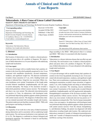
- 1. Annals of Clinical and Medical Case Reports Case Report ISSN 2639-8109 Volume 8 Tuberculosis: A Rare Cause of Linear Labial Ulceration Saoud ZT*, Hali F, Elfatoiki FZ and Chiheb S Department of Dermatology and Venerology, Ibn Rochd University Hospital, Casablanca, Morocco * Corresponding author: Zineb Tazi Saoud, Department of Dermatology and Venerology, Ibn Rochd University Hospital, University Hassan II, Casablanca, Morocco, Tel: +212670934582, E-mail address: z.tazisaoud@gmail.com Received: 16 Feb 2022 Accepted: 04 Mar 2022 Published: 11 Mar 2022 J Short Name: ACMCR Copyright: ©2022 Saoud ZT. This is an open access article distribut- ed under the terms of the Creative Commons Attribution License, which permits unrestricted use, distribution, and build upon your work non-commercially. Citation: Saoud ZT, Tuberculosis: A Rare Cause of Linear Labial Ulceration. Ann Clin Med Case Rep. 2022; V8(13): 1-3 Keywords: Tuberculosis; Oral tuberculosis; Mucous membrane; Lip; Lymph node tuberculosis http://www.acmcasereport.com/ 1 Abreviations: TB: tuberculosis; PCR: polymerase chain reaction 1. Abstract 1.1. Introduction Oral location of tuberculosis is rare. It admits a clinical polymor- phism and poses above all a problem of diagnosis. We report a case of labial tuberculosis in a 16-year-old patient with underlying lymph node localization. 1.2. Case report A 16-year-old teenager with no notable history had a painless ul- ceration in the lower lip which gradually progressed in size. It was associated with mandibular tumefaction, elevated temperature, weakness, and significant weight loss. The physical examination revealed a linear ulceration of 3 cm, with a budding border and sitting on indurated base. Gingivitis was associated. Others mu- cous membranes were normal. Examination of the lymph nodes revealed bilateral mandibular lymphadenopathy and left jugu- lo-carotid adenopathy. The biopsy of the ulceration revealed a tuberculoid granuloma without caseous necrosis. The hemogram and chest x-ray were normal. The tuberculin intra-dermoreaction was positive at 17mm. An adenectomy with histological study was performed objectifying epithelio-giganto-cellular granulomas with central caseous necrosis. The diagnosis of tuberculous chan- cre was retained associated with secondary lymph node location. The patient underwent anti-bacillary treatment (2RHZ / 7RH) with good progress and healing of ulceration. 1.3. Discussion The originality of our work lies in the rarity of labial localization during tuberculosis and its association with an underlying lymph node location. The oral mucosa localization is rare represents only 0.1 to 5%. The diagnosis is confirmed on clinical, biological and histological arguments. The treatment is based on anti-bacillary drugs according to the 2RHZ / 4RH protocol when it is isolated and 2RHZ / 7RH when it is associated with other locations. 2. Introduction Tuberculosis is a chronic infectious disease that can affect any part of the body. The oral location is rare. It admits a clinical polymor- phism and poses above all a problem of diagnosis. We report a case of labial tuberculosis in a 16-year-old patient with underlying lymph node localization. 3. Case Report A 16-year-old teenager with no notable history had a painless ul- ceration on her lower lip gradually increasing in size. It was asso- ciated with elevated temperature, weakness, and significant weight loss. There were no pulmonary or gastrointestinal symptoms. Der- matological examination revealed a linear ulceration, of approxi- mately 3 cm, with a budding border and sitting on indurated base. Gingivitis was associated. The remaining mucous membranes were normal. Examination of the lymph nodes revealed bilateral mandibular lymphadenopathy and left jugulocarotid adenopathy. Histopathological examination of the lip lesion revealed a tuber- culoid granuloma without caseous necrosis. The hemogram and chest x-ray were normal. A tuberculin skin test was positive at 17 mm induration (evaluated after 72h). Test for human immunodefi- ciency virus was negative. An adenectomy with histological study was performed objectifying epithelio-giganto-cellular granulomas with central caseous necrosis. From the clinical, histopatholog- ic, and laboratory findings, the patient was diagnosed with labial tuberculosis revealing lymph node location. She underwent anti- tuberculosis treatment (2 months of rifampicin/isoniazid/pyrazin- amide and 7 months of rifampicin/isoniazid) with good progress and fibrous scarring of the ulceration.
- 2. http://www.acmcasereport.com/ 2 Volume 8 Issue 13 -2022 Case Report 4. Discussion The originality of our work lies in the rarity of primary labial tu- berculosis rarely seen and mostly recognized through case reports [1]. It is estimated that only 0.05% to 5% of total TB cases may present with oral manifestations [2]. It remains rare even in a coun- try where the disease is widespread, such as Morocco where the prevalence is reported to 30897 cases in 2017 [3]. Oral TB lesions may be either primary or secondary in occurrence [4]. Primary lesions are uncommon, seen in younger patients, and present as single painless ulcer with regional lymph node enlargement. The secondary lesions are common, often associated with pulmonary disease, usually present as single, indurated, irregular, painful ul- cer covered by inflammatory exudates in patients of any age group but relatively more common in middle-aged and elderly patients [5]. It is believed that the intact oral mucosa, constant flow of sa- liva and its antibacterial properties protect from tubercle bacilli invasion to the oral tissues [6]. However, any local trauma can promote infection. Other local predisposing factors include poor oral hygiene, hyperkeratosis disorders such as leukoplakia, oral mucosa inflammation or even tooth extraction [7-9]. In our case, poor oral hygiene is incriminated. Histopathological assessment may reveal the presence of granu- lomatous inflammatory infiltration with Langhans giant cells and lymphocytes. Foci of caseous necrosis of the tissue can be ob- served. Mycobacteria can be demonstrated in the collected spec- imen [7,8,10-12]. Microbiological culture of sputum and of the material taken from the surface of the oral lesion should be done, but the results are obtained after 10 weeks. According to various studies only a small percentage (7.8%) of histopathology speci- mens stain positive for acid fast bacilli [13]. Therefore, a nega- Figure 1: Linear labial ulceration of the lower lip tive result does not rule out completely the possibility of TB. In doubtful cases, molecular tests (PCR) may be helpful. In our case, histopathological examination of the lymph node biopsy was an important aid in the diagnosis of the disease because the finding of caseous necrosis was highly suggestive of tuberculosis. In our country, the treatment requires a combination of 3 drugs (ri- fampicin, isoniazid and pyrazinamide) administered daily for the first 2 months, followed by an additional 4 months with 2 drugs (ri- fampicin, isoniazid). In our case, the patient underwent 9 months of treatment because she had associated lymph node location. The purpose of this case report is to highlight the rare clinical pre- sentation of tuberculosis and bring to the attention a differential diagnosis of tuberculosis while dealing with chronic oral ulcers. It is particularly relevant in a country like Morocco with one of the highest tuberculosis burdens. 5. Acknowledgment None. 6. Conflict of Interest None. References 1. Vucicevic Boras V, Gabric D, Smiljanic Tomicevic L, Seiwerth S, Grcic K, Sarcevic B, et al. Tuberculosis of the Oral Cavity Misdi- agnosed as Precancerous Lesion. Acta Stomatol Croat. 2017; 51(4): 326‑331. 2. Aoun N, El-Hajj G, El Toum S. Oral ulcer: an uncommon site in primary tuberculosis. Aust Dent J. 2015; 60(1): 119‑122. 3. Ministry of Health National strategic plan for the prevention and control of tuberculosis in Morocco 2012-2018. 4. Jain P. Oral Manifestations of Tuberculosis: Step towards Early Di- agnosis. J Clin Diagn Res. 2014. 5. Mignogna M, Muzio L, Favia G, Ruoppo E, Sammartino G, Zarrelli C, et al. Oral tuberculosis: a clinical evaluation of 42 cases. Oral Dis. 2008; 6(1): 25‑30. 6. Krawiecka E, Szponar E. Tuberculosis of the oral cavity: an uncom- mon but still a live issue. Adv Dermatol Allergol Dermatol Alergol. 2015; 32(4): 302‑306. 7. Dixit R, Sharma S, Nuwal P. Tuberculosis of Oral Cavity. Indian J Tuberc. 2008; 55(1): 51-53. 8. Nanda KDS, Mehta A, Marwaha M, Kalra M, Nanda J. A Dis- guised Tuberculosis in Oral Buccal Mucosa. Dent Res J. 2011; 8(3): 154‑159. 9. Von Arx DP, Husain A. Oral tuberculosis. Br Dent J. 2001; 190(8): 420‑422. 10. Kakisi OK, Kechagia AS, Kakisis IK, Rafailidis PI, Falagas ME. Tuberculosis of the oral cavity: a systematic review. Eur J Oral Sci. 2010; 118(2): 103‑109. 11. Wang WC, Chen JY, Chen YK, Lin -M. Tuberculosis of the head and neck: a review of 20 cases. Oral Surg Oral Med Oral Pathol Oral Radiol Endodontology. 2009; 107(3): 381‑386.
- 3. http://www.acmcasereport.com/ 3 Volume 8 Issue 13 -2022 Case Report 12. Rowinska-Zakrzewska E, Korzeniewska-Kosela M, Roszkows- ki-Sliz K. Extrapulmonary tuberculosis in Poland in the years 1974- 2010. Pneumonol Alergol Pol. 2013; 81(2): 121‑129. 13. Thilander H, Wennestrom A. Tuberculosis of mouth and the sur- rounding tissues. Oral Surgery, Oral Medicine, Oral Pathology, Oral Radiology, and Endodontology. 1956; 858‑870.