
Recommended
Recommended
More Related Content
Similar to ENDOSCOPIC TREATMENT OF PILONIDAL SINUS IN EGYPTIAN PATIENTS
Similar to ENDOSCOPIC TREATMENT OF PILONIDAL SINUS IN EGYPTIAN PATIENTS (19)
More from indexPub
More from indexPub (20)
Recently uploaded
Recently uploaded (20)
ENDOSCOPIC TREATMENT OF PILONIDAL SINUS IN EGYPTIAN PATIENTS
- 1. Hunan Daxue Xuebao/Journal of Hunan University Natural Sciences ISSN:1674-2974 | CN 43-1061 / N DOI: 10.5281/zenodo.10152961 Vol: 60 | Issue: 11 | 2023 Nov 2023 | 15 ENDOSCOPIC TREATMENT OF PILONIDAL SINUS IN EGYPTIAN PATIENTS MOHAMED A.ELASHRY* Researcher, Department of Surgery, Theodor Bilharz Research Institute, Giza, Egypt. *Corresponding Author Email: ashry2020hana@gmail.com, m.elashry@tbri.gov.eg M EMAD ESMAT Professor, Department of Surgery, Theodor Bilharz Research Institute, Giza, Egypt. Email: emadesmat2000@yahoo.com MOHAMED S HEDAYA Professor, Department of Surgery, Theodor Bilharz Research Institute, Giza, Egypt. Email: hedayams@gmail.com Abstract Background: Treatment for pilonidal disease using minimally invasive methods is a reliable and successful alternative to conventional surgery, with quicker recovery, better cosmetic outcomes, and better pain management. The primary goals of this study are to assess the early outcomes of endoscopic pilonidal sinus treatment and to demonstrate the surgical approach and its adaptations. Materials and Methods: Our study was conducted on 30 patients with pilonidal sinus disease as a prospective cohort study for endoscopic treatment of the pilonidal sinus, from October 2021 to October 2022, in our surgical department at Theodor Bilharz Research Institute (TBRI). Surgical outcomes of sinus healing, pain, and discharge were reviewed in the outpatient clinic, and patient satisfaction levels were assessed through a standardized phone interview. Results: There were 24 males and 6 females, with a median age of 21.87±1.85 years (ranging from 16 to 57 years). The mean operative time was 44.17 (35-55) ±1.26 min. During the follow-up period of 24 weeks, wound closure was seen after a median of 4 weeks. Wounds were closed in 72% of patients after one month and 93% of patients after two months. 2 patients had to be re-operated due to failure: one had persistence of discharge, and the other had recurrence after 3 months. The satisfaction rate was 93.3%. Conclusions: Endoscopic pilonidal sinus treatment is a minimally invasive and cosmetically favorable procedure. To find out if it reduces recovery time and the long-term recurrence rate, a larger sample size and a longer follow-up are needed. Keywords: Pilonidal Sinus, Endoscopy, and Treatment. INTRODUCTION Pilonidal sinus (PS) is a frequent sacrococcygeal health issue that primarily affects young males (1). It is linked to obesity, sedentary jobs, local irritation, and hirsutism (2). Several ideas were created to support the idea that the disease is congenital, however, in the last 50 years, it has been claimed that the disorder is acquired (3) due to the blocking of hair or hair follicles in the natal cleft (4). The symptoms might range from asymptomatic pits to pain, reddening of the skin, discharge of pus or blood from a skin orifice, a foul odor, an acute abscess, and a chronic cyst (5). Pilonidal illness incidence peaks between the ages of 16 and 20 and continues to be high until the age of 25, at which point it starts to rapidly diminish (6). Regarding economics and human resources, it has raised serious concerns (7).
- 2. Hunan Daxue Xuebao/Journal of Hunan University Natural Sciences ISSN:1674-2974 | CN 43-1061 / N DOI: 10.5281/zenodo.10152961 Vol: 60 | Issue: 11 | 2023 Nov 2023 | 16 Pilonidal sinus treatment aims to offer a good likelihood of cure with a low recurrence rate, prevent hospitalisation and general anaesthesia, and cause the patient the least amount of discomfort, complications, and time away from work (8). There have been numerous therapies recommended for the pilonidal sinus during the past 25 years, but no agreement has developed, and there have been few studies comparing the relative expenses, discomfort, and time off work associated with various treatments (9). There is still a significant opportunity for improvement given the number of recorded recurrences and the variety of surgical alternatives, which point to the fact that the perfect surgical treatment is still being sought after (10). The best surgical method would eliminate the cyst and remove and clean the sinus tract or tracts, resulting in thorough and long-lasting healing and pleasing aesthetic outcomes (11). The treatment for PS involves open excision and healing by secondary intention; however, the literature reports that this method has a poor postoperative quality of life and necessitates frequent clinical surveillance (12). In contrast, PS excision with primary closure employing a variety of methods, primarily midline closure or flap-based surgeries, appears to be the gold standard. These surgical techniques produce varying results, as well as varying healing durations and consequences. For the treatment of PS, various minimally invasive procedures have been developed, including radiosurgery, fibrin glue injection, and more recently, endoscopy (13). Meinero et al. devised a special fistuloscope with the potential to remove the sinus cavity and sinus tracts while viewing them directly through an operating channel, and they suggested using this method (14), also using a hysteroscopy and the video-assisted anal fistula therapy (VAAFT) technique, both of which produced outstanding results (15). It is established that alternative, more complex techniques of pilonidal sinus surgery have a high recurrence and morbidity rate (16). Aim of work In this study, we sought to document our experience treating the pilonidal sinus endoscopically using this method, with healing rate as the primary outcome and hospitalization, morbidity, healing duration, return to work, and satisfaction rate as secondary endpoints. MATERIALS AND METHODS A prospective cohort study was conducted on 30 candidates for pilonidal sinus from October 2021 to October 2022 in the Theodor Bilharz Research Institute. Demographic data (age, sex, and BMI) were documented. All patients are candidates for endoscopic treatment of PNS. Patients under the age of 16 and those with acute pilonidal abscess were excluded from the study, but those with symptomatic chronic or recurrent PNS were included sequentially. Ethical approval The study's protocol was approved by the TBRI institutional review board under Federal Wide Assurance (FWA 00010609), and the work was carried out in accordance with the World Medical Association's Code of Ethics for Human Experiments (Declaration of Helsinki) and its later amendments (GCP guidelines) or comparable ethical standards. Examination and diagnosis All cases are diagnosed by taking the patient's medical history, performing an examination. All patients underwent routine laboratory tests according to their ASA anaesthesia score (complete blood picture, coagulation profile, random blood sugar, renal, and liver function tests). Patients were informed about the procedures and signed consent forms before surgery.
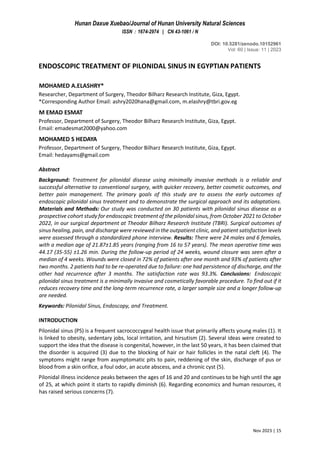
- 3. Hunan Daxue Xuebao/Journal of Hunan University Natural Sciences ISSN:1674-2974 | CN 43-1061 / N DOI: 10.5281/zenodo.10152961 Vol: 60 | Issue: 11 | 2023 Nov 2023 | 17 Operation protocol All patients underwent a day surgery procedure under local anaesthesia and sedation while prone, with the buttocks separated by two large plasters. All patients are instructed to remove surrounding hairs by shaving or using depilatory cream (at least an area 10 cm in diameter). A single dose of antibiotic prophylaxis (Ceftriaxone 1 g) was administered 30 minutes before surgery. Informed consent was obtained from all patients before the surgical procedure. The surgical technique of endoscopic treatment of the pilonidal sinus is performed with a fistuloscope (sinoscope) manufactured by Karl Storz (Southbridge, Massachusetts, USA). In the kit (Figure 1), the fistuloscope has an 8° angle eyepiece and is equipped with an optical channel 14 cm long with a handle, an operative channel, and an irrigation channel. The latter channel is connected to a 5000-mL bag containing a solution of glycine and 1% mannitol. If the external orifice is too small, it is enlarged with a scalpel or diathermy to allow introduction of the fistuloscope. In our procedure, we use 2 openings, one for the fistuloscope and the other for extraction of hairs if present in the pilonidal cavity, curettage with a spoon, and diathermy. (Figure 2). The procedure begins from the first opening with a diagnostic step, which is necessary to characterise the anatomy of the tracts, followed by the operative step from the second opening to achieve intraluminal destruction and the removal of waste material (Figure 3). During the diagnostic phase, the fistuloscope (sinoscopy) is introduced through the external opening into the sinus cavity. Hair and all fistula tracts or abscess cavities clearly appear on the screen (Fig. 3). By making slow up-and-down and side-to-side movements, the infected area can be clearly delineated. The progression of the fistuloscope is easy, even through long and angled secondary tracts. In the operative phase, all hair debris was removed by mosquito forceps (Figure 3), then all the granulation tissue was destroyed and removed by a brush or curettage spoon. A diathermy electrode was then introduced, and the cavity and fistula’s track were ablated. The continuous lavage of the washing solution allows full elimination of debris and blood. Lastly, the opening of the endoscope was closed with prolene suture, but the second opening was left for repeated dressing. Postoperative Discharge and Follow-up Patients were encouraged to mobilize immediately after surgery, with the goal of discharge within 4 hours after treatment. After discharge, patients were instructed to irrigate the cavity through the external opening with 5 mL of saline solution twice a day and to keep the surrounding area clean and dry. All patients were asked to report daily the pain experienced on the visual analogue scale (VAS 0– 10), as well as to take a 1 g paracetamol tablet if necessary. Severe pain was represented by a VAS score greater than 7. After surgery, patients were seen in the outpatient clinic at 1, 2, and 4 weeks and then routinely every 6 months. During the follow-up, patients were monitored for healing, pain, complications, recurrence, and satisfaction (satisfied or not satisfied). Healing of the PS was defined as complete wound healing with closure of all external openings during the first 60 postoperative days. The presence of an external opening or secretion after 2 months was considered wound persistence, and recurrence was defined as the return of symptoms 90 days after complete healing. Statistical analysis Study population in prospective Cohort a total of patients 30, two tail, effect size dz = 0.69 a err prob = 0.5 power (1- β err prob ) = 0.9 Total sample size = 30 The data was computerized and statistically analyzed was performed using (IBM SPSS version 25). Descriptive statistics will be presented in tables. The results are reported as counts and percentages for categorical variables, as the means ±SEM (range) for continuous normally distributed variables. The difference between the two values will be analyzed using a two-tailed unpaired t test. Significance
- 4. Hunan Daxue Xuebao/Journal of Hunan University Natural Sciences ISSN:1674-2974 | CN 43-1061 / N DOI: 10.5281/zenodo.10152961 Vol: 60 | Issue: 11 | 2023 Nov 2023 | 18 levels were assessed at p value ˂0.05. The work will be reported in line with the TBRI criteria (strengthening the reporting of Cohort studied in surgery). Figure 1: the endoscope used in our procedure Figure 2: shows the openings used during the procedure Figure 3: shows hairs and granulation tissue inside pilonidal cavity
- 5. Hunan Daxue Xuebao/Journal of Hunan University Natural Sciences ISSN:1674-2974 | CN 43-1061 / N DOI: 10.5281/zenodo.10152961 Vol: 60 | Issue: 11 | 2023 Nov 2023 | 19 RESULTS The patients’ characteristics are shown in Table 1. From October 2021 through October 2022, we enrolled 30 consecutive patients: 24 males and 6 females, median age 21 (range, 16–57) years. There were follow-ups at 1, 2, 4, and 6 weeks. Twenty seven patients had a primary sacrococcygeal pilonidal sinus, and three had a recurrent pilonidal sinus. The mean operative time was 44.17 (35–55) ±1.26 min. The average hospital stay was 8 hours, with a range of 6–10 hours. All patients were discharged as planned after the end of the procedure. No patients required admission for an overnight stay or emergency room readmission after the discharge. According to the VAS score, at 1 week after surgery, 24 patients (80%) reported a VAS between 1 and 3, and 4 (13.33%) reported a score between 4 and 6, while only 2 patients (6.66%) reported a score between 7 and 10. The average time off work and return to daily activities (mean ±SD) was 5.62±2.55 days. During the follow-up period of 24 weeks, wound closure was seen after a median of 4 weeks. Wounds were closed in 72% of patients after one month and 93% of patients after two months. Four patients experienced more bloody discharge after surgery and required unplanned outpatient visits. The overall healing rate was 92%. We reported 2 failures (6.66%): 1 patient reported incomplete healing (persistence), and 1 reported recurrence. The satisfaction rate was 93.3% (Table 2). Table 1: patient characteristics Characteristic Value Patients enrolled 30 Male/female 24/6 Age (years) 21.87±1.85 Presentation, n (%) De novo Recurrent 27 (90.0%) 3 (10.0%) Mean operative time, min (range) 44.17 (35-55)±1.26 Number of external openings; n (%) 1 2 ≥3 16 (53.3) 11 (36.67) 3 (10.0) Discharge, n (%) No Yes 22 (73.3%) 8 (28.7%) Mean operative time, min (range) 44.17 (35-55)±1.26
- 6. Hunan Daxue Xuebao/Journal of Hunan University Natural Sciences ISSN:1674-2974 | CN 43-1061 / N DOI: 10.5281/zenodo.10152961 Vol: 60 | Issue: 11 | 2023 Nov 2023 | 20 Table 2: intraoperative and post-operative charctaristics. 1-3 vas score ***p<0.001 significant decrease than 4-6 and 7-10; 4-6 was score **p<0.01 significant decrease than 7-10. DISCUSSION A safe and successful substitute for traditional surgery for the treatment of pilonidal disease is minimally invasive endoscopic procedures (17). Our study's objectives are to describe the surgical procedure and its adjustments while assessing the early outcomes of endoscopic pilonidal sinus treatment in Egyptian patients. Our study shows that 93.3% of the patients achieved complete long-term healing, a quick return to normal daily activities, a short time off work, and high patient satisfaction after endoscopic treatment of the pilonidal sinus. Under direct vision, the endoscopic technique allows for the removal of the infected area, hair, and hair follicles, as well as the vision of fistula tracts, side tracts, or any abscess cavities. This is in line with a study conducted on 42 patients by Foti, N., et al., who state that patient satisfaction, time off work, and failure rate were similar to those of excisional surgery (18). In our study, the aesthetic result appears to be good, and the procedure is well tolerated with a prompt return to work. There is no need for painful dressings, and healing occurs within 2–4 weeks. This is consistent with a study done on 32 patients by Gallo Gaetano et al., who report no painful repeated dressings with excellent aesthetic results (19). All patients were discharged within 4 hours. In the first postoperative week, we found a mean VAS score of 1.79±0.19, and that 2 patients needed painkillers for 4 weeks (7.5 %). This is similar to what Emile et al. reported, which stated that the mean VAS score was 1.35±0.8 (20). Our follow-up for this endoscopic technique of the pilonidal sinus showed 2 cases of recurrence, one with persistence of discharge and the other with recurrence after 3 months. The failure rate was higher in recurrent disease and when lateral openings were present. This is in line with a prospective international multicenter study done by Meinero P, et al., who concludes that recurrence is more common with recurrent cases and multiple openings (21). Characteristic Value Sinus openings One Two More than two 19 (63.33%) 8 (26.66%) 3 (10.0%) Removed hair 30 (100%) No hair 0 VAS score (1 week after surgery) (%) 1–3 n=24 4–6 n=4 7–10 n = 2 1.79±0.19*** 5.25±0.25** 7.5±0.50 Post-operative Infection None Wound closure 28 (93.3%) Recurrence 2 (6.7%) Time off work (day) (mean ±SD) 5.62±2.55 satisfaction satisfied Not satisfied 28 (93.3%) 2 (6.7%)
- 7. Hunan Daxue Xuebao/Journal of Hunan University Natural Sciences ISSN:1674-2974 | CN 43-1061 / N DOI: 10.5281/zenodo.10152961 Vol: 60 | Issue: 11 | 2023 Nov 2023 | 21 In our study, operative costs in endoscopic treatment of the pinonidal sinus were significantly higher compared to open surgery, but hospital costs were significantly higher for open surgery than for endoscopic surgery due to the increased hospital stay. In open surgery, the benefits from the delay in returning to normal activity, both at work and at home, are greater, increasing the daily losses per person in our government. But in endoscopic surgery, based on the lower rate of relapse, the lower average length of stay, the shorter duration of sick leave, and the duration of the work sick leave period, this endoscopic technique would provide more financial benefits. Milone et al., reported lower costs for control visits and dressing on one side, and for time off work on the other (22). CONCLUSIONS Endoscopic treatment of the pilonidal sinus is associated with a short hospital stay, a low level of pain, limited painkiller usage, fast recovery, a return to work, optimal cosmetic results, and high patient satisfaction. The favorable postoperative outcomes suggest that surgeons should be familiar with this technique. Acknowledgment General: Not applicable Funding statement No funding received. Author information Affiliations Department of Surgery, Theodor Bilharz Research Institute, Giza, Egypt Mohamed A. Elashry*, Emad Esmat, and Mohamed S Hedaya Corresponding author Correspondence to Mohamed A. Elashry. Conflicts of interest Authors declared that they have no competing interests Availability of the data and materials All relevant data analyzed during this study are presented in tabular form in this published article. The original datasets used during the current study are available from the corresponding author on reasonable request Informed consent statement was obtained from all the participants in the study. Authors' contribution Mohamed Elashry, M Emad Esmat sharing in design of the study. Mohamed Elashry, and Mohamed S Hedaya were involved data analysis, interpretation and manuscript writing and Mohamed Elashry collected the data from medical records. All authors shared in reviewing and approval of the final manuscripts. Consent for publication Not applicable
- 8. Hunan Daxue Xuebao/Journal of Hunan University Natural Sciences ISSN:1674-2974 | CN 43-1061 / N DOI: 10.5281/zenodo.10152961 Vol: 60 | Issue: 11 | 2023 Nov 2023 | 22 Reference 1) Kepenekci I, Demirkan A, Celasin H, Gecim IE. Unroofing and curettage for the treatment of acute and chronic pilonidal disease. World journal of surgery. 2010 Jan; 34:153-7. 2) Uğurbaş MV. Management of pilonidal sinus, 2019. 3) Berkem H, Topaloglu S, Ozel H, Avsar FM, Yildiz Y, Yuksel BC, Hengirmen S, Akyurek N. V–Y advancement flap closures for complicated pilonidal sinus disease. International journal of colorectal disease. 2005 Jul; 20:343-8. 4) Aydede H, Erhan Y, Sakarya A, Kumkumoglu Y. Comparison of three methods in surgical treatment of pilonidal disease. ANZ journal of surgery. 2001 Jun 8; 71(6):362-4. 5) Giarratano G. Endoscopic pilonidal sinus treatment (EP Si. T): a new minimally invasive technique for the treatment of sacrococcygeal pilonidal sinus, 2014. 6) Azhough R, Azari Y, Taher S, Jalali P. Endoscopic pilonidal sinus treatment: a minimally invasive surgical technique. Asian Journal of Endoscopic Surgery. 2021 Jul; 14(3):458-63. 7) Khanna A, Rombeau JL. Pilonidal disease. Clinics in colon and rectal surgery. 2011 Mar; 24(01):046-53. 8) el-Khadrawy O, Hashish M, Ismail K, Shalaby H. Outcome of the rhomboid flap for recurrent pilonidal disease. World journal of surgery. 2009 May; 33:1064-8. 9) Sahasrabudhe P, Panse N, Waghmare C, Waykole P. VY advancement flap technique in resurfacing postexcisional defect in cases with pilonidal sinus disease—Study of 25 Cases. Indian Journal of Surgery. 2012 Oct; 74:364-70. 10) Chia CL, Tay VW, Mantoo SK. Endoscopic pilonidal sinus treatment in the Asian population. Surgical Laparoscopy Endoscopy & Percutaneous Techniques. 2015 Jun 1; 25(3):e95-7. 11) Soll C, Dindo D, Steinemann D, Hauffe T, Clavien PA, Hahnloser D. Sinusectomy for primary pilonidal sinus: less is more. Surgery. 2011 Nov 1; 150(5):996-1001. 12) Giarratano G, Toscana C, Shalaby M, Buonomo O, Petrella G, Sileri P. Endoscopic pilonidal sinus treatment: long-term results of a prospective series. JSLS: Journal of the Society of Laparoendoscopic Surgeons. 2017 Jul; 21(3). 13) Enriquez-Navascues JM, Emparanza JI, Alkorta M, Placer C. Meta-analysis of randomized controlled trials comparing different techniques with primary closure for chronic pilonidal sinus. Techniques in coloproctology. 2014 Oct; 18:863-72. 14) Meinero P, Mori L, Gasloli G. Endoscopic pilonidal sinus treatment (EP Si. T.). Techniques in coloproctology. 2014 Apr; 18(4):389-92. 15) Milone M, Musella M, Sardo AD, Bifulco G, Salvatore G, Fernandez LM, Bianco P, Zizolfi B, Nappi C, Milone F. Video-assisted ablation of pilonidal sinus: a new minimally invasive treatment—a pilot study. Surgery. 2014 Mar 1; 155(3):562-6. 16) Rabie ME, Al Refeidi AA, Al Haizaee A, Hilal S, Al Ajmi H, Al Amri AA. Sacrococcygeal pilonidal disease: sinotomy versus excisional surgery, a retrospective study. ANZ Journal of Surgery. 2007 Mar; 77(3):177-80. 17) Sequeira JB, Coelho A, Marinho AS, Bonet B, Carvalho F, Moreira-Pinto J. Endoscopic pilonidal sinus treatment versus total excision with primary closure for sacrococcygeal pilonidal sinus disease in the pediatric population. Journal of Pediatric Surgery. 2018 Oct 1; 53(10):2003-7. 18) 18 - Foti N, Passannanti D, Libia A, Campanile FC. A minimally invasive approach to pilonidal disease with endoscopic pilonidal sinus treatment (EPSiT): a single-center case series with long-term results. Techniques in Coloproctology. 2021 Sep; 25(9):1045-54. 19) Gallo G, Carpino A, De Paola G, Fulginiti S, Novelli E, Ferrari F, Sammarco G. Endoscopic pilonidal sinus treatment: a tertiary care academic center experience. Frontiers in Surgery. 2021 Aug 9; 8:723050.
- 9. Hunan Daxue Xuebao/Journal of Hunan University Natural Sciences ISSN:1674-2974 | CN 43-1061 / N DOI: 10.5281/zenodo.10152961 Vol: 60 | Issue: 11 | 2023 Nov 2023 | 23 20) Meinero P, La Torre M, Lisi G, Stazi A, Carbone A, Regusci L, Fasolini F. Endoscopic pilonidal sinus treatment (EPSiT) in recurrent pilonidal disease: a prospective international multicenter study. International journal of colorectal disease. 2019 Apr 1; 34:741-6. 21) Emile SH, Elfeki H, Shalaby M, Sakr A, Giaccaglia V, Sileri P, Wexner SD. Endoscopic pilonidal sinus treatment: a systematic review and meta-analysis. Surgical Endoscopy. 2018 Sep; 32:3754-62. 22) Milone M, Velotti N, Manigrasso M, Vertaldi S, Di Lauro K, De Simone G, Cirillo V, Maione F, Gennarelli N, Fernandez LM, De Palma GD. Long-term results of a randomized clinical trial comparing endoscopic versus conventional treatment of pilonidal sinus. International Journal of Surgery. 2020 Feb 1; 74:81-5.