
Recommended
Recommended
More Related Content
What's hot
What's hot (20)
Similar to Acc
Similar to Acc (20)
Recently uploaded
Recently uploaded (20)
Acc
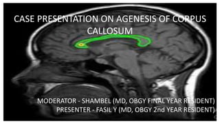
- 1. - CASE PRESENTATION ON AGENESIS OF CORPUS CALLOSUM MODERATOR - SHAMBEL (MD, OBGY FINAL YEAR RESIDENT) PRESENTER - FASIL Y (MD, OBGY 2nd YEAR RESIDENT)
- 3. Outline • Objectives • Case Summary • Discussion • Comments • References
- 4. Objective • To use the case as entry point and to discuss on management of fetal ACC • To continue the rewarding management, learn from possible pitfalls and have common understanding on management of similar cases subsequently • To be familiar with neurosonographic evaluation of ACC for prognostication and counseling of similar cases for the future
- 5. Summary of the case Identification • MRN - 763677 • Name - S.T • Age - 31 • Address - Gondar K. 03 • Marital Status - Married • Occupation -House Wife • Date of admission - 19/12/13E.C
- 6. Anatomic scan- 22/10/13 at 32W3D •Singlton, cephalic •GA by BPD, HC-36 wk; TCD-29W6D; AC &FL-31W6D •There is Lt venticulomegally (19mm) with colpocephaly •CSP & part of CC seen on brain axial view •Difficult to visualize CC on sagital view with both 2D &PD •No midline or posterior fossa defect •CVS,GZ,MSS appear normal •Mild left kidney hydronephrosis-8.9 mm INDEX - 3rd TM Px + ?CC agenesis + mildleft kidney hydronephrosis PLAN - fetal brain MRI & counseling of the mother
- 7. Pre and Post contrast Brain MRI-25/10/13 • There is no well visualized corpus callosum with paralleling of the anterior horns of the lateral ventricles and disproportinate enlargment of the occipital horns and atria Conclusion - Corpus Callosum Agenesis
- 8. History on 19/12/13 ec • 31 years old G IV PII (both alive) and I SA mother • GA-40 Wks (13W and 3D US) • ANC- at Private clinic and GUH BG & RH-B+ NR for RVI & VDRL •1st delivery 11 yrs back- told to have a form of anomaly in the CNS and alive but have difficulty in communication •2nd pregnancy- spontaneous abortion at 3 month of px •3rd delivery 3 years back- uneventful
- 9. Physical Examination • GA- Comfortable • V/S- BP-100/60 mmhg PR-84 RR-22 T-36.8OC • HEENT -pink & non icteric sclera • LGS - no LAP • CHEST - clear & resonant • CVS - S1& S2 well heard • ABD - Term sized gravid uterus • lontudinal lie , cephalic ,no tenderness or contraction • FHB -148
- 10. . • GUS - no CVAT • Cx-closed,posterior,uneffaced,firm in consistency,station -1 • IGS - no pallor • MSS - no edema • CNS - conscious & oriented
- 11. obs US by consultant- 19/12/13 • Singlton, cephalic, FHB - +ve • AGA - 38 W, EFW - 3680 gm, HC - 34 cm • Fundal Placenta , AFI - 2.5 cm • GBM, FT,FBM sen • CNS - colpocephaly with unilateral ventriculomegally (LT=23 mm) On midsagital view- difficult to appreciate the whole segment of CC and difficult to visualize pericallosal vessels on CF • Renal- left moderate hydronephrosis INDEX 3rd TM px + Agenesis of CC + Lt moderate hydronephrosis + Oligohydraminos PLAN- Admit for Induction
- 12. Done • Admitted to maternity ward • Cervix ripened with transcervical foley catheter • Induction started per protocol and followed ?? • Emergency CS was done for the indication of Failed Induction - 3.5 kg M neonate with 8&9 APGAR score •Neonate transfered to NICU for evaluation
- 13. Admission NST
- 15. Discharge Summary 23/12/13 • A 31 yrs old PIII 1 SA mother on her 2nd post op day after ETLUS CS was done for the indication of Failed Induction to effect in the delivery of a 3.5 kg male alive neonate with 8&9 APGAR score after she was admmited with the diagnosis of full term px + oligohydraminos + ACC + unfavorable bishop score • GA- comfortable, Stable vital sign •Post op HCT - 34% • Good Post OP condition • PLAN-advice on contraceptive, sunlight exposure, EBF, danger signs link the neonate to PEDI side for furthur evaluation
- 16. Year 1 Resident Neonatal Evaluation at NICU 21/12/13 • This is a 1hr M alive neonate born to a 31 yrs old PIII mother • Told to have fetal ACC by perinatal US of the fetus • Labor onset is by induction with a total duration of labor 16 hrs and duration of ROM of 8 hrs • Mode of delvery was via CS for failed induction to effect in the delivery of 3.5 kg M with 8&9 APGAR score
- 17. • VS- AHR-146 RR-44 T-36.8 WT - 3500gm L- 54cm HC-37cm → AGA • HEENT-pink conj & NIS deformed and low seated ear •LGS - well formed breast buds •CHEST- good air entry, no SC or IC retraction •GUS - there is ureatheral opening on the ventral part of penis near the scrotum •MSS - Rocker Bottom Feet ASST- Edward Syndrome + Hypospadiasis PLAN- CBC, BG, RBS, RFT, Trans fontanel US, Abd US, ECHO, Consult surical side
- 18. • CBC - • RFT - Normal • RBS - • Abd US ?? • Trans fontanel US & Echo? • Linked to physiotherapy
- 20. Agenesis of the Corpus Callosum
- 21. Corpus Callosum: • Major interhemispheric bundle of commissural fibers in the brain that allows the transfer of motor, sensory, and cognitive information between the two hemispheres • Contain about 200 million axons that connect the left and right cerebral hemisphere • It is one of the five main cerebral commissures • Consist approximately 2–3% of all cortical fibers • Unique to placental mammals
- 22. Traditionally been divided into four anatomically defined regions: The genum, rostrum, body (divided into anterior, middle, and posterior segments), and the splenium
- 23. • It appears by the 10th week of development and connects the nonolfactory areas of the right and the left cerebral cortices • First appears in the genu region at, and follows a cranio‐caudal progression • The anterior part of the rostrum appears last • By 18–22 weeks the final shape of the CC is apparent on imaging although further thickening occurs throughout pregnancy and infancy
- 26. STRUCTURAL ANOMALIES OF THE CORPUS CALLOSUM • Malformations of corpus callosum can occur in isolation; in association with chromosomal, syndromic, or monogenic disorders; or, rarely, secondary to infectious, ischemic, or teratogenic causes • Congenital structural abnormalities of the CC include: ACC - total Vs partial Hypoplasia - refers to a thinner CC that has a normal anterior‐posterior extent Dysgenesis - refers to the CC being present but malformed Hyperplasia - which may result from reduced postnatal axonal pruning
- 28. PREVALENCE • Among live births, the reported prevalence is 1.8: 10,000. • In children with developmental disabilities, it is 2 : 100 to 3 :100, and • In patients with an existing CNS anomaly, it is 47 :100
- 29. ETIOLOGY • Approximately 30%-45% have an identifiable cause • The most frequent causes of corpus callosum agenesis (ACC) are gene mutations • 10%-17% have chromosomal anomalies such as deletions, trisomies (13, 18, 21), and duplications • 20%-35% have recognizable genetic syndromes such as Anderman, L1CAM, and Aicardi • Fetal alcohol syndrome (FAS) is the most important non-genetic congenital cause of ACC, with an incidence of approximately 7% in FAS cases
- 31. Possible causes include: 1. Chromosomal (genetic) abnormalities (e.g, trisomy 8 and 18, Andermann syndrome ) 2. Prenatal infections or viruses (e.g, rubella) 3. Exposure to certain medications ( e.g, valproate, an epilepsy medication ) 4. Toxic metabolic conditions (e.g, Fetal Alcohol Syndrome) 5. Blockage of the growth of the corpus callosum (e.g , cysts) 6. Metabolic disorders 7. Other unknown factors
- 32. Diagnosis of ACC The diagnosis of intracranial anomalies depends on 3 important factors: 1.Accurate determination of the gestational age of the fetus, 2.Knowledge of the developmental embryology of the central nervous system, and 3.Use of a systematic approach for the evaluation of the fetal brain.
- 33. BASIC EVALUATION OF THE FETAL BRAIN • Transabdominal approach • Used in order to screen for CNS malformations in the second and third trimesters of gestation in low-risk patients • Sensitivity of 80% for the detection of CNS malformations •Conventionally, the ultrasound evaluation of brain development during pregnancy is performed in the axial planes of the fetal skull
- 34. (a) Transventricular plane: includes the lateral ventricles (b) Transcerebellar plane:the cerebellum and cisterna magna are measured (c) Transthalamic plane:measure the BPD and HC and includes the midline falx, cavum septum pellucidum (CSP), and thalami
- 35. Transthalamic plane Transventricular plane Transcerebellar plane
- 36. NEUROSONOGRAM: 1)Is often performed using transvaginal ultrasonography (if the fetus is in a cephalic presentation) to improve imaging 2)Adds the coronal and sagittal planes 3)Uses color Doppler to clarify the nature of cystic structures or document brain vessels 4) Adds 3-dimensional ultrasonography (if available) •NB obtained by positioning the transducer in the cranial sutures and fontanelles
- 40. Indirect US features • Absence of the cavum septi pellucidi, • Colpocephaly - dilatation of the atria and occipital horns of the lateral ventricles • Abnormal course of the pericallosal artery, • Widening of the interhemisphere fissure, and • Radial disposition of the sulci on the internal aspects of the hemispheres In our case
- 42. • The definitive diagnosis of complete or partial ACC is made in the directly acquired 2-dimensional median plane or in the median plane of an axially acquired 3-dimensional volume • Fetal MRI is particularly useful in the workup of isolated ACC • Ultrasound imaging has a false positive rate of 20% • Prenatal MRI is preferable after 22 weeks as it provides improved detection of additional abnormal gyral patterns and heterotopia in up to 22.5% of cases
- 43. • Fourteen studies (798 fetuse) were included RESULTS • In cases with isolated cACC, 10.9% and 4.3% of associated anomalies were detected during fetal MRI and postnatal , respectively • in cases with isolated pACC, 13.4% and 16.2% of associated anomalies were detected during fetal MRI and postnatal, respectively Conclusion: The rate of associated anomaly detected exclusively with fetal MRI for isolated neurosonographical ACC is lower than previously reported.
- 44. Associated Anomalies CNS anomalies are seen in up to 85% of cases such as • interhemispheric cysts, arachnoid cysts, Chiari II malformation, migration disorders, and schizencepha Non-CNS anomalies are seen in up to 65% of ACC cases: • craniofacial (macrocephaly, hypertelorism, broad and depressed nasal bridge, and cleft lip/palate), • skeletal (hand malformations, scoliosis), • cardiac (ventricular septal defect [VSD], patent ductus arteriosus [PDA]), • ocular (coloboma, anopthalmia), • genital (cryptorchism), and • renal abnormalities (hydronephrosis)
- 45. • Genetic Evaluation:Chromosomal abnormalities occur in 17% of ACC cases,and even isolated complete or partial ACC carries a 4.8% and 7.5% risk for aneuploidy, respectively • most commonly trisomies 18 and 13 and mosaic trisomy 8 • Serologic testing for cytomegalovirus or Zika virus is rarely useful unless suggested by other findings • amniocentesis for CMA should be offered when ACC is detected
- 48. Signs and symptoms Vary greatly among individuals • Some characteristics common in individuals with callosal disorders include Poor motor coordination, Delays in motor milestones such as sitting and walking, Delayed toilet training, Chewing and swallowing difficulties Vision impairments, Hypotonia Low perception of pain, sometimes seizures, spasticity, early feeding difficulties
- 49. Management
- 50. OBSTETRIC MANAGEMENT •Delivery in a tertiary care facility with access to NICU and pediatric subspecialties is suggested •Vaginal delivery is not contraindicated •Cesarean delivery for obstetric indications
- 51. NEONATAL MANAGEMENT • Consider ultrasound and/or MRI to better define the anomaly and to look for associated malformations • Consultation with genetics to exclude genetic syndrome • Neurology and neurosurgery consultations depending on associated anomalies
- 52. Prognosis • Complete and partial ACCs associated with coexisting brain abnormalities or genetic syndromes tend to lead to poor neurodevelopmental outcomes • In contrast, isolated complete and partial ACCs have a better prognosis and appear to have similar outcomes • Normal development, in about 75% of individuals • Borderline-moderate and severe neurodevelopmental delays are observed in 14.9% to 16.0% and 8.2% to 12.5% of isolated cases, respectively • Epilepsy in 35%-severity depends on the presence of other CNS anomalies • Recurrence - Isolated: 3-5%.
- 53. • Controlled clinical trials published between May 23, 2009, and May 23, 2019, using the term “agenesis of corpus callosum” were reviewed • A total of 942 articles were identified, and 8 studies were included in the systematic review depending on the inclusion criteria • These studies included 217 fetuses with isolated CCA and no other anomalies at prenatal assessment • In this review, neurodevelopment was favorable in two-thirds of the cases (IQ>85) • They could not determine any difference, between cCCA and pCCA, possibly due to the small number of patients with pCCA
- 54. Comments • Fetal surveilance were good with better FM foolowup; • Timing of Neurosonography? • Evaluation by NICU side, better to involve consultants • Post partum investigations are incomplete • Poor documentation by NICU side • Proper counseling postpartally were not provided • No surgical side evaluation for the hypospadiasis
- 55. Reference • Creasy and Resnik’s Maternal-Fetal Medicine,7th ed,n2014 • Callen’s Ultrasound in Obstetrics and Gynecology, 6th ed,n2017 • Ultrasound in Obstetrics & Gynecology, a practical approach.1sted,2014 • ISUOG practice guidelines: performance of first trimester fetal ultrasound scan. Ultrasound Obstet Gynecol 2013; 41:102-113 • American Institute of Ultrasound in Medicine practice guidelines on the performance the obstetric ultrasound examination, 2013. • Salomon LJ, Alfirevic Z, Brghalla C, Bilardo C, Hernandez – Andrade E, Jhonsen SL Kalache K. Practice Guidelinefor performance of the routine mid-trimester fetal ultrasound scan . Ultrasound Obstet Gynecol 2011;37;116-126 • Sunita Dashottar1, Krishna Pratap Singh Senger2*, Yashashvi Shukla1, Ankita Singh3, Surabhi Sharma. Transcerebellar diameter: an effective tool in predicting gestational age in normal and IUGR pregnancy. Int J Reprod Contracept Obstet Gynecol. 2018 Oct;7(10):4190-4196
- 56. THANK YOU
Editor's Notes
- commissures: (bundles of nerve fibres that cross the midline of the human brain at the level of their origin) anterior, posterior, hippocampal, and habenular commissures
- Antenatal imaging earlier than or at 18–20 weeks (the usual timing of standard US screening) does not reliably detect total agenesis, partial agenesis, or hypoplasia of the CC
- corpus-body callosum-tough or hard......latin word
- s. In complete agenesis of the corpus callosum (ACC), there is a total failure of the commissure to develop. In partial agenesis (hypo_x0002_plasia), the anterior parts (beak, genu, and anterior part of the body) are developed, but the posterior aspects (posterior part of the body, isthmus, and splenium) are absent hypoplasia is sometimes used to describe a corpus callosum that is full in its rostrocaudal extent but narrow or thin in cross-section, presumably due to decreased numbers of crossing axons.
- ACC results from an insult during embryogenesis. Conse_x0002_quently, there is failure of the callosal axons to pass across the midline Partial ACC results from an insult affecting a previously normal corpus callosum.
- transthalamic plane, the biometry of the skull (biparietal diameter and cranial circumference) transventricular plane, the posterior horn of the lateral ventricle (atrium) is measured transcerebellar plane, the posterior fossa structures (cerebellum and cisterna magna) are assessed
- In high-risk pregnancies, a detailed obstetrical ultrasound examination that includes a more extensive evaluation of the intracranial anatomy may be performed, usually by an expert in fetal imaging, such as a maternal-fetal medicine subspecialist. In low-risk pregnancies, the fetal brain is evaluated as part of the anatomy scan during the standard second_x0002_trimester ultrasound examination, which is typically per_x0002_formed between 18 and 22 weeks of gestation.
- 3 Coronal views of the fetal head. (a) Transfrontal plane; (b) transcaudate plane; (c) transthalamic plane; (d) transcerebellar plane
- (a) Midsagittal plane; (b) parasagittal plane
- Dilation of the occipital horns is caused by reduced white matter formation in the absence of callosal axons, whereas dilation of the frontal horns is limited by the firm caudate nuclei at their lateral borders The absence of the CC results in abnormal induction of the medial cerebral convolutions, determining a radiate arrangement ofcerebral sulci around the roof of the third ventricle
- • Partial ACC is more commonly associated with posterior fossa abnormalities. • Complete ACC is more commonly
- NB - 15.1% of cases thought to be isolated prenatally were found to have associated abnormalities after birth