Call Girls Mussoorie Just Call 8854095900 Top Class Call Girl Service Available
ultrasound in 1st TM.pdf
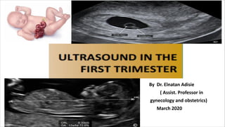
1. By Dr. Elnatan Adisie
( Assist. Professor in
gynecology and obstetrics)
March 2020
2. Outline
• Introduction
• Sonographic Land marks in 1st TM
• Major Fetal Anomaly that can be diagnosed in early Gestation
• Chorionicity
• Pregnancy Dating
• Nuchal Translucency
• Elements Of Pregnancy Failure
• Summary
3. Introduction
• Accurate performance of an ultrasound examination in the 1st TM is
important given its ability to confirm an intrauterine gestation, assess
viability and number of embryo(s) and accurately date a pregnancy, all
of which are critical for the course of pregnancy.
7. Gestational Sac
• Also called chorionic sac
• First seen at 4-4.5 wks from
LNMP by TVS
• Circular to Ellipsoid shape
• Intradecidual sac sign
• Double decidual sac sign
• Echogenic ring
8.
9. Pseudo Gestational Sac
• occurs in 10- 20% of ectopic
pregnancy
• fluid or blood accumulation in
the endometrial cavity
• acute angle/tear drop shape
• absence of double echogenic
ring
11. Yolk Sac
• seen at 5.5wks
• Confirm IUP
• diamond engegement ring
• 3mm - 6mm in size
• provide nutrients for embryo
12.
13.
14. Amnion
• thin echogenic structure
surrounding the embryo
• occur following the yolk sac and
just before the embryo
• growth is closely related to the
embryo
15. Embryo
• seen at 5th wks
• focal thickening on top of the
yolk sac (diamond ring)
• cardiac activity seen around 6-
6.5 wks
• fialed pregnancy diagnosed if no
cardiac activity at CRL >=7mm.
16. • The Grain Of Rice appearance
• thin cylinder with no discernible
body parts
17. the Gummy Bear appearance
- body curvature and clear delination of head,
chest, abdomen and extremity.
19. Major Congenital Malformations in early Gesation
• can be diagnosed b/n 11-13 wks of GA
• close observation of anatomic details with transvaginal ultrasound
• includes abnormalities of HEAD , CHEST, ABDOMEN, and EXTREMITY.
20. HEAD
Anencephaly
• loss of brain tissue and the
skull bone
• Acrania-Exencephaly-
Anenecphaly sequence
• frog sign
36. Multiple Pregnancy and Chorionicity
• Dizygotic twins have Dichorionic and Diamniotic placenta
• Monozygotic twins will have:
- 75% are MC & DA
- 25% are DC & DA
- < 1% are MC & MA
37. Chorionicity can be diagnosed after 08 wks of
gestation when yolk sac is present
DC & DA
• thick dividing membrane
• delta, lambda, or twin
pick sign
38. MC & DA
• thin dividing membrane
• T- configuration
• two yolk sac present
39.
40. MC & MA
• no separating membrane
• one yolk sac is present
• two embryo is seen
42. Pregnancy dating in the first TM
1, Gestational Sac Diameter(GSD)
2, Crown-Rump Length(CRL)
3, Bi-Parietal Diameter(BPD)
43. Mean Sac Diamater
• Use it when GA is < 6 wks or
when the embryo is absent
• measure the mean of
greatest sagital, transverse
and coronal view
• GA = MSD in mm + 30
44.
45. CRL
• actual measurment
corresponds to the longest
straight line distance
• use three discrete
measurment and take their
mean
• GA = CRL in mm + 42
• Integrated software
46. Nuchal Translucency(NT)
• Measurment of collection of fluid under the skin behind the fetal neck
• measured b/n 11-13 wks or a CRL of 45-84mm
• provide risk assesment for chromosomal abnormalities
• for efficiency in screening, it should be combined with:
- maternal age
- maternal blood biochemical markers( HCG & PAPP-A)
47.
48.
49.
50. Elements of Pregnancy Failure
• 10-15% of pregnancy will fail during the first TM
• The diagnosis can be made by u/s before the mother is symptomatic
• Different scenarios are possible:
- Gestational sac absent with +ve HCG
- GS with no yolk sac or embryo
- GS and Embryo visible but no cardiac activity
- GS + Embryo + cardiac activity present but various measurment
are out of range for yolk sac, embryo, or amniotic sac
- presence of subchorionic bleeding
- Abnormal anatomical appearance of the embryo
51.
52.
53. Summary
• Ultrasound in the 1st TM is important step in the evaluation of
pregnancy b/c it allows for pregnancy confirmation and accurate
dating.
• Significant change occur during the 1st TM which can be noticed by
Trans vaginal ultrasound
• Steps of development form early gestation to late should be known to
better understand and differentiate the normal from abnormal
growth.