2. Understandings
Statement Guidance
6.1 U.1
The contraction of circular and
longitudinal muscle of the small
intestine mixes the food with enzymes
and moves it along the gut.
6.1 U.2
The pancreas secretes enzymes into
the lumen of the small intestine.
Students should know that amylase, lipase
and an endopeptidase are secreted by the
pancreas. The name trypsin and the method
used to activate it are not required.
6.1 U.3
Enzymes digest most macromolecules
in food into monomers in the small
intestine.
Students should know that starch, glycogen,
lipids and nucleic acids are digested into
monomers and that cellulose remains
undigested.
6.1 U.4
Villi increase the surface area of
epithelium over which absorption is
carried out.
6.1 U.5
Villi absorb monomers formed by
digestion as well as mineral ions and
vitamins.
6.1 U.6
Different methods of membrane
transport are required to absorb
different nutrients.
3. Applications and Skills
Statement Guidance
6.1 A.1
Processes occurring in the small
intestine that result in the digestion of
starch and transport of the products of
digestion to the liver.
6.1 A.2
Use of dialysis tubing to model
absorption of digested food in the
intestine.
6.1 S.1
Production of an annotated diagram of
the digestive system.
6.1 S.2
Identification of tissue layers in
transverse sections of the small
intestine viewed with a microscope or
in a micrograph.
Tissue layers should include longitudinal
and circular muscles, mucosa and
epithelium.
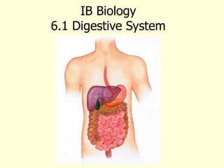
4. 6.1 S.1 Production of an annotated diagram of the
digestive system.
Use the animation and video to learn about the digestive system and how to draw it.
http://highered.mheducation.com/sites/0072495855/stu
dent_view0/chapter26/animation__organs_of_digestion.
html
https://youtu.be/Nm-pT7fk6gs
5. 6.1 S.1 Production of an annotated diagram of the digestive system.
Plus add in the
accessory organs:
the gall bladder,
liver and pancreas.
7. 6.1 S.1 Production of an annotated diagram of the digestive system.
Now add the annotations to show what happens in digestion.
8. Digesting large molecule
• Most food molecules are large polymers and insoluble. These molecules are
broken down so they can be absorbed and assimilated by the body.
• They must first be digested to smaller soluble molecules before they can be
absorbed into the blood this occurs in 3 ways, mechanical (chewing),
chemical (enzymes) and acids (stomach glands).
6.1 U.3 Enzymes digest most macromolecules in food into
monomers in the small intestine.
9. Enzymes and digestion
•Enzymes are biological catalysts that increase the rate of reaction
•Digestive enzymes are secreted into the lumen of the gut
•Digestive enzyme increase the rate of reaction of the hydrolysis of
insoluble food molecules to soluble end products
•Digestive enzymes increase the rate of reaction at body temperature
6.1 U.3 Enzymes digest most macromolecules in food into
monomers in the small intestine.
10. 6.1 U.3 Enzymes digest most macromolecules in food
into monomers in the small intestine.
11. 6.1 U.3 Enzymes digest most macromolecules in food into monomers in the small intestine.
Human Digestive Enzymes
Amylases break down carbohydrates
Example: salivary amylase
Substrate: starch Product: maltose
Source: mouth (salivary glands)
Optimum pH: 7-7.8
diagram from: http://www.teachervision.fen.com/digestive-system/printable/57730.html
12. 6.1 U.3 Enzymes digest most macromolecules in food into monomers in the small intestine.
Human Digestive Enzymes
Proteases break down polypeptides
Example: pepsin
Substrate: polypeptides
Product: amino acids
Source: stomach
Optimum pH: 2
diagram from: http://www.teachervision.fen.com/digestive-system/printable/57730.html
13. 6.1 U.3 Enzymes digest most macromolecules in food into monomers in the small intestine.
Human Digestive Enzymes
The Pancreases produces lipases break down
fats and lipids
Example: pancreatic lipase
Substrate: triglycerides Product: fatty acids
& glycerol
Source: pancreas, delivered into small intestine
Optimum pH: 7.2 - 7.5
15. • Digestion begins in the oral cavity (mouth).
• Saliva begins the chemical digestion of food.
Saliva contains the protein mucin to lubricate
the food for swallowing.
• Amylase breaks down starch and glycogen.
• Food is shaped into a ball (bolus) and is then
swallowed.
*
16. • Peristalsis: is the shortening of muscle by contraction. These
muscles provide the force to move food the esophagus to the
stomach while mixing food with enzymens.
*
6.1 U.1 The contraction of circular and longitudinal muscle of the small intestine mixes the food
with enzymes and moves it along the gut
17. • Pharynx (throat): is where the esophagus and the windpipe
meet.When food is swallowed, the epiglottis closes the passage
to the windpipe.
• Esophagus: Conducts food from the pharynx to the
stomach.The mouth, pharynx and esophagus are responsible for
carbohydrate digestion.
• Stomach: produces gastric juice which has a pH of 2 aiding in
digestion. In addition it contains pepsin that breaks down
proteins.
*After chemical digestion in the
stomach, the food has been
turned into a nutrient broth called
chyme. It takes 2to 6 hours for
the stomach to empty.
18. Structure: Small Intestine digestion is completed in
the SI. The products of digestion are absorbed into the blood
stream.
*
6.1 U.4 Villi increase the surface area of epithelium over
which absorption is carried out.
19. Structure: Small Intestine
(A)Blood Capillaries move digested food molecules into the
blood from the lumen of the ileum.
B) Lacteals are connect to the lymphatic system for the
transport of lipids.
(C) Mitochondria Providing ATP for active transport of
the products of digestion
(D) Microvilli border of the epithelial cell increases the
surface are for absorption.
20. 6.1 S.2 Identification of tissue layers in transverse sections of the small
intestine viewed with a microscope or in a micrograph
The small intestine contains four
distinct tissue layers from the lumen
Muscular layer
1. Mucosa – inner lining, includes villi
2. Submucosa – connective tissue
(between the mucosa and muscle)
3. Muscular layer –
inner circular and outer
longitudinal muscle
perform peristalsis
4. Serosa – protective
outer layer
Serosa layer
Mucosa layer
Submucosa layer
21. • Pancreas: produces the enzymes amylase to digest
carbohydrates, lipases to digest lipids, and proteases
to digest polypeptides. In addition it produces an alkaline
solution made of bicarbonate. The bicarbonate acts as a buffer
to help neutralize the acidic chyme.
6.1 U.2 The pancreas secretes enzymes into the lumen of the
small intestine.
22. Liver: produces bile
which contains no digestive
enzymes, but helps in the
absorption of fats.
6.1 A.1 Processes occurring in the small intestine that result in the
digestion of starch and transport of the products of digestion to the
liver.
Emulsification
23. Liver: In addition, working
with the pancreas the liver
removes excess monosaccharides
from the blood after they have
been broken down from
disaccharides and polysaccharides
in the small intestines. Excess
sugars are converted back into a
polysaccharide (Glycogen)
6.1 A.1 Processes occurring in the small intestine that result in the
digestion of starch and transport of the products of digestion to the
liver.
Two Steps
1. Glycogenesis Excess glucose in
the blood is convert into
glycogen.
2. Glycogenolysis conversion of
glycogen into glucose and then
released into the blood
24. 6.1 A.1 Processes occurring in the small intestine that result in the digestion of
starch and transport of the products of digestion to the liver.
The digested glucose is absorbed and then transported to various body tissues
1. Glucose is co-transported*
with sodium ions into the
epithelial cells (of the villus).
2. Glucose moves by facilitated
diffusion into the lumen of the
villus.
3. Glucose then diffuses a short
distance into the adjacent
capillaries where it dissolves
into the blood plasma.
4. Blood in the capillaries moves
to to venules then to the
hepatic portal vein which
transports the glucose to the
liver.
5. The liver absorbs excess
glucose which it converts to
glycogen for storage.
25. 6.1 U.6 Different methods of membrane transport are required to
absorb different nutrients.
Method of
transport
Nutrients Outline
Simple
diffusion
Lipids Lipids are non-polar and therefore can pass
freely through hydrophobic core of the plasma
membrane into the epithelial cells (down the
concentration gradient )
Facilitated
Diffusion
Fructose,
vitamins
Water-soluble (hydrophilic) molecules use
channel proteins to pass phospholipid bilayer
and enter the epithelial cells (down the
concentration gradient)
Active
Transport
Glucose, amino
acids and mineral
ions
Protein pumps use ATP to move molecules
against the concentration gradient into the
epithelial cells
Endocytosis
(Pinocytosis)
Antibodies from
breast milk
The plasma membrane folds inward to form
vesicles to absorb larger molecules without
digesting them
How is membrane transport involved in absorption of
nutrients from the small intestine?
26.
27. Large Intestine (LI):
• Is also called the colon.
• The primary job of the LI is to reabsorb
water.
• Together, the SI and the LI reabsorb ~
90% of the water that was used for
digestion.
• Wastes become more solid as they
move through the LI through peristalsis
and results in feces.
*
28. Absorption and assimilation: insoluble food molecules are
digested to soluble products in the lumen of the gut.
Absorption:
1.The soluble products are first taken up by various
mechanisms into the epithelial cells that line the gut.
2.These epithelial cells then load the various absorbed molecules
into the blood stream.
Assimilation:
1.The soluble products of digestion are then transported to the
various tissues by the circulatory system.
2.The cells of the tissues then absorb the molecules for use
within this tissues
6.1 U.5 Villi absorb monomers formed by digestion as well as
mineral ions and vitamins.
30. 6.1 A.2 Use of dialysis tubing to model absorption of digested food in the intestine.
Dialysis tubing can be used to model absorption
The tubing is semi-permeable and
contains pores typically ranging 1 – 10 nm
in diameter. Predict what will happen to
the glucose and starch after 15 minutes
.