
Recommended
More Related Content
What's hot
What's hot (20)
Similar to Facial Nerve Anatomy
Similar to Facial Nerve Anatomy (20)
Recently uploaded
Recently uploaded (20)
Facial Nerve Anatomy
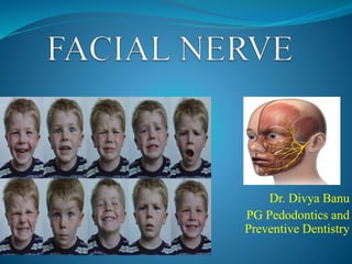
- 1. Dr. Divya Banu PG Pedodontics and Preventive Dentistry
- 2. CONTENTS Introduction Functional Components Nuclei Ganglia Course And Relations Branches And Distribution Clinical Co-relation References
- 3. INTRODUCTION 7th cranial nerve Mixed nerve, predominantly motor. Most frequently paralyzed peripheral nerve. Supplies muscles of facial expression.
- 4. Embryology/Development Developmentally derived from hyoid arch (second branchial arch). Motor division: Derived from basal plate of embryonic pons. Sensory division: Originates from cranial neural crest.
- 5. The nerve course, branching pattern, and anatomical relationships: First trimester Nerve is not fully developed until about 4 years of age. Third week of gestation: Facioacoustic primordium or crest : First identifiable facial nerve tissue
- 6. Motor Nucleus (Branchiomotor) Superior Salivatory Nucleus (Parasympathetic) Lacrimatory Nucleus (Parasympathetic) Nucleus of Tractus Solitarius (Gustatory) NUCLEI (In lower pons)
- 7. FUNCTIONAL COMPONENTS EFFERENT Special visceral or branchial efferent General visceral efferent or parasympathetic AFFERENT Special visceral afferent General somatic afferent
- 8. • Taste sensations from palate and anterior two-thirds of tongue except vallate papillae. • Terminates in nucleus of tractus solitarius. SVA • General sensations from skin of auricle. • Terminates in spinal nucleus of trigeminal nerve. GSA
- 9. • Arise from motor nucleus. • Supplies muscles of facial expression. • Elevation of hyoid bone. SVE • Arise from lacrimatory and superior salivatory nuclei • Pre-ganglionic parasympathetic fibers. • Supply secretomotor fibers to lacrimal, submandibular and sublingual glands. GVE
- 12. Geniculate Ganglion Located on first bend of facial nerve. Sensory ganglion. The taste fibres present in nerve are peripheral processes of pseudounipolar neurons present in geniculate ganglion.
- 13. Submandibular ganglion Parasympathetic ganglion for relay of secretomotor fibres to submandibular and sublingual glands. Preganglionic fibres come from chorda tympani nerve.
- 14. Pterygopalatine ganglion Parasympathetic ganglion. Secretomotor fibres for lacrimal gland relay in this ganglion. The fibres reach ganglion from nerve to pterygoid canal.
- 15. COURSE AND RELATIONS Attachment of facial nerve to brain stem Motor Root Sensory Root (Nervus Intermedius) Lateral part of lower border of pons just medial to 8th cranial nerve
- 16. Motor and sensory root Internal Acoustic Meatus Motor Root lies in a groove on 8th cranial nerve Sensory Root intervening Single trunk in FACIAL CANAL of petrous part of temporal bone Laterally and Forwards Fundus of meatus
- 18. Facial Canal Two Bends Laterally above the vestibule Backwards irt medial wall of middle ear above promontary Vertically downwards behind promontary Facial Nerve leaves skull through STYLOMASTOID FORAMEN
- 19. EXTRACRANIAL COURSE Facial nerve crosses lateral side of base of styloid process Posteromedial surface of parotid gland Crosses retromandibular vein and ECA Terminal branches along anterior border of parotid gland Behind neck of mandible
- 21. Within Facial Canal: 1. Greater petrosal nerve. 2. Nerve to stapedius. 3. Chorda tympani. At its exit from Stylomastoid Foramen: 1. Posterior auricular. 2. Digastric. 3. Stylohyoid. Terminal Branches within Parotid Gland: 1. Temporal. 2. Zygomatic. 3. Buccal. 4. Marginal mandibular. 5. Cervical. Communicating Branches with adjacent cranial and spinal nerves Branches And Distribution
- 23. Greater Petrosal Nerve Greater petrosal nerve and deep petrosal nerve Nerve of pterygoid canal Postganglionic for lacrimal gland join zygomatic nerve Pass through communicating branch Lacrimal nerve Lacrimal gland RELAY
- 24. Nerve to Stapedius Arises opposite the pyramid of middle ear. Supplies stapedius muscle The muscle dampens excessive vibrations of stapes caused by high-pitched sounds.
- 25. Chorda Tympani Arises in vertical part of facial canal about 6 mm above stylomastoid foramen. Runs upwards and forwards Middle ear Runs forwards in close relation to tympanic membrane Passes through petrotympanic fissure Medial to spine of sphenoid Infratemporal fossa Joins lingual nerve through which it is distributed
- 27. Chorda tympani carries: Preganglionic Secretomotor Fibres • To submandibular ganglion. • Supplies submandibular and sublingual salivary glands. Taste Fibres • From anterior two- thirds of tongue except circumvallate papillae.
- 28. Posterior Auricular Nerve Arises just below stylomastoid foramen Ascends between mastoid process and external acoustic meatus Auricularis Occipitalis Intrinsic posterior muscles on the back of auricle
- 29. • Arises close to posterior auricular nerve. • Short. • Supplies posterior belly of digastric. Digastric Branch • Arises with the digastric branch. • Long. • Supplies stylohyoid muscle. Stylohyoid Branch
- 31. Terminal Branches of Parotid Gland
- 33. Temporal Branches Crosses zygomatic arch Supplies: 1. Auricularis anterior 2. Auricularis superior 3. Intrinsic muscles on the lateral side of the ear 4. Frontalis 5. Orbicularis oculi 6. Corrugator supercilii
- 34. Zygomatic Branches • Run across zygomatic bone. • Supplies orbicularis oculi. Buccal Branches • Two in number. • Upper: Runs above the parotid duct. • Lower: Below the duct. • Supply muscles in that vicinity, especially the buccinator.
- 36. Marginal Mandibular Branch • Runs below angle of mandible deep to platysma. • Crosses body of mandible and supplies muscle of lower lip and chin. Cervical Branch • Emerges from apex of parotid gland. • Runs downwards and forwards in the neck to supply the platysma.
- 37. Communicating Branches Motor nerves of first, second and third branchial arches communicate with each other for effective coordination between movements of muscles of these arches. The facial nerve also communicates with the sensory nerves distributed over its motor territory.
- 38. Nervus Intermedius Formed of central processes of unipolar cells of geniculate ganglion. Also contains efferent preganglionic parasympathetic fibers from parasympathetic nuclei.
- 39. CLINICAL CORRELATION Lesions of facial nerve Supranuclear Infranuclear
- 41. Conclusion Functional and esthetic outcomes in children. Bell’s palsy (idiopathic) is most common in children too. Careful diagnostic workout and differential diagnosis is required. Hopefully, regenerative medicine could offer new options for the treatment of this condition.
- 43. References Grey’s Anatomy Carpenter’s Neurophysiology Burkit’s Oral Medicine B.D. Chaurasia’s Human Anatomy Vishram Singh Anatomy of Head, Neck and Brain. Transient Delayed Facial Nerve Palsy After Inferior Alveolar Nerve Block Anesthesia Fotios H. Tzermpos, DMD, MD, PhD,* Alina Cocos, Matthaios Kleftogiannis. Facial nerve paralysis in children Andrea Ciorba, Virginia Corazzi, Veronica Conz, Chiara Bianchini, Claudia Aimoni Hadlock TA, Malo JS, Cheney ML, Henstrom DK. Free gracilis Transfer for smile in children: the Massachusetts Eye and Ear Infirmary Experience in excursion and quality-of-life changes. Arch Facial Plast Surg 2011; Malik S, Bhandekar HS, Korday CS. Traumatic peripheral neuropraxias in neonates: a case series. J Clin Diagn Res 2014;
- 44. Shahidullah M, Haque A, Islam MR, Rizvi AN, Sultana N, Mia BA, Hussain MA. Comparative study between combination of famciclovir and prednisolone with prednisolone alone in acute Bell’s palsy. Mymensingh Med J 2011 Goudakos JK, Markou KD. Corticosteroids vs corticosteroids plus antiviral agents in the treatment of Bell palsy: a systematic review and meta-analysis. Arch Otolaryngol Head Neck Surg 2009; 135: 558-564 Numthavaj P, Thakkinstian A, Dejthevaporn C, Attia J. Corticosteroid and antiviral therapy for Bell’s palsy: a network meta-analysis. BMC Neurol 112011 Pitaro J, Waissbluth S, Daniel SJ. Do children with Bell’s palsy benefit from steroid treatment? A systematic review. Int J Pediatr Otorhinolaryngol 2012 Chen WX, Wong V. Prognosis of Bell’s palsy in children--analysis of 29 cases.
- 45. Thank You