Intracranial inflammatory cystic lesion Dr Ahmed Esawy CT MRI part 3

Intracranial inflammatory cystic lesion dr ahmed esawy ct mri part 3 • 1-arachnoid cysts • 2-porencephalic cyst, • 4-multicystic encephalomalacia • 3-hydranencephaly • 5-holoprosencephaly • 6-hydrocephalus (aqueduct stenosis) • 7-periventricular leukomalacia (PVL), • 8-septum pellucidum changes CSP CV CI • 9-dandy walker malformation • 10-dandy walker varaint • 11-mega cisterna magna • 12-schizencephaly • 13-conatal cysts • 14-subependymal cysts • 15-encephalomalacia, • 16-subarachnoid cyst • 17-cystic lesions after brain surgery and radiation injury to the brain. • 18-Leptomeningeal cyst • 19-Post traumatic porenencephally • 20-brain abscess • 21-cysticercosis • 22-hydatid cyst. • 23-amoebic abscess • 24-Aneurysm • 25-Parenchymal Perianeurysmal Cystic Changes in the Brain • 26-Vein of Galen malformation • 27-epidermoid cysts • 28-dermoid cyst (cystic teratoma) • 29-craniopharyngioma • 30-cystic astrocytoma • 31-cystic meningioma • 32-cystic shwannoma • 33-hemangioblastoma • 34-cystic metastasis • 35-cystic pituitary adenoma • 36-Cystic degeneration / necrotic neoplasm • 37-colloid cysts • 38-Rathke’s cleft cysts, • 39-neuroepithelial cysts • 40-neuroenteric cysts • 41-pineal cysts. • 42-Choriod plexus cyst • 43-CSF-Iike Choroidal Fissure and Parenchymal Cysts of the Brain • 44-Trigonal cyst • 45-Interhemispheric cyst • 46-Dorsal cyst • 47-Ependymal cysts • 48-Enlarged VRS • 49-Cystic trapped 4th ventricle • 50-Diverticulation of 3rd , lateral ventricle

Recommended
More Related Content
What's hot
What's hot (20)
Similar to Intracranial inflammatory cystic lesion Dr Ahmed Esawy CT MRI part 3
Similar to Intracranial inflammatory cystic lesion Dr Ahmed Esawy CT MRI part 3 (20)
More from AHMED ESAWY
More from AHMED ESAWY (20)
Recently uploaded
Recently uploaded (20)
Intracranial inflammatory cystic lesion Dr Ahmed Esawy CT MRI part 3
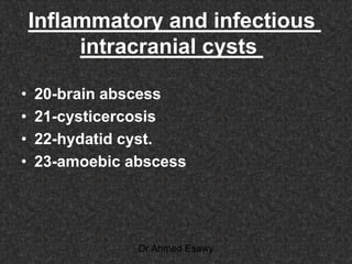
- 1. Inflammatory and infectious intracranial cysts • 20-brain abscess • 21-cysticercosis • 22-hydatid cyst. • 23-amoebic abscess Dr Ahmed Esawy
- 2. Brain Abscess Dr Ahmed Esawy
- 3. Brain abscess.. poorly defined area of posterior parietal brain edema (arrows). Early cerebritis may not outline a focal mass clearly Dr Ahmed Esawy
- 4. Brain abscess. a poorly defined pattern of mass effect and low attenuation in the left temporal lobe. Of early cerebritis Dr Ahmed Esawy
- 5. Brain abscess. An area of ring like enhancement (yellow arrow) is noted within a much larger pattern of edema (white arrow). The central core of the abscess (black arrow) does not enhance (central necrosis) Dr Ahmed Esawy
- 6. temporal lobe abscess, extracranial, subdural, and intracerebral abscesses Dr Ahmed Esawy
- 7. Brain abscess. depressed skull fracture. The left parietal cranial injury an abscess of the subgaleal space (SGA) the epidural space (EDA) the left cerebral hemisphere (CA). Dr Ahmed Esawy
- 8. Brain abscess. Axial T1 +C ,T2-weighted MRI in a patient with a right frontal abscess. Dr Ahmed Esawy
- 9. The right frontal lobe of the brain is shifted across the midline (double arrow) by an intracranial abscess (single black arrow) that has extended upward from the medial right orbit and medial ethmoid air cells (curved dotted arrow). T1-contras Brain abscess T1-contras the enhancement within the right ethmoid sinuses from which the infection arose. The medial superior right maxillary sinus has been destroyed (yellow arrow). T1-contras An abscess is noted within the medial inferior right orbit. The right maxillary sinus (double white arrows) contains infected secretions and mucus Dr Ahmed Esawy
- 10. Brain abscess. (FLAIR) MRI in a patient with abscess of the cerebellar vermis (black arrow). T2- MRI abscess of the midline cerebellum. the large area of increased signal, both within the abscess and within the surrounding cerebellum (black arrow). Dr Ahmed Esawy
- 11. Brain abscess. T1-enhanced central zone of enhancement within the abscess, with a zone of decreased brightness (edema, white arrow). Brain abscess. T1enhanced enhanced mass within the right medial cerebellum (yellow arrow). The thick- walled cystic mass was opened. Dr Ahmed Esawy
- 12. CEREBRAL ABSCESS ON DW MRI On trace DWI abscesses are typically hyperintense, indicating decreased diffusion of water. – This is secondary to increased viscosity of pus which contains, in addition to cellular debris and bacteria, large molecules such as fibrinogen, which bind water molecules and add to the effect of restricted diffusion. – This can be confirmed with an apparent diffusion coefficient (ADC) map where abscesses are of low signal ,markedly reduced ADCDr Ahmed Esawy
- 13. Diffusion-weighted Imaging ADC maps are of great value in distinguishing neoplasms in ADC maps is more often have facilitated diffusion, Dr Ahmed Esawy
- 14. CEREBRAL THALAMIC ABSCESS ON MRI Post-Gd T1WI: WI2T DWI Dr Ahmed Esawy
- 15. Left and right frontal abscesses: 35-year-old male. DWI ADCWI2TWI1T Dr Ahmed Esawy
- 16. Pyogenic Abscess T2 T1 T1/Gd DWI bright on DWI Dr Ahmed Esawy
- 17. Abscess (purulent) ADC decreased dark on ADC mapDr Ahmed Esawy
- 18. 7. 8. DD : tumour central hypointensity on diffusion-weighted image and hyperintensity on ADC map, consistent with the diagnosis of tumor. Dr Ahmed Esawy
- 19. 7. 8. DD : tumour Central hypointensity is seen on the diffusion-weighted image and hyperintensity on the ADC map, consistent with the diagnosis of tumor. Dr Ahmed Esawy
- 20. Brain abscess primary and secondary (daughter Fluid and necrotic tissue (bright area) . edema surrounds the abscess cavities (black arrows). surrounding the abscess does not enhance (white arrows). DWI T1/Gd Dr Ahmed Esawy
- 21. Brain abscess (FLAIR) left occipital-parietal brain abscess. Dr Ahmed Esawy
- 23. MR Spectroscopy • .Typical MR spectroscopic features of brain abscesses include • elevated peaks of amino acid, lactate, alanine, acetate, pyruvate, and succinate • absent signals of NAA, creatine, and choline. Dr Ahmed Esawy
- 24. MR spectroscopy • shed light on which organism is responsible for the abscess • because the presence of anaerobic bacteria tends to cause elevated acetate and succinate peaks. Dr Ahmed Esawy
- 25. DD : NEOPLASM • Elevation of choline and absence of signal from a variety of amino acids, acetate and succinate favours neoplastic process Dr Ahmed Esawy
- 26. Dr Ahmed Esawy
- 27. Dr Ahmed Esawy
- 28. necrotic or cystic neoplasmsPyogenic brain abscesses Elevated choline , decrease NAA elevated peaks of amino acid, lactate, alanine, acetate, pyruvate, and succinate absent signals of NAA, creatine, and choline MRS facilitate diffusion dark restricted diffusion bright DW Bright on ADC map The walls of necrotic or cystic tumors have a lower ADC value than of an abscess markedly reduced ADC maps.ADC wall of necrotic or cystic neoplasms tends to have higher rTBV capsule of an abscess tends to have lower rTBV MR PERFUSION Dr Ahmed Esawy
- 29. Signal volume MR spectra of abscess Short-echo MRS shows depression of the NAA, choline (Cho) and creatine (Cr) as well as elevation of the amino acid, lactate (Lac), acetate and succinate.Dr Ahmed Esawy
- 30. T2 T1+C Single voxel MRS peaks representing alanine, lactate and amino acids DW hyperintense signal in centre ADC decrease signal in centre Brain abscess Dr Ahmed Esawy
- 31. brain abscess Dr Ahmed Esawy
- 32. Brain abscess in a 28-week gestation preterm newborn well-defined cystic structure with low- level echoes (arrowheads) in the left posterior parietal region abscess has ring enhancement (arrowheads).Dr Ahmed Esawy
- 34. Cystercercus cellulosae - (3-20 mm) regular round thin walled cyst, produces only mild inflammation larva in cyst Dr Ahmed Esawy
- 35. Calcification in cysticercosis • Calcification in burned out residues of cysticercosis scattered throughout the brain in later stagesDr Ahmed Esawy
- 36. NEUROCYSTICERCOSIS Multiple neurocysticercosis cysts of various sizes. Some contain visible scolices (arrows). MR image shows T1 innumerable tiny low-signal-intensity neurocyticercosis cysts in brain parenchyma and subarachnoid spaces. Most contain small “dot” that represents the scolex (arrows Dr Ahmed Esawy
- 37. Intraparenchymal cysticercal cyst Scolex within each cyst Dr Ahmed Esawy
- 38. Differential Diagnosis • abscess (T2-hypointense rim ( • Tuberculosis (profoundly hypointense on T2 ,meningitis) • toxoplasmosis • neoplasm primary or metastatic • enlarged PVSs same appearance as CSF at all MR sequences and do not enhance) • NEUROCYSTICERCOSIS characteristic “cyst with dot” appearance . Dr Ahmed Esawy
- 39. multiloculated amebic abscess partially cystic mixed-signal-intensity subcortical mass (arrow)T1. some enhancement around complex cystic mass (arrow)T1+CONTRASTDr Ahmed Esawy
- 40. Differential Diagnosis • Complex conglomerated parasitic cysts of any origin may mimic primary or metastatic brain tumor . Dr Ahmed Esawy
- 41. hydatid cyst CT Unilocular cyst CSF density No edema no enhancement ± calcification MRI low signal T1 , high signal T2 Dr Ahmed Esawy
- 42. hydatid cyst T1+C T1 T2 Dr Ahmed Esawy
- 43. HYDATID CYSTS • 5 year child very large nonenhancing cystic mass without surrounding edema (arrows). Dr Ahmed Esawy
- 44. Differential Diagnosis • arachnoid cyst • epidermoid cyst • neurocysticercosis Dr Ahmed Esawy
- 45. Tuberculous abscesses T1- multiple scattered ring-enhancing lesions Dr Ahmed Esawy
- 46. MRS • Tuberculous abscesses typically have high lipid and lactate peaks. • These abscesses have no peaks for amino acids (leucine, isoleucine, and valine) at 0.9 ppm, succinate at 2.41 ppm, acetate at 1.92 ppm, and alanine at 1.48 ppm, • in contrast to pyogenic abscesses, which have peaks for all these metabolites. Dr Ahmed Esawy